cytoskeleton - · pdf file3/12/2016 · overview •cytoskeleton is a complex...
TRANSCRIPT

CYTOSKELETON(pp 35-46)
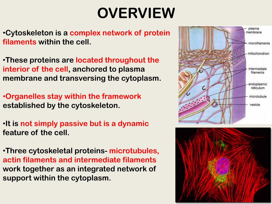
OVERVIEW•Cytoskeleton is a complex network of protein filaments within the cell.
•These proteins are located throughout the interior of the cell, anchored to plasma membrane and transversing the cytoplasm.
•Organelles stay within the framework established by the cytoskeleton.
•It is not simply passive but is a dynamic feature of the cell.
•Three cytoskeletal proteins- microtubules, actin filaments and intermediate filaments work together as an integrated network of support within the cytoplasm.
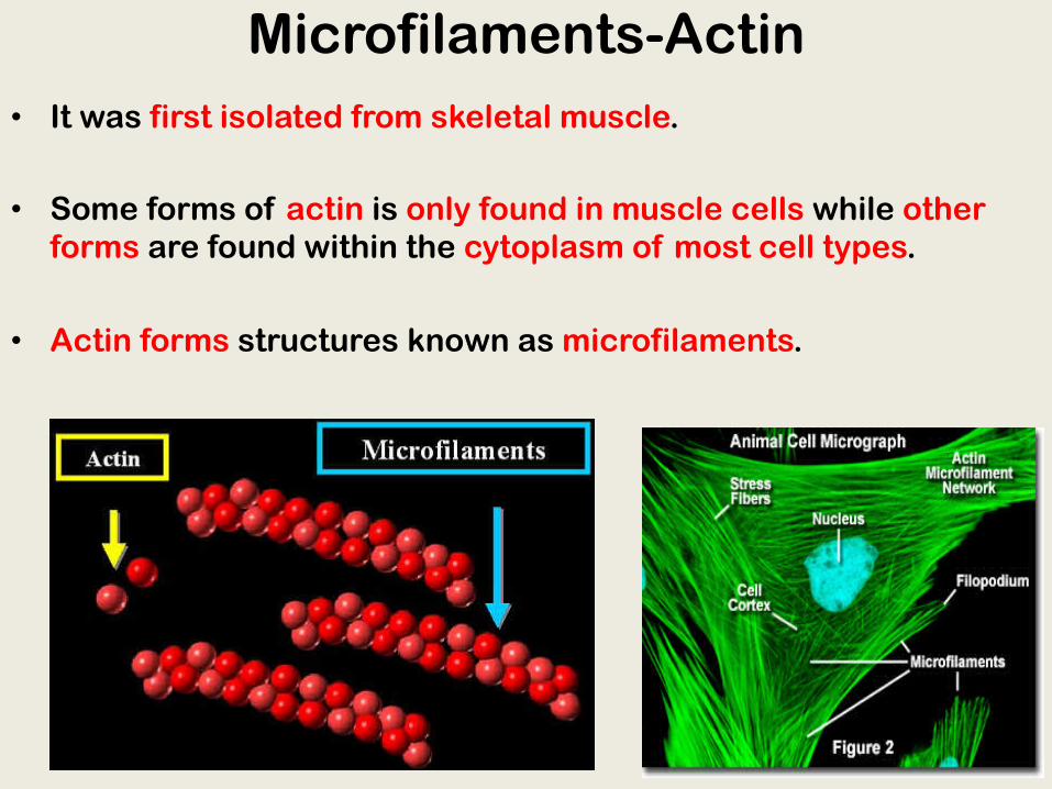
Microfilaments-Actin• It was first isolated from skeletal muscle.
• Some forms of actin is only found in muscle cells while other forms are found within the cytoplasm of most cell types.
• Actin forms structures known as microfilaments.

Functions of Actin• Actin helps to establish a cytoplasmic
protein framework radiating out of nucleus to the plasma membrane.
• Actin is often localized to regions near the plasma membrane-cell cortex.
• It is important in inducing contraction in muscle cells.
• In nonmuscle cells, regulate cell movement and formation of contractile rings in cell division.
• In nucleus, actin is important for chromatin and nuclear structure and regulation of gene processing.
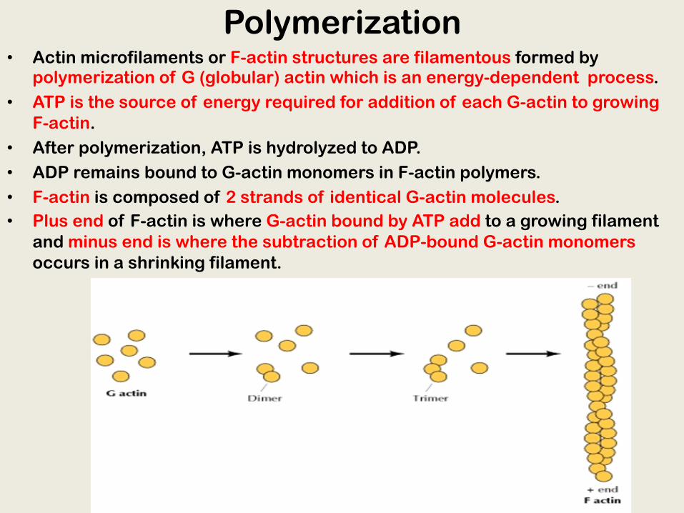
Polymerization• Actin microfilaments or F-actin structures are filamentous formed by
polymerization of G (globular) actin which is an energy-dependent process.• ATP is the source of energy required for addition of each G-actin to growing
F-actin.• After polymerization, ATP is hydrolyzed to ADP.• ADP remains bound to G-actin monomers in F-actin polymers.• F-actin is composed of 2 strands of identical G-actin molecules.• Plus end of F-actin is where G-actin bound by ATP add to a growing filament
and minus end is where the subtraction of ADP-bound G-actin monomers occurs in a shrinking filament.
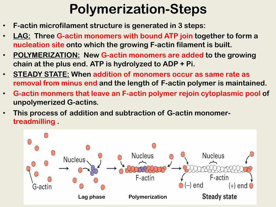
Polymerization-Steps• F-actin microfilament structure is generated in 3 steps:• LAG: Three G-actin monomers with bound ATP join together to form a
nucleation site onto which the growing F-actin filament is built.• POLYMERIZATION: New G-actin monomers are added to the growing
chain at the plus end. ATP is hydrolyzed to ADP + Pi.• STEADY STATE: When addition of monomers occur as same rate as
removal from minus end and the length of F-actin polymer is maintained.• G-actin monmers that leave an F-actin polymer rejoin cytoplasmic pool of
unpolymerized G-actins.
• This process of addition and subtraction of G-actin monomer-treadmilling .
Lag phase Polymerization

Myosin in Muscle Cells
• In muscle cells, thousands of actin filaments are arranged parallel to one another.
• Thicker filaments, composed of a motor protein, myosin, interconnected with the thinner actin fibers.
• Myosin molecules walk along the actin filament, pulling stacks of actin fibers together and shortening the cell.

Intermediate Filaments
• Intermediate filaments are larger than actin microfilaments and smaller than microtubules.
• Most intermediate filaments are present in the cytosol ,between nuclear envelope and the plasma membrane.
• Provide structural stability to cytoplasm somewhat similar to what steel rods can do to reinforce concrete .
• In the nucleus, intermediate filaments along with nuclear lamina provide strength and support to the nucleus.

• They are formed by α helical rod like protein subunits that have globular domains at both their ends.
• Two rod-like subunits combine to form dimers-Coiled coils.
• One coiled-coil dimer self associates with another coiled-coil dimer, in a staggered pattern, to form a tetramer.
• Tetramers attach to each other in a side-to-side array of eight tetramers that wind together to form rope-like structure of mature intermediate filament.
• No energy is required for the assembly and lack polarity.
Intermediate Filaments-Structure

Microtubules• They are involved in chromosomal
movements during nuclear division (mitosis and meosis).
• Also in formation of cilia and flagella in certain cell types.
• To coordinate and regulate microtubules, cells contain a structure –centrosome, which organize microtubules by controlling their number, location and cytoplasmic orientation.
• 2 type of microtubules in cells:
• Stable and long-lived (found in cilia, flagella, neurons).
• Unstable and short-lived (found in mitosis)

Microtubules-Structure
• The structure resembles a hollow cylindrical tube.
• Basic structural unit is protein heterodimer, composed of α and βtubulin molecule.
• These heterodimer self assemble to form protofilaments.
• Thirteen protofilaments form the outer wall of the cylindrical microtubule structure.

Microtubules-Assembly•Polymerization of microtubules is a complex process because of their continuous switch between growing and shrinking phases- dynamic instability.
•Tubulin heterodimers are GTP-bound which interact with other GTP-bound tubulin heterodimers to form protofilaments.
•A GTP cap is formed at + or the leading end (opposite to the anchored end) of microtubule representing the newly added tubulin heterdimers .
•Microtubules continues to grow its length in search of cellular structures such as organelles to which it can bind.

Microtubules-Disassembly•Soon after polymerization GTP on the tubulin is hydrolyzed to GDP and GTP cap is lost.
•New tubulin heterodimers cannot bind to GDP-containing tubulin heterodimers which stops assembly and also destabilizes the structure.
•Individuals protofilaments peel back and curve away .
•Tubulin heterodimers containing GDP dissociate from protofilament quickly and microtubule disassembles rapidly.

Microtubule Motor Proteins
• Organelles as well as vesicles can be observed to travel along microtubules within cells with the help of two motor proteins-Dynein and Kinesin.
• They have ATP-binding heads and tails that bind stably with intracellular cargo.
• Dynein move along the microtubule toward the - end and kinesin travels toward the + end.
• Both proteins hydrolyse ATP for their movement while pulling their cargo along the network provided by microtubules.

Summary• Cytoskeleton is a complex network of protein
filaments found throughout the interior of cells.
• Three main types of protein filaments-microfilaments, intermediate filaments and microtubules.
• Accessory proteins bind and regulate function of these filaments.
• Microfilaments: in nonmuscle cells actin regulatecell movement and formation of contractile rings in cell division .
• Actin polymerization occurs by the addition of G-actin monomers onto F-actin polymers, in an ATP dependent manner.• Treadmilling is a dynamic process of addition of a new G-actin monomer to a growing chain followed by displacement along F-actin polymer and its removal from the chain.• In muscle cells, myosin molecules walk along the actin filament, pulling stacks of actin fibers together and shortening the cell.

Summary
• Intermediate filaments: stable, rope like cytoskeletal structures that provide strength and support.
• Microtubules: involved in chromosomal movements during nuclear division, in formation of cilia and flagella and in intracellular transport.
• Composed of tubulin heterodimers, microtubules have a dynamic instability and continue to assemble and disassembleaccording to the needs of the cell.