deciphering the functional role of ripk4 in melanoma
TRANSCRIPT
International Journal of
Molecular Sciences
Article
Deciphering the Functional Role of RIPK4 in Melanoma
Ewelina Madej 1 , Damian Ryszawy 2,†, Anna A. Brozyna 3 , Malgorzata Czyz 4 , Jaroslaw Czyz 2
and Agnieszka Wolnicka-Glubisz 1,*
�����������������
Citation: Madej, E.; Ryszawy, D.;
Brozyna, A.A.; Czyz, M.; Czyz, J.;
Wolnicka-Glubisz, A. Deciphering the
Functional Role of RIPK4 in
Melanoma. Int. J. Mol. Sci. 2021, 22,
11504. https://doi.org/10.3390/
ijms222111504
Academic Editor: Silvana Morello
Received: 23 September 2021
Accepted: 22 October 2021
Published: 25 October 2021
Publisher’s Note: MDPI stays neutral
with regard to jurisdictional claims in
published maps and institutional affil-
iations.
Copyright: © 2021 by the authors.
Licensee MDPI, Basel, Switzerland.
This article is an open access article
distributed under the terms and
conditions of the Creative Commons
Attribution (CC BY) license (https://
creativecommons.org/licenses/by/
4.0/).
1 Faculty of Biochemistry, Biophysics and Biotechnology, Department of Biophysics, Jagiellonian University,7 Gronostajowa Street, 30-387 Krakow, Poland; [email protected]
2 Faculty of Biochemistry, Biophysics and Biotechnology, Department of Cell Biology, Jagiellonian University,7 Gronostajowa Street, 30-387 Krakow, Poland; [email protected] (D.R.); [email protected] (J.C.)
3 Faculty of Biological and Veterinary Sciences, Institute of Biology, Department of Human Biology, NicolausCopernicus University, 1 Lwowska Street, 87-100 Torun, Poland; [email protected]
4 Department of Molecular Biology of Cancer Lodz, Medical University of Lodz, 6/8 Mazowiecka Street,92-215 Lodz, Poland; [email protected]
* Correspondence: [email protected]; Tel.: +48-12-664-65-26; Fax: +48-12-664-69† Damian Ryszawy, passed away on 11 September 2020.
Abstract: The receptor-interacting protein kinase 4 (RIPK4) plays an important role in the devel-opment and maintenance of various tissues including skin, but its role in melanoma has not beenreported. Using patient-derived cell lines and clinical samples, we show that RIPK4 is expressedin melanomas at different levels. This heterogenous expression, together with very low level ofRIPK4 in melanocytes, indicates that the role of this kinase in melanoma is context-dependent. Whilethe analysis of microarray data has revealed no straightforward correlation between the stage ofmelanoma progression and RIPK4 expression in vivo, relatively high levels of RIPK4 are in metastaticmelanoma cell lines. RIPK4 down-regulation by siRNA resulted in the attenuation of invasivepotential as assessed by time-lapse video microscopy, wound-healing and transmigration assays.These effects were accompanied by reduced level of pro-invasive proteins such as MMP9, MMP2,and N-cadherin. Incubation of melanoma cells with phorbol ester (PMA) increased PKC-1β leveland hyperphosphorylation of RIPK4 resulting in degradation of RIPK4. Interestingly, incubation ofcells with PMA for short and long durations revealed that cell migration is controlled by the NF-κBsignaling in a RIPK4-dependent (RIPK4high) or independent (RIPK4low) manner depending on cellorigin (distant or lymph node metastasis) or phenotype (mesenchymal or epithelial).
Keywords: invasive potential; melanoma; MMPs; NF-κB; RIPK4
1. Introduction
The receptor-interacting serine/threonine kinase protein 4 (RIPK4, also known asDIK or PKK) is a highly conserved member of the RIP family of serine-threonine kinasescontaining an N-terminal RIP-like kinase domain and a C-terminal region characterized by11 ankyrin repeats [1,2]. In humans, RIPK4 is widely expressed in numerous tissues [1,3],and two isoforms of 86 and 92 kDa are found as the result of alternative splicing. Mutationsin RIPK4, including loss-of-function mutation, have been detected in keratinocytes ofpatients with Bartsocas-Papas syndrome [4,5]. Deletion of RIPK4 in mice leads to deathin the first hours of postnatal life. Interestingly, the observed deformations of RIPK4 KOmice are phenotypically similar to IKK (I kappa B kinase) KO mice, suggesting that RIPK4is involved in NF-κB-dependent keratinocyte differentiation [6,7]. These findings are incontrast to conflicting data on the RIPK4 role in cancer [7]. Depending on the context,RIPK4 acts as a cancer “suppressor” or oncogene [8–13]. While RIPK4 down-regulation wasobserved during the development of neoplasms of liver [8], tongue [9], and lung [10], highRIPK4 levels are correlated with progression and poor prognosis in cervical squamous cellcarcinoma [11], lymph node metastasis of cervical cancer [12], and increased invasiveness
Int. J. Mol. Sci. 2021, 22, 11504. https://doi.org/10.3390/ijms222111504 https://www.mdpi.com/journal/ijms
Int. J. Mol. Sci. 2021, 22, 11504 2 of 17
of pancreatic cancer [13]. Despite the role of RIPK4 in skin homeostasis [7], its function inmelanoma progression has not yet been investigated.
Melanoma that arises from transformed melanocytes represents the most lethal typeof skin tumor. Although a decreasing trend in fatal melanoma cases is observed in theUnited States and the majority of European countries [14,15], as a result of early detectionand the clinical use of the BRAFV600/MEK inhibitors and immune checkpoint inhibitors foradvanced stage melanomas [16,17], the incidence and mortality rates are still unacceptablyhigh [14,18]. Numerous questions about melanoma diagnosis, development of metastasis,lack of response and resistance to immune- and targeted- therapy still need to be ad-dressed [18]. Melanoma initiation, progression, and resistance to treatment are determinedby various signaling pathways, including RAF/MEK/ERK [19], PKC/NF-κB [20–23], andWnt/β-catenin signaling [24]. Notably, these pathways have also been identified as targetsof RIPK4 [25–27].
We investigated interactions between RIPK4, PKC and NF-κB pathways in melanomaand the role of RIPK4 in regulating the invasive potential of melanoma cells. To thisend, we used transcriptomic analyses of RIPK4 expression levels in melanoma specimensfrom clinical trials, IHC in melanoma biopsies, and protein level in patient-derived andcommercially available melanoma cell lines. The results indicate that RIPK4 contributes tomelanoma invasive potential in a lineage-specific/NF-κB-dependent manner.
2. Results2.1. RIPK4 Is Heterogeneously Expressed in Melanoma Cells
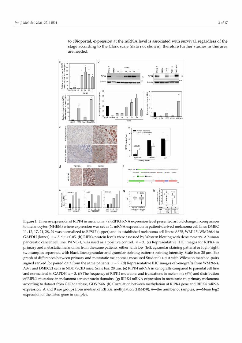
To date, there is no evidence that RIPK4 plays any role in melanoma, thus we firstinvestigated its level in established melanoma cell lines, patient-derived melanoma cell linesand clinical samples. Expression of RIPK4 in melanoma cells at the mRNA and protein levelswas higher in melanoma cells than in normal melanocytes (Figure 1a,b). Various levels ofRIPK4 were detected in melanoma cells, from slightly above levels in melanocytes of DMBC11,DMBC12 and WM115 cell lines to highly elevated values in DMBC28, DMBC29 and WM266.4cell lines. Interestingly, much higher mRNA and protein levels of RIPK4 were observed in themelanoma cells derived from lymph node metastasis (WM266.4) when compared to WM115cells that were derived from primary melanoma of the same patient.
To preliminarily assess the significance of RIPK4 in melanoma development, we per-formed immunohistochemical (IHC) staining on primary and metastatic melanoma tissues,including samples from the same patients. We found that RIPK4 expression is also het-erogeneously expressed in the clinical material, from low to high intensities (Figure 1c).Interestingly, we observed two different types of staining within the cytoplasm, agranularand granular, both with different intensities. A similar difference in staining was seen inxenografts obtained from melanoma cell lines with high levels of the RIPK4 kinase suchas A375, WM266.4 or DMBC21 (Figure 1d). We also found that RIPK4 expression was up-regulated in xenografts when compared to original melanoma cell lines (Figure 1e). To furtherexplore the involvement of RIPK4 in human cutaneous melanoma progression we conductedmetadata analysis of RIPK4 mRNA levels in 83 clinical specimens (GDS 3966), 31 primarymelanoma and 52 metastatic melanoma specimens using the Gene Expression Omnibus(GEO) database (https://www.ncbi.nlm.nih.gov/geo, accessed data 15 June 2020). This re-vealed similar RIPK4 mRNA levels (normalized to the log2 value) in the melanoma biopsiesfrom primary and metastatic tumors (Figure 1g), similarly to IHC staining data in this study.While we observed no statistically significant differences between primary and metastaticmelanomas, a much broader scatter of values was found within biopsies derived frommetastatic melanomas than primary melanomas. Further queries of datasets for melanoma-related genetic/epigenetic changes in RIPK4 (http://www.cbioportal.org/data_sets.jsp, in-cluding the TCGA through the cBIO Portal for Cancer Genomics, accessed data 15 June 2020)demonstrated a low (6%) incidence of missense RIPK4 mutations in melanoma (Figure 1f andSupplementary Figure S1) and a reverse correlation between RIPK4 levels and methylationlevels of RIPK4-encoding regions in melanoma samples (Figure 1h). Interestingly, according
Int. J. Mol. Sci. 2021, 22, 11504 3 of 17
to cBioportal, expression at the mRNA level is associated with survival, regardless of thestage according to the Clark scale (data not shown); therefore further studies in this areaare needed.
Figure 1. Diverse expression of RIPK4 in melanoma. (a) RIPK4 RNA expression level presented as fold change in comparisonto melanocytes (NHEM) where expression was set as 1. mRNA expression in patient-derived melanoma cell lines DMBC11, 12, 17, 21, 28, 29 was normalized to RPS17 (upper) and in established melanoma cell lines: A375, WM115, WM266.4 toGAPDH (lower). n = 3. * p < 0.05. (b) RIPK4 protein levels were assessed by Western blotting with densitometry. A humanpancreatic cancer cell line, PANC-1, was used as a positive control. n = 3. (c) Representative IHC images for RIPK4 inprimary and metastatic melanomas from the same patients, either with low (left; agranular staining pattern) or high (right;two samples separated with black line; agranular and granular staining pattern) staining intensity. Scale bar: 20 µm. Bargraph of differences between primary and metastatic melanomas measured Student’s t-test with Wilcoxon matched-pairssigned ranked for paired data from the same patients. n = 7. (d) Representative IHC images of xenografts from WM266.4,A375 and DMBC21 cells in NOD/SCID mice. Scale bar: 20 µm. (e) RIPK4 mRNA in xenografts compared to parental cell lineand normalized to GAPDH. n = 3. (f) The frequency of RIPK4 mutations and truncations in melanoma (6%) and distributionof RIPK4 mutations in melanoma across protein domains. (g) RIPK4 mRNA expression in metastatic vs. primary melanomaaccording to dataset from GEO database, GDS 3966. (h) Correlation between methylation of RIPK4 gene and RIPK4 mRNAexpression. A and B are groups from median of RIPK4: methylation (HM450), n—the number of samples, µ—Mean log2expression of the listed gene in samples.

Int. J. Mol. Sci. 2021, 22, 11504 4 of 17
2.2. RIPK4 Down-Regulation Interferes with the Invasive Phenotype of WM266.4 Cells
WM266.4 cells have mesenchymal morphology (Figure 2a), considerably higher N-cadherin levels (Figure 2a) and more pronounced motile activity than WM115 and A375cells (Figure 2b). WM266.4 cell speed was significantly higher than the speed of theirprimary counterparts, WM115 cells (p < 0.001).
Figure 2. Melanoma cells with elevated levels of RIPK4 exert enhanced motile activity. (a) Morphology and the levels ofN-cadherin and E-cadherin in WM115, WM266.4, and A375 melanoma cells. GAPDH was used as a loading control inWestern blotting. (b) The motile activity of melanoma cells vs. WM115. Statistical analysis is based on a collection of 60 cellsfrom three independent experiments by the Mann-Whitney Rank Sum Test * p < 0.05, ** p < 0.001.
To address the role of RIPK4 in melanoma progression more directly, we furtherinvestigated the effects of ectopic RIPK4 down-regulation on the phenotype of highlyinvasive WM266.4 melanoma cells. Using RIPK4-specific small interference RNAs (siRNAs)we were able to inhibit RIPK4 levels in these cells by 70–90% (Figure 3a). Down-regulationof RIPK4 only slightly interfered with WM266.4 proliferation (Supplementary Figure S2)without causing a significant effect on apoptosis (data not shown). RIPK4 silencing resultedin decreased transcript levels of fibronectin, MMP9 and MMP2 (Figure 3b) and inhibition ofWM266.4 motility (Figure 3c). Estimation of single WM226.4 cell trajectories by time-lapsevideo microscopy along with the results of wound healing assay demonstrated the reducedmotility and displacement of melanoma cells after the ectopic RIPK4 down-regulation(Figure 3c). These data are in concordance with the results of Transwell assays (Figure 3d),which showed attenuated transmigration capacity of WM266.4 cells after RIPK4 down-regulation. The visualization of the cytoskeleton (Figure 3e, Supplementary Figure S3)revealed the F-actin rearrangements, from well-organized cytoskeletal architecture withthe signs of actin polymerization at the leading edges towards thin microfilament bundles,

Int. J. Mol. Sci. 2021, 22, 11504 5 of 17
which were weakly anchored to non-matured focal adhesions. Concomitantly, we observeda slight down-regulation of CD44, which is a receptor for hyaluronic acid (HA) involvedin cell–cell interactions, cell adhesion and migration (Figure 3f). On the other hand, nosignificant effects of RIPK4 down-regulation on the levels of N-cadherin, Snail-1 and Twist-1 were seen (Figure 3g). These observations indicate the potential role of RIPK4 in theregulation of the invasive potential of WM266.4 cells. They also suggest the involvementof other signaling pathways in the regulation of post-EMT phenotype of melanoma cells.
Figure 3. Downregulation of RIPK4 reduces WM266.4 cells motile activity. Cells were transfected with RIPK4.si1, RIPK4.si2or neg.siRNA and induced changes were analyzed after 48 h. (a) RIPK4 levels in WM266.4 cells were assessed by Westernblotting with densitometry and qRT-PCR normalized to GAPDH. n = 3. (b) Transcript levels of Fibronectin, MMP2 andMMP9 normalized to GAPDH. n = 3. (c) Cell motility. Statistical analysis is based on a collection of 60 cells from threeindependent experiments by Mann-Whitney Rank Sum Test. * p < 0.05, ** p < 0.001. Wound healing, n = 3. (d) Time-dependent migration of cells by Transwell assay. n = 3. (e) Global architecture of F-actin (red) and vinculin (green). Scalebar: 25 µm. (f) CD44 expression by flow cytometry. (g) N-cadherin and E-cadherin levels in WM266.4 cells along withdensitometry. n = 3. All samples of RIPK4.si transfected cells were compared to neg.si (scrambled control) samples * p < 0.05,** p < 0.001.

Int. J. Mol. Sci. 2021, 22, 11504 6 of 17
2.3. RIPK4 Remains under the Control of Protein Kinase C-Dependent Signalingin WM266.4 Cells
Phosphorylation of RIPK4 by PKC has previously been shown to direct RIPK4 towardsproteolytic degradation [7]. To estimate the significance of PKC signaling for RIPK4 activity inWM266.4 cells and its consequences for cell motility, we treated melanoma cells with 150 nMphorbol-12-myristate-13-acetate (PMA). As expected, WM266.4 cells showed lower PKC-1βlevel than their WM115 counterparts (Figure 4a). PMA increased PKC1β level and reducedRIPK4 level in WM266.4 melanoma cells after 12–48 h of treatment (Figure 4b), and concomi-tantly inhibited the WM266.4 cell motility (Figure 4c). This reduction was even more pronouncedthan that caused by RIPK4 siRNA. Along with PMA-induced inhibition of the motile activity,reduced p65 phosphorylation at Ser 536, and impaired wound healing of WM266.4 cells wasobserved (Figure 4c). PMA treatment also induced the morphological shifts of WM266.4 cellstowards non-polarized epithelioid shape and decreased the level of N-cadherin, but had noeffect on Snail-1 and Twist-1 levels (Figure 4d). These analyses further confirmed the role ofRIPK4 in the regulation of melanoma motile capacity in vitro and showed the functional linksbetween RIPK4- and PKC-dependent signaling in WM266.4 cells.
Figure 4. PMA decreases RIPK4 levels in WM266.4 melanoma cells. (a) PKC-1β level in A375, WM115 and WM266.4 cells wasassessed by Western blotting. (b) Protein levels of PKC-1β, RIPK4 and GAPDH in WM266.4 cells treated with DMSO (control) orPMA (150 nM) for 12–48 h along with densitometry. The samples were compared to the DMSO control samples. n = 3 * p < 0.05,** p < 0.001. (c) Motility of WM266.4 cells treated with DMSO (control) or PMA (150 nM) for 48 h. Statistical analysis is based ona collection of 150 cells from three independent experiments by the Mann-Whitney Rank Sum Test. ** p < 0.001. Protein levelsof RIPK4, IκBα, P-p65, p65 were assessed by Western blotting. GAPDH was used as a loading control. Representative imagesfrom three independent wound healing experiments. (d) Protein levels of N-cadherin, Twist-1, Snail-1 and β-Tubulin along withdensitometry. The samples were compared to the DMSO control samples. n = 3. ** p < 0.001. Cell morphology of cells treated withDMSO (control) or PMA (150 nM) for 48 h.
Int. J. Mol. Sci. 2021, 22, 11504 7 of 17
2.4. Concerted RIPK4/PKC Signaling Regulates the Invasive Potential of A375 Cells
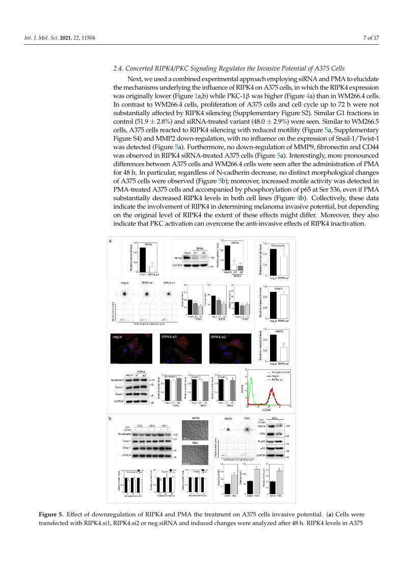
Next, we used a combined experimental approach employing siRNA and PMA to elucidatethe mechanisms underlying the influence of RIPK4 on A375 cells, in which the RIPK4 expressionwas originally lower (Figure 1a,b) while PKC-1β was higher (Figure 4a) than in WM266.4 cells.In contrast to WM266.4 cells, proliferation of A375 cells and cell cycle up to 72 h were notsubstantially affected by RIPK4 silencing (Supplementary Figure S2). Similar G1 fractions incontrol (51.9± 2.8%) and siRNA-treated variant (48.0± 2.9%) were seen. Similar to WM266.5cells, A375 cells reacted to RIPK4 silencing with reduced motility (Figure 5a, SupplementaryFigure S4) and MMP2 down-regulation, with no influence on the expression of Snail-1/Twist-1was detected (Figure 5a). Furthermore, no down-regulation of MMP9, fibronectin and CD44was observed in RIPK4 siRNA-treated A375 cells (Figure 5a). Interestingly, more pronounceddifferences between A375 cells and WM266.4 cells were seen after the administration of PMAfor 48 h. In particular, regardless of N-cadherin decrease, no distinct morphological changesof A375 cells were observed (Figure 5b); moreover, increased motile activity was detected inPMA-treated A375 cells and accompanied by phosphorylation of p65 at Ser 536, even if PMAsubstantially decreased RIPK4 levels in both cell lines (Figure 4b). Collectively, these dataindicate the involvement of RIPK4 in determining melanoma invasive potential, but dependingon the original level of RIPK4 the extent of these effects might differ. Moreover, they alsoindicate that PKC activation can overcome the anti-invasive effects of RIPK4 inactivation.
Figure 5. Effect of downregulation of RIPK4 and PMA the treatment on A375 cells invasive potential. (a) Cells weretransfected with RIPK4.si1, RIPK4.si2 or neg.siRNA and induced changes were analyzed after 48 h. RIPK4 levels in A375
Int. J. Mol. Sci. 2021, 22, 11504 8 of 17
cells were assessed by qRT-PCR and Western blotting with densitometry normalized to GAPDH. n = 3. Cell motility.Statistical analysis is based on a collection of 90 cells from three independent experiments using the Mann-Whitney RankSum Test. * p < 0.05, ** p < 0.001. Global architecture of F-actin (red) and vinculin (green). Scale bar: 25 µm. Transcript levelsof Fibronectin, MMP2 and MMP9 normalized to GAPDH. n = 3. Protein levels of N-cadherin, Twist-1, Snail-1 and GAPDHalong with densitometry. All samples of the RIPK4.si transfected cells were compared to neg.si (scrambled control) samples,** p < 0.001. CD44 expression by flow cytometry. (b) Cells were treated with DMSO (control) or PMA (150 nM) for 12-48 h.Protein levels of N-cadherin, Twist-1, Snail-1 and β-Tubulin along with densitometry. n = 3. Cell morphology and motility.Statistical analysis is based on a collection of 90 cells from three independent experiments. ** p < 0.001. Protein levels ofRIPK4, IκBα, P-p65, p65 were assessed by Western blotting. GAPDH was used as a loading control. The samples werecompared to the DMSO control samples. n = 3. ** p < 0.001.
2.5. Immediate vs. Long-Term Effect of RIPK4 Down-Regulation on NF-κB Activity in WM266.4and A375 Cells
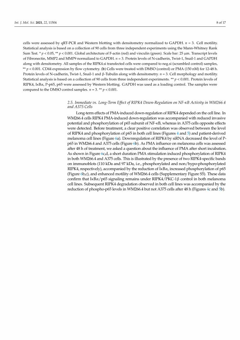
Long-term effects of PMA-induced down-regulation of RIPK4 depended on the cell line. InWM266.4 cells RIPK4 PMA-induced down-regulation was accompanied with reduced invasivepotential and phosphorylation of p65 subunit of NF-κB, whereas in A375 cells opposite effectswere detected. Before treatment, a clear positive correlation was observed between the levelof RIPK4 and phosphorylation of p65 in both cell lines (Figures 4 and 5) and patient-derivedmelanoma cell lines (Figure 6a). Downregulation of RIPK4 by siRNA decreased the level of P-p65 in WM266.4 and A375 cells (Figure 6b). As PMA influence on melanoma cells was assessedafter 48 h of treatment, we asked a question about the influence of PMA after short incubation.As shown in Figure 6c,d, a short duration PMA stimulation induced phosphorylation of RIPK4in both WM266.4 and A375 cells. This is illustrated by the presence of two RIPK4-specific bandson immunoblots (110 kDa and 97 kDa, i.e., phosphorylated and non/hypo-phosphorylatedRIPK4, respectively), accompanied by the reduction of IκBα, increased phosphorylation of p65(Figure 6b,c), and enhanced motility of WM266.4 cells (Supplementary Figure S5). These dataconfirm that IκBα/p65 signaling remains under RIPK4/PKC-1β control in both melanomacell lines. Subsequent RIPK4 degradation observed in both cell lines was accompanied by thereduction of phospho-p65 levels in WM266.4 but not A375 cells after 48 h (Figures 4c and 5b).
Int. J. Mol. Sci. 2021, 22, 11504 9 of 17
Figure 6. Effect of PMA short time treatment on NF-κB signaling in WM266.4 and A375 cells. (a) The level of phosphorylatedp65 in relation to RIPK4 level in patient-derived melanoma cells with densitometry. PANC-1 cells was used as a positivecontrol. (b) The level of phosphorylated p65 in cells transfected with RIPK4.si1, RIPK4.si2 or neg.si RNA by Westernblotting with densitometry. The samples were compared to neg.si (scrambled control) samples. n = 3. * p < 0.05, ** p < 0.001.(c,d) Cells were treated with DMSO (control) or PMA (150 nM) for 0.5–2 h. (c) WM266.4 cell. (d) A375 cells. Protein levelsof RIPK4, IκBα, P-p65, p65 were assessed by Western blotting. GAPDH was used as a loading control. The samples werecompared to the DMSO control samples. n = 3. * p < 0.05, ** p < 0.001.
Int. J. Mol. Sci. 2021, 22, 11504 10 of 17
3. Discussion
RIPK4 has been implicated in the progression of numerous tumors [8–13,28,29]; how-ever its role in melanoma remained unresolved. Our study fills this gap because it is thefirst to reveal the functional role of RIPK4 in melanoma. This role is illustrated by (i) therelatively high levels of RIPK4 in metastatic melanoma cell lineages and tumor specimensand (ii) the reduction of motility of melanoma cells by RIPK4 down-regulation. Further-more, we show (iii) the modulatory effect of PKC that differentially influences the impact ofRIPK4/NF-κB axis on the invasive potential of melanoma cell lineages. Thus, RIPK4 mightplay a pivotal role in the formation of a melanoma invasive front. However, in conjunctionwith the lack of correlation between melanoma stage and RIPK4 expression levels in exvivo melanoma biopsies, these data also indicate the complexity of the consequences ofRIPK4-dependent signaling for this process.
Abnormal cell proliferation decreased cell apoptosis and induction of cell motilityplay pivotal roles in skin tumorigenesis. De-Qing Liu et al. [11], demonstrated that RIPK4knockdown significantly inhibited cell proliferation and clone formation capacity in SiHaand Caski cells. The possible role of RIPK4 in the regulation of invasive potential ofmelanoma was suggested by the current findings by the high RIPK4 levels in metastatic(DMBC21, DMBC28, DMBC29, WM266.4) melanoma cell lines. Further analyses of theconsequences of RIPK4 down-regulation in melanoma cells revealed its clear impact onthe invasive potential manifested by decreased transmigratory potential and motility ofWM266.4 cells, which was also observed in A375 cells. Moreover, RIPK4-induced changesof F-actin and focal contact architecture correlated with their pro-epithelial morphologicalshifts, cytoskeletal rearrangements and CD44 down-regulation in WM266.4 cells. These ob-servations correspond to the results of studies on keratinocytes, where RIPK4 participatedin the maintenance of cortical F-actin organization [30,31] and intercellular junctions [32],whereas CD44 expression apparently participated to tumor invasion [33,34]. Thus, ourresults indicate that RIPK4 is involved in melanoma cell invasiveness by influencing celladhesion and actin dynamics. In osteosarcomas, silencing of RIPK4 inhibited EMT by inac-tivating the Wnt/β-catenin signaling pathway [28]. In melanoma cells, down-regulation ofRIPK4 expression by siRNA or PMA did not completely abolish the expression of EMTmarkers including Twist-1 and Snail-1 and N-cadherin. Thus, the lack of RIPK4 effects onSnail-1/Twist-1 levels indicates that RIPK4 exerts its pro-invasive effects downstream ofEMT master regulators and seems to be related to cell adhesion.
Our mechanistic studies demonstrated that the function of RIPK4 in melanoma cellsis predominantly executed through the activation of NF-κB signaling. This is illustratedby a correlation between RIPK4 and P-p65 levels in patient-derived cell lines and reducedphosphorylation of p65 in RIPK4 siRNA-treated WM266.4 and A375 cells, along with theirreduced motility. The relationship between NF-κB activity and melanoma cell motilityis well established [20]. RIPK4 has also been shown to promote bladder urothelial carci-noma cell aggressiveness by NFκB-induced EMT [29]. Our results also correspond to theprevious reports on the down-regulation of vimentin, MMP2 and fibronectin after RIPK4knockdown in cervical squamous carcinoma cells [11]. NF-κB has also been identified as aregulator of CD44 expression in melanocytes [33]. Thus, RIPK4/NF-κB-dependent signal-ing axis at least partly sustains mesenchymal phenotype of melanoma cells as additionallyrevealed by morphological shifts of WM266.4 cells towards an epithelioid phenotype uponRIPK4 inhibition.
Our data also show that IκBα/p65-dependent signaling remains under the controlof RIPK4/PKC-1β in both melanoma cell lines. Actually, short-term PMA stimulationinduced RIPK4 phosphorylation in both WM266.4 and A375 cells. This was accompaniedby reduced IκBα and elevated p65 phosphorylation and induction of WM266.4 motility,additionally confirming the notion that RIPK4 activates the NF-κB signaling cascadeupstream of IKKβ and IκBα [35]. These results are consistent with previous observationsindicating interactions of RIPK4 with PKCβ1 and PKCδ and a role for PKC activity in the

Int. J. Mol. Sci. 2021, 22, 11504 11 of 17
activation of NF-κB and other signaling molecules (including JNK-AP-1) [1,35]. A similarinterrelation has been found in keratinocytes [36].
We also observed differences in the sensitivity of melanoma cell lines to RIPK4-dependent signaling. These are manifested by less pronounced effects of RIPK4 silencingon morphology and expression of pro-invasive markers in A375 cells than in WM266.4cells, and by differential effects of PKC on RIPK4/NF-κB axis and the invasive potentialof melanoma cell lines. Apparently, the level of phospho-p65 in WM266.4 and A375 cellsafter their 48 h-long PMA treatment correlated with the inhibition of WM266.4 and theinduction of A375 motility. PMA reduced RIPK4 in A375 cells while retaining NF-κBactivity and invasiveness of these cells. This observation indicates the direct effect ofPKC on NF-κB activity and the invasive potential of A375 cells. It also shows that PKCcan differentially affect NF-κB activity in discrete melanoma cell lineages. Accordingly,PKC can modulate NF-κB signaling in a RIPK4-dependent manner and/or via the RIPK4-independent pathways.
It appears that further studies are needed to fully assess the long-term consequencesof RIPK4 involvement in the regulation of melanoma phenotype and microevolution ofmelanoma cells.
On the other hand, our data show that RIPK4 activity contributes to the invasivepotential of melanoma cells. However, together with the relatively low RIPK4 levels in thebiopsies of prospectively malignant melanoma tumors, they also indicate the stage-specificfunction of RIPK4 in melanoma development. Corresponding tumor stage-specificity haspreviously been documented for several other factors, including Cx43 [37–39]. RIPK4exerts its inducing effect on melanoma invasiveness via NF-κB signaling; however, highsusceptibility of NF-κB pathway to other signaling pathways (incl. PKC) may also deter-mine the recruitment of RIPK4-depressed cells to the invasive melanoma front. This cellcontext-dependent involvement of RIPK4 in melanoma cell invasiveness, together withthe suppressive function(s) of RIPK4 at the early stages of melanoma, apparently underliethe lack of straightforward correlation between RIPK4 levels and melanoma progression.Differential interplay between PKC and RIPK4 expression in discrete melanoma cells partic-ipates in this process. Further in vivo studies on clinical material from oncological patientsshould help to further decipher these interrelations.
4. Materials and Methods4.1. Clinical Samples
Preliminary analysis of RIPK4 expression in human clinical samples was performedon formalin-fixed, paraffin-embedded (FFPE) samples from seven patients with diagnosedcutaneous melanoma, including samples of the primary melanomas and lymph nodemetastases from the same patients. The study was carried out following the rules of theDeclaration of Helsinki of 1975 (revised in 2008) and the study was approved by the Institu-tional Review Board of Collegium Medicum, Nicolaus Copernicus University (KB136/2016)and the Bioethics Committee of the Jagiellonian University (no. 1072.6120.125.2017, date ofapproval 25 November 2020).
4.2. Geo Database Analysis
The RIPK4 expression profile deposited in Gene Expression Omnibus (GEO) (accessionnumber GDS3966 was analyzed [40]. The microarray platforms used in these datasets wereHUI133A Affymetrix DNA chips. The GDS3966 dataset comprises 83 melanoma samples(31 primary and 52 metastatic tumors) collected at the Massachusetts General Hospital andHarvard Medical School from 1992 to 2001 as a part of the diagnostic workup or planningfor therapy. As claimed in the original papers, all the studies had been approved by thelocal ethical committees and all participants gave written informed consent [40]. The geneexpression level was normalized to the log2 value.

Int. J. Mol. Sci. 2021, 22, 11504 12 of 17
4.3. Xenografts
The experiments were carried out according to the guidelines of the I Local EthicsCommittee of the Institute of Pharmacology of the Polish Academy of Sciences (approval no.411/2020 and 461/2020, date of approval 24 June 2020, 25 November 2020). The mice werehandled according to the regulations of the national and local animal welfare bodies underSPF (Specific-pathogen-free) conditions, with sufficient water and food provided at alltimes. The 4- to 6-week old female NOD-SCID mice (non-obese diabetes severe combinedimmunodeficiency mice) were injected subcutaneously with 3 × 10⁶ A375, WM266.4 andDMBC21 cells/mouse. Experiments were carried out with three NOD-SCID mice/group.Mice were subjected to anesthesia and xenografts were harvested after 36 days (A375 cells),41 days (WM266.4 cells), and 55 days (DMBC21). Tumor samples were divided into twopieces, one group was fixed in 10% formalin and the second was frozen at −80 ◦C forRNA isolation.
4.4. Cell Culture and Treatment
Normal, human, adult melanocytes were obtained from Lonza in 2020 (Lonza, Basel,Switzerland). Melanocytes were cultured in M254 medium supplemented with humanmelanocytes growth supplement (HMGS, Invitrogen, Thermo Fisher Scientific, Waltham,MA, USA). Human melanoma cell lines: Wistar Melanoma collection cells: WM115(RRID:CVCL_0040; primary melanoma, VGP—vertical growth phase) and WM266.4(RRID:CVCL_2765; metastatic human melanoma cell line, derived from the same pa-tient as WM115) kindly provided by Dept. of Medical Biochemistry, Jagiellonian UniversityMedical College (Kraków, Poland) in 2006 and have been authenticated using STR profiling.A375 cells (RRID:CVCL_0132; metastatic human melanoma cell line) were obtained fromATCC (Manassas, VA, USA) in 2020. Melanoma cells were cultured in RPMI1640 mediumsupplemented with 10% FBS (Gibco, Paisley, UK) and antibiotics (penicillin 150 U/mL,streptomycin 100 µg/mL, Sigma-Aldrich, Saint Louis, MO, USA) at 37 ◦C at 5% CO2 and95% humidity. DMBC cell lines (Department of Molecular Biology of Cancer, DMBC) wereobtained from drug-naïve melanoma patients during surgical interventions. The studywas approved by the Ethical Commission of Medical University of Lodz (RNN/84/09/KE)and informed consent was obtained from all patients. Cells were culture in condition de-scribed previously [41]. Whole-Exome Sequencing raw data of DMBC cell lines are publiclyavailable under the accession numbers E-MTAB-6978. Data were mapped to the referencegenome GRCh37/hg19 using BWA package (version bwa-0.7.12) [41]. Phenotypes of thesecells were also extensively characterized [42]. PMA (150 nM, Sigma-Aldrich, Saint Louis,MO, USA) was dissolved in DMSO and added to cells culture for 0.5–48 h. DMSO concen-tration does not exceed 0.05%. All experiments were performed with mycoplasma-freecells (Lonza, Basel, Switzerland).
4.5. Immunostaining
4µm FFPE sections, after deparaffinization, rehydration, and heat-induced antigenretrieval using low pH buffer (Vector Laboratories, Inc., Burlingame, CA, USA) and quench-ing the endogenous peroxidase with 3% H2O2, were incubated with primary anti-RIPK4antibody (recognizing recombinant fusion protein containing a sequence correspondingto amino acids 240–520 of human RIPK4; ABclonal, Woburn, MA, USA) overnight at4 ◦C. Next, the HRP-conjugated secondary antibody (ImmPRESS HRP REAGENT KITanti-Rabbit IgG, Vector Laboratories, Inc., Burlingame, CA, USA) followed by ImmPACTNovaRED (Vector Laboratories Inc., Burlingame, CA, USA) and counterstaining withhematoxylin were applied. Duodenum served as positive control. In negative controls,the primary antibodies were omitted and replaced with antibody diluent. The sectionswere evaluated semi-quantitatively, as previously described [43,44]. Since we observedheterogenous: agranular and granular staining, the assessment was performed separatelyfor each staining pattern.

Int. J. Mol. Sci. 2021, 22, 11504 13 of 17
4.6. Cell Transfection
For RIPK4 downregulation two types of small interfering RNA (siRNA) were used:RIPK4-specific Silencer Select siRNAs (ID: s28865 and s28863, Thermo Fisher Scientific,Waltham, MA, USA) or Silencer Select Negative Control No. 2 (cat.no 4390846, ThermoFisher Scientific, Waltham, MA, USA), serving as a negative control. The transfection wasperformed, as previously described [45]. The levels of RIPK4 in siRNA transfected cellswas verified by Western blot or qRT-PCR analysis.
4.7. Western Blot
Western blot analysis was carried out as described previously [46], except for lysatesfrom DMBC lineage cells which were analyzed as described [41]. The blots were cut priorto hybridization with antibodies. Labelled bands were detected with ChemiDoc ImagingSystem (BioRad, Hercules, CA, USA). Densitometry analysis was performed using ImageJsoftware or ImageLab 5.2.1. software. We used the following antibodies and dilutions:rabbit IgG anti-human: RIPK4, N-cadherin, P-p65 (Ser-536), p65, GAPDH, IκBα (1:2000,Cell Signaling Technology, Danvers, MA, USA), PKC1β (1:2000, Invitrogen, Thermo FisherScientific, Waltham, MA, USA), E-cadherin, Twist-1, Snail-1 (1:2000, Sigma-Aldrich, SaintLouis, MO, USA), β-Tubulin (1:2000, Abcam, Cambridge, UK), β-Actin (1:4000, SantaCruz Biotechnology, Dallas, CA, USA), goat HRP-conjugated anti-rabbit (1:4000, CellSignaling Technology Danvers, MA, USA) and goat HRP-conjugated anti-mouse (1:4000,BP Pharmingen, NJ, USA).
4.8. RNA Isolation and Quantitative RT-PCR
Total RNA was isolated using the Total RNA Mini Plus (A&A Biotechnology, GdanskPoland) accordingly to the manufacturer’s protocol. RNA (1µg) was reverse-transcribedinto cDNA using oligo(dT)18 and TranScriba Kit (A&A Biotechnology, Gdansk Poland) orusing SuperScript II Reverse Transcriptase (Invitrogen, Thermo Fisher Scientific, Waltham,MA, USA). qRT-PCR reactions were performed using Sensitive RT HS-PCR Mix (A&ABiotechnology, Gdansk, Poland) and the qTOWER3 real-time PCR thermal cycler (AnalytikJena, Jena, Germany) or the KAPA SYBR FAST qPCR and Rotor-Gene 3000 Real-Time DNAanalysis system (Corbett Research, Mortlake, Australia). Primer sequences were as follows:RIPK4 forward: 5′-ATG CCC ACT ACC ACG TCA AG-3′ and reverse 5′-TCT TCT CATCTG CAA ACG GCT-3′; RPS17 forward 5′-AAT CTC CTG ATC CAA GGC TG-3′ andreverse 5′-CAA GAT AGC AGG TTA TGT CAC G-3′. A mathematical model including anefficiency correction was used to calculate relative expression of selected genes versus areference gene RPS17. All TaqMan primers were from Thermo Fisher Scientific/Invitrogen:RIPK4 (Hs01062501_m1), Fibronectin-1 (Hs01549976_m1), MMP2 (Hs01548727_m1), MMP9(Hs00957562_m1), GAPDH (cat. No. 4326317E). The relative levels of transcripts werequantified by the 2−∆∆Ct method, using GAPDH as a reference gene.
4.9. Immunofluorescence
2× 10⁴ cells were seeded on coverslips in a 12-well plate and incubated for 24–96 h. Forimmunolocalization of cytoskeletal proteins (vinculin, F-actin) cells were fixed and labeledas described previously [47] and stained with primary antibodies rabbit anti-vinculin (1:200Sigma-Aldrich, Saint Louis, MO, USA), secondary antibodies AlexaFluor488-conjugatedchicken anti-rabbit and TRITC-conjugated phalloidin (1:50). For DNA visualization the cellswere stained with Hoechst 33258 (Sigma-Aldrich, Saint Louis, MO, USA). The specimenswere visualized by TIRF imaging using a DMI6000B microscope.
4.10. Time-Lapse Video Microscopy
Analysis of cells’ motility was performed using time-lapse video microscopy. Cellswere seeded in a 24-well plate and subjected to microscopic analysis 40 h after PMA orsiRNA treatment, within environmental control chamber custom adjusted to the procedure.Images were registered every 5 min within 8 h span. Images were acquired using fully
Int. J. Mol. Sci. 2021, 22, 11504 14 of 17
automatic DMI6000B (Leica AF7000 version, Wetzlar, Germany) and analyzed with Hiroprogram [47].
4.11. Transmigration Tests
15 × 103 cells were seeded on the upper side of trans-well migration inserts (Corning,NJ, USA) in a 24-well plate and allowed to transmigrate in hemodynamic conditions for24 h. Afterward, the inserts were transferred to another well for 24 h and the procedurewas repeated 4 times. Cells in the wells were counted to determine the initial number oftransmigrating melanoma cells [48].
4.12. Wound Healing Assay
The scratch wound was made using 200 µL sterile pipette tip in a confluent cell culturepre-treated with DMSO, PMA or transfected with negative siRNA (scrambled control)or RIPK4.siRNAs The scratch area was washed, and cells were re-incubated in the samecondition for additional 48 h. The images were taken at 0, 24 and 48 h time points. TheImageJ software was used to draw the lines of the healing wound areas.
4.13. Flow Cytometry Analysis of CD44
Cells were washed with PBS containing 0.1 mM EDTA and counted with a Bürkerhemocytometer. Then 5 × 10⁵/mL cells were incubated for 30 min on ice in PBS/EDTAsupplemented with 1% BSA and rat anti-CD44-FITC conjugated antibodies (clone IM7,1:600 Thermo Fisher, Waltham, MA, USA) or its isotype match control (Rat IgG2bκ IsotypeControl) and washed twice. Afterward, viable cells were gated based on FSC/SSC scatterand 10,000 cells were collected by FACS Calibur instrument (BD Biosciences, San Jose,CA, USA) and analyzed with CellQuest (BD Biosciences) software. The measurement wascarried out using 488 nm excitation and a 510–570 nm band-pass emission filter for thedetection of fluorescein isothiocyanate (FITC).
4.14. Cell Cycle
Cells were trypsinized and washed with PBS followed by fixation with ice-cold EtOH(70%) and stored at −20 ◦C. Cells were washed and stained as described [49]. Viablecells were gated based on FSC/SSC scatter and collected by FACS Calibur instrument (BDBiosciences, San Jose, CA, USA). Data were analyzed using the ModFit LT 5 software.
4.15. Statistical Analysis
Graphs are presented as a mean of at least three independent experiments± SD unlessstated otherwise. To compare two independent samples, the Student’s t-test was used.The differences between primary and metastatic melanomas were measured Student’st-test with Wilcoxon matched-pairs signed ranked for paired data from the same patients.Differences were considered significant at p < 0.05.
5. Conclusions
Our data show that RIPK4 contributes to melanoma development. Apparently, thiseffect is achieved in a manner dependent on NF-kB signaling. A selective recruitment ofRIPK4low and RIPK4high discrete melanoma cell lineages to the invasive front of melanomais determined by a differential sensitivity of NF-κB signaling to alternative (PKC-related)activators. Our data also confirm that PKC-dependent signaling can affect the invasivepotential of discrete melanoma cell lineages in a manner dependent on the duration of PKCactivation. Therefore, the interaction between the RIPK4, PKC and NF-κB pathways [25,27]is involved in the progression of melanoma [50].
Supplementary Materials: The following are available online at https://www.mdpi.com/article/10.3390/ijms222111504/s1.

Int. J. Mol. Sci. 2021, 22, 11504 15 of 17
Author Contributions: Conceptualization, E.M., A.W.-G.; methodology, E.M., A.W.-G., D.R.; valida-tion, E.M., D.R.; A.A.B.; formal analysis, E.M., A.W.-G., D.R., A.A.B.; investigation, E.M., A.W.-G.;resources, A.A.B., A.W.-G.; data curation, E.M.; writing—original draft preparation, E.M., A.W.-G.,D.R.; writing—review and editing, J.C., M.C., A.W.-G.; visualization, E.M., supervision, A.W.-G.;project administration, E.M.; A.W.-G.; funding acquisition, E.M., A.W.-G. All authors have read andagreed to the published version of the manuscript.
Funding: This research was funded by the National Science Centre, Poland, grant numbers 2018/31/N/NZ3/02625 and 2018/31/B/NZ5/01423. The open-access publication of this article was fundedby the Priority Research Area BioS under the program “Excellence Initiative—Research University”at the Jagiellonian University in Krakow.
Institutional Review Board Statement: The clinical study was carried out following the rules of theDeclaration of Helsinki of 1975 (revised in 2008). The study on FFPE samples was approved by theInstitutional Review Board of Collegium Medicum, Nicolaus Copernicus University (KB136/2016)and the Bioethics Committee of the Jagiellonian University (no. 1072.6120.125.2017, date of approval25 December 2020). DMBC cell lines were obtained from drug-naïve melanoma patients duringsurgical interventions. The study was approved by the Ethical Commission of Medical Universityof Lodz (RNN/84/09/KE, date of approval 17 March 2009). Animal experiments were carriedout according to accordance with the guidelines of the I Local Ethics Committee of the Institute ofPharmacology of the Polish Academy of Sciences (approval no. 411/2020 date of approval 24 June2020 and no. 461/2020, date of approval 25 December 2020).
Informed Consent Statement: Informed consent was obtained from all subjects involved in the study.
Data Availability Statement: Data sharing not applicable. No new data were created or analyzed inthis study. Data sharing is not applicable to this article.
Acknowledgments: The authors thank Monika Zywicka and Lukasz Czarnecki for IHC staining,Sara Seweryn for GEO analysis, Maciej Pudelek deconvolution of image A375 cells, Marcin Wozniakfor cell authentication, and Malgorzata Hajduk and Katarzyna Dylanowska from Animal House. Theauthors thank Russel Reiter for excellent proofreading.
Conflicts of Interest: The authors declare no conflict of interest.
References1. Chen, L.; Haider, K.; Ponda, M.; Cariappa, A.; Rowitch, D.; Pillai, S. Protein kinase C-associated kinase (PKK), a novel membrane-
associated, ankyrin repeat-containing protein kinase. J. Biol. Chem. 2001, 276, 21737–21744. [CrossRef] [PubMed]2. Huang, C.S.; Oberbeck, N.; Hsiao, Y.C.; Liu, P.; Johnson, A.R.; Dixit, V.M.; Hymowitz, S.G. Crystal Structure of Ripk4 Reveals
Dimerization-Dependent Kinase Activity. Structure 2018, 26, 767–777.e5. [CrossRef] [PubMed]3. Meylan, E.; Tschopp, J. The RIP kinases: Crucial integrators of cellular stress. Trends Biochem. Sci. 2005, 30, 151–159. [CrossRef]4. Mitchell, K.; O’Sullivan, J.; Missero, C.; Blair, E.; Richardson, R.; Anderson, B.; Antonini, D.; Murray, J.C.; Shanske, A.L.;
Schutte, B.C.; et al. Exome sequence identifies RIPK4 as the Bartsocas-Papas syndrome locus. Am. J. Hum. Genet. 2012, 90, 69–75.[CrossRef] [PubMed]
5. Kalay, E.; Sezgin, O.; Chellappa, V.; Mutlu, M.; Morsy, H.; Kayserili, H.; Kreiger, E.; Cansu, A.; Toraman, B.; Abdalla, E.M.; et al.Mutations in RIPK4 cause the autosomal-recessive form of popliteal pterygium syndrome. Am. J. Hum. Genet. 2012, 90, 76–85.[CrossRef]
6. Holland, P.; Willis, C.; Kanaly, S.; Glaccum, M.; Warren, A.; Charrier, K.; Murison, J.; Derry, J.; Virca, G.; Bird, T.; et al. RIP4 is anankyrin repeat-containing kinase essential for keratinocyte differentiation. Curr. Biol. 2002, 12, 1424–1428. [CrossRef]
7. Xu, J.; Wei, Q.; He, Z. Insight Into the Function of RIPK4 in Keratinocyte Differentiation and Carcinogenesis. Front. Oncol. 2020,10, 1562. [CrossRef]
8. Heim, D.; Cornils, K.; Schulze, K.; Fehse, B.; Lohse, A.W.; Brümmendorf, T.H.; Wege, H. Retroviral insertional mutagenesis intelomerase-immortalized hepatocytes identifies RIPK4 as novel tumor suppressor in human hepatocarcinogenesis. Oncogene2015, 34, 364–372. [CrossRef]
9. Wang, X.; Zhu, W.; Zhou, Y.; Xu, W.; Wang, H. RIPK4 is downregulated in poorly differentiated tongue cancer and is associatedwith migration/invasion and cisplatin-induced apoptosis. Int. J. Biol. Mark. 2014, 29, e150–e159. [CrossRef]
10. Kopparam, J.; Chiffelle, J.; Angelino, P.; Piersigilli, A.; Zangger, N.; Delorenzi, M.; Meylan, E. RIP4 inhibits STAT3 signaling tosustain lung adenocarcinoma differentiation. Cell Death Differ. 2017, 24, 1761–1771. [CrossRef]
11. Liu, D.Q.; Li, F.F.; Zhang, J.B.; Zhou, T.J.; Xue, W.Q.; Zheng, X.H.; Chen, Y.B.; Liao, X.Y.; Zhang, L.; Zhang, S.D.; et al. IncreasedRIPK4 expression is associated with progression and poor prognosis in cervical squamous cell carcinoma patients. Sci. Rep. 2015,5, 11955. [CrossRef]

Int. J. Mol. Sci. 2021, 22, 11504 16 of 17
12. Azizmohammadi, S.; Azizmohammadi, S.; Safari, A.; Kaghazian, M.; Sadrkhanlo, M.; Behnod, V.; Seifoleslami, M. High-LevelExpression of RIPK4 and EZH2 Contributes to Lymph Node Metastasis and Predicts Favorable Prognosis in Patients WithCervical Cancer. Oncol. Res. 2017, 25, 495–501. [CrossRef]
13. Qi, Z.H.; Xu, H.X.; Zhang, S.R.; Xu, J.Z.; Li, S.; Gao, H.L.; Jin, W.; Wang, W.Q.; Wu, C.T.; Ni, Q.X.; et al. RIPK4/PEBP1 axispromotes pancreatic cancer cell migration and invasion by activating RAF1/MEK/ERK signaling. Int. J. Oncol. 2018, 52,1105–1116. [CrossRef]
14. Siegel, R.L.; Miller, K.D.; Jemal, A. Cancer statistics, 2020. CA Cancer J. Clin. 2020, 70, 7–30. [CrossRef]15. Hawthorne, S.; Zhao, L.; Hanson, M.; Kanas, G.; Davis, C.; Robinson, D.; Turnure, M.; Clark, O. Treatment of Advanced/Metastatic
Melanoma in the United States and Western Europe: Results of the CancerMPact Survey. Cancer Manag. Res. 2020, 12, 5633–5639.[CrossRef]
16. Sacchetto, L.; Rosso, S.; Comber, H.; Bouchardy, C.; Broganelli, P.; Galceran, J.; Hackl, M.; Katalinic, A.; Louwman, M.;Robsahm, T.E.; et al. Skin melanoma deaths within 1 or 3 years from diagnosis in Europe. Int. J. Cancer 2021, 148, 2898–2905.[CrossRef]
17. Bai, X.; Flaherty, K.T. Targeted and immunotherapies in BRAF mutant melanoma: Where we stand and what to expect. Br. J.Dermatol. 2021, 185, 253–262. [CrossRef]
18. Atkins, M.B.; Curiel-Lewandrowski, C.; Fisher, D.E.; Swetter, S.M.; Tsao, H.; Aguirre-Ghiso, J.A.; Soengas, M.S.; Weeraratna, A.T.;Flaherty, K.T.; Herlyn, M.; et al. The State of Melanoma: Emergent Challenges and Opportunities. Clin. Cancer Res. 2021, 27,2678–2697. [CrossRef]
19. Dumaz, N.; Lebbé, C. New perspectives on targeting RAF, MEK and ERK in melanoma. Curr. Opin. Oncol. 2021, 33, 120–126.[CrossRef]
20. Madonna, G.; Ullman, C.D.; Gentilcore, G.; Palmieri, G.; Ascierto, P.A. NF-κB as potential target in the treatment of melanoma. J.Transl. Med. 2012, 10, 53. [CrossRef]
21. Gallagher, S.J.; Mijatov, B.; Gunatilake, D.; Gowrishankar, K.; Tiffen, J.; James, W.; Jin, L.; Pupo, G.; Cullinane, C.;McArthur, G.A.; et al. Control of NF-kB activity in human melanoma by bromodomain and extra-terminal protein inhibitorI-BET151. Pigment Cell Melanoma Res. 2014, 27, 1126–1137. [CrossRef]
22. Ratnayake, W.S.; Apostolatos, C.A.; Apostolatos, A.H.; Schutte, R.J.; Huynh, M.A.; Ostrov, D.A.; Acevedo-Duncan, M. OncogenicPKC-ι activates Vimentin during epithelial-mesenchymal transition in melanoma; a study based on PKC-ι and PKC-ζ specificinhibitors. Cell Adh. Migr. 2018, 12, 447–463. [CrossRef]
23. Denning, M.F. Specifying protein kinase C functions in melanoma. Pigment Cell Melanoma Res. 2012, 25, 466–476. [CrossRef]24. Gajos-Michniewicz, A.; Czyz, M. WNT Signaling in Melanoma. Int. J. Mol. Sci. 2020, 21, 4852. [CrossRef]25. Muto, A.; Ruland, J.; McAllister-Lucas, L.M.; Lucas, P.C.; Yamaoka, S.; Chen, F.F.; Lin, A.; Mak, T.W.; Núñez, G.; Inohara, N.
Protein kinase C-associated kinase (PKK) mediates Bcl10-independent NF-kappa B activation induced by phorbol ester. J. Biol.Chem. 2002, 277, 31871–31876. [CrossRef]
26. Huang, X.; McGann, J.C.; Liu, B.Y.; Hannoush, R.N.; Lill, J.R.; Pham, V.; Newton, K.; Kakunda, M.; Liu, J.; Yu, C.; et al.Phosphorylation of Dishevelled by protein kinase RIPK4 regulates Wnt signaling. Science 2013, 339, 1441–1445. [CrossRef]
27. Cariappa, A.; Chen, L.; Haider, K.; Tang, M.; Nebelitskiy, E.; Moran, S.T.; Pillai, S. A catalytically inactive form of protein kinaseC-associated kinase/receptor interacting protein 4, a protein kinase C beta-associated kinase that mediates NF-kappa B activation,interferes with early B cell development. J. Immunol. 2003, 171, 1875–1880. [CrossRef]
28. Yi, Z.; Pu, Y.; Gou, R.; Chen, Y.; Ren, X.; Liu, W.; Dong, P. Silencing of RIPK4 inhibits epithelial-mesenchymal transition byinactivating the Wnt/β-catenin signaling pathway in osteosarcoma. Mol. Med. Rep. 2020, 21, 1154–1162. [CrossRef]
29. Liu, J.Y.; Zeng, Q.H.; Cao, P.G.; Xie, D.; Chen, X.; Yang, F.; He, L.Y.; Dai, Y.B.; Li, J.J.; Liu, X.M.; et al. RIPK4 promotes bladderurothelial carcinoma cell aggressiveness by upregulating VEGF-A through the NF-κB pathway. Br. J. Cancer 2018, 118, 1617–1627.[CrossRef]
30. De Groote, P.; Tran, H.T.; Fransen, M.; Tanghe, G.; Urwyler, C.; De Craene, B.; Leurs, K.; Gilbert, B.; Van Imschoot, G.; De Rycke, R.;et al. A novel RIPK4-IRF6 connection is required to prevent epithelial fusions characteristic for popliteal pterygium syndromes.Cell Death Differ. 2015, 22, 1012–1024. [CrossRef]
31. Urwyler-Rösselet, C.; Tanghe, G.; Leurs, K.; Gilbert, B.; De Rycke, R.; De Bruyne, M.; Lippens, S.; Bartunkova, S.; De Groote, P.;Niessen, C.; et al. Keratinocyte-Specific Ablation of RIPK4 Allows Epidermal Cornification but Impairs Skin Barrier Formation. J.Investig. Dermatol. 2018, 138, 1268–1278. [CrossRef]
32. Lee, P.; Jiang, S.; Li, Y.; Yue, J.; Gou, X.; Chen, S.Y.; Zhao, Y.; Schober, M.; Tan, M.; Wu, X. Phosphorylation of Pkp1 by RIPK4regulates epidermal differentiation and skin tumorigenesis. EMBO J. 2017, 36, 1963–1980. [CrossRef] [PubMed]
33. Damm, S.; Koefinger, P.; Stefan, M.; Wels, C.; Mehes, G.; Richtig, E.; Kerl, H.; Otte, M.; Schaider, H. HGF-promoted motility inprimary human melanocytes depends on CD44v6 regulated via NF-kappa B, Egr-1, and C/EBP-beta. J. Investig. Dermatol. 2010,130, 1893–1903. [CrossRef] [PubMed]
34. Smith, S.M.; Lyu, Y.L.; Cai, L. NF-κB affects proliferation and invasiveness of breast cancer cells by regulating CD44 expression.PLoS ONE. 2014, 9, e106966. [CrossRef] [PubMed]
35. Kim, S.W.; Schifano, M.; Oleksyn, D.; Jordan, C.T.; Ryan, D.; Insel, R.; Zhao, J.; Chen, L. Protein kinase C-associated kinaseregulates NF-κB activation through inducing IKK activation. Int. J. Oncol. 2014, 45, 1707–1714. [CrossRef]

Int. J. Mol. Sci. 2021, 22, 11504 17 of 17
36. Adams, S.; Pankow, S.; Werner, S.; Munz, B. Regulation of NF-kappaB activity and keratinocyte differentiation by the RIP4protein: Implications for cutaneous wound repair. J. Investig. Dermatol. 2007, 127, 538–544. [CrossRef] [PubMed]
37. Czyz, J. The stage-specific function of gap junctions during tumourigenesis. Cell Mol. Biol. Lett. 2008, 13, 92–102. [CrossRef]38. Czyz, J.; Szpak, K.; Madeja, Z. The role of connexins in prostate cancer promotion and progression. Nat. Rev. Urol. 2012, 9,
274–282. [CrossRef]39. Czyz, J.; Piwowarczyk, K.; Paw, M.; Luty, M.; Wróbel, T.; Catapano, J.; Madeja, Z.; Ryszawy, D. Connexin-dependent intercellular
stress signaling in tissue homeostasis and tumor development. Acta Biochim. Pol. 2017, 64, 377–389. [CrossRef]40. Xu, L.; Shen, S.S.; Hoshida, Y.; Subramanian, A.; Ross, K.; Brunet, J.P.; Wagner, S.N.; Ramaswamy, S.; Mesirov, J.P.; Hynes, R.O.
Gene expression changes in an animal melanoma model correlate with aggressiveness of human melanoma metastases. Mol.Cancer Res. 2008, 6, 760–769. [CrossRef]
41. Czyz, M.; Sztiller-Sikorska, M.; Gajos-Michniewicz, A.; Osrodek, M.; Hartman, M.L. Plasticity of Drug-Naïve and Vemurafenib-or Trametinib-Resistant Melanoma Cells in Execution of Differentiation/Pigmentation Program. J. Oncol. 2019, 2019, 1697913.[CrossRef]
42. Hartman, M.L.; Gajos-Michniewicz, A.; Talaj, J.A.; Mielczarek-Lewandowska, A.; Czyz, M. BH3 mimetics potentiate pro-apoptoticactivity of encorafenib in BRAFV600E melanoma cells. Cancer Lett. 2021, 499, 122–136. [CrossRef]
43. Brozyna, A.A.; Józwicki, W.; Jetten, A.M.; Slominski, A.T. On the relationship between VDR, RORα and RORγ receptorsexpression and HIF1-α levels in human melanomas. Exp. Dermatol. 2019, 28, 1036–1043. [CrossRef] [PubMed]
44. Brozyna, A.A.; Kim, T.K.; Zabłocka, M.; Józwicki, W.; Yue, J.; Tuckey, R.C.; Jetten, A.M.; Slominski, A.T. Association amongVitamin D, Retinoic Acid-Related Orphan Receptors, and Vitamin D Hydroxyderivatives in Ovarian Cancer. Nutrients 2020, 12,3541. [CrossRef] [PubMed]
45. Skalniak, L.; Smejda, M.; Cierniak, A.; Adamczyk, A.; Konieczny, P.; Madej, E.; Wolnicka-Glubisz, A. p38 but not p53 is responsiblefor UVA-induced MCPIP1 expression. Mech. Ageing Dev. 2018, 172, 96–106. [CrossRef] [PubMed]
46. Kuryłowicz, K.; Cierniak, A.; Madej, E.; Skalniak, Ł.; Wolnicka-Głubisz, A. Resveratrol enhances apoptosis induced by theheterocyclic aromatic amines in p53-wt LoVo cells, but not in p53-deficient HaCaT cells. Acta Biochim. Pol. 2020, 67, 605–611.[CrossRef]
47. Ryszawy, D.; Pudełek, M.; Kochanowski, P.; Janik-Olchawa, N.; Bogusz, J.; Rapała, M.; Koczurkiewicz, P.; Mikołajczyk, J.; Borek, I.;Kedracka-Krok, S.; et al. High bisphenol A concentrations augment the invasiveness of tumor cells through Snail-1/Cx43/ERRγ-dependent epithelial-mesenchymal transition. Toxicol. In Vitro 2020, 62, 104676. [CrossRef]
48. Ryszawy, D.; Pudełek, M.; Catapano, J.; Ciarach, M.; Setkowicz, Z.; Konduracka, E.; Madeja, Z.; Czyz, J. High doses of sodiumascorbate interfere with the expansion of glioblastoma multiforme cells in vitro and in vivo. Life Sci. 2019, 232, 116657. [CrossRef]
49. Wolnicka-Glubisz, A.; King, W.; Noonan, F.P. SCA-1+ cells with an adipocyte phenotype in neonatal mouse skin. J. Investig.Dermatol. 2005, 125, 383–385. [CrossRef] [PubMed]
50. Hartman, M.L.; Sztiller-Sikorska, M.; Czyz, M. Whole-exome sequencing reveals novel genetic variants associated with diversephenotypes of melanoma cells. Mol. Carcinog. 2019, 58, 588–602. [CrossRef]