deep learning in radiology - jrzech.github.io filehuman-level performance? auc of rajpurkar et al....
TRANSCRIPT
Deep Learning in RadiologyPromise and Caveats
John R. Zech, M.D., M.A.PGY-1 Prelim Medicine, CPMC
About Me: John Zech
Preliminary medicine intern, CPMC
Future radiology resident, Columbia
Studied machine learning at Columbia (M.A. Statistics)
Prior to medicine: developed quantitative models in investment management
The rise of the machines
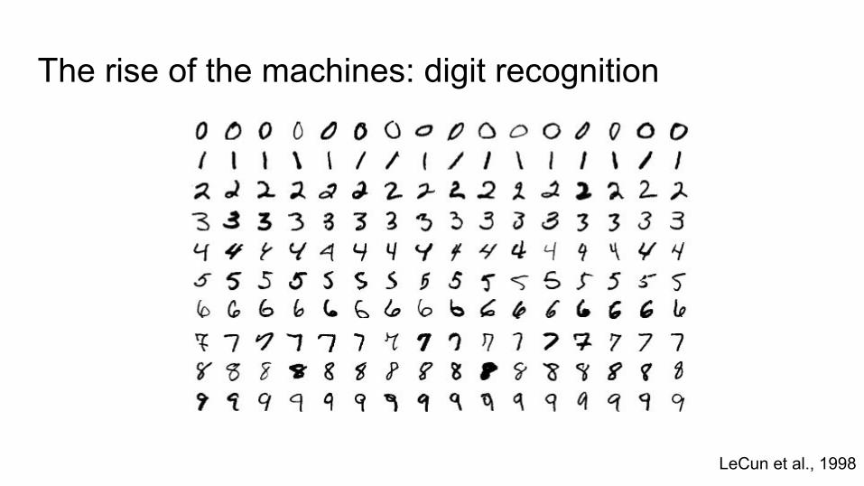
The rise of the machines: digit recognition
LeCun et al., 1998
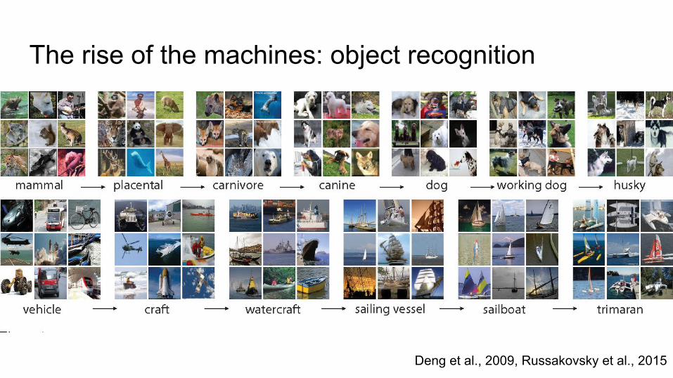
The rise of the machines: object recognition
Deng et al., 2009, Russakovsky et al., 2015

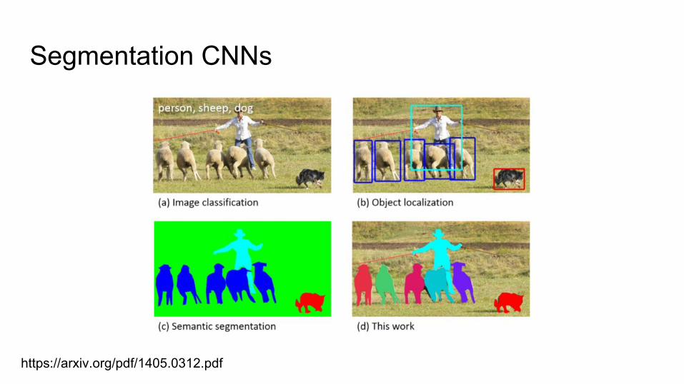
Segmentation CNNs
https://arxiv.org/pdf/1405.0312.pdf
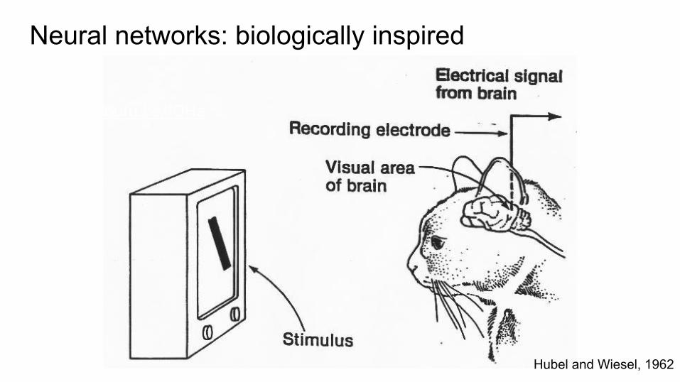
Neural networks: biologically inspired
Hubel and Wiesel, 1962
https://youtu.be/IOHayh06LJ4
Neural networks: biologically inspired
Hubel and Wiesel, 1962 - https://youtu.be/IOHayh06LJ4
https://youtu.be/IOHayh06LJ4
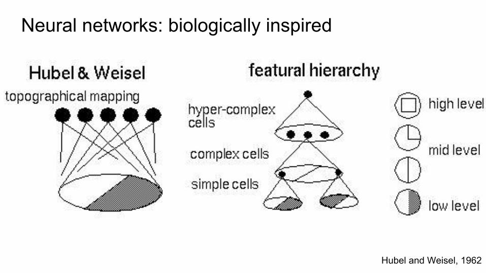
Neural networks: biologically inspired
Hubel and Weisel, 1962
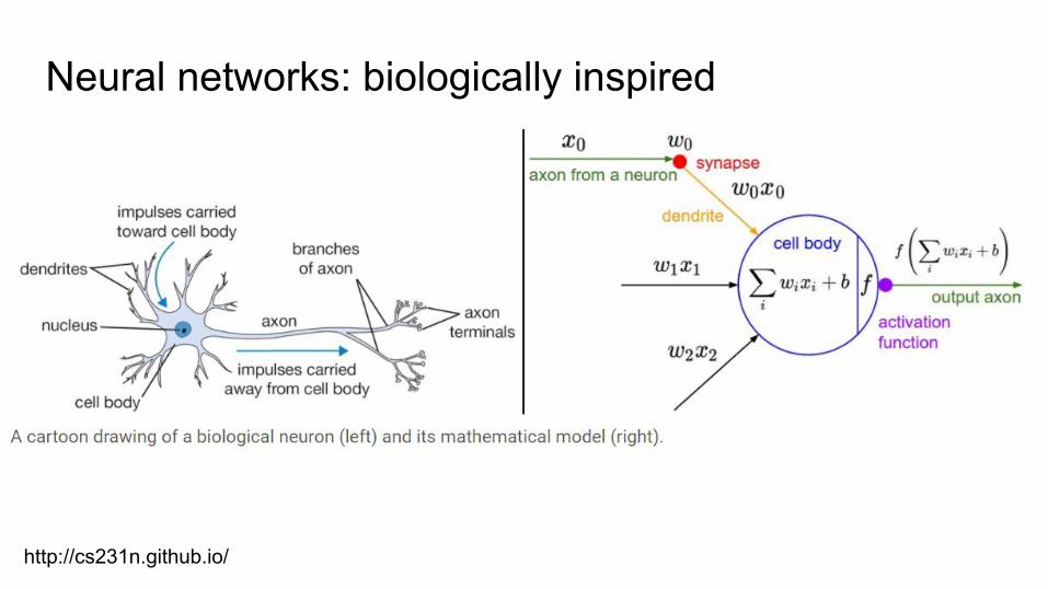
Neural networks: biologically inspired
http://cs231n.github.io/
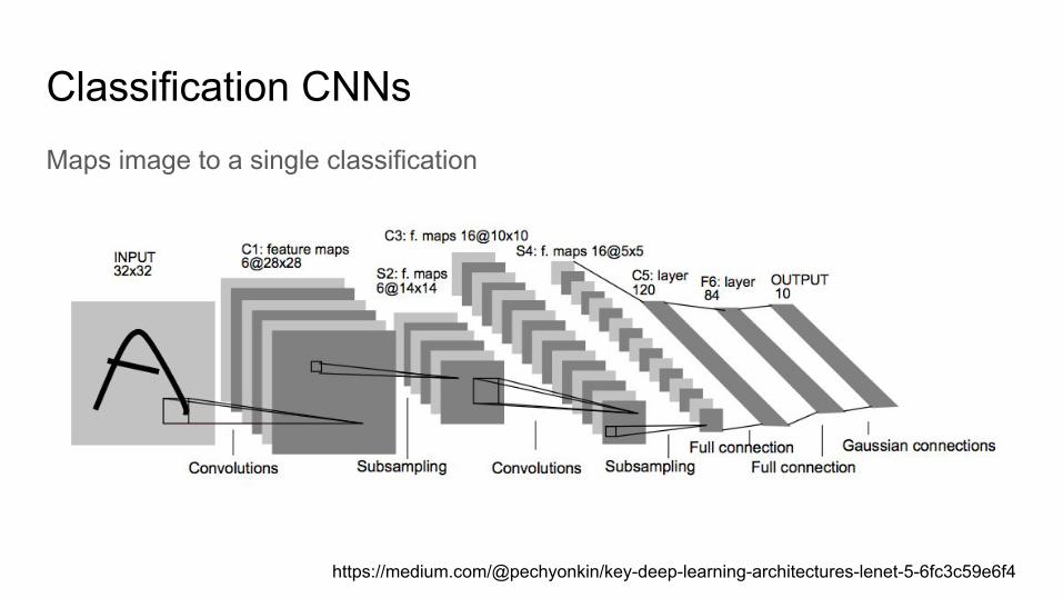
Classification CNNsMaps image to a single classification
https://medium.com/@pechyonkin/key-deep-learning-architectures-lenet-5-6fc3c59e6f4
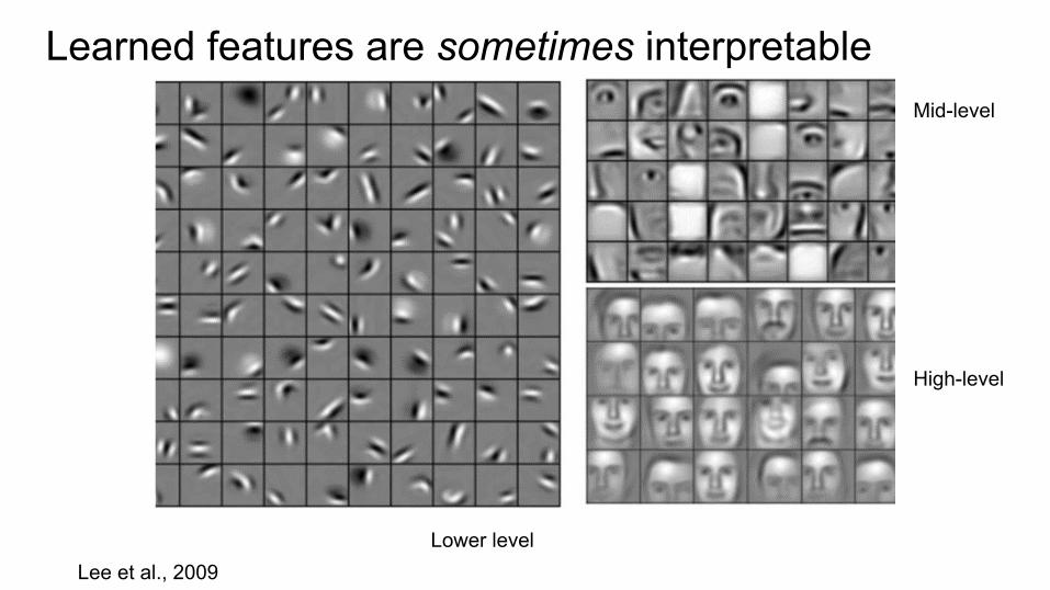
Lee et al., 2009
Learned features are sometimes interpretable
Lower level
Mid-level
High-level
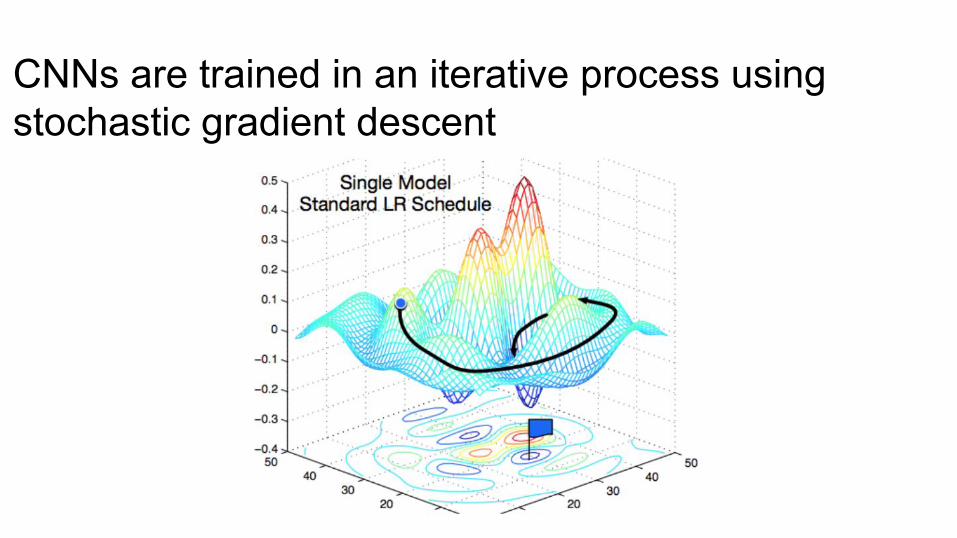
CNNs are trained in an iterative process using stochastic gradient descent
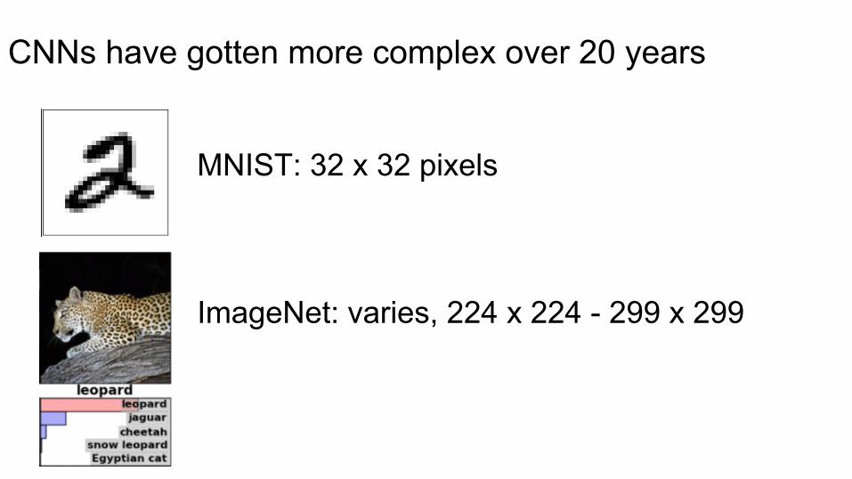
MNIST: 32 x 32 pixels
ImageNet: varies, 224 x 224 - 299 x 299
CNNs have gotten more complex over 20 years
CNNs have gotten more complex over 20 years
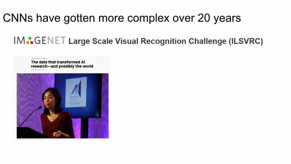
CNNs have gotten more complex over 20 years
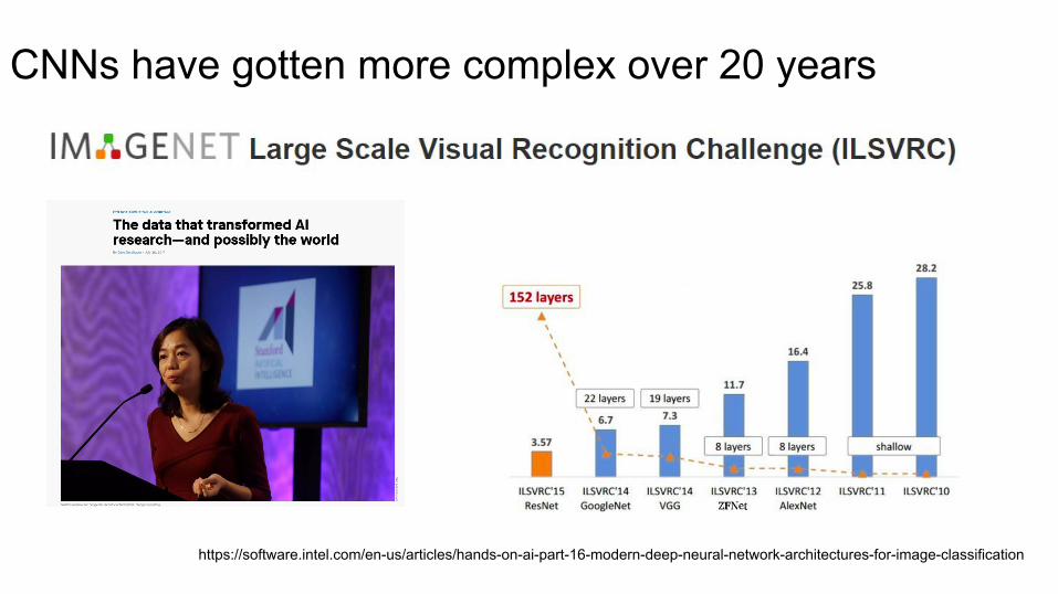
CNNs have gotten more complex over 20 years
https://software.intel.com/en-us/articles/hands-on-ai-part-16-modern-deep-neural-network-architectures-for-image-classification
CNNs are challenging to train, but...
You can start with pre-trained model and ‘fine-tune’ to your problem
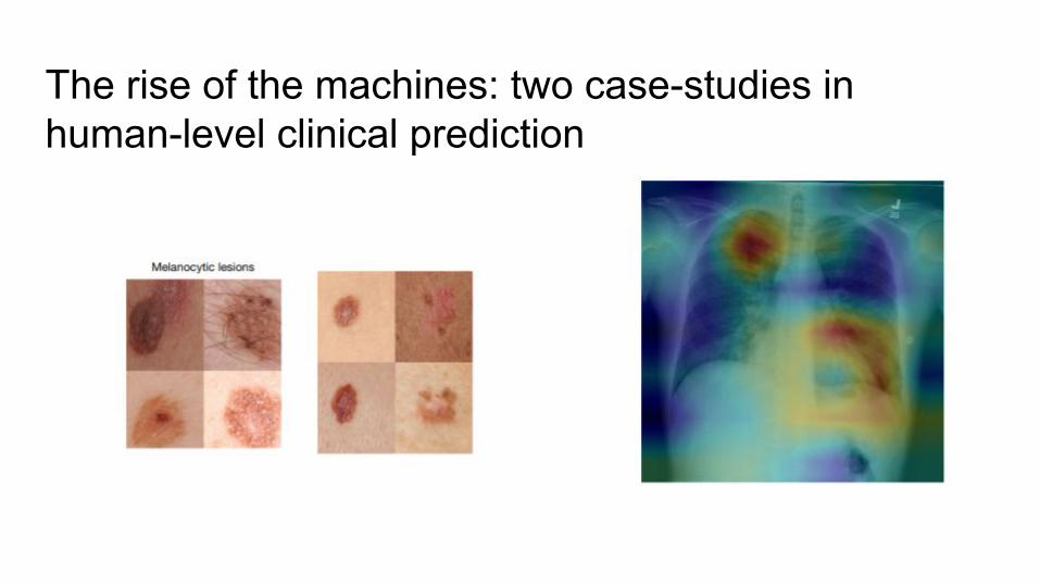
The rise of the machines: two case-studies in human-level clinical prediction
Esteva et al. (2017)
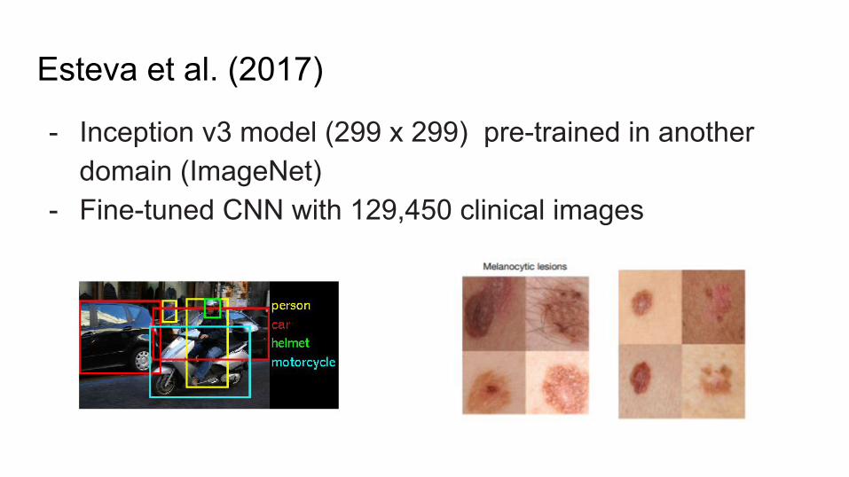
Esteva et al. (2017)
- Inception v3 model (299 x 299) pre-trained in another domain (ImageNet)
- Fine-tuned CNN with 129,450 clinical images
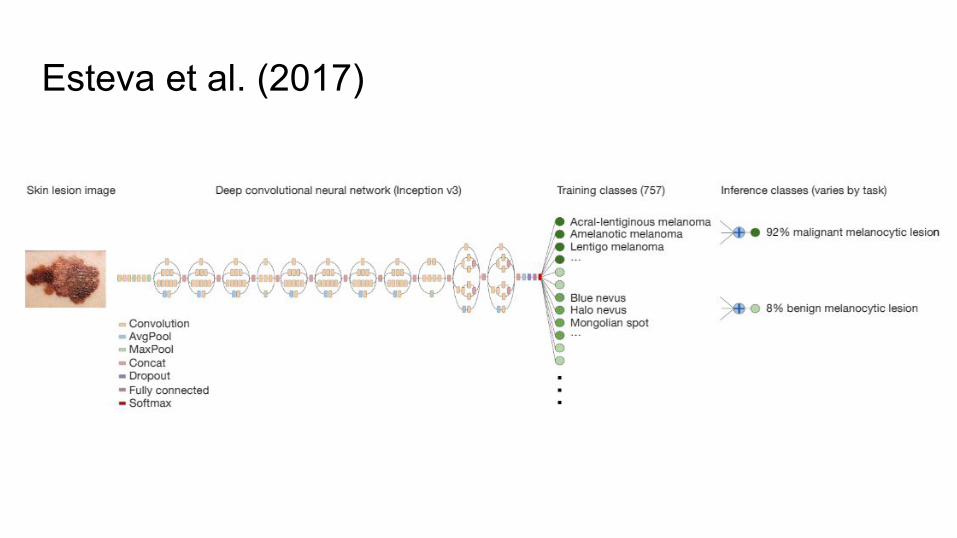
Esteva et al. (2017)
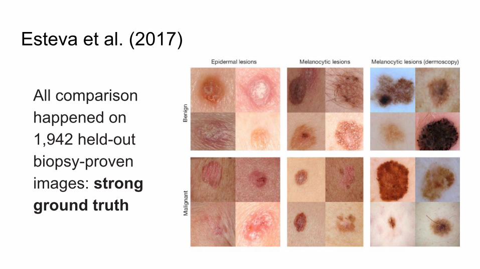
Esteva et al. (2017)
All comparison happened on 1,942 held-out biopsy-proven images: strong ground truth
Esteva et al. (2017)
Compare to 21 dermatologists on:
1. Keratinocyte carcinomas vs benign seborrheic keratoses (most common skin cancer)
2. Malignant melanomas versus benign nevi (most deadly skin cancer)
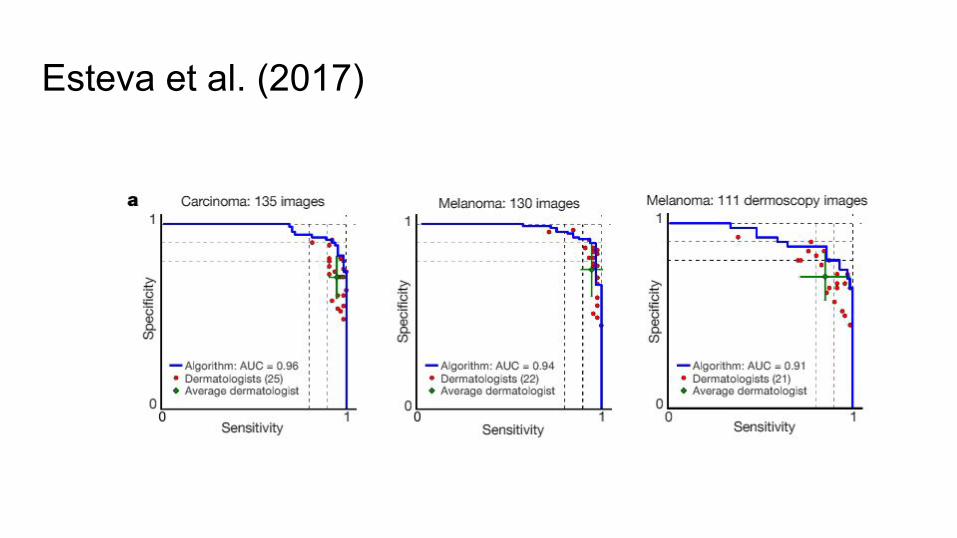
Esteva et al. (2017)
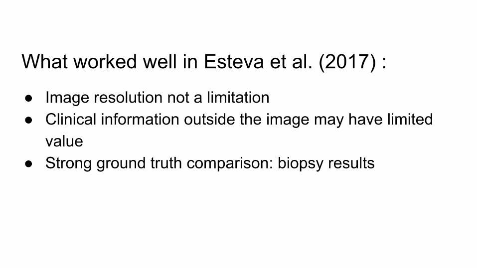
What worked well in Esteva et al. (2017) :
● Image resolution not a limitation● Clinical information outside the image may have limited
value● Strong ground truth comparison: biopsy results
Rajpurkar et al. (2017)
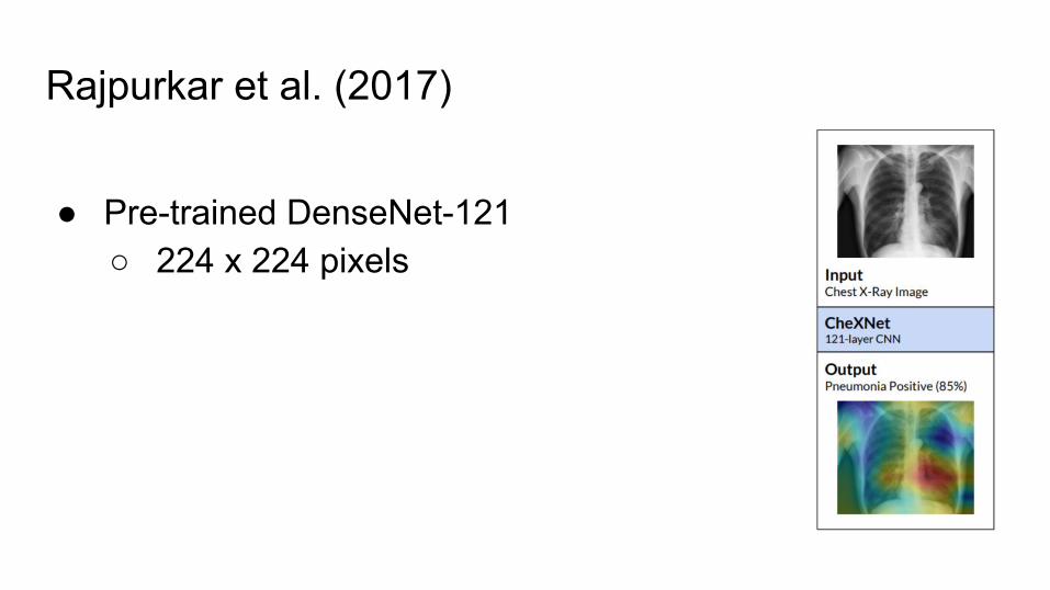
Rajpurkar et al. (2017)
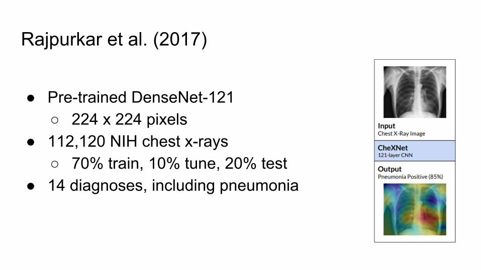
● Pre-trained DenseNet-121○ 224 x 224 pixels
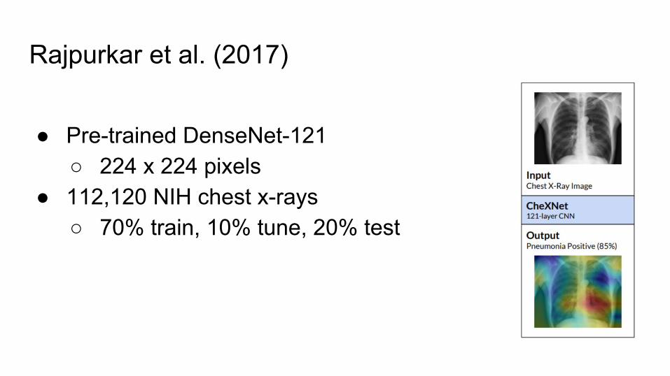
Rajpurkar et al. (2017)
● Pre-trained DenseNet-121○ 224 x 224 pixels
● 112,120 NIH chest x-rays○ 70% train, 10% tune, 20% test
Rajpurkar et al. (2017)
● Pre-trained DenseNet-121○ 224 x 224 pixels
● 112,120 NIH chest x-rays○ 70% train, 10% tune, 20% test
● 14 diagnoses, including pneumonia

Rajpurkar et al. (2017)
● 14 diagnoses, including pneumonia● AUC for pneumonia: 0.7680

Rajpurkar et al. (2017)
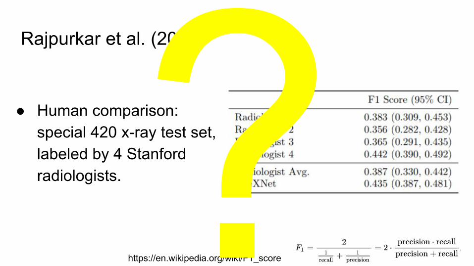
● Human comparison: special 420 x-ray test set, labeled by 4 Stanford radiologists.
https://en.wikipedia.org/wiki/F1_score
Rajpurkar et al. (2017)
● Human comparison: special 420 x-ray test set, labeled by 4 Stanford radiologists.
https://en.wikipedia.org/wiki/F1_score
Human-level performance?
● AUC of Rajpurkar et al. (2017): for pneumonia 0.7680○ By comparison, AUC of Esteva et al. (2017): 0.91-0.96
● Why did the Rajpurkar et al. (2017) compare using 4 radiologists and F1 score?○ Low radiologist agreement
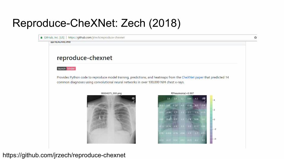
Reproduce-CheXNet: Zech (2018)
https://github.com/jrzech/reproduce-chexnet

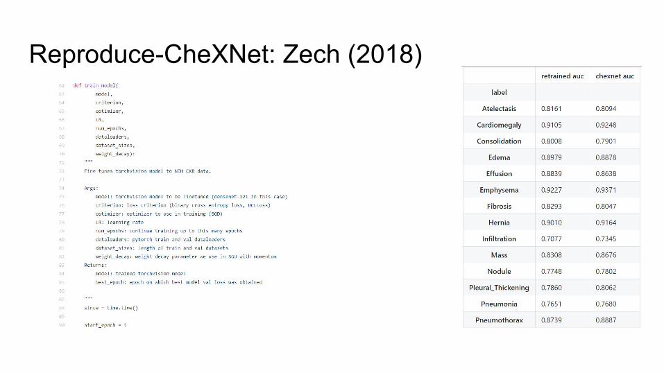
Reproduce-CheXNet: Zech (2018)
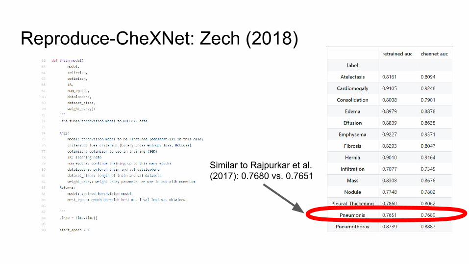
Reproduce-CheXNet: Zech (2018)
Similar to Rajpurkar et al. (2017): 0.7680 vs. 0.7651

Will CheXNet generalize?
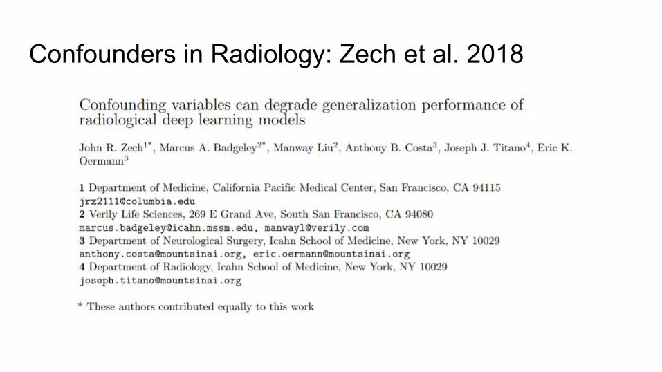
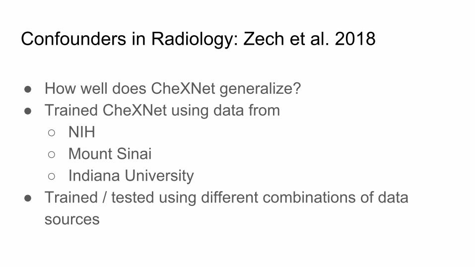
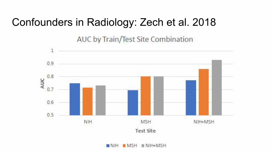
Confounders in Radiology: Zech et al. 2018
Confounders in Radiology: Zech et al. 2018
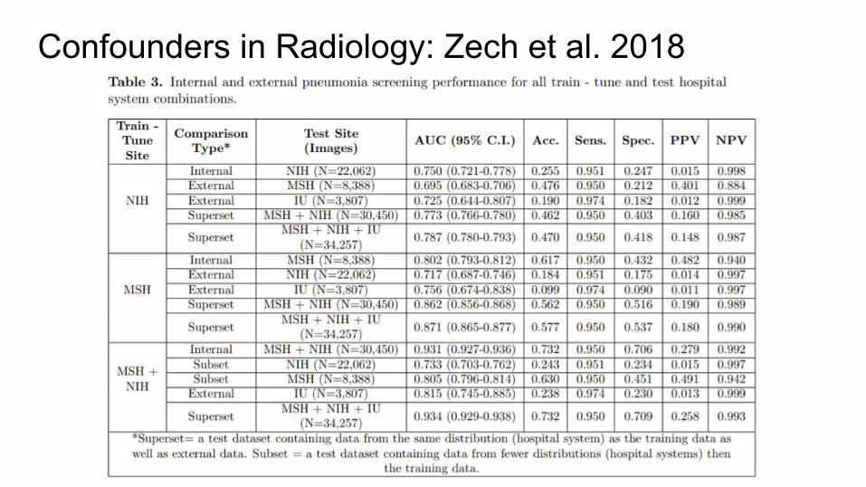
● How well does CheXNet generalize?● Trained CheXNet using data from
○ NIH○ Mount Sinai○ Indiana University
● Trained / tested using different combinations of data sources
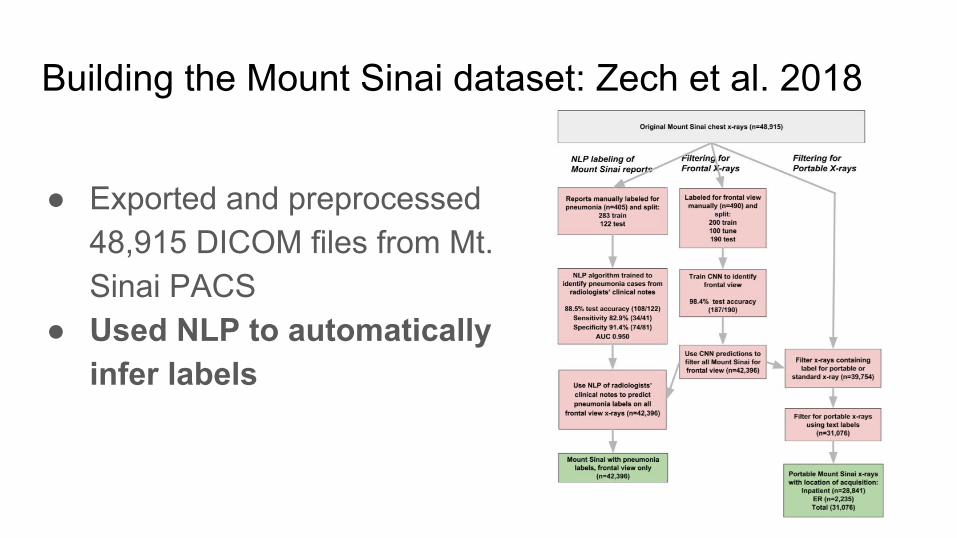
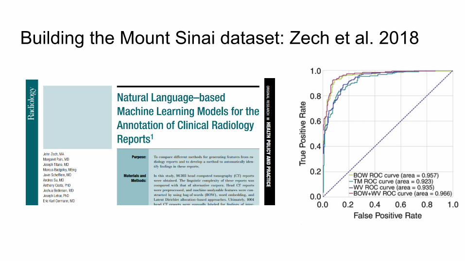
Building the Mount Sinai dataset: Zech et al. 2018
● Exported and preprocessed 48,915 DICOM files from Mt. Sinai PACS
● Used NLP to automatically infer labels
Building the Mount Sinai dataset: Zech et al. 2018
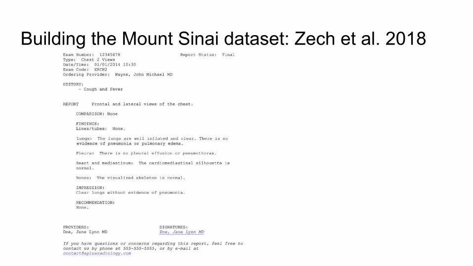
Building the Mount Sinai dataset: Zech et al. 2018
Building the Mount Sinai dataset: Zech et al. 2018
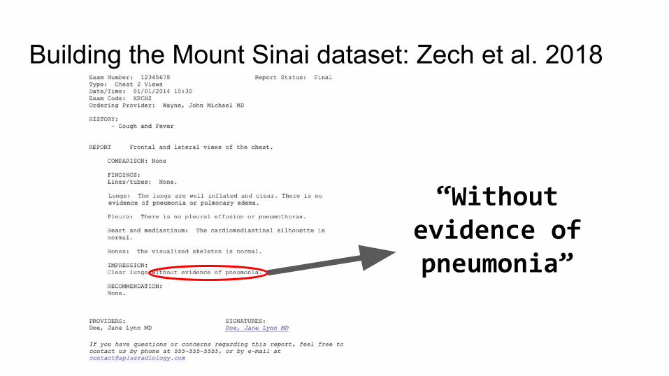
“Without evidence of pneumonia”
Building the Mount Sinai dataset: Zech et al. 2018
● These are imperfect labels ○ ~90% sensitivity, specificity

Building the Mount Sinai dataset: Zech et al. 2018
● These are imperfect labels ○ ~90% sensitivity, specificity
● What could introduce biases into these labels?
Building the Mount Sinai dataset: Zech et al. 2018
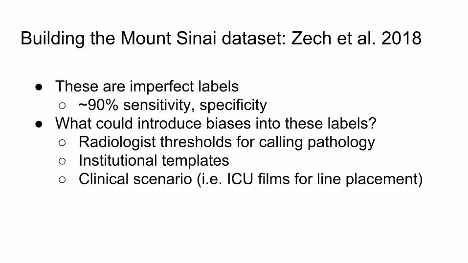
● These are imperfect labels ○ ~90% sensitivity, specificity
● What could introduce biases into these labels?○ Radiologist thresholds for calling pathology○ Institutional templates○ Clinical scenario (i.e. ICU films for line placement)
Confounders in Radiology: Zech et al. 2018
Confounders in Radiology: Zech et al. 2018
Confounders in Radiology: Zech et al. 2018
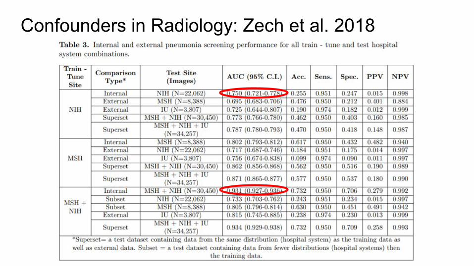
Why better performance on joint NIH+Mount Sinai dataset?
Confounders in Radiology: Zech et al. 2018
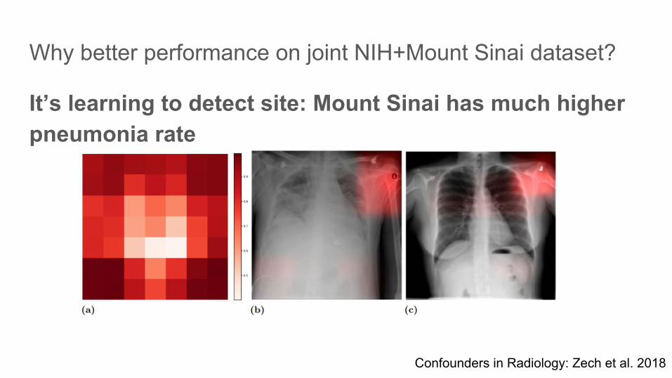
Why better performance on joint NIH+Mount Sinai dataset?
It’s learning to detect site: Mount Sinai has much higher pneumonia rate
Confounders in Radiology: Zech et al. 2018
Confounders in Radiology: Zech et al. 2018
● CNN learned something very useful in making predictions, but not clinically helpful.
Confounders in Radiology: Zech et al. 2018
● CNN learned something very useful in making predictions, but not clinically helpful.
● CNNs are hard to interpret: >6 million parameters
CNNs can an detect hospital system:
can it detect department within hospital?
CNNs can an detect hospital system:
can it detect department within hospital?
Yes.
At Mount Sinai, CNNs could detect portable x-ray scanner department (inpatient vs. ED) with near-perfect accuracy
We don’t have metadata for NIH, but...
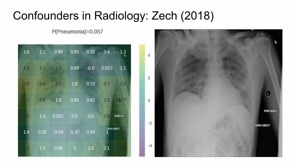
Confounders in Radiology: Zech (2018)
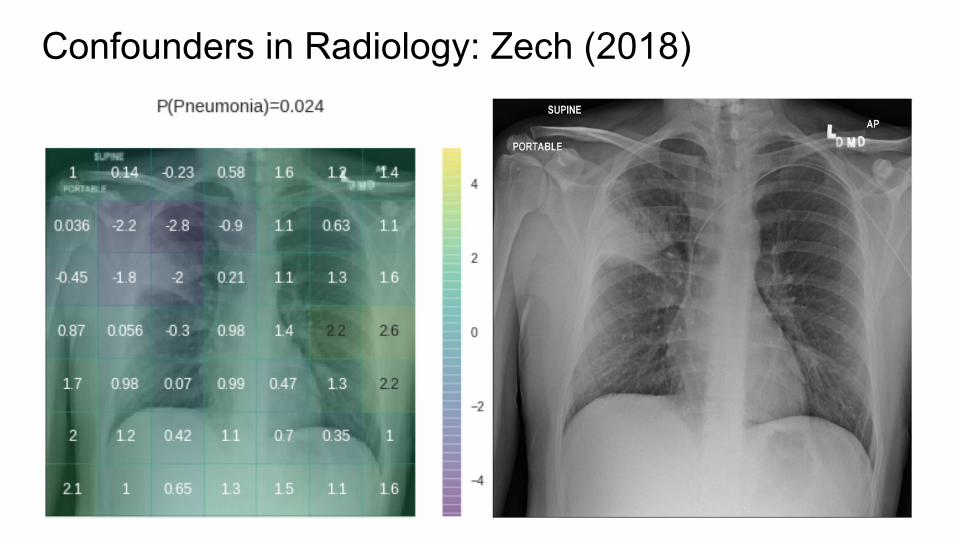
Confounders in Radiology: Zech (2018)
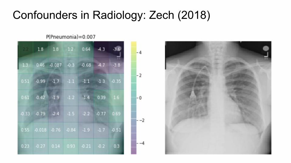
Confounders in Radiology: Zech (2018)
Confounders in Radiology: Zech et al. 2018● CNNs appear to exploit information beyond specific disease-related
imaging findings on x-rays to calibrate their disease predictions.
Confounders in Radiology: Zech et al. 2018● CNNs appear to exploit information beyond specific disease-related
imaging findings on x-rays to calibrate their disease predictions● Scanner type (especially portable vs regular PA/lateral) is easily exploited

Confounders in Radiology: Zech et al. 2018● CNNs appear to exploit information beyond specific disease-related
imaging findings on x-rays to calibrate their disease predictions● Scanner type (especially portable vs regular PA/lateral) is easily exploited● Whole-image, low-res classification is especially vulnerable
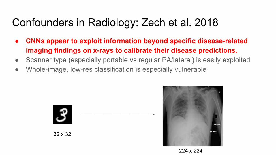
Confounders in Radiology: Zech et al. 2018● CNNs appear to exploit information beyond specific disease-related
imaging findings on x-rays to calibrate their disease predictions.● Scanner type (especially portable vs regular PA/lateral) is easily exploited.● Whole-image, low-res classification is especially vulnerable
32 x 32
224 x 224
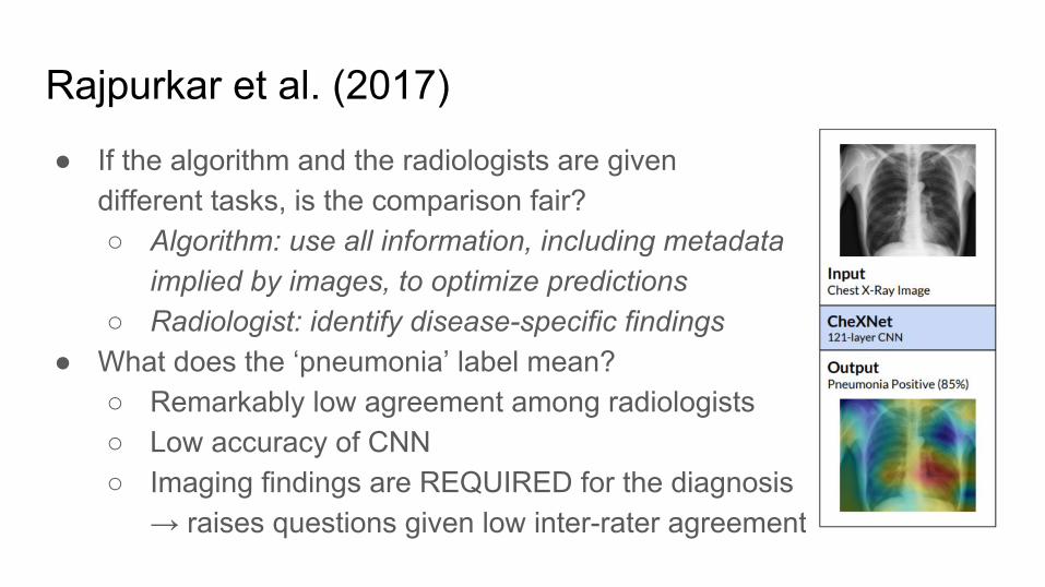
Rajpurkar et al. (2017)
● If the algorithm and the radiologists are givendifferent tasks, is the comparison fair?○ Algorithm: use all information, including metadata
implied by images, to optimize predictions○ Radiologist: identify disease-specific findings
● What does the ‘pneumonia’ label mean?○ Remarkably low agreement among radiologists ○ Low accuracy of CNN○ Imaging findings are REQUIRED for the diagnosis
→ raises questions given low inter-rater agreement
How do we move forward from weakly-supervised ImageNet-based transfer learning?
How do we move forward from weakly-supervised ImageNet-based transfer learning?
Domain adapted approaches that use segmentation
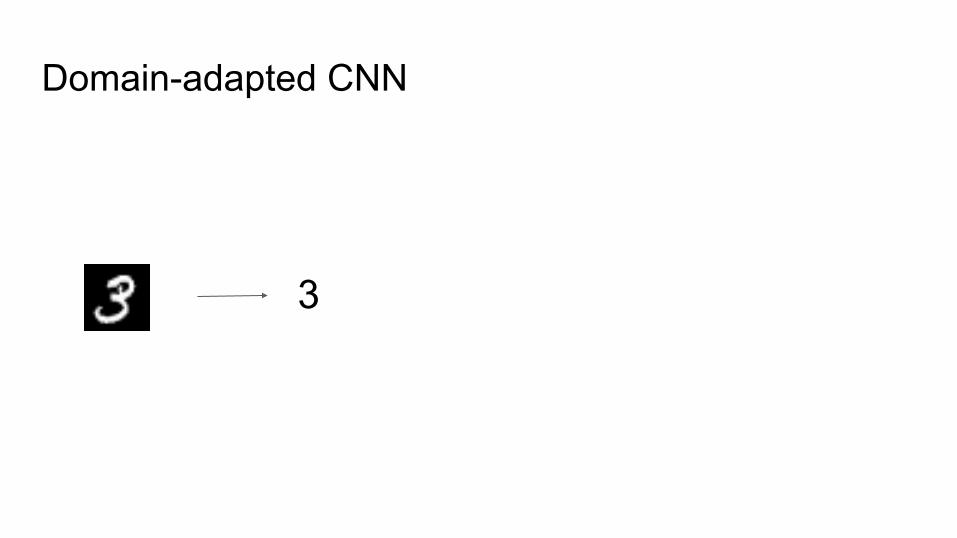
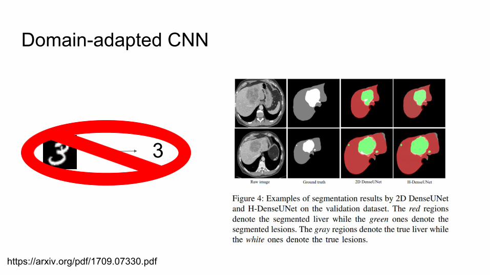
Domain-adapted CNN
3
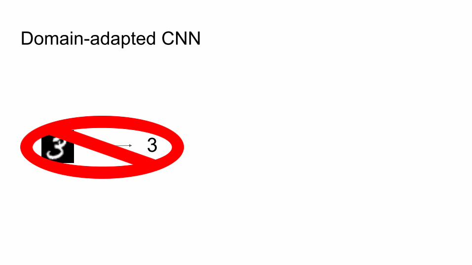
Domain-adapted CNN
3
Domain-adapted CNN
3
https://arxiv.org/pdf/1709.07330.pdf

Classification CNNsMaps image to a single classification
https://medium.com/@pechyonkin/key-deep-learning-architectures-lenet-5-6fc3c59e6f4
Segmentation CNNs
https://arxiv.org/pdf/1405.0312.pdf
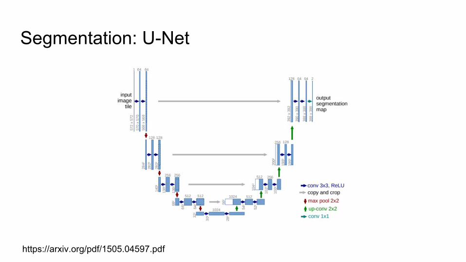
Segmentation: U-Net
https://arxiv.org/pdf/1505.04597.pdf
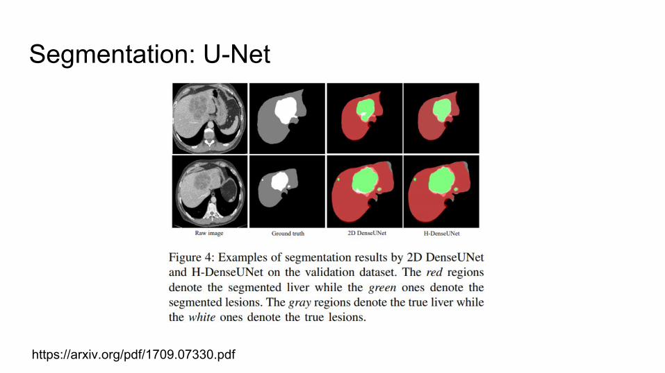
Segmentation: U-Net
https://arxiv.org/pdf/1709.07330.pdf
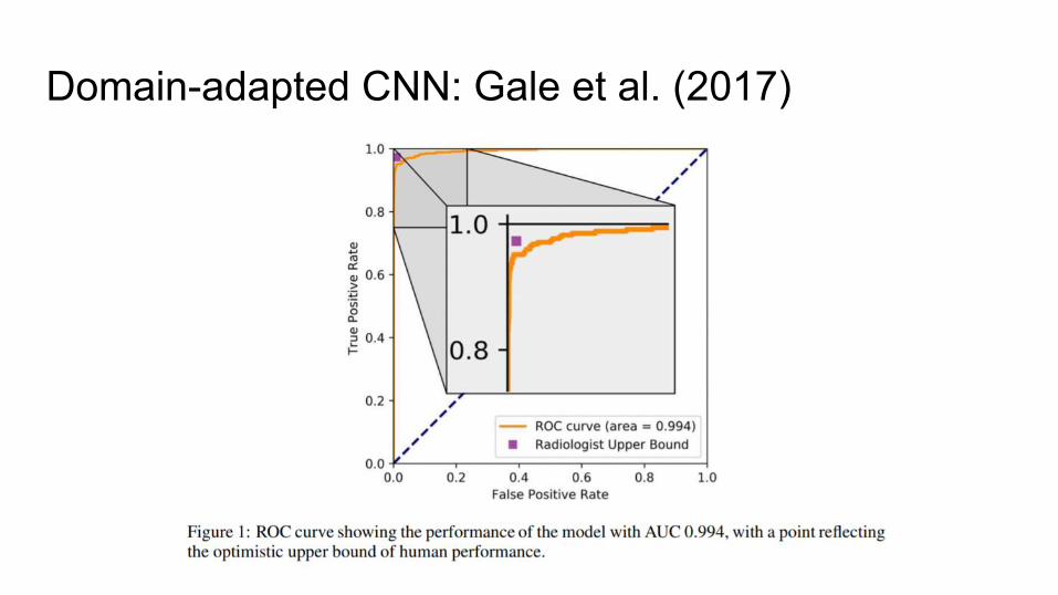
Domain-adapted CNN: Gale et al. (2017)
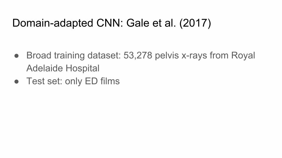
Domain-adapted CNN: Gale et al. (2017)
● Broad training dataset: 53,278 pelvis x-rays from Royal Adelaide Hospital
● Test set: only ED films
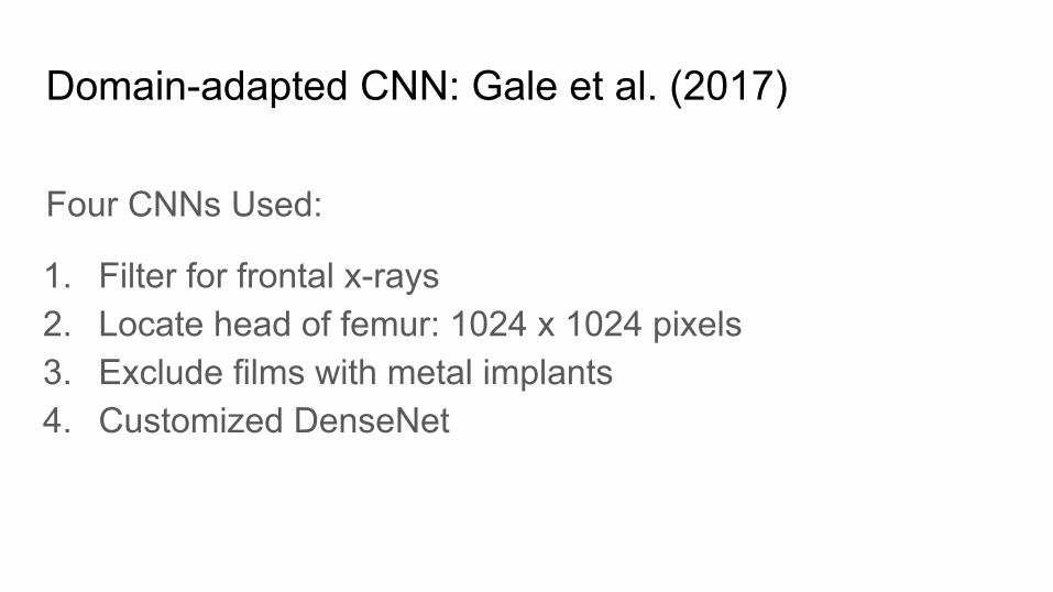
Domain-adapted CNN: Gale et al. (2017)
Four CNNs Used:
1. Filter for frontal x-rays2. Locate head of femur: 1024 x 1024 pixels3. Exclude films with metal implants4. Customized DenseNet
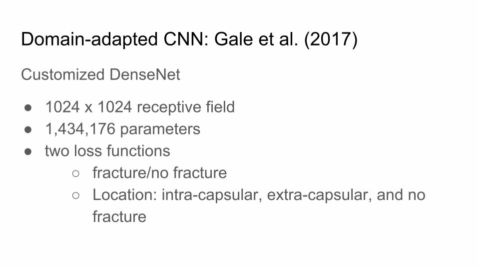
Domain-adapted CNN: Gale et al. (2017)
Customized DenseNet
● 1024 x 1024 receptive field● 1,434,176 parameters● two loss functions
○ fracture/no fracture○ Location: intra-capsular, extra-capsular, and no
fracture
Domain-adapted CNN: Gale et al. (2017)
Domain-adapted CNN: Gale et al. (2017)
● Careful data cleaning to avoid confounding variables○ Normalization○ No metal
● Chosen test set reflecting real clinical use scenario: ED● Followed a radiologist’s process
○ zooming in on femur○ maintain high resolution
Domain-adapted CNN: Chang et al. (2018)
● Identify hemorrhage on 10,159 head CTs● Used segmentation-based approach● Results in challenging ED environment in true forward out
of sample testing○ 0.989 AUC○ 97.2% accuracy○ 0.951 sensitivity ○ 0.973 specificity
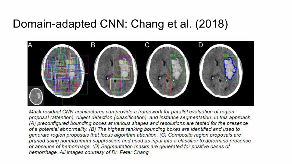
Domain-adapted CNN: Chang et al. (2018)
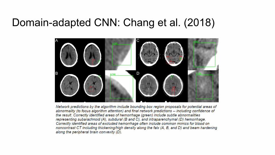
Domain-adapted CNN: Chang et al. (2018)
Stronger approach and results,but needs generalization testing on new sites
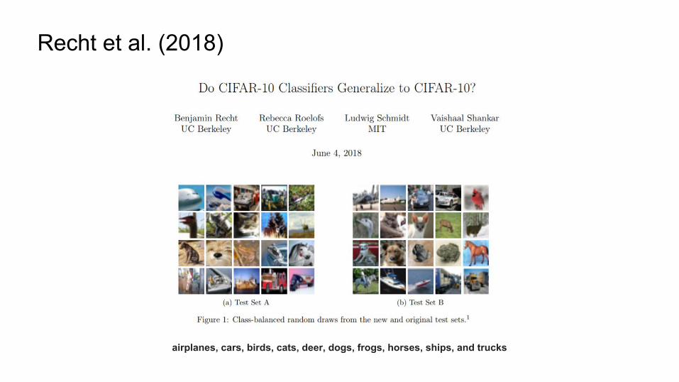
Recht et al. (2018)
airplanes, cars, birds, cats, deer, dogs, frogs, horses, ships, and trucks
“Current accuracy numbers are brittle and susceptible to even minute natural
variations in the data distribution.”
Recht et al. (2018)
How will we use deep learning in radiology?
How will we use deep learning in radiology?
● Can perform well at well-specified, clearly-designed imaging tasks: fracture, hemorrhage detection
How will we use deep learning in radiology?
● Can perform well at well-specified, clearly-designed imaging tasks: fracture, hemorrhage detection○ but must be carefully designed
How will we use deep learning in radiology?
● Can perform well at well-specified, clearly-designed imaging tasks: fracture, hemorrhage detection○ but must be carefully designed
● Could flag important information that affects interpretation, e.g., structured EHR data, text of physician notes
Are they truly ‘artificially intelligent’?
Or a (really intriguing) statistical model?
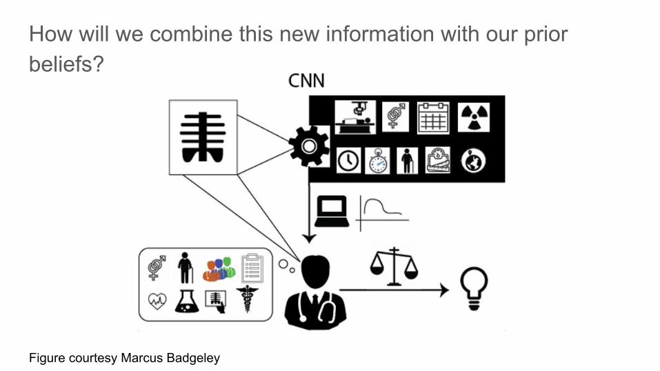
Figure courtesy Marcus Badgeley
How will we combine this new information with our prior beliefs?
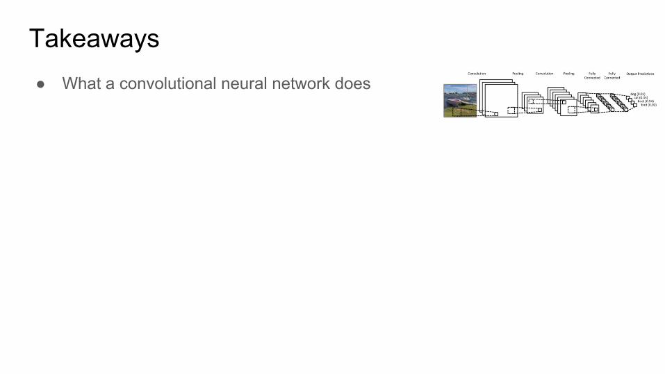
Takeaways● What a convolutional neural network does
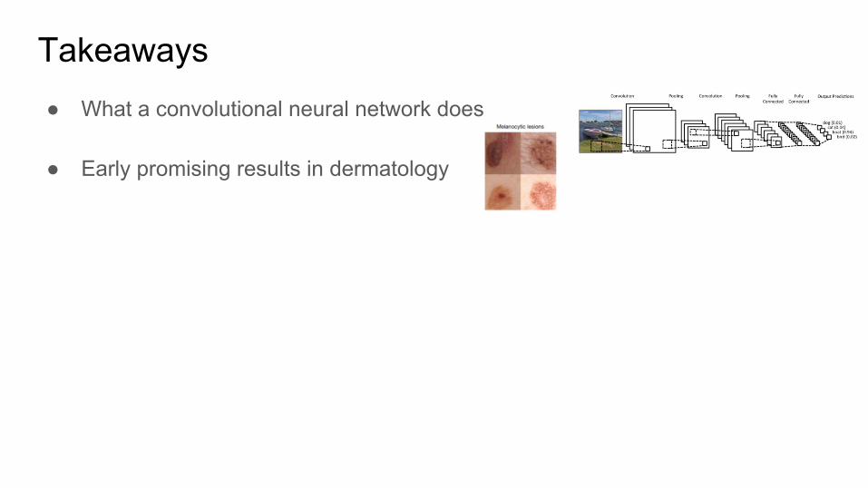
Takeaways● What a convolutional neural network does
● Early promising results in dermatology
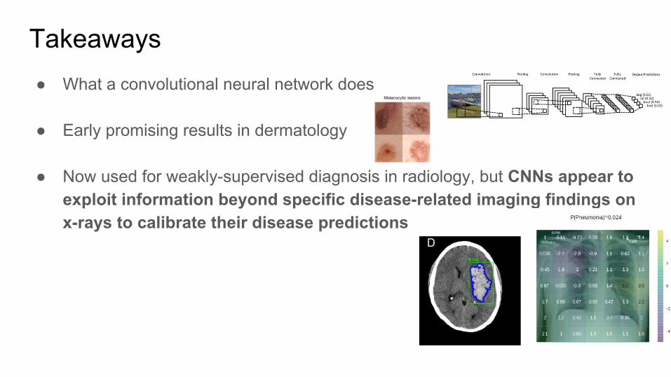
Takeaways● What a convolutional neural network does
● Early promising results in dermatology
● Now used for weakly-supervised diagnosis in radiology, but CNNs appear to exploit information beyond specific disease-related imaging findings on x-rays to calibrate their disease predictions
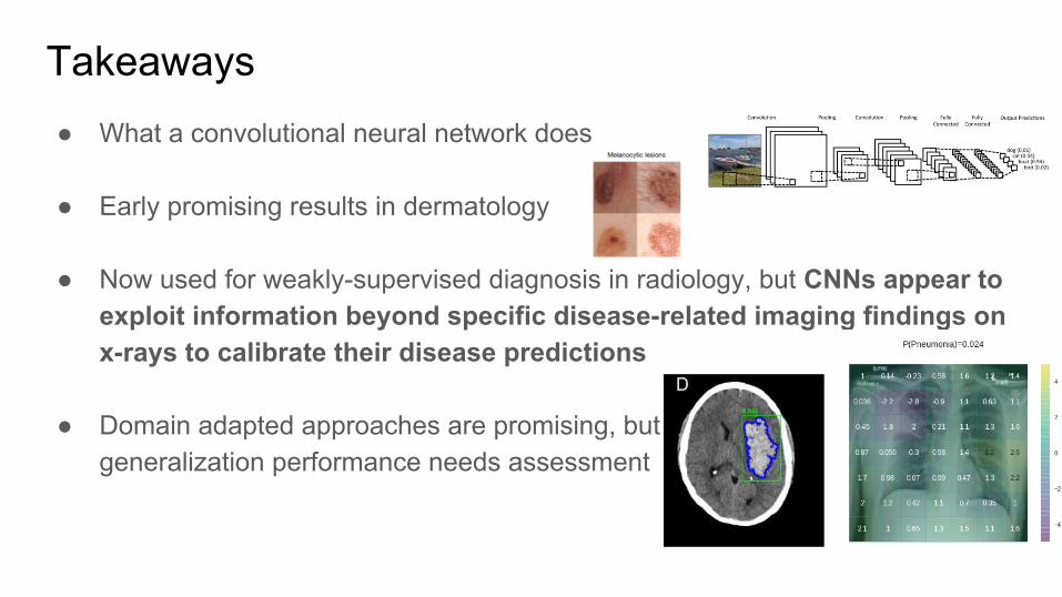
Takeaways● What a convolutional neural network does
● Early promising results in dermatology
● Now used for weakly-supervised diagnosis in radiology, but CNNs appear to exploit information beyond specific disease-related imaging findings on x-rays to calibrate their disease predictions
● Domain adapted approaches are promising, butgeneralization performance needs assessment
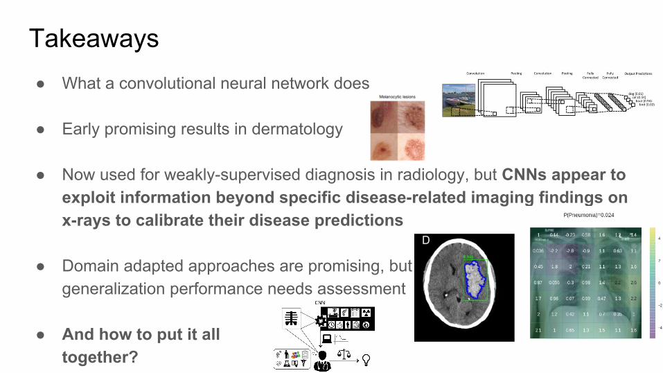
● And how to put it all together?
Thank you!
...and everyone else who contributed to these projects!