dehydration, muscle damage, and exercise in the heat
TRANSCRIPT
University of Arkansas, FayettevilleScholarWorks@UARK
Theses and Dissertations
8-2018
Dehydration, Muscle Damage, and Exercise in theHeat: Impacts on Renal Stress, Thermoregulation,and Muscular Damage RecoveryCory L. ButtsUniversity of Arkansas, Fayetteville
Follow this and additional works at: http://scholarworks.uark.edu/etd
Part of the Exercise Science Commons, and the Other Kinesiology Commons
This Dissertation is brought to you for free and open access by ScholarWorks@UARK. It has been accepted for inclusion in Theses and Dissertations byan authorized administrator of ScholarWorks@UARK. For more information, please contact [email protected], [email protected].
Recommended CitationButts, Cory L., "Dehydration, Muscle Damage, and Exercise in the Heat: Impacts on Renal Stress, Thermoregulation, and MuscularDamage Recovery" (2018). Theses and Dissertations. 2847.http://scholarworks.uark.edu/etd/2847

Dehydration, Muscle Damage, and Exercise in the Heat:
Impacts on Renal Stress, Thermoregulation, and Muscular Damage Recovery
A dissertation submitted in partial fulfillment
of the requirements for the degree of
Doctor of Philosophy in Kinesiology
by
Cory L. Butts
Colorado State University – Pueblo
Bachelor of Science in Exercise Science, 2011
University of Texas at Arlington
Master of Science in Exercise Physiology, 2013
August 2018
University of Arkansas
This dissertation is approved for recommendation to the Graduate Council.
____________________________________ Brendon P. McDermott, Ph.D.
Dissertation Director
____________________________________ ____________________________________
Matthew S. Ganio, Ph.D. Stavros A. Kavouras, Ph.D.
Committee Member Committee Member
____________________________________ ____________________________________
Nicholas P. Greene, Ph.D. Ronna C. Turner, Ph.D.
Committee Member Committee Member
Abstract
Purpose: The purpose was to identify the combined influence of dehydration, muscle damage,
and exertional hyperthermia on biological markers of acute kidney injury and renal function. We
also investigated the effects of performing muscle damaging exercise during mild hypohydration
on muscle damage biomarkers and muscular strength recovery. Methods: Eighteen
recreationally-active males (age 24 ± 5 y, body fat 17.3 ± 6.2%) completed a familiarization visit
and two experimental trials separated by ≥28 days. The two experimental conditions consisted of
either euhydration (EU; maintaining hydration, -1.2 ± 0.8%) or hypohydration (HY; restricting
fluid consumption for 24 hours prior to and during the trial, -4.4 ± 1.9%). Participants completed
a unilateral eccentric knee flexion muscle damaging protocol, 60-minute treadmill exercise in the
heat, 30-minute passive recovery, and a rehydrated 24-h follow-up visit, respectively. Results:
Strength was reduced across time independent of trial for isometric strength at 70° (P<0.001),
isometric strength at 90° (P=0.001), and isokinetic strength at 60°·sec-1 (P=0.001). Serum
creatine kinase increased regardless of trial (P<0.001), with the 24-h follow-up greater (grand
mean; 58.7 ± 25.1 U/L) than at baseline (grand mean; 35.7 ± 23.1 U/L, P<0.001) and post-
exercise (grand mean; 51.6 ± 23.2 U/L, P=0.009). Percent change in plasma neutrophil
gelatinous associated lipocalin was greater in the HY trial post-exercise (EU 28.0 ± 15.2%, HY
41.8 ± 17.5%, P<0.001), but not at 24-h follow-up (P=0.39). Serum creatinine was increased in
the HY trial regardless of time (EU 0.97 ± 0.14, HY 1.04 ± 0.15, mg/dL, P=0.025). Urine
NGAL and urine creatinine were also elevated in the HY trial pre-exercise and post-exercise (all,
P<0.05) but were returned to EU levels by 24-h follow-up (all, P>0.05). Conclusions: We
demonstrated no significant impact of hydration status when performing muscle damaging
exercise, followed by exercise in the heat, on indices of muscle damage recovery. Exercise in

the heat with muscle damage increased physiological and renal strain when HY, but the
rehydration protocol ameliorated differences between trials by the 24-h follow-up. These
findings highlight the importance of proper fluid intake following exercise to mitigate renal
stress.

©2018 by Cory L. Butts
All Rights Reserved
Acknowledgements
I would like to first acknowledge my loving wife, Amanda. For all the late nights, early
mornings, and long weekends, your amazing support, devotion, and confidence provided an
unparalleled motivation that allowed me to succeed.
To my tremendous advisor, Dr. Brendon McDermott. Your willingness to discuss ideas
and deep passion for science are contagious, and made it easy to stay motivated while working
with you. I have greatly valued your leadership and I am fortunate to have had you as my
advisor.
To my committee, thank you for your guidance and wisdom along this journey. Dr.
Ganio, Dr. Kavouras, and Dr. Greene, your expertise has aided in my development as a scientist,
educator, and professional. Dr. Turner, your ability to explain statistical principles made every
course and conversation enjoyable, interesting, and provided a lasting effect on my interest in
statistics.
I would like to thank everyone in the Exercise Science Research Center for everything
they have done for me. Thank you all for the conversations and opportunity to work with such a
motivated and dedicated group of scientists.
Lastly, to my family and friends, thank you for your endless love and support. Thank
you to my brother and sisters for supporting my goals. To my parents, much of my educational
success is because of you. I would like to thank my mother, for staying home to help with
prepare me for school and always challenge me to be better. To my father, thank for you for
showing what it means to work hard. Watching you work long days, while still being involved
in our lives showed me what it meant to stay focused, driven, and persistent.
Table of Contents
I. Introduction ................................................................................................................................ 1
Specific Aims .............................................................................................................................. 5
II. Literature Review ....................................................................................................................... 7
Renal Function & Biomarkers..................................................................................................... 7
Renal Function During Exercise ............................................................................................... 18
Renal Function with Passive and Active Heat Stress ................................................................ 28
Influence of Hydration on Renal Function ................................................................................ 31
Modifiers of Exercise Induced Muscle Damage ....................................................................... 35
Renal Function with Exercise-Induced Muscle Damage .......................................................... 37
Beneficial Effects of Exercise on Renal Function .................................................................... 41
Cardiovascular and Thermoregulatory Responses to Exercise in the Heat .............................. 42
Cardiovascular and Thermoregulatory Responses Following Exercise in the Heat ................. 44
Summary ................................................................................................................................... 46
III. Methods................................................................................................................................... 48
IV. Manuscript #1: Combined Effects of Hypohydration, Muscle Damage, and Exertional
Hyperthermia on Biomarkers of Acute Kidney Injury ................................................................. 56
Abstract ..................................................................................................................................... 57
Introduction ............................................................................................................................... 58
Methods ..................................................................................................................................... 60
Results ....................................................................................................................................... 67
Discussion ................................................................................................................................. 74
References ................................................................................................................................. 80
Figure Legends .......................................................................................................................... 85

V. Manuscript #2: Influences of Hypohydration During Exercise-Induced Muscle Damage on
Recovery ....................................................................................................................................... 91
Abstract ..................................................................................................................................... 92
Introduction ............................................................................................................................... 93
Methods ..................................................................................................................................... 95
Results ..................................................................................................................................... 100
Discussion ............................................................................................................................... 102
References ............................................................................................................................... 106
Figure Legends ........................................................................................................................ 110
IV. Conclusions........................................................................................................................... 114
References ............................................................................................................................... 116
Appendix ................................................................................................................................. 135
List of Figures
Study 1
Figure 1 ………………………………………………………………………………….87
Figure 2……………………………………………………………………………….….88
Figure 3…………………………………………………………………………………..89
Figure 4…………………………………………………………………………………..90
Study 2
Figure 1 ………………………………………………………………………………...111
Figure 2…………………………………………………………………………………112
Figure 3…………………………………………………………………………………113

1
I. Introduction
Exposure to heat stress yields a spectrum of responses ranging from positive
cardiovascular adaptations (e.g. plasma volume expansion, improved vascular function) to the
potential life-threatening risk of heat illness (e.g. exertional heat stroke) (Casa et al., 2015a;
Epstein & Roberts, 2011; Laukkanen, Khan, Zaccardi, & Laukkanen, 2015; Lorenzo, Halliwill,
Sawka, & Minson, 2010; Lorenzo & Minson, 2010; Nadel, Pandolf, Roberts, & Stolwijk, 1974;
Patterson, Stocks, & Taylor, 2004; Xiang, Hansen, Pisaniello, & Bi, 2015). When exposed to
heat stress, increases in skin blood flow and sweating occur to maintain thermoregulatory
homeostasis, leading to reductions in splanchnic and renal blood flow (Hohimer, Hales, Rowell,
& Smith, 1983; Rowell, 1974; Sawka, Leon, Montain, & Sonna, 2011). The reductions in blood
flow to vital organs at rest and during exercise in the heat are often transient attenuations in
perfusion, resulting in minor functional alterations as evidenced by a return to baseline function
shortly after exposure (e.g. within hours) (Junglee et al., 2013; Melin et al., 1997; Radigan &
Robinson, 1949). However, heat stress is often experienced in combination with other
physiological challenges, such as dehydration and muscle damage. Hypohydration, muscle
damage, and environmental heat stress are commonly experienced in athletic, military, and
occupational settings (Armstrong et al., 2010; Godek, Bartolozzi, Burkholder, Sugarman, &
Dorshimer, 2006; Johnson et al., 2016; Knochel, Dotin, & Hamburger, 1974; Meade, Lauzon,
Poirier, Flouris, & Kenny, 2015; Poirier et al., 2015; Schlader et al., 2017; Schrier et al., 1970;
Smoot, Cavanaugh, Amendola, West, & Herwaldt, 2014; Yeargin et al., 2010). The combined
effects of these stressors may compromise renal function and increase acute kidney injury risk,
however, long term consequences remain unknown (Johnson et al., 2016; Junglee et al., 2013;
Schrier et al., 1970; Smith, Robinson, & Pearcy, 1952).
2
Adequate perfusion of the renal vasculature is vital for maintaining optimal kidney
function (e.g. fluid homeostasis and filtration of waste products from the blood). Reductions in
renal blood flow may occur in response to a variety of physiological and thermoregulatory
challenges, such as exercise (Tidgren, Hjemdahl, Theodorsson, & Nussberger, 1991), heat stress
(Minson, Wladkowski, Cardell, Pawelczyk, & Kenney, 1998), or hypohydration (Melin et al.,
1997). Schlader et al. (2017) recently found greater increases in biomarkers of renal stress with
longer duration treadmill exercise in the heat. Preliminary field work from our laboratory
(unpublished) has investigated the renal responses to a 100 mile or 100 km cycling event in the
heat (22 – 34°C) in recreational riders (age 52 ± 9 y) completing the race in ~5.7 hours. We
demonstrated significant elevations in the acute kidney injury biomarker neutrophil gelatinase
associated lipocalin (NGAL), as well as increases in the renal function biomarker, serum
creatinine, immediately following the ride. Bongers et al. (2017) also showed elevations in
urinary markers of renal stress after one and three days of long distance walking, however, these
increases were relatively low and likely not indicative of serious complications. The exercise in
this study was walking, therefore the intensity (average heart rate = 112 bpm) may not have been
severe enough to induce reductions in renal perfusion and subsequent kidney stress. Studies of
marathon (McCullough et al., 2011) and ultramarathon runners (Hoffman & Weiss, 2016; Lippi
et al., 2012; Skenderi, Kavouras, Anastasiou, Yiannakouris, & Matalas, 2006) have also revealed
significant elevations in biomarkers of muscle damage and acute kidney injury immediately post-
race. Interestingly, McCullough et al. (2011) demonstrated that 24 hours post-race, NGAL and
creatinine had returned to near-baseline levels. Hoffman and Weiss (2016) reported similar renal
stress responses during a subsequent ultramarathon, importantly noting that these individuals did
not appear to experience lasting effects from the first ultramarathon. Thus, stress induced during

3
the marathon may only transiently alter renal function. These findings suggest mild acute kidney
injury and reduced renal function may also be related to the duration of the exposure to
exertional hyperthermia.
Melin et al. (1997) demonstrated significant reductions in creatinine clearance (a marker
of renal function) with dehydration compared to a euhydrated control during treadmill walking in
the heat. Reductions in plasma volume with hypohydration lead to blood volume attenuations,
which can increase cardiovascular strain and compromise thermoregulation (González-Alonso,
Mora-Rodríguez, & Coyle, 2000). Hypohydration with concomitant exercise in the heat
challenges thermoregulation and performance due to the competition for blood flow between
active skeletal muscle and the skin for heat dissipation (Casa et al., 2010; González-Alonso,
Calbet, & Nielsen, 1998; González-Alonso et al., 1999). Thus, to compensate for increased
demand for blood flow in the cutaneous vasculature, blood flow is further reduced to inactive
tissues (i.e. splanchnic and renal vasculature), potentially compromising function in these areas.
Heat stress, hypohydration, and muscle damage are factors commonly found in preseason
athletic practices, such as in American football (Yeargin et al., 2010). Smoot et al. (Smoot et al.,
2014) demonstrated elevated serum creatine kinase, a marker of muscle damage, throughout
preseason football practices in NCAA Division I football players. These findings have been
further confirmed in several observational studies of offseason, pre-season, and in-season play as
well (Ehlers, Ball, & Liston, 2002; J. R. Hoffman, Kang, Ratamess, & Faigenbaum, 2005;
Kraemer et al., 2013; Kraemer et al., 2009; Smoot et al., 2013). Severe skeletal muscle damage
(i.e. exertional rhabdomyolysis) may lead to acute kidney injury due to the nephrotoxic effects of
intracellular contents (i.e. myoglobin) entering the circulation from skeletal muscle cell
breakdown. However, in settings of optimal hydration and thermoneutral environmental

4
temperatures, muscle damage does not appear to alter renal function. Therefore, the implications
for sustained mild muscle damage throughout preseason practices are not yet known.
Athletes also often report to activities in a water conserving state (i.e. sub-optimally
hydrated) as evidenced by urinary markers (Godek, Godek, & Bartolozzi, 2005; Phillips, Sykes,
& Gibson, 2014; Yeargin et al., 2010), potentially increasing physiological strain and decreasing
performance during exercise (Bardis, Kavouras, Arnaoutis, Panagiotakos, & Sidossis, 2013;
Casa et al., 2010). Furthermore, football players were found to progressively dehydrate
throughout preseason practices (Godek et al., 2006; Godek et al., 2005; Stover, Zachwieja,
Stofan, Murray, & Horswill, 2006; Yeargin et al., 2010). Concomitant muscle damage and
dehydration may only be exacerbated by the high ambient temperatures often experienced during
preseason American football practices. Junglee et al. (2013) revealed elevations in biomarkers
of acute kidney injury with muscle damage during exercise in the heat. Another study (Fortes et
al., 2013), also demonstrated muscle damage to elicit elevations in thermal strain during
subsequent exercise in the heat. However, both studies maintained hydration state to a similar
degree in all trials, thus the impact of hypohydration compared to euhydration is unknown.
Furthermore, the renal responses among many other sporting activities (e.g. American football,
soccer, rugby) have received little investigation. Thus, sports that require individuals to exercise
regularly (i.e. several times per week) in high ambient temperatures and humidity, when muscle
damage and dehydration are present, may provide a unique stress to renal function, of which the
consequences remain unknown. Elucidating the role of adequate hydration may be pivotal to
improving the overall safety of athletics, especially since factors such as muscle damage (often
induced by strength training) and heat stress are not easily avoidable and inherent in typical
conditioning regimens.
5
In working populations, it has been suggested that the regular exposure to heat stress with
concomitant dehydration and mild muscle damage may increase the risk of chronic kidney
disease (García-Trabanino et al., 2015; Johnson et al., 2016; Moyce, Joseph, Tancredi, Mitchell,
& Schenker, 2016; Roncal-Jimenez et al., 2016). The recent rise in chronic kidney disease in
Mesoamerican sugar cane workers has been related to physiological responses to environmental
working conditions (Bodin et al., 2016; Crowe, Nilsson, Kjellstrom, & Wesseling, 2015; García-
Trabanino et al., 2015; Laws et al., 2015, 2016; Roncal-Jimenez et al., 2016). As glomerular
filtration rate, a marker of kidney function, has been shown to decrease throughout the work day,
the increased prevalence of chronic kidney disease may be due to the additive effects of
repetitive kidney stress from the concomitant environmental and physiological strain experienced
by these workers (Bodin et al., 2016; Crowe et al., 2015; García-Trabanino et al., 2015; Laws et
al., 2015, 2016; Roncal-Jimenez et al., 2016). Furthermore, these workers also experience
progressive dehydration throughout the workday through elevated urine specific gravity and
decreased glomerular filtration rates (García-Trabanino et al., 2015; Roncal-Jimenez et al., 2016;
Wesseling et al., 2016). The combination of heat stress with strenuous exercise and gradual
dehydration throughout the workday places a high demand on the kidneys to retain fluid while
clearing excess waste from potential muscle damage. These characteristics may apply to many
other occupations as well, such as firefighters, military, agricultural and industrial settings.
Specific Aims
Aim #1: Identify the combined influence of dehydration, exercise in the heat, and muscle
damage on biomarkers of acute kidney injury and renal function.
Research Hypothesis #1: The thermoregulatory strain associated with dehydration during
exercise in the heat would augment renal biomarker elevations immediately post-exercise as

6
compared to the euhydrated trial, however, these differences would be transient with returns to
baseline 24 hours post.
Aim #2: Identify the effects of performing muscle damaging exercise during mild hypohydration
on muscle damage biomarkers (creatine kinase) and muscular strength recovery indices (i.e.
isometric and isokinetic strength).
Research Hypothesis #2: There would be no differences in muscle damage biomarkers with
hypohydration, however, muscle strength recovery would be modestly impaired as compared to a
euhydrated state.

7
II. Literature Review
Renal Function & Biomarkers
The homeostatic role of the renal system in maintaining fluid balance, waste elimination,
acid-base balance, and blood pressure, is vital to the preservation of normal health and function
(Poortmans & Vanderstraeten, 1994). As such, physiological and environmental challenges may
provide stress to the kidneys to maintain optimal function. Reports of renal compromise in
athletics primarily focus on renal trauma, unless the individual has experienced exertional
rhabdomyolysis or heat illness (Bosch, Poch, & Grau, 2009; Brophy et al., 2008; Gerstenbluth,
Spirnak, & Elder, 2002; Grinsell, Butz, Gurka, Gurka, & Norwood, 2012). It was found that of
the 52 kidney injuries reported in the National Football League from 1984 to 2004, only two
were caused by dysfunction, with trauma (contusion or laceration) being most common (Brophy
et al., 2008). In contrast, 30-80% of ultra-marathon runners are suspected to develop transient
acute kidney injury (AKI) following a race (M. D. Hoffman & Weiss, 2016). Renal function and
acute renal failure have received much investigation in clinical populations, however, the renal
responses during and following exercise with environmental stress and muscle damage has
received considerably less attention.
The role of the kidneys in fluid balance is necessary for maintaining optimal hydration,
both during resting and exercise conditions. Losses in total body water (i.e. dehydration) can be
detrimental to physiological and psychological performance (Bardis et al., 2013; Casa et al.,
2015a; Casa et al., 2010; Cheuvront & Kenefick, 2014; Cheuvront, Kenefick, Montain, &
Sawka, 2010; Distefano et al., 2013; Judelson et al., 2007; Lopez et al., 2011; McDermott, Casa,
Lee, Yamamoto, Beasley, Emmanuel, Pescatello, et al., 2013; Yamamoto et al., 2008). Further,
operating in a state of low body water (i.e. hypohydration) for chronic periods of time has been
linked to several negative health consequences (Clark et al., 2016; Clark et al., 2014; García-
8
Trabanino et al., 2015; Glaser et al., 2016; Johnson et al., 2016; Rosinger, Lawman, Akinbami,
& Ogden, 2016).
Fluid homeostasis is maintained through an intricate balance between behavioral (e.g.
water-seeking, removal from challenging thermal environments) and hormonal mechanisms.
When fluids are inadequately consumed (i.e. drinking or food intake), water retention is
dependent on hormonal release. A hormone vital to the process of fluid maintenance is arginine
vasopressin (AVP), also known as antidiuretic hormone. AVP is produced in the paraventricular
nucleus and supraoptic nucleus of the hypothalamus and secreted by the posterior pituitary gland.
The primary drivers of AVP release are osmoreceptor and baroreceptor feedback in response to
osmolality and blood pressure changes, respectively (Bankir, 2013; Baylis & Robertson, 1980;
Koshimizu et al., 2012; Robertson, 1984; Robertson & Athar, 1976; Robertson, Shelton, &
Athar, 1976; Share, 1996). AVP release typically occurs at a plasma osmolality of ~280
mOsm/kg (Robertson, 1984; Robertson et al., 1976). Bayliss and Robertson (1980) also
demonstrated a similar release threshold and further showed that every 1% increase in plasma
osmolality induced a 1.8 pg/mL increase in AVP. Therefore, when fluid losses are greater than
gains (i.e. dehydration), plasma osmolality increases and subsequently AVP is released.
Similarly, blood volume decreases, causing reductions in blood pressure, also stimulates AVP
release. However, greater blood volume reductions (10-20%) are typically necessary for the
stimulation of AVP as compared to osmotic regulation (Share, 1996). The action of AVP is
widespread, however, arguably the most important is water conservation at the kidney. AVP
acts on V2 receptors in the renal tubules and collecting duct, which, stimulates the action of
aquaporin channels to reabsorb water into the vasculature, producing a concentrated urine
(Johnson et al., 2016; Koshimizu et al., 2012). The action of AVP on V1a receptors located in

9
the walls of the vasculature also causing increases in blood pressure, subsequently increasing
cardiovascular stability (Koshimizu et al., 2012). AVP also stimulates water-seeking behaviors
through thirst, therefore, once blood volume and osmolality are increased, individuals will drink
fluids causing a decrease in osmolality and AVP secretion. AVP has many other non-fluid
regulatory actions (e.g. stimulating release of ACTH through V1b receptor in anterior pituitary),
thus it has also been termed a survival hormone (Johnson et al., 2016; Koshimizu et al., 2012).
As such, chronically elevated levels of AVP due to improper hydration strategies have been
suggested to have significant health consequences (Bankir, 2013; Bouby, Bachmann, Bichet, &
Bankir, 1990; Bouby, Hassler, & Bankir, 1999; Clark et al., 2016; Clark et al., 2014; Johnson et
al., 2016; Kuwabara et al., 2017; Roussel et al., 2014; Share, 1996).
Others hormones are also responsible for fluid balance as urine concentration and fluid
conservation has been shown to occur in the absence of AVP (Gellai, Edwards, & Valtin, 1979).
As renal perfusion is reduced, the juxtaglomerular apparatus detects these changes, and releases
renin (Sparks, Crowley, Gurley, Mirotsou, & Coffman, 2014). Renin then acts to convert
angiotensinogen to angiotensin I, a biologically inert hormone (Sparks et al., 2014). Angiotensin
I is converted to angiotensin II through angiotensin converting enzyme, which directly induces
blood pressure increases through actions on the smooth muscle of the vasculature (Sparks et al.,
2014). Angiotensin II also stimulates the release of aldosterone from the adrenal glomerulosa
and thirst centers in the brain (Sparks et al., 2014; Thornton, 2010). Aldosterone increases blood
pressure through actions in the vasculature, however, is also known for stimulating the
reabsorption of sodium from the kidney. This is an important mechanism to aid in fluid
preservation, as the increased sodium retention allows for greater water movement into the
vasculature as a result of increased osmolality (Thornton, 2010). Further, the actions of the
10
renin-angiotensin-aldosterone system play a vital role in the maintenance of fluid balance
through thirst stimulation to increase water seeking behaviors, which are essential to proper
hydration (Thornton, 2010). Evidence is continually increasing to support the role of proper
water consumption to mitigate reliance on hormonal fluid regulation and prevent renal
dysfunction and disease (Clark et al., 2016; Wang, Grantham, & Wetmore, 2013).
Identifying renal dysfunction may be vital to improving safety in athletic, military, and
working populations alike. Furthermore, elucidating the effects of exertion, with or without
environmental and physiological stressors (i.e. heat stress, dehydration, and muscle damage),
may also provide implications for the development of acute kidney injury (AKI) or chronic
kidney disease (CKD). The classic clinical definition for AKI involves the decrease in
glomerular filtration rate (GFR) over a period of hours to days leading to the buildup of
creatinine and blood urea nitrogen (Basile, Anderson, & Sutton, 2012). However, the
mechanism eliciting these elevations may drastically alter clinical treatment and the definition of
this injury. Prerenal AKI occurs as a consequence of renal perfusion alterations, leading to
changes in filtration through the glomeruli (Basile et al., 2012). In contrast, impedances to
normal urinary tract flow may induce postrenal AKI. Lastly, renal AKI encompasses etiologies
that compromise tissue structure, such as tubular or glomerular damage (Basile et al., 2012).
Renal compromise by any mechanism is of serious clinical concern as the development of AKI is
associated with morbidity and mortality rates of 40-60% (Schiffl & Lang, 2012).
A primary focus in the literature regarding AKI is patients in hospital settings with
serious illness or injury. Therefore, it should be noted that the term AKI discussed in this review
with respect to exercise, may be misleading given the severity and duration of the renal
compromise. In certain clinical situations associated with exercise (e.g. exertional

11
rhabdomyolysis), the risk of AKI may place the individual at an increased potential for negative
outcomes. Accordingly, hospitalizations due to rhabdomyolysis have been reported to develop
AKI in 13 to 50% of cases (Bosch et al., 2009). However, the use of AKI to describe renal
responses to exercise and environmental stress in otherwise healthy individuals may be
inappropriate. This is not to infer that the renal function alterations described in this review do
not provide the potential for development of CKD, but rather the epidemiological data is absent
and therefore cannot be exclusively stated at this time. Regardless, the depth of literature on this
topic is relatively lacking, thus comparison to clinical standards for AKI are commonly used
throughout the exercise renal physiology literature and will be used in this review.
Assessment of short and long-term detriments to renal function and health are essential in
identifying AKI and CKD. The risk for CKD increases with AKI occurrences in clinical
settings, however, the risk of CKD following elevations in AKI markers induced by exercise or
environmental stress remains relatively unstudied. Mesoamerican nephropathy may perhaps be
the closest human model to athletics to represent the impact of recurrent AKI induced via
physiological and environmental stress, however, this is still limited due to several confounding
variables not often present in organized sport. Nonetheless, this population is experiencing CKD
at alarming rates, hypothesized to be driven by recurrent dehydration with concomitant
subclinical rhabdomyolysis and heat stress (García-Trabanino et al., 2015; Johnson et al., 2016;
Roncal-Jimenez et al., 2016). The mechanisms for acute renal stress leading to CKD in this
population will be further detailed later in this literature review. While the long-term health
complications associated with acute renal stress remain unknown, it is well demonstrated that
acute renal failure occurs on a spectrum, and if improperly managed, may result in sequela and
potential fatality.
12
Distinguishing between appropriate biological markers in both urine and blood samples is
essential to proper diagnosis of AKI and establishing practical treatment or prevention strategies.
The use of different biomarkers also allows for the specific identification of renal injury or
dysfunction location, as well as provide clarity for the functional significance in these elevations.
While all biomarkers have pitfalls and benefits, understanding the mechanism of action for each
is pertinent for identifying details regarding the location of renal dysfunction (Vanmassenhove,
Vanholder, Nagler, & Van Biesen, 2013). This is of importance in cases of sub-clinical AKI
often shown with exercise, as the impact of transient renal dysfunction in this instance is not well
understood.
The assessment of kidney function via glomerular filtration rate (GFR) is a primary
assessment in renal health, as failure to properly filter the plasma through the glomerulus or
reabsorb molecules in the tubules will alter the excretion of substances. Glomerular filtration has
been assessed with a variety of markers, both exogenous and endogenous (Beierwaltes, Harrison-
Bernard, Sullivan, & Mattson, 2013). The gold standard assessment of GFR is performed
through inulin injection into the circulation combined with collection in the urine. Because
inulin is readily filtered by the glomerulus, with no reabsorption, anything collected in the urine
can be compared to what is left in the plasma to identify excretion rates (Beierwaltes et al.,
2013). The use of inulin however, requires time and expenses that may not be available in
clinical or field settings. Hence, the use of an endogenously produced marker may be favored in
clinical practice.
Creatinine is commonly used to assess GFR as it is endogenously produced and can be
measured in the blood and urine (Beierwaltes et al., 2013). Further, creatinine is freely filtered
by the glomerulus, and when excretion rates are high, minimal reabsorption occurs with only
13
slight secretion by the proximal tubule (Beierwaltes et al., 2013). Similar to inulin, the
assessment of GFR with creatinine uses collection in the blood and urine over a period of time to
identify excretion rates. Typical values for blood creatinine range from (0.8-1.4 ml/dL) while
urine values provide a much greater range (Beierwaltes et al., 2013). Because creatinine is
produced as a byproduct of the reaction between phosphocreatine and ADP, there is a large
release by skeletal muscle and can be dependent on muscle mass (Beierwaltes et al., 2013). This
also creates an issue regarding the steady state values of creatinine in the blood. If rises in
creatinine are found in the blood, it is difficult to ascertain whether the increases occurred due to
decreases in GFR or increased production by other tissues.
Serum creatinine (SCr) can also be utilized to estimate GFR, independent of the urinary
collection (Beierwaltes et al., 2013; Poortmans, Gulbis, De Bruyn, Baudry, & Carpentier, 2013).
Poortmans et al. (2013) demonstrated a lower estimated GFR from SCr alone compared to GFR
measured using both urine and serum creatinine. Further, when creatinine clearance via urine
and serum samples returned to baseline values, the estimated GFR via SCr was still reduced
below baseline by ~10% (Poortmans et al., 2013). The assumption that GFR is altered when it
has returned to normal may impact clinical decision-making, however, the use of this marker in
research may still be implicated in instances when urine creatinine assessment is unavailable.
SCr can also be used to classify levels of AKI. Many foundations have guidelines
regarding stages of AKI and CKD, however two commonly used in clinical and exercise settings
are the RIFLE criteria (Risk, Injury, Failure, Loss, End-stage kidney disease) and AKIN (acute
kidney injury network) (Mehta et al., 2007; Vaidya, Ferguson, & Bonventre, 2008). According
to the AKIN classifications, there are three stages of AKI, including stage one, which occurs
with an increase in SCr ≥0.3 mg/dl or 150-200% increase from baseline. Stage two requires a
14
200-300% increase from baseline and stage three necessitates >300% increase from baseline or
>4.0 mg/dl with an acute 0.5 mg/dl increase (Mehta et al., 2007; Vaidya et al., 2008). These
stages can also use urine output of <0.5 ml/kg per six hours, <0.5ml/kg per 12 hours, and
<0.3ml/kg per 24 hours or anuria for 12 hours, for stages one, two and three respectively (Mehta
et al., 2007; Vaidya et al., 2008). Stages one through three also correspond to the first three
stages according to the RIFLE criteria (i.e. risk, injury, failure). The RIFLE criteria also includes
a Loss stage (stage four) which indicates a complete loss of function greater than four weeks and
an end stage renal disease stage (stage five), which is a greater than three month loss of kidney
function (Vaidya et al., 2008). The RIFLE criteria also includes reductions in GFR, allowing for
use with different biomarkers (Mehta et al., 2007; Ricci, Cruz, & Ronco, 2011). A concern with
the AKIN and RIFLE criteria, however, is the mandate for a baseline sample of SCr, which,
clinically, may be very challenging. Further, reductions in renal perfusion with exercise induce
elevations in SCr that may be misinterpreted as AKI, when they are instead transient alterations
in GFR, potentially with minimal negative outcomes.
Interestingly, certain disease states and illnesses also induce hyperfiltration (i.e. increased
GFR), which may result in renal injury and GFR impairments long term (Palatini, 2012). It has
been suggested that glomerular hyperfiltration may even be a risk factor in pre-diabetes or pre-
hypertension, due to the potential development of microalbuminuria (Palatini, 2012). Oxidative
stress and inflammation are also suggested with hyperfiltration, leading to potential nephropathy
(Palatini, 2012). These conditions are often associated with underlying etiologies, but are
thought to arise from endothelial dysfunction or altered tubuloglomerular feedback, causing
vasodilation of the afferent arteriole and increased permeability through the glomerulus. Thus,
15
high GFR in patients is also detrimental to renal health, and as such, requires immediate medical
attention.
The use of creatinine to assess renal function has received much criticism due to the
delayed response time and lack of sensitivity, thus the use of novel biomarkers has received
much attention (Ferguson, Vaidya, & Bonventre, 2008). Cystatin C (CyC) is a 13 kD protein
that has been suggested for use instead of creatinine for assessment of GFR, due to a greater
ability to detect acute renal failure (Charlton, Portilla, & Okusa, 2014; Colombini et al., 2012;
Herget-Rosenthal, Metzger, Albalat, Bitsika, & Mischak, 2012). CyC is produced by all
nucleated cells, and similar to creatinine, is freely filtered by the glomerulus (Charlton et al.,
2014). However, CyC is reabsorbed at the proximal tubule, therefore, excretion of CyC in the
urine is indicative of tubular damage (Charlton et al., 2014). It is because of this mechanism that
CyC is suggested to be a better marker in the detection of AKI than creatinine. Also, in contrast
to creatinine, CyC estimates of GFR are affected by obesity, whereas creatinine is affected by
muscle mass (Chew-Harris, Florkowski, George, Elmslie, & Endre, 2013).
Interestingly, the use of serum CyC demonstrated underestimation of GFR compared to
creatinine clearance measured in the urine and serum (Poortmans et al., 2013). Further, CyC was
also ~10% below baseline values when GFR returned to baseline (Poortmans et al., 2013).
Mingels et al. (2009) found CyC to produce lower elevations compared to SCr immediately
following a marathon and returned to baseline values by one day post-race, which SCr did not.
These findings suggest that CyC may be better for the evaluation of GFR as it is less affected by
confounding factors, such as muscle mass or breakdown. These findings have also been
mirrored in rugby populations, where muscle mass may considerably impact creatinine
assessment by underestimating GFR as compared to actual creatinine clearance (Banfi, Del

16
Fabbro, d'Eril, & Melegati, 2009; Banfi et al., 2012). Further, CyC estimated GFR was less
correlated with creatine kinase (a marker of muscle breakdown) during a 3-week endurance
cycling event than GFR estimated by creatinine (Colombini et al., 2012). This also lends to the
argument that creatinine may be altered by muscle mass.
GFR only provides assessment of the functional status of the glomerulus and is largely
altered by differences in renal perfusion. However, renal health assessment should also analyze
the renal tissue, which includes cells of the renal tubules where reabsorption and excretion are
regulated. Stress to the tubular cells provides an alternative view of the effects of different
stresses and recovery status. With this, several different biological markers (i.e. biomarkers)
have been assessed for validity and usefulness in the evaluation of the state of the tubules. While
there have been many biomarkers suggested for use in clinical settings of AKI (e.g. IL-18,
FABP, NAG), this review will focus on those also being utilized in the renal responses to
exercise literature.
Neutrophil-gelatinase associated lipocalin (NGAL), a 25 kD protein measured in both
urine and plasma has been found to be a reliable and accurate predictor of AKI in clinical
settings (Alge & Arthur, 2015; Charlton et al., 2014; Ferguson et al., 2008; Mårtensson,
Martling, & Bell, 2012). NGAL is produced in many tissues (e.g. bone marrow, epithelial cells)
throughout the body in response to inflammation, however, it is also readily expressed in the
proximal tubule cells (Ferguson et al., 2008; Mishra et al., 2003; Mårtensson et al., 2012).
NGAL secretion is increased following ischemia or nephrotoxic injury, with urine value
increases in as little as three hours post-insult (Alge & Arthur, 2015; Mishra et al., 2003). In a
study of intensive care unit patients, NGAL diagnosed AKI in less than six hours with an area
under the curve of 0.82 in patients with estimated GFR values of 90-120 ml/min (i.e. normal)

17
(Endre et al., 2011). However, when patients had low estimated GFR (<60 ml/min), NGAL only
predicted AKI in less than six hours with an area under the curve of 0.45 (Endre et al., 2011). As
ischemia is a driver for NGAL production, the use of this biomarker to evaluate the renal
response to exercise may be beneficial due to the reductions in renal blood flow that are
commonly associated with exertion. ICU patients with prerenal etiologies of AKI (e.g.
perfusion) demonstrated elevations in urinary NGAL (Nejat et al., 2012), which may provide
some extrapolation to exercise due to reduced blood flow as a potential prerenal cause. As such,
many investigations have evaluated urinary and blood NGAL responses following exercise
(Junglee et al., 2013; Junglee et al., 2012; Lippi et al., 2012; Mansour et al., 2017; McCullough
et al., 2011; Schlader et al., 2017). Further, NGAL has been shown to have a relationship to the
development of acute mountain sickness and the negative response to altitude (Mellor et al.,
2013). NGAL is also involved in the repair process from renal injuries such as ischemia-
reperfusion (Alge & Arthur, 2015). The differentiation of progenitor cells in the renal tubules is
thought to be caused by NGAL (Mårtensson et al., 2012). Therefore, the elevation of NGAL
following ischemic injury may indicate a repair mechanism rather than continued damage.
Using NGAL elevations post-insult may be beneficial in understanding long-term renal tissue
responses to potential ischemic activities such as exercise.
Kidney Injury Molecule 1 (KIM-1) is another marker that has shown promise in clinical
and exercise settings to evaluate AKI (Alge & Arthur, 2015; Nejat et al., 2012). Expressed in the
epithelial cells of the proximal tubules of the kidney in response to ischemic injury, KIM-1 is a
38.7 kD protein that provides implications for injury when measured in the urine (Alge &
Arthur, 2015; Charlton et al., 2014). KIM-1 has been found to elicit phagocytic activities to aid
in the removal of cellular debris following AKI. KIM-1 is primarily used as a urinary target with

18
peak values usually occurring at ~48 hours post injury (Alge & Arthur, 2015; Nejat et al., 2012).
KIM-1 identified AKI with an area under the curve of 0.85 in six to 12 hours in intensive care
unit patients with normal estimated GFR (90-120 ml/min) (Endre et al., 2011). Urinary KIM-1
also increased in ICU patients with pre-renal causes of AKI. While use of KIM-1 in clinical
practice is somewhat controversial, it may have benefit in the recognition of kidney stress with
exertion or thermal challenges (Ferguson et al., 2008; McCullough et al., 2011; Vaidya et al.,
2008; Vaidya et al., 2010). As with NGAL, KIM-1 provides information regarding the recovery
state of the renal tissue, which following exertion driven renal ischemia, may alert clinicians to
potential negative health outcomes.
Renal Function During Exercise
Exercise poses a transient challenge to renal function (e.g. GFR), driven by renal
perfusion decreases during exertion. However, upon cessation of activity, kidney blood flow and
subsequent function returns to normal. Therefore, renal blood flow is a pivotal driver in
mediating functional response with exercise.
At the onset of exercise, the increase in sympathetic nervous system activity mandates a
redirection of blood flow to the active tissue (Hohimer & Smith, 1979). Vasoconstriction of the
renal and splanchnic vasculature greatly reduce blood flow to these organs in direct relation to
exercise intensity (Grimby, 1965; Rowell, 1974). Grimby (1965) assessed renal clearance of
inulin and para-aminohippuric acid during exercise intensities from 150 to 900 kpm/min, noting
greater reductions in clearance at higher workloads. It was further determined that the fraction of
cardiac output directed toward the renal vasculature was reduced from ~17% at rest to <5% at
oxygen uptakes of 2.0 to 2.5 L/min. Baboons conducting dynamic leg exercise also
demonstrated decreases in renal blood flow of ~19% (Hohimer & Smith, 1979). Further, one

19
kidney in the baboons was denervated, which exhibited increased blood flow during exercise,
confirming that the vasoconstriction of the renal vasculature with exercise is neurally mediated
(Hohimer & Smith, 1979). Renal vascular conductance also decreases during moderate intensity
dynamic exercise (Pricher, Holowatz, Williams, Lockwood, & Halliwill, 2004). Exercise
induced renal blood flow reductions are suggested to be attenuated following endurance training
(McAllister, 1998). The mechanism for this is not well described, however, it is likely the result
of alterations in sympathetically mediated vasoconstriction (McAllister, 1998).
Renal function during exercise may also exhibit a mode dependent effect. Many of the
aforementioned studies have consisted of dynamic, aerobic endurance exercise, however, static
exercise also mediates kidney function. Both passive stretch and static contraction of the triceps
surae in rats induced renal sympathetic nervous system increases, subsequently reducing renal
cortical vascular conductance and renal cortical blood flow (Koba, Yoshida, & Hayashi, 2006).
The control of renal blood flow due to electrically stimulated contractions suggests that the
exercise pressor reflex may mediate the renal response to an exercise stimulus (Koba et al.,
2006). Static handgrip exercise performed by healthy controls and kidney transplant patients,
elicited much greater reductions in renal blood flow velocity assessed by Doppler ultrasound in
the healthy controls compared to the renal transplant group (Momen et al., 2005). These support
the vital role of sympathetic neural mediated mechanisms in altering renal blood flow during
exercise rather than autoregulatory mechanisms (Momen et al., 2005). Interestingly, neither
gender nor muscle mass engaged (i.e. leg vs arm) impacted the renal vascular resistance
increases or renal blood flow reductions during static exercise (Momen, Handly, Kunselman,
Leuenberger, & Sinoway, 2006). Further, baroreceptor unloading via orthostatic stress did not
significantly alter the renal vascular response to handgrip exercise, again supporting that the
20
primary regulation of renal vasoconstrictor tone with exercise occurs via central command and
the exercise pressor reflex (Momen, Thomas, et al., 2006). It should be noted that orthostatic
stress induced using lower body negative pressure increased renal vascular resistance in the
absence of exercise (Momen, Thomas, et al., 2006).
Renal blood flow and renal vascular conductance following moderate intensity dynamic
exercise has been shown to return to baseline levels within 20 minutes of exercise completion
(Pricher et al., 2004). This is particularly interesting given the exercise induced systemic
hypotension that can last at least two hours following exercise (Pricher et al., 2004). As muscle
blood flow is still elevated due to a reduced sympathetic activity post exercise, it would be
expected that vasoconstriction of the splanchnic and renal vasculature would occur to prevent
marked reductions in mean arterial pressure (Pricher et al., 2004). However, because there is a
lack in sympathetic activity to induce vasoconstriction, the renal vascular conductance returns to
resting levels (Pricher et al., 2004).
In contrast, renal function remains reduced immediately following exhausting exercise.
Suzuki et al. (Suzuki et al., 1996) utilized a radioactive tracer (technetium 99m phytate) to
identify changes in renal blood flow up to 60 minutes after a graded maximal cycling test. Renal
blood flow immediately post-exercise was determined to be 53% reduced compared to a resting
baseline (Suzuki et al., 1996). Further, at 30 and 60 minutes, renal blood flow was still reduced
17.5% and 21.1%, respectively. The reductions in renal blood flow were mirrored by reductions
in creatinine clearance of similar magnitudes from immediately after exercise through 60
minutes (Suzuki et al., 1996). Given that the exercise performed only lasted an average of 11.4
minutes in this protocol, the delayed return to normal clearance may be of impact when

21
exhaustive exercise lasts longer or additional stressors are present (e.g. heat stress, dehydration,
muscle damage).
Dr. Poortmans and colleagues have conducted a multitude of studies investigating the
effects of exercise on renal function, particularly the consequences of protein in the urine
(Poortmans, 1977, 1984, 1985, 1995; Poortmans, Auquier, et al., 1997; Poortmans, Blommaert,
Baptista, De Broe, & Nouwen, 1997; Poortmans et al., 1988; Poortmans et al., 2013; Poortmans
& Haralambie, 1979; Poortmans, Jeannaud, Baudry, & Carpentier, 2015; Poortmans & Labilloy,
1988; Poortmans, Mathieu, & De Plaen, 1996; Poortmans, Rampaer, & Wolfs, 1989; Poortmans
& Vancalck, 1978; Poortmans & Vanderstraeten, 1994). Protein found in the urine (i.e.
proteinuria) has been well documented following exercise and is implicated as a marker of renal
function alterations (Junglee et al., 2012; Poortmans, 1984, 1985; Poortmans, Blommaert, et al.,
1997; Poortmans et al., 1988; Poortmans & Haralambie, 1979; Poortmans et al., 2015;
Poortmans & Labilloy, 1988; Poortmans et al., 1989; Poortmans & Vancalck, 1978; Poortmans
& Vanderstraeten, 1994; Schrier et al., 1970). The presence of proteinuria can indicate increased
glomerular permeability, tubular dysfunction, or both. Recently, proteinuria has been linked to
mTOR-mediated autophagy impairments in the proximal tubule of mice, potentially leading to
tubular injury and the progression of disease (Nolin et al., 2016). However, this model did not
involve exercise, limiting the extrapolation to exercising humans.
Male participants running distances from 100 meters to 3000 meters at maximal effort
displayed increases in total protein excreted for all events, however, the greatest increases were
found with 400 and 800 meter events (Poortmans et al., 1996). This pattern was also shown with
individual proteins assessed (e.g. albumin, β2-microglobulin, retinol-binding protein) and plasma
lactate values. Furthermore, there was a direct relationship (R2 = 0.996) between protein
22
excretion and plasma lactate. These findings demonstrate that supramaximal intensity races (400
and 800 meter) produce the greatest protein clearance, indicating increased glomerular
permeability, as well as tubular reabsorption limitations with increases in exercise intensity. The
increased excretion of protein may have also contributed to the greater reductions in plasma
volume with these events due to reductions in oncotic forces. Interestingly, excretion of
creatinine was not altered with shorter and middle distance events, but the 1500 and 3000 meter
runs both demonstrated reductions in urine creatinine, in a dose-dependent manner (Poortmans et
al., 1996). These races also exhibited the greatest increases in plasma creatinine. It is likely that
the reductions in creatinine clearance (i.e. glomerular filtration) occurred in the longer duration
activities due to the length of time reductions in renal perfusion were present. While there were
likely marked renal blood flow reductions with the 400 and 800-meter events, these races were
short enough in duration that the glomerular filtration rate was not affected, but rather
permeability increases (as evidenced by greater protein excretion) were possibly driven by higher
blood pressures with these events (not measured). The findings of increased protein excretion
without creatinine clearance alterations have also been demonstrated in women conducting one
minute interval sprints (Poortmans & Vancalck, 1978). Regardless, increased protein excretion at
higher intensities merely indicates the tubular cells of the kidneys were not able to meet the
demands for reabsorption. However, the findings from 1500 and 3000 meter races provide
greater evaluation of changes in renal function, as glomerular filtration was decreased with
concomitant permeability increases and tubular reabsorption saturation for protein (Poortmans et
al., 1996). These events only lasted between 5 and 12 minutes as well, which could provide
greater challenges with longer events.
23
In addition to protein excretion and creatinine clearance, other markers of renal tissue
stress have been investigated during exercise. During 400 and 3000-meter maximal effort
running exercise, N-acetyl-β-D-glucosaminidase (NAG) and tissue-nonspecific alkaline
phosphatase (TNAP) were significantly elevated above resting levels, with greater increases in
the 400-meter run. Increases in these markers indicate changes to the proximal tubule cells,
however, the extent the alteration in these related to kidney function is not well understood
(Poortmans, Blommaert, et al., 1997). When evaluating increased expression of renal tubular
enzymes, glomerular permeability should also be considered. Augmented glomerular
permeability evidenced by increased total protein excretion also challenges the tubular
reabsorption. Plasma proteins of high molecular weight, such as albumin, may saturate the renal
tubular ability for reabsorption (Poortmans, Blommaert, et al., 1997). This may subsequently
stress the cells of the proximal tubule, therefore eliciting the release of these tubular markers.
Junglee and colleagues (2012) also evaluated proteinuria inducing exercise (800-meter
run) effects on NGAL production, demonstrating transient elevations in urinary NGAL, peaking
at 25 minutes post-exercise and returning to baseline by two hours post-exercise. Interestingly,
plasma NGAL levels slightly decreased following exercise, providing conflicting evidence
regarding the expression of NGAL in response to a high intensity bout of exercise. However, all
participants were well hydrated and performed the exercise bout only one time (Junglee et al.,
2012). Therefore, it is difficult to ascertain whether elevations in urinary NGAL immediately
post-exercise occurred due to plasma NGAL reductions (i.e. increased filtration and excretion of
plasma NGAL) or increased expression of NGAL in the proximal tubule.
Urea and uric acid clearance also decrease significantly during exercise (Poortmans,
1984; Poortmans & Vanderstraeten, 1994). This results in a greater reabsorption for urea and

24
uric acid in the tubule, causing plasma elevations (Poortmans & Vanderstraeten, 1994). While
likely unsubstantial, low urea may be linked to the formation of casts in renal tissue (Poortmans,
1984). Uric acid has also been implicated in the development of CKD with certain working
populations (Johnson et al., 2016; Roncal-Jimenez et al., 2016).
Marathon, ultramarathon, and triathlon races also provide a unique model for evaluation
of renal function due to the long duration of exercise. Protein excretion in the urine during
marathon running has been shown to be elevated relative to pre-race values, however, total
serum protein remained unchanged relative to pre-race values (Poortmans & Haralambie, 1979).
The day following the race also revealed a decreased total serum protein compared to pre-race
and race values, yet urinary protein was only slightly elevated above baseline (57 vs 50 µg/min).
These findings indicate that marathon running only slightly increases glomerular permeability,
however, filtration was not assessed. Poortmans et al. (2015) also evaluated the renal response
after each event of a half triathlon (swim, cycle, run). Interestingly, the total protein excretion
was the greatest (~10-fold increase above baseline) after the first event (i.e. swimming), with ~2-
3 fold elevations above baseline during the two subsequent events. In line with findings from
previous work (Poortmans et al., 1996), the plasma lactate levels were also the greatest following
the swim, indicating that the greatest excretion of protein occurred with the highest intensity
activity. Urine creatinine was decreased continuously throughout the triathlon with greatest
reductions in the last event, however, there were no changes in plasma creatinine throughout the
event (Poortmans et al., 2015). Thus, the glomerular filtration may have been maintained,
despite likely perfusion reductions. It should be noted that the environmental conditions were
cool ~16°C, therefore thermal stress and dehydration may have been minimal.
25
Findings from 2001 Boston Marathon runners demonstrated only minimal increases in
SCr from prerace values (4 hours post-, 1.3 vs pre-race 1.0 mg/dL) (Kratz et al., 2002). Further,
total protein only increased by 0.3 g/dL four hours post-marathon and returned to baseline values
within 24 hours (Kratz et al., 2002). Therefore, renal function was stressed, yet only transiently
by marathon running in cool weather conditions. Clarkson (2007) suggested that acute renal
failure in marathon runners generally requires a cumulative effect of several physiological and
environmental factors (i.e. rhabdomyolysis, heat stress, dehydration) concomitant with prior
illness or medication use (e.g. viral infection or non-steroidal anti-inflammatory drugs).
In contrast, Mansour and colleagues (Mansour et al., 2017) recently assessed biomarkers
of renal function and cellular stress immediately and 24-hours following the Hartford marathon
(race temperature ~16°C). Per AKIN criteria, 82% of runners developed stage one AKI, with
one runner developing stage two AKI. SCr, urinary albumin, and biomarkers of renal stress and
inflammation (NGAL, IL-18, IL-6, TNF-α) were all significantly elevated immediately
following the race, however, by 24-hours post-race, these markers had returned to baseline or
near-baseline levels (Mansour et al., 2017). Interestingly, KIM-1 remained significantly elevated
24-hours post-race, potentially indicating a supporting role of this biomarker in cellular repair. It
should be noted that the elevations in KIM-1, even immediately post-race, were minor compared
to reference ranges for AKI, thus the extent of the damage or stress expressed by this marker
should be interpreted with caution.
McCullough et al. (2011) mirrored the findings of transient elevations in biomarkers of
AKI and renal function (i.e. NGAL, KIM-1, SCr, CyC) following a marathon in a cool climate
(~1°C). Approximately 40% of runners also met the criteria for stage one AKI per AKIN,
however, no runners were identified for stages two or three (McCullough et al., 2011). The

26
cooler climate in this investigation may explain the reduction in AKI occurrence compared to
Mansour et al. (1°C vs 16°C). In a slightly warmer (24-28°C) 100 km ultramarathon, 22 of the
26 study participants demonstrated at least stage one AKI, with significant increases in SCr and
NGAL. However, by one day post-race, the SCr values had already returned to near baseline
(Kao et al., 2015). Lippi et al. (2012) also demonstrated acute elevations in NGAL and
creatinine following an ultramarathon race completed in high humidity (54-87%) albeit cooler
temperatures (6-8°C).
The long term consequences of marathon and ultramarathon running are relatively
unknown, though it has been suggested that completing ultramarathon running does not impact
future renal responses (Hoffman & Weiss, 2016). Hoffman et al. (2016) found renal responses
following an ultramarathon race did not differ from those in subsequent races. Interestingly,
individuals who experienced marked elevations in SCr in the first race also experienced similar
magnitude increases in the race the following year (Hoffman & Weiss, 2016). A key finding,
however, is that participation in ultramarathon running did not cause more severe renal responses
during subsequent races. Therefore, it is possible that the transient elevations in renal
biomarkers merely reveal a stressed kidney, and, as such, indicate a natural recovery process.
These findings are limited to ultramarathon runners as the physiological and cardiovascular
fitness is much different than in other sports (e.g. soccer, American football). Bongers et al.
(Bongers et al., 2017) also showed elevations in urinary markers of renal stress after one and
three days of long distance walking at a light intensity (average heart rate = 112 bpm). Further,
the intensity may have been light enough to minimize reductions in renal perfusion and
subsequent kidney stress, as the biomarker increases were relatively low and likely not indicative
of serious complications.
27
Resistance exercise also impacts renal function, with SCr increases and estimated GFR
decreases demonstrated up to 72 hours post-exercise (Machado et al., 2012). A strong
correlation (-0.92) was found between changes in estimated GFR and changes in serum creatine
kinase (Machado et al., 2012). This is in contrast to other work (Clarkson, Kearns, Rouzier,
Rubin, & Thompson, 2006), demonstrating no relationship (r = 0.23) between SCr and creatine
kinase following exercise induced muscle damage. It should be noted that Clarkson et al. (2006)
conducted elbow flexor exercises, whereas Machado et al. (2012) conducted resistance exercises
typically performed by athletes. The difference in muscle mass engaged, as well as exercise
duration and intensity, may explain variations in these findings. Additionally, the practical
implications for the Clarkson et al. findings that SCr is not directly impacted by creatine kinase
elevations are limited, as most athletes are not conducting exercise on a unilateral single muscle
group (i.e. elbow flexors). The participants in this study were also well hydrated with no
environmental stress or prior exercise, not commonly experienced in athletics.
Regular resistance training may also impact renal health. A murine model assessing renal
outcomes after 12 weeks of high intensity training compared with no training found lower
plasma creatinine levels with high intensity exercise (Aparicio et al., 2014). Interestingly,
negative morphological renal effects were found in the high intensity exercise intervention,
which the authors suggest could lead to long-term kidney disease (Aparicio et al., 2014).
Unfortunately, there is little epidemiological evidence to support the negative aspects of this
hypothesis. Apoptosis of renal tubular cells has been shown following exercise to exhaustion in
rats, however, regular endurance training reduced the number of apoptotic cells compared to a
sedentary group following exhaustive exercise (Podhorska-Okolow et al., 2007). These results
suggest that the type of exercise training program may affect the outcomes of renal health, yet
28
further evaluation is necessary to ensure individuals completing long-term high intensity exercise
are not increasing risk for renal disease.
Renal Function with Passive and Active Heat Stress
Increases in global temperatures have resulted in heat waves, subsequently increasing the
thermoregulatory strain in populations across the world (Glaser et al., 2016; Kjellstrom, Butler,
Lucas, & Bonita, 2010). In Florida from 2005 to 2012, there were nearly 24,000 heat related
illnesses not related to work treated in the emergency department (Harduar Morano, Watkins, &
Kintziger, 2016). This is a rate of 33.11 visits per 100,000 person-years (Harduar Morano et al.,
2016). Analysis from 12 years of hospital admissions in South Australia, revealed increases in
hospital admissions during heat waves for renal disease and acute renal failure (IRR; 1.13 and
1.25, respectively) (Hansen et al., 2008). Further, prolonged occupational exposure to heat stress
in Thailand led to CKD at odds 2.22 times greater than men without exposure (Tawatsupa et al.,
2012). High skin temperature induced by heat stress causes significant impact on renal perfusion
(Wilson, 2017). Similar to exercise, heat stress causes redistribution of blood flow away from
vital organs such as the splanchnic and renal vasculatures (Radigan & Robinson, 1949; Rowell,
Brengelmann, Blackmon, & Murray, 1970; Wilson, 2017). Rowell and colleagues demonstrated
progressive decreases in renal blood flow as skin temperature and rectal temperature increased
via passive heating (Rowell et al., 1970). Hales et al. (1979) confirmed these findings with
reduced renal blood flow by ~27% during passive heat stress in baboons.
Hyperthermia has also been found to induce heat shock protein 72 upregulation in renal
cells (Borkan, Emami, & Schwartz, 1993; Emami, Schwartz, & Borkan, 1991). This is thought
to occur as a mechanism to induce thermal protection for subsequent bouts of heat stress (Borkan
et al., 1993; Emami et al., 1991). Further, this may be beneficial to prevent mitochondrial
29
function impairments that can occur with extreme levels of heat stress (Borkan et al., 1993). The
upregulation of heat shock proteins has also shown to be promising in reducing the negative
effects of ischemia/reperfusion injuries (Harrison et al., 2008).
The addition of heat stress to exercise provides a further challenge to maintain cardiac
output in the face of cutaneous vasodilation and increased perfusion to active skeletal muscle.
As such, blood flow to the splanchnic and renal vasculature may be further attenuated (Radigan
& Robinson, 1949; Rowell, 1974). Radigan and Robinson reported renal plasma flow and GFR
decreases of 38% and 25% from resting values, respectively, during treadmill walking in a hot
environment (50°C) (Radigan & Robinson, 1949). Renal function following a soccer match
played in 27°C was also found to be compromised with significant elevations in SCr and
substantial reductions in estimated GFR (Colombini, Machado, Lombardi, Lanteri, & Banfi,
2014). Schlader et al. (2017) recently evaluated different durations of treadmill walking (two 20
minute bouts vs three 20 minute bouts) in the heat on biomarkers of renal function. The authors
revealed greater changes in plasma NGAL and augmented creatinine responses during the long
protocol (Schlader et al., 2017). In congruence with previous literature from marathon and
ultramarathons, the NGAL and creatinine values returned to baseline by 24 hours post exercise
(Schlader et al., 2017). The authors also demonstrated a weak, but significant relationship (r =
0.32), between core temperature and plasma NGAL (Schlader et al., 2017). This could suggest
that higher body temperatures, which in this protocol, increased with protocol duration, may be a
contributor to the extent of NGAL expression. This hypothesis also fits with data from our own
laboratory regarding a relationship between NGAL and exercise finishing time at an endurance-
cycling event (100 km or 100 mile) in the heat. As exercise in the heat may induce an ischemic-
like event in the kidneys, the duration of exercise may elicit greater stress on the tubules.
30
A common risk with exercise in the heat is the development of exertional heat illness,
with exertional heat stroke representing the most severe condition (Casa et al., 2015b; Leon &
Bouchama, 2015). If treated improperly, heat stroke can result in multiple organ failure, renal
compromise, and potential fatality (Leon & Bouchama, 2015; Leon & Helwig, 2010; Sawka et
al., 2011). Furthermore, heat stroke is commonly associated with a systemic inflammatory
response, thought to potentially leading to the development of multiple organ dysfunction or
failure (Leon & Bouchama, 2015; Leon & Helwig, 2010). One factor thought to induce this
inflammatory response is the release of endotoxins (e.g. lipopolysaccharide, LPS) into the blood
from the intestinal tract caused by severe reductions in splanchnic blood flow (Leon & Helwig,
2010). The impact of LPS on renal function has also received investigation in the absence of
heat stroke. In rats injected with LPS, SCr was significantly elevated from three to 12 hours
post-injection, meeting guidelines for AKI (Han, Li, Liu, & Cong, 2012). Further, at three hours
and six hours post-administration, plasma NGAL and urinary NGAL reached peak values,
indicating stress in the proximal tubule (Han et al., 2012). Interestingly, TNF-α mRNA were
strongly correlated with NGAL mRNA (r = 0.99), suggesting that the upregulation of NGAL
following sepsis may be regulated by a TNF-α cytokine response (Han et al., 2012). However,
NGAL was not related to IL-6 expression, which has been shown in other models of AKI (Han et
al., 2012; Junglee et al., 2013).
In addition to dynamic exercise, the effects of forearm heating on renal vascular
responses to static handgrip exercise have also been evaluated (Kuipers, Sauder, Kearney, &
Ray, 2007). The exercise-induced reductions in renal blood flow velocity were augmented with
forearm heating, potentially indicating a greater activation of the exercise pressor reflex with
heat stress. The authors suggested the increased renal vasoconstriction with heating likely

31
occurred due to enhanced mechanoreceptor sensitivity, as post-exercise ischemia (i.e.
metaboreceptor stimulation) did not increase renal vasoconstriction (Kuipers et al., 2007). In
contrast, cooling the forearm reduced vasoconstriction in the renal vasculature via a dampened
metaboreflex response (Kuipers et al., 2007).
Influence of Hydration on Renal Function
Hydration also affects renal structure and function both acutely and chronically.
Dehydration is defined as the process of losing total body water and can be divided into
extracellular (e.g. diuretics, diarrhea) and intracellular (e.g. thermoregulatory sweating) deficits
(Cheuvront & Kenefick, 2014). Common measures of dehydration include blood (serum or
plasma), urine, and body mass, however, the proper assessment depends largely on the
mechanism of fluid loss (Cheuvront & Kenefick, 2014). Regardless, deficits in total body water,
acute and chronically, necessitate return to homeostasis through fluid retention strategies via
renal mechanisms. As such, proper renal function with suboptimal hydration is of serious
concern to prevent negative consequences related to physiological, performance, or health
outcomes.
Acutely changing an individual’s drinking pattern to high fluid volumes has been shown
to reduce the kidney’s ability to concentrate urine following subsequent fluid deprivation (DE
WARDENER & HERXHEIMER, 1957). Short-term dehydration (60 hours) in rats has been
shown to upregulate aquaporin-2 mRNA expression, sodium chloride creatine transporter mRNA
expression, and creatine uptake compared to a water loaded animal (Garcia-Miranda, Peral, &
Ilundain, 2010). In clinical settings, the use of early hydration has been found to reduce the
incidence of contrast-induced AKI (Rihal & Kashani, 2011).
32
Heat stress following 48 hours of water deprivation in rats was shown to induce
substantial reductions in renal and mesenteric blood flow (Massett, Johnson, & Kregel, 1996).
Interestingly, the change in renal blood flow during heating in the euhydrated rats were greater
than those in the 48-hour water deprivation trials. The altered pressor response following water
deprivation may have been due to adrenergic receptor sensitivity or lower cardiac output
(Massett et al., 1996).
Smith et al. (1952) conducted early work evaluating the influence of dehydration on renal
function during treadmill walking in the heat. High ambient temperatures induced marked
reductions in GFR and renal plasma flow, with greater attenuations found when work was
conducted in the heat while dehydrated (Smith et al., 1952). This work was pivotal in
demonstrating that dehydration during exercise in the heat induces substantial GFR and renal
blood flow reductions, with some greater than 50% of those during exercise in a cool
environment (Smith et al., 1952). These interruptions in renal plasma flow may have a
substantial impact on the ischemic response to exercise. The response noted by Smith and
colleagues were during light activity as well, therefore it would be expected that a graded
decrease in renal blood flow during dehydration would occur as exercise intensity increases.
Melin et al. (1997) also evaluated the influence of dehydration on renal responses during one
hour of treadmill walking in the heat. Compared with a euhydrated control trial, dehydration
reduced creatinine clearance, urine volume, and free water clearance.
Method of dehydration also impacts renal and hormonal responses (Melin et al., 2001).
Melin et al. (2001) evaluated similar levels of dehydration induced by passive heating or
exertional hyperthermia, demonstrating greater plasma levels of renin and aldosterone as well as
larger reductions in creatinine clearance with exercise induced dehydration. The reduction in
33
creatinine clearance likely contributed to the reduced osmolar clearance and free water clearance
with exercise (Melin et al., 2001). Although not directly measured, the renal hormonal and
filtration responses suggest a greater reduction in renal blood flow during the exercise trial
compared to passive heating.
Renal GFR during recovery from dehydrating exercise is also significantly reduced,
however by 240 minutes post-exercise GFR had returned to baseline values (Stachenfeld, Gleim,
Zabetakis, & Nicholas, 1996). Subsequently, urine volume was also reduced from the end of
exercise through 240 minutes of recovery, even though the participants had consumed to within
1% of their baseline mass (Stachenfeld et al., 1996). Interestingly, a gender comparison revealed
males to have slower recovery of aldosterone at one and two hours post exercise. Yet, males
also had greater osmolar clearance than females at two hours post exercise (Stachenfeld et al.,
1996). These findings suggest an influence of gender (potentially oestrogen driven) on the
hormonal regulation of fluid post dehydrating exercise (Stachenfeld et al., 1996).
Beverage choice during rehydration following exercise induced dehydration also impacts
the renal functionality. Kamijo et al. (Kamijo et al., 2012) provided participants with high
carbohydrate, low carbohydrate, or control following mild dehydration induced by exercise in
the heat. The consumption of a high carbohydrate beverage, increased sodium reabsorption in
the kidney and decreased urine volume. Interestingly, GFR (assessed by inulin) was greater in
the high carbohydrate trial during and immediately after drinking. The authors suggest that this
increase was due to greater insulin with the high glucose concentration, inducing vasodilation
and thus increased blood flow through the glomerulus (Kamijo et al., 2012). Further, the
increased sodium reabsorption was suggested to occur via insulin stimulation in the proximal
tubule (Kamijo et al., 2012).

34
Acute intense exercise also enhances proximal tubular sodium reabsorption during the
following day (Nagashima, Wu, Kavouras, & Mack, 2001). The reabsorption of sodium then
aids to enhance plasma volume expansion. This is potentially due to reduced renal blood flow
driving a decreased hydrostatic pressure in the peritubular capillaries (Nagashima et al., 2001).
These findings provide a mechanism for improvements in plasma volume expansion with
endurance exercise training.
Hydration is also suggested to play a role in the development and progression of CKD
(Clark et al., 2016; Kuwabara et al., 2017). A recent retrospective analysis of over 12,000
subjects over a 5 year period evaluated the effect of elevated serum sodium, a potential indicator
of inadequate hydration, on CKD development (Kuwabara et al., 2017). Regression analysis
revealed every 5 mmol/L increase in serum sodium was associated with an 18% increase in risk
of CKD (Kuwabara et al., 2017). Roussel et al. (2014) evaluated the relationship between
copeptin or vasopressin and CKD in a sample of ~2300 participants. The authors determined a
strong relationship between AVP and copeptin, supporting the use of copeptin as a surrogate of
AVP (Roussel et al., 2014). Elevated copeptin levels have also been related to greater
prevalence of renal cyst formation and number of cysts (Ponte et al., 2015). Using water
restriction to induce recurrent dehydration in spontaneously hypertensive rats has recently been
found to increase renal fibrosis, pro-inflammatory cytokine release and urinary NGAL levels
(Hilliard et al., 2016). These findings support the work from Bouby et al. (Bouby et al., 1990),
augmenting the progression of CKD as well as hypertension with poor drinking habits (Hilliard
et al., 2016).
35
Modifiers of Exercise Induced Muscle Damage
Exercise induced muscle damage may range from asymptomatic increases in damage
biomarkers to exertional rhabdomyolysis requiring medical attention. Subclinical
rhabdomyolysis can easily be treated with rest and hydration (Tietze & Borchers, 2014).
Rhabdomyolysis is characterized as the breakdown of skeletal muscle, resulting in the release of
intracellular components such as creatine kinase and myoglobin in the circulation (Tietze &
Borchers, 2014). Exertional rhabdomyolysis is characterized as an overuse injury, often due to
unaccustomed exercise. Exertional rhabdomyolysis is typically defined as a creatine kinase
response greater than 5 to 10 times the upper limit or values >1000 U/L (Tietze & Borchers,
2014).
While creatine kinase may not be an ideal marker of muscle damage, it does provide
indication that there is a disturbance to the skeletal muscle. This information is important given
the presence of creatine kinase elevations and possible hospitalizations for rhabdomyolysis in
preseason and offseason athletes, military settings, and working populations, both with and
without exertional rhabdomyolysis (Aizawa, Morita, Minami, Sasaki, & Tobise, 1995; Bhalla &
Dick-Perez, 2014; M. A. Cleary, Sadowski, Lee, Miller, & Nichols, 2011; Ehlers et al., 2002;
Galvez, Stacy, & Howley, 2008; Hummel, Gregory, Desai, & Diamond, 2016; Kahanov,
Eberman, Wasik, & Alvey, 2012; Smoot et al., 2013; Smoot et al., 2014; "Update: Exertional
rhabdomyolysis, active component, U.S. Armed Forces, 2011," 2012). Creatine kinase
responses are criticized in clinical diagnosis of exertional rhabdomyolysis as the values exhibit
large inter-individual variabilities, even when individuals perform similar exercises (Lin, Chie, &
Lien, 2006). Individuals completing a 246 km road race demonstrated dramatic increases in

36
creatine kinase (>43000 U/L), yet were essentially asymptomatic for exertional rhabdomyolysis
and did not require clinical treatment (Skenderi et al., 2006).
Castellani et al. (2016) conducted eccentric elbow flexor exercise with and without heat
stress, demonstrating that muscle temperature >40°C does impact injury biomarkers or skeletal
muscle strength assessment. However, these findings are limited as only skeletal muscle
temperature was increased (via short-wave diathermy), which would not occur in exercise
settings without core body temperature increases as well.
Hydration impacts resistance exercise performance, however has no impact on circulating
markers of muscle damage (i.e. creatine kinase and myoglobin) with exercise (Yamamoto et al.,
2008). Moderate hypohydration (5%) was successful at inducing slight increases in myoglobin
one and two hours post-exercise, however, total work was not affected compared to an
euhydrated condition. In contrast, work in wrestlers found the greatest creatine kinase levels in
those who reported the greatest body mass change to compete in a certain weight class (Ozkan &
Ibrahim, 2016). These data were purely observational, and body mass changes were reported by
the athletes. The impact of dehydration on anaerobic exercise performance remains somewhat
controversial. An excellent review of the literature on this topic, concluded that the overall
effect of dehydration on muscular strength and power was negative (Judelson et al., 2007).
Finally, dehydration combined with hyperthermia may impact recovery from muscle
damaging exercise (M. A. Cleary, Sweeney, Kendrick, & Sitler, 2005). Cleary et al. (2005)
investigated the effects of fluid restriction on delayed onset muscle soreness (DOMS) recovery
from downhill running in the heat. Perceptions of muscle pain were found to be significantly
elevated 24- and 48 hours post exercise, with nonsignificant elevations through 96 hours post
exercise (M. A. Cleary et al., 2005). Overall perceptions of pain were elevated throughout the 96
37
hour recovery (M. A. Cleary et al., 2005). However, there were no differences between trials in
strength decreases, indicating no impacts on the severity of muscle damage. Interestingly, when
the downhill running was performed in a thermoneutral environment, the effects of dehydration
on DOMS were ameliorated (M. A. Cleary, Sitler, & Kendrick, 2006). Therefore, the addition of
hyperthermia with concomitant dehydration may impact perceptions of skeletal muscle recovery,
yet, the authors did not evaluate biomarkers of muscle damage (creatine kinase levels) at any
time point. In organized sport, when compared with a normothermic soccer match, there was no
impact of heat stress on markers of muscle damage recovery, however perceptions were not
assessed (Nybo et al., 2013). Furthermore, this was post-soccer match therefore, the hydration
and damage responses may have differed significantly between individuals. Regardless, the
impact of muscle damaging exercise with concomitant dehydration may exacerbate symptoms of
DOMS, potentially due to delayed recovery induced via hyperthermia and dehydration.
Renal Function with Exercise-Induced Muscle Damage
Exertional rhabdomyolysis is the breakdown of skeletal muscle, causing the release of
cellular contents, such as creatine kinase and myoglobin (a nephrotoxic substance), into
circulation (Huerta-Alardín, Varon, & Marik, 2005). Acute renal failure with exertional
rhabdomyolysis has been well documented. Athletes, soldiers, or workers completing
unaccustomed strenuous activity induce significant muscle damage which then overwhelms the
kidneys, potentially inducing AKI (Bach & Clement, 1980). Furthermore, the presence of heat
stress and dehydration often create the “perfect storm” to accentuate the severity of the muscle
damage and prompt AKI (Clarkson, 2007; M. Cleary, Ruiz, Eberman, Mitchell, & Binkley,
2007; Kodama et al., 1985). This is often transient with aggressive fluid resuscitation and rest
resulting in full recovery of function after the event (M. Cleary et al., 2007). In certain cases,

38
however, rhabdomyolysis severity may induce significant AKI resulting in renal replacement
therapy to return renal function. The presence of genetic predispositions, such as sickle cell trait,
also facilitate the risk of rhabdomyolysis and AKI, therefore, precautions are necessary to
prevent negative health outcomes in challenging physiological and environmental conditions
(Anzalone et al., 2010; O'Connor et al., 2012)
The risk of exertional rhabdomyolysis is of concern for athletes, particularly those
completing unaccustomed exercise. However, the impact of subclinical rhabdomyolysis (i.e.
low-grade muscle injury) on renal function and structure has received considerably less attention
as this event does not require medical attention and likely goes unnoticed by medical
professionals. After exercise, creatine kinase levels in the blood can rise considerably without
any clinical symptoms, making this marker rather unreliable in the assessment of risk for
immediate negative renal outcomes (Clarkson & Eichner, 2006). Furthermore, this suggests
that, for the development of AKI, other stressors (i.e. environmental heat, dehydration) are
necessary to compromise renal function (Clarkson & Eichner, 2006). Renal stress induced by
subclinical rhabdomyolysis with concomitant heat stress and dehydration may have substantial
impact on AKI and the development of CKD.
Knochel and colleagues evaluated military recruits undergoing physical training at Fort
Sam Houston in San Antonio, Texas during hot, warm, and cool weather (Knochel et al., 1974).
Uric acid excretion continuously increased through study day 11 in recruits while creatinine
clearance fell during the initial days of observation, before returning to basal levels during
training in the hot weather. Further, urine volume in hot weather was nearly half the production
in cool weather, while urine creatinine initially decreased through day four, but then steadily
increased through day 25 of training (Knochel et al., 1974).
39
The assessment of AKI in military recruits was also assessed by another study (Schrier et
al., 1970). The findings demonstrated similar creatinine elevations to severe exercise at 10 days
and three weeks of training. The authors showed similar increases in uric acid at these time
points (Schrier et al., 1970). Further, the authors provided a schematic suggesting contributing
mechanisms leading to acute renal failure following exercise in the heat. Interestingly, this
model was proposed in 1970, yet the recent rise in CKD associated with Mesoamerican
sugarcane workers has revealed a model that provides nearly identical mechanisms (dehydration,
heat stress, physical exertion, subclinical rhabdomyolysis) for the development of AKI and CKD
in this population (Johnson et al., 2016; Roncal-Jimenez et al., 2016; Schrier et al., 1970).
To date, there have been few controlled laboratory investigations of the cumulative
effects of dehydration, heat stress, and muscle damage on renal function during and following
exercise. Junglee and colleagues investigated the renal responses to exertional hyperthermia
following 40 minutes of moderate intensity treadmill running in the heat with muscle damage
previously induced via downhill running (Junglee et al., 2013). Compared with the no muscle
damage trials, exercise in the heat following muscle damage elicited elevations in urinary
NGAL, plasma NGAL, and SCr. The hydration status of individuals was not well controlled,
likely resulting in minor dehydration (1-2%) in these individuals. Additionally, no follow-up
measures were collected, therefore it is difficult to ascertain whether these responses followed a
similar transient path shown in marathon running (Mansour et al., 2017; McCullough et al.,
2011). Thus, the impact of dehydration with muscle damage during exercise in the heat remains
a heretofore unstudied phenomenon.
Unfortunately, the long-term impact of low-grade muscle injury on the development of
CKD is relatively unknown in athletic or working populations in the United States. Agricultural

40
workers in central California demonstrated renal dysfunction after a single shift (Moyce et al.,
2016). Using SCr measurements before and after a work shift, 11.8% of the 295 workers
assessed met the criteria for stage one AKI (Moyce et al., 2016). Mesoamerican sugarcane
workers represent one such population that regularly experiences subclinical rhabdomyolysis
with extreme heat stress and dehydration (García-Trabanino et al., 2015; Glaser et al., 2016;
Paula Santos, Zanetta, Terra-Filho, & Burdmann, 2015; Peraza et al., 2012; Roncal-Jimenez et
al., 2016). The recent rise in CKD in Mesoamerican sugar cane workers has been largely related
to physiological responses to environmental working conditions (Bodin et al., 2016; Crowe et al.,
2015; García-Trabanino et al., 2015; Laws et al., 2015, 2016; Roncal-Jimenez et al., 2016).
These workers experience progressive dehydration throughout the work day through elevated
urine specific gravity and decreased glomerular filtration rates (García-Trabanino et al., 2015;
Roncal-Jimenez et al., 2016; Wesseling et al., 2016). The combination of heat stress with
strenuous exercise and gradual dehydration throughout the work day places a high demand on
the kidneys to retain fluid while clearing excess waste from potential muscle damage. As GFR
has been shown to decrease throughout the work day, the increased prevalence of CKD may be
due to the additive effects of repetitive kidney stress from the concomitant environmental and
physiological strain experienced by these workers (Bodin et al., 2016; Crowe et al., 2015;
García-Trabanino et al., 2015; Laws et al., 2015, 2016; Peraza et al., 2012; Roncal-Jimenez et al.,
2016). Cutters regularly experience uncompensable heat stress with wet bulb globe temperatures
>30°C by 9:00 AM in coastland working areas (Bodin et al., 2016). Interestingly, coastland
areas tend to have greater elevations in creatinine as well, suggesting a link to environmental
heat stress (Peraza et al., 2012). Furthermore, metabolic loads in the Costa Rican sugarcane
cutters have been estimated to be 261 W/m2, which, using recommendations from the National

41
Institute of Occupational Safety and Health in Spain, is suggested to be conducted at a maximum
wet bulb globe temperature of 26°C (Crowe et al., 2013). The characteristics of the heat stress
experienced may apply to many other occupations as well, such as firefighters, military,
agricultural and industrial settings.
Interestingly, implementation of a simple water (CamelBaks), shade, and rest break
intervention by Bodin and colleagues in sugar cane workers in El Salvador, not only increased
water consumption, but also improved worker productivity and reported symptoms of heat stress
and dehydration (Bodin et al., 2016). However, renal function in these individuals was not
evaluated therefore it is difficult to ascertain whether the increased fluid consumption or rest
breaks resulted in substantial improvements in the development of CKD.
Beneficial Effects of Exercise on Renal Function
It should be clarified that the aim of this review is not to indemnify the beneficial effects
of exercise on long term renal functionality. Following 16 weeks of treadmill running, blood
pressure increases and renal function impairments were prevented in spontaneously hypertensive
rats compared to their sedentary counterparts (Agarwal et al., 2012). Furthermore, the exercise
training program prevented elevations in inflammatory markers such as TNF-α or NF-κB
(Agarwal et al., 2012). Twelve weeks of resistance training in patients receiving hemodialysis
was found to have no impact on circulating pro-inflammatory markers (e.g. TNF-α, IL-8) with a
slight positive impact on anti-inflammatory markers (IL-6) associated with muscular adaptations
(Cheema et al., 2011). Skeletal muscle growth induced in Akt-1 transgenic mice has also shown
mitigation of renal damage, inflammation, and fibrosis following surgery (Hanatani et al., 2014).
These findings are substantial as the benefit of skeletal muscle growth occurred independent of
exercise. While clinically less applicable than an exercise model, the improvement in skeletal

42
muscle mass inducing renal protective effects, further supports the need for exercise and other
benefits associated with exertion. Renal resistance artery sensitivity to vasoconstrictor stimuli
was increased following an exercise training protocol in rats (Koçer et al., 2011).
A concern with elevations in kidney injury biomarkers or changes in renal function is the
development of chronic kidney disease. Hiraki and colleagues utilized chronic kidney disease
patients to evaluate the effects of an acute bout of moderate intensity treadmill walking on renal
biomarkers (Hiraki et al., 2013). The authors demonstrated slight, but non-significant, increases
in L-type fatty acid binding protein (L-FABP) with no changes in NAG or estimated GFR
(Hiraki et al., 2013). Thus, moderate intensity treadmill walking in this population provides a
safe modality for physical activity. These findings are also confirmed in healthy populations,
with lower intensity exercise producing no changes in GFR (Poortmans & Vanderstraeten,
1994). The aforementioned return of renal blood flow shortly after ceasing moderate intensity
exercise (Pricher et al., 2004) likely plays an important role in the ability for individuals to
tolerate or recover renal function from exercise.
Cardiovascular and Thermoregulatory Responses to Exercise in the Heat
The challenge of exercising in the heat requires marked cardiovascular and
thermoregulatory adjustments to maintain both core body temperature and mean arterial pressure
(González-Alonso, Mora-Rodríguez, Below, & Coyle, 1997; González-Alonso et al., 2000;
González-Alonso et al., 1999; Montain & Coyle, 1992; Montain, Latzka, & Sawka, 1995;
Montain, Sawka, Latzka, & Valeri, 1998; Rowell, 1974; Sawka, Cheuvront, & Kenefick, 2012;
Sawka et al., 2011). During the initial transition to exercise, feed-forward signals from central
command and feedback from the exercise pressor reflex (i.e. mechanoreflex and metaboreflex)
dictate an increase in heart rate and blood pressure to maintain perfusion of cerebral vasculature
43
and exercising tissue. Further, the cutaneous vasculature experiences marked vasoconstriction to
aid in the maintenance of central venous pressure (i.e. cardiac filling). As exercise continues,
especially in the heat, cutaneous blood flow and sweating increase as core temperature and skin
temperature rise (Nadel, 1979; Nadel, Bullard, & Stolwijk, 1971; Rowell, 1974; Sawka et al.,
2011). The redistribution of blood flow to the cutaneous vasculature causes a subsequent
reduction in central blood volume. To maintain cardiac output, heart rate and cardiac
contractility are increased, and blood flow to the splanchnic and renal vasculature is decreased
(Rowell, 1974). As intensity increases during exercise in the heat, however, the competition for
blood flow between the cutaneous and skeletal muscle vasculature induces greater cardiovascular
strain. As a result, performance is attenuated, particularly during endurance events (Ely,
Cheuvront, Roberts, & Montain, 2007; Sawka et al., 2011).
When dehydration is combined with exercise in the heat, there is substantial impairment
in stroke volume due to blood volume reductions (González-Alonso et al., 2000; González-
Alonso et al., 1999). Cardiac output is therefore reduced compared to a euhydrated state
(González-Alonso et al., 1997). Further, there is a reduction in muscle blood flow which leads to
sacrificed performance (Casa et al., 2010; González-Alonso et al., 1998). Dehydration has also
been found to impair skin blood flow and sweating (González-Alonso et al., 2000). Therefore,
there is a decreased ability to dissipate heat, resulting in elevated core temperatures during
exercise as compared to a euhydrated state (Casa et al., 2010; Cheuvront & Kenefick, 2014;
González-Alonso, Mora-Rodríguez, Below, & Coyle, 1995; González-Alonso et al., 1997).
Thermal strain is also increased with prior muscle damage (Fortes et al., 2013). Fortes et
al. (2013) induced muscle damage via a downhill running protocol, followed by exercise in the
heat either 30 minutes or 24 hours post-muscle damage. Interestingly, rectal temperature during

44
exercise in the heat was elevated above the non-muscle damage trial when exercise was
completed 30 minutes post damage. There were no differences in sweat rate in any of the trials,
however IL-6, an inflammatory cytokine, was significantly greater in the muscle damage trial
versus no damage. Interestingly, when the exercise was performed 24 hours post muscle damage
there were no differences as compared to no damage. Hence, the acute inflammatory
immediately post-muscle damage may have led to the decreases in thermoregulation (Fortes et
al., 2013). However, the authors did not assess skin blood flow, thus potential differences in heat
losses via convective mechanisms may have contributed. There were also no differences in skin
temperature, so it is likely that the cutaneous blood flow responses were similar between trials
(Fortes et al., 2013). Additionally, the authors provided fluids during the muscle damaging
exercise but not during exercise in the heat, allowing for potential hydration changes. Therefore,
the impact of dehydration in thermal strain during exercise in the heat following exercise induced
muscle damage has not been fully investigated.
Cardiovascular and Thermoregulatory Responses Following Exercise in the Heat
Upon completion of exercise, there is a marked decrease in sympathetic nervous system
activity. This may induce post-exercise hypotension due to the continued vasodilation of skeletal
muscle vasculature in combination with increases in blood flow to splanchnic and renal systems
(Charkoudian, Halliwill, Morgan, Eisenach, & Joyner, 2003; Halliwill, 2001; Pricher et al.,
2004). Interestingly, skin blood flow and sweating also decrease following exercise completion,
despite continued elevations in core body temperature (Kenny et al., 2008; Kenny, Jay, &
Journeay, 2007; Kenny et al., 2006). It has been suggested that these decreases in
thermoregulatory mechanisms are largely baroreflex driven as adjusting body posture to the
supine position (i.e. baroreflex loading) improves sweating and skin blood flow (Jay et al., 2008;
45
Kenny et al., 2008; Kenny et al., 2007; Kenny et al., 2006). As dehydration impairs
thermoregulatory mechanisms, cardiovascular stability, and blood flow to the muscle during
exercise, it has been shown that some of these mechanisms continue to be altered during
immediate recovery as well (Charkoudian et al., 2003; Gagnon, Lynn, Binder, Boushel, &
Kenny, 2012; González-Alonso et al., 1998; González-Alonso et al., 1997; González-Alonso et
al., 2000; González-Alonso et al., 1999; McDermott, Casa, Lee, Yamamoto, Beasley,
Emmanuel, Anderson, et al., 2013; McDermott, Casa, Lee, Yamamoto, Beasley, Emmanuel,
Pescatello, et al., 2013). Lower mean arterial pressures were demonstrated when dehydrated
following exercise, with greater effects reductions in trained individuals (Gagnon et al., 2012).
Further, esophageal temperature was elevated by ~ 0.8°C in untrained and trained individuals
with dehydration after 210 minutes of recovery from exercise (Gagnon et al., 2012). However,
esophageal temperature at the end of exercise was greater with dehydration than the euhydrated
condition. Consequently, the rate of passive cooling may have been similar. Our laboratory has
demonstrated small but significant reductions in cooling rates during cold water immersion with
mild dehydration (Butts et al., 2016). McDermott et al. also revealed a slowed recovery rate of
rectal temperature when dehydrated compared to conditions of post-exercise rehydration
(McDermott, Casa, Lee, Yamamoto, Beasley, Emmanuel, Anderson, et al., 2013). Interestingly,
skin temperature was similar between dehydrated and rehydrated individuals, suggesting a
possible mechanism that would lead to similar skin blood flow responses.
Lynn and colleagues found a slower core temperature recovery with elevated skin blood
following exercise in the heat without fluid replacement (Lynn, Minson, & Halliwill, 2009). As
hyperosmolality associated with dehydration is shown to induce alterations in skin blood flow
and sweating, it is possible that this mechanism would continue to alter thermoregulatory

46
responses post-exercise (Charkoudian, 2010; Charkoudian et al., 2003; Fortney, Wenger, Bove,
& Nadel, 1984; Shibasaki, Aoki, Morimoto, Johnson, & Takamata, 2009). These responses were
only compared to compared to fluid and no fluid replacement exercise bouts in thermoneutral
environments, thus the impact of elevated skin and core temperature on these responses cannot
be stated (Lynn et al., 2009). Paull et al. (2016) recently assessed the effects of plasma
osmolality on heat loss mechanisms post-exercise (i.e. skin blood flow and sweating) using
hypertonic and isotonic saline infusion during exercise. Interestingly, hypertonic saline during
exercise resulted in a slower recovery of skin temperatures, with elevations through 60 minutes.
However, there were no differences in skin blood flow, esophageal temperature, or local sweat
rate between isotonic and hypertonic saline infusion. Conversely, hypertonic saline resulted in
lower sweat rates on the upper back and chest. Thus, the effects of hyperosmolality appear to
moderately impact heat loss mechanisms post-exercise. However, the post-exercise responses of
skin blood flow and sweating when fluid replacement during exercise was provided by oral
drinking compared with no drinking has not been exclusively investigated.
Summary
There is overwhelming evidence to suggest a negative role of dehydration in renal
function, thermoregulation, performance, and cardiovascular stability. The concomitant
exposure to physiological (i.e. exercise, muscle damage) and environmental (high ambient
temperature and humidity) stressors commonly experienced by athletes, military, and
occupational populations may augment the deleterious responses to dehydration. The recent use
of novel biological markers (e.g. NGAL, KIM-1) to evaluate renal function during and following
exercise has also provided the ability to evaluate stress placed on renal components outside of
the glomerulus, further detailing the response of renal health with exertion. However, the role of

47
optimal hydration during exercise on renal function and the expression of AKI biomarkers, has
not been exclusively investigated. Elucidating the acute effects of dehydration with concomitant
exercise, muscle damage, and heat exposure may provide insight for long-term consequences in
populations regularly exposed to these combined stressors.
48
III. Methods
Eighteen healthy, recreationally active males (age 24 ± 5 y, wt 75.9 ± 10.0 kg, ht 1.79 ±
0.05m, body fat 17.3 ± 6.2%, VO2max, 51.0 ± 6.0 ml/kg/min) were recruited from the University
and surrounding areas to participate in this randomized, crossover, counterbalanced design study.
All procedures were approved by the University Institutional Review Board and written
informed consent was acquired from all individuals prior to participation. Participants
completed five total visits including one familiarization day and two experimental days (one
hypohydrated, HY, and one euhydrated, EU) each with 24-h follow-up visits. Experimental
visits were separated by ≥28 days (average; 41 ± 16 days) to allow for muscle damage recovery
and prevent acclimation to the heat. Exclusionary criteria included previous heat exhaustion or
heat stroke within the past 3 years, current musculoskeletal injury, hypertension where vigorous
exercise is contraindicated, diagnosed sickle cell trait, use of medications that may alter
thermoregulation or kidney function, current use of creatine supplementation, and a history of
kidney disease. All participants were asked to refrain from alcohol use for 24-h, caffeine use for
12-h, resistance training for 5-days and exercise for 24-h prior to each trial. Body composition
was assessed via dual energy x-ray absorptiometry (DXA, Lunar Prodigy, General Electric,
Madison, WI, USA).
Familiarization Day:
During the initial familiarization visit, participants signed an informed consent form and
completed a medical history questionnaire. Upon approval, baseline demographic information
was collected and body composition assessed via DXA. Participants then completed a five-
minute warm-up on a cycle ergometer (~50W) and were fitted to the isokinetic dynamometer
(Biodex System 3, Biodex Medical Systems, New York) with seat and leg positions recorded for

49
future testing. Baseline isometric strength at 70° and 90°, and isokinetic strength at 60°·sec-1
were competed in triplicate. Because these were used as a familiarization, these measures were
not included in analysis. Participants then underwent a graded maximal exercise test on a
treadmill to assess maximal oxygen uptake (VO2max) to establish exercise intensities for
subsequent trials. The test consisted of a constant jogging speed (selected by the participant)
with grade increasing by 2% every two minutes until volitional exhaustion.
Experimental Days:
Prior to arrival, participants completed three-day euhydrated baseline body weights with
a provided scale (BalanceFrom High Accuracy Bathroom Scale, BalanceFrom LLC, China) for
both trials, as well as a diet record on a standard log for 24-h prior to each trial. Additionally,
participants collected all urinations for the 24-h prior to the start of trial, which was subsequently
analyzed for 24-hr urine osmolality (freezing point depression, Model 3250, Advanced
Instruments Inc., Norwood, MA). For the HY trial, the dehydration protocol consisted of 24-h
fluid restriction in addition to fluid restriction during the protocol. Prior to the euhydrated
protocol, participants were instructed to consume fluids prior to arrival, while water was
provided during the trial to ensure less than 2% body mass loss.
Upon arrival, participants completed a 24-h history, provided a spot urine sample, and
completed a nude body mass. The 24-h urine collection and spot urine were analyzed for urine
specific gravity (refractometer, model Master-SUR,NM, Atago Co Ltd, Tokyo, Japan) and
osmolality (freezing point depression, Model 3250, Advanced Instruments Inc., Norwood, MA)
to confirm hydration status. Spot urine was also stored for later assessment of the acute kidney
injury biomarker, (UNGAL) and creatinine (UCr) via their respective assays. Participants were
provided a rectal thermometer (RET-1, Physitemp Instruments Inc, Clifton, NJ, USA) to insert

50
~15 cm past the anal sphincter to assess rectal temperature (Tre). Participants were also
instrumented with a Polar heart rate monitor (FT1/T31, Polar Inc, Lake Success, NY, USA),
automated blood pressure cuff (Tango+, Suntech, Medical Inc., Morrisville, NC, USA) and skin
temperature thermochrons (iButton, Maxim Integrated, San Jose, CA, USA) to assess four-site
mean weighted skin temperature (Tsk) (Ramanathan, 1964). Participant attire consisted of
running shorts, socks, and shoes. prior to arrival will also be analyzed.
Participants completed a 20-minute semi-recumbent baseline rest in a thermoneutral
environment (~20°C). During this time, participants were informed on the perceptual scales for
rating of perceived exertion (RPE) (Borg, 1970), thermal sensation (Toner, Drolet, & Pandolf,
1986), and perceived thirst (Engell et al., 1987), as well as visual analog scales to identify overall
and leg-specific muscle pain. Following the 20 minutes of rest, baseline physiological and
perceptual measures were recorded and a baseline blood draw via venipuncture was conducted to
collect serum and sodium heparin plasma vacutainers (BD, Ontario, Canada
Participants moved to a cycle ergometer (Monark 828E, Monark Exercise AB, Sweden)
to complete a 5-minute warm-up at 50W before completing the muscle damaging protocol on an
isokinetic dynamometer (Biodex System 3, Biodex Medical Systems, New York) (Xin, Hyldahl,
Chipkin, & Clarkson, 2014). The muscle damaging procedure involved unilateral eccentric knee
extension exercise, with the contralateral leg utilized during the second trial to minimize the
potential repeated bout effects. The leg used during the first trial was randomized and
counterbalanced for hydration and dominance between participants. Prior to, and immediately
following the eccentric protocol, isometric strength (i.e. peak torque) measures were performed
via three 5-second maximal voluntary isometric knee extensor contractions at 70° and 90° knee
flexion with one minute of rest between repetitions. Isokinetic knee extensor strength (i.e. peak

51
torque) was also completed in triplicate at 60°·sec-1. For each trial, participants completed 10
sets of 10 eccentric maximal effort knee flexion repetitions at a speed of 30°/s with sets separated
by one-minute (Xin et al., 2014). Perceptions of muscle pain and RPE were recorded following
the exercise induced muscle damage. Decreases in strength, as well as elevations in SCK and
muscle pain, served as indices of muscle damage (Damas, Nosaka, Libardi, Chen, &
Ugrinowitsch, 2016). While it is recognized that there are contralateral adaptations following
single leg eccentric exercise, the 4 weeks trial separation should minimize many of the
responses. Xin et al. (2014) demonstrated no differences in muscle soreness or creatine kinase
following contralateral eccentric exercise separated by 4 weeks, while isometric strength was
lower in the second bout from 24 to 96 hours post exercise. Further, the purpose of the eccentric
exercise protocol was to induce mild muscle damage similar to athletic practices or labor-
intensive occupational settings, thus physiological adaptations were not expected to alter the
findings given our counterbalanced, crossover study design. Also, the participants were directed
to avoid changes in their exercise regimen between trials to minimize changes in fitness.
Next, the participants transitioned to an environmental chamber (33.0 ± 0.3°C, 54 ± 2%
relative humidity) and complete a 10-minute up-right seated acclimation period. During this
period sweat patches (Tegaderm+Pad, 3M, St. Paul, MN, USA) were applied to the dorsal or
ventral forearm (depending on the forearm hair) and superior scapula (back) (Baker, Stofan,
Hamilton, & Horswill, 2009). Physiological and perceptual measures were assessed at the end of
the ten minutes, at which time a body mass was obtained and the participant moved to a treadmill
to begin a 5-minute walking warm-up (1.3 m/s) followed by running at 60% VO2max (no
difference between trials; EU 61.8 ± 3.9%, HY 62.5 ± 3.85%; P = 0.18) (1% grade) for 60
minutes with a 5- minute walking (1.3 m/s) break at 30 minutes and 5-minute cool down (1.3
52
m/s). Heart rate, Tre, Tsk, and perceptual measures were recorded every 10 minutes. Exercise
was paused every 10 minutes to complete a body mass, which was subsequently used to provide
water (warmed to 38°C) to replace sweat losses within 2% body mass loss in the EU trial
(average intake; 0.96 ± 0.63 L). In the HY trial, small amounts of water were provided every 10-
20 minutes to prevent excessive dehydration and improve participant comfort (average intake;
0.19 ± 0.12 L). Exercise was terminated early in 3 individuals due to rectal temperature equal to
40°C in the HY trial and in one individual in the EU trial due to trial time.
Immediately following exercise, a body mass was obtained and participants remained in
the chamber to complete a 30-minute semi-recumbent recovery period with physiological (heart
rate, Tre, Tsk) and perceptual measures (thirst, thermal sensation, muscle pain) recorded every 10
minutes. A blood draw was also collected at 20 minutes of recovery. The participants then
exited the chamber, removed instrumentation, and provided a final nude body mass and urine
sample.
Participants were provided with a 24-hour urine collection container, diet log, and
rehydration instructions such that 100% of fluid losses were replaced within four hours of
completion and an additional 2.5 L were consumed before arriving for the follow-up visit ~24-h
later (actual time; 21.5 ± 0.9 h). Compliance was verbally confirmed upon arrival at the 24-h
follow-up visit.
At the 24-h follow-up visit, participants provided a nude body mass and spot urine
sample, followed by a 20-minute semi-recumbent rest. At the end of the rest, physiological and
perceptual measures were recorded and a blood draw was collected. Participants then completed
the five-minute warm-up on a cycle ergometer at 50W before moving to the isokinetic
dynamometer. Knee extension strength was once again recorded for isometric contractions at
53
70° and 90° of knee flexion as well as isokinetic knee extension contractions at 60°·sec-1.
Participants were then instructed to resume normal exercise routines and second visits were
scheduled, if applicable.
Blood analysis
Serum collected at each time point clotted at room temperature followed by
centrifugation at 1000g and 4°C for 15 minutes. Serum was then used to assess osmolality via
freezing point depression, sodium (ion-selective electrode, EasyElectrolyte, Medica Corporation,
Bedford, MA, USA), and protein (refractometer, model Master-SUR,NM, Atago Co Ltd, Tokyo,
Japan) in duplicate. Serum was also aliquoted and stored at -80°C for subsequent analysis of
creatine kinase (SCK) and creatinine (SCr) performed per manufacturer’s instructions via their
respective commercially available colorimetric assays (BioAssay Systems, Hayward, CA, USA).
Plasma collected at each time point was used to assess hemoglobin (Hb) in duplicate
(HemoCueHb 201+, HemoCue, Angelholm, Sweden) and hematocrit read in triplicate using
microcapillary tubes with a Micro-Capillary Reader (International Equipment Company,
Needham Heights, MA). Plasma was also aliquoted and stored at -80°C for subsequent analysis
of NGAL (PNGAL) performed per manufacturer’s instructions via a commercially available
enzyme linked immunosorbent assay kit (R&D Systems Inc, Minneapolis, MN, USA). UNGAL
assessment was also completed per manufacturer’s instructions via a commercially available
enzyme linked immunosorbent assay kit (R&D Systems Inc, Minneapolis, MN, USA). UCr was
assessd performed per manufacturer’s instructions via a commercially available colorimetric
assay (BioAssay Systems, Hayward, CA, USA). The coefficients of variation for SCK, SCr,
UCr, PNGAL, and UNGAL were 2.1%, 2.8%, 4.9%, 3.2%, and 3.8% respectively.
Statistical Analysis
54
All statistical analyses were completed using SPSS version 24 (IBM Corporation,
Somers, NY). Normality was assessed via Shapiro-Wilks test and histogram analysis and
outliers were identified as three x interquartile range. Statistical analyses were initially
completed with outliers removed. Outliers were then returned to the data to identify the impacts
on statistical outcomes. Results were reported with outliers maintained if they did not impact
statistical conclusions. Statistical findings that required outlier removal (i.e. decreasing sample
size) are noted in the results. All partial eta squared (ηp²) and Hedge’s g values were calculated
using a spreadsheet from Lakens (2013). Based on a .80 power calculation using the primary
outcome variable NGAL (Junglee et al., 2013; Melin et al., 1997) with a correlation between
time points of 0.42, a 2-standard deviation effect size, β of 0.20, and α of 0.05, it was determined
17 participants would be sufficient to complete this study. An increased experiment-wise type I
error rate is acknowledged due to the multiple multivariate and univariate analyses conducted.
Because the experimental protocol is time, resource, and cost intensive, power estimates were
calculated based on singular analyses to provide initial experimental outcome indicators and
guide future research.
For paper number one, repeated measures multivariate analysis of variance were used to
assess blood NGAL and creatinine differences with a 3-way repeated measures analysis.
Repeated measures multivariate analysis of variance were also used to assess urine NGAL and
creatinine differences with a 3-way repeated measures analysis. All thermoregulatory,
cardiovascular, hydration, and muscle damage variables (i.e. Tre, Tsk, thermal sensation, RPE,
thirst, heart rate, blood pressure, body mass, urine specific gravity, urine osmolality, and
isometric strength changes) were analyzed using two-way (time x hydration) repeated measures
analysis of variance. When sphericity was violated, Greenhouse-Geisser adjustments were used
55
in the omnibus test. Post-hoc analyses involved pairwise comparisons with an appropriate
Bonferroni corrected alpha to identify significant time point differences. Data that failed
normality tests (thirst sensation and muscle pain) were analyzed with a Friedman test across time
and between hydration states. Follow-up pairwise analysis were conducted using a Wilcoxon
signed rank test for individual time point differences. Alpha of 0.05 was set a priori to determine
significance at the omnibus level for each analysis.
For paper number two, repeated measures multivariate analysis of variance was used to
assess absolute peak torque (i.e. strength) across time (pre-muscle damage, post-muscle damage,
and the 24-h follow-up) and between hydration (EU and HY) for isometric peak torque at 70°
and 90° knee flexion and 60°·sec-1 isokinetic peak torque with a 3-way repeated measures
analysis. Additional variables (body mass, creatine kinase, and muscle pain) were analyzed using
two-way (time x hydration) repeated measures analysis of variance. When sphericity was
violated, Greenhouse-Geisser adjustments were used in the omnibus test. Post-hoc analyses
involved pairwise comparisons with an appropriate Bonferroni corrected alpha to identify
significant time point differences. Dependent t-tests were used to assess differences in 24-h
urine osmolality, total eccentric work, average eccentric peak torque and ratings of perceived
exertion between trials. Data that failed normality tests (muscle pain) were analyzed with a
Friedman test across time and between hydration states. Follow-up pairwise analysis were
conducted using a Wilcoxon signed rank test for individual time point differences. Alpha of 0.05
was set a priori to determine significance at the omnibus level for each analysis.
56
IV. Manuscript #1: Combined Effects of Hypohydration, Muscle Damage, and Exertional
Hyperthermia on Biomarkers of Acute Kidney Injury
Cory L. Butts, Aaron R. Caldwell, Samantha A. Mohler, Matthew S. Ganio, Nicholas P. Greene,
Stavros A. Kavouras, Ronna C. Turner, Brendon P. McDermott.
Exercise Science Research Center, University of Arkansas, Fayetteville, Arkansas, USA

57
Abstract
Purpose: We investigated the combination of dehydration, muscle damage, and exercise in the
heat on biomarkers of renal stress. Methods: Eighteen male participants (age 24±5 y, mass
75.9±10.0 kg, body fat 17.3±6.2%, VO2peak 51.0 ± 6.0 ml/kg/min) completed two trials, one
euhydrated (EU; fluid replacement ≤2% body mass loss; actual loss -1.2 ± 0.8%) and one
hypohydrated (HY; fluid restriction 24-h prior to and throughout exercise; total loss -4.4 ±
1.9%), separated by ≥28 days. Trials consisted of muscle damaging unilateral eccentric knee
flexion, 60 minutes of treadmill running (~60% VO2peak) in the heat (33.0 ± 0.3°C, 54 ± 2%
RH), and 30 minutes of passive recovery. Participants were provided a rehydration protocol in
both trials and returned 24-h later for a follow-up visit. Results: The HY trial caused greater
changes in rectal temperature during exercise (1.8 ± 0.5°C) compared to the EU trial (1.5 ±
0.4°C, P = 0.04). Percent change in plasma neutrophil gelatinous associated lipocalin (NGAL, a
biomarker of acute kidney injury) was greater in the HY trial post-exercise (EU 28.0 ± 15.2%,
HY 41.8 ± 17.5%, P <0.001), but not at 24-h follow-up (P = 0.39). Serum creatinine also
exhibited a main effect of trial (EU 0.97 ± 0.14, HY 1.04 ± 0.15, mg/dL, P = 0.025). Urine
NGAL and urine creatinine were also elevated in the HY trial pre-exercise and post exercise (all,
P<0.05) but were returned to EU levels by 24-h follow-up (all, P > 0.05). Conclusion: These
findings suggest that improper fluid consumption prior to and during exercise may augment renal
stress, yet the long-term consequences of these detriments require further investigation.
Key Words: acute kidney injury, hyperthermia, dehydration
58
Introduction
Heat stress and humidity can place high levels of physiological and perceptual strain on
individuals conducting physical labor and exercise (1-5). Poor hydration practices in these
environments, whether due to unavailability of fluids or voluntary under-consumption, may
result in progressive dehydration (2, 3, 5). If individuals are not able to replace fluids
adequately, this may lead to a cycle in which people report for subsequent bouts of work or
exercise sub-optimally hydrated (5). As such, populations including agricultural workers,
military members, and athletes may be at risk for negative health outcomes in these settings. For
example, Mesoamerican nephropathy, also known as chronic interstitial nephritis of agricultural
communities, occurring in Central American agricultural workers has been suggested to occur
partially due to recurrent dehydration, exertional hyperthermia, and subclinical rhabdomyolysis
(3, 6-8). From 1997 to 2013, an estimated 47,885 deaths occurred in Costa Rica, El Salvador,
Nicaragua and Panamá because of chronic kidney disease, with ~40% of deaths occurring in
persons aged 60 years or younger (6). While certain aspects of these occupations are
unavoidable (i.e. muscle damaging labor, heat stress, pesticides), understanding the role of sub-
optimal hydration in the elevation of biomarkers of acute kidney injury may allow for proper
recommendations in fluid intake that would mitigate long term health consequences.
During exercise and exposure to heat stress, blood flow to the splanchnic and renal
regions decrease, allowing for increased perfusion of the skin for thermoregulation (9, 10). This
often results in elevated blood markers of renal function (i.e. creatinine), however, these
alterations are transient and a consequence of reduced glomerular filtration associated with the
lower renal perfusion (11, 12). Due to the energetic demands of the renal tubules, this reduction
in blood flow during exercise in the heat may cause an ischemic atmosphere and subsequent

59
oxidative stress. Ischemic damage and nephrotoxicity in the renal tubules lead to the release of
biological markers in the urine and blood (13). Novel biomarkers, such as neutrophil gelatinase
associated lipocalin (NGAL), have been implicated as an alternative to traditional creatinine
measurement due the expression of these markers in the renal tubules (13, 14). Schlader et al.
(15) showed augmented levels of NGAL by extending the duration of exercise in the heat.
Further, NGAL elevations have been shown immediately following distance running events (12,
16). However, these studies did not evaluate the influence of hydration on NGAL responses with
exercise.
The addition of other physiological stressors such as muscle damage has been shown to
cause further strain during exercise in the heat (17, 18). Muscle damage can be a consequence of
exercise, particularly when individuals are unaccustomed to the activity or an eccentric
component is present in the movements. Biological markers of muscle damage (e.g. serum
creatine kinase) can be elevated in military training (19), agriculture workers (20), and athletes
during preseason American football practices (21). Intramuscular contents leaked following
muscle damage (e.g. myoglobin) may have nephrotoxic effects therefore, managing breakdown
is necessary to mitigate renal stress (22). Junglee et al. (18) demonstrated significantly greater
increases in NGAL and creatinine when muscle damaging exercise was performed before a bout
of exertional hyperthermia compared with no damage. Thus, the presence of heat stress and
exercise increase the nephrotoxic effects associated with muscle damage (18). However,
hydration status of these individuals was maintained similarly between trials, therefore the
potential protective effect of proper fluid intake when completing exercise in the heat with
concomitant muscle damage is unclear.

60
Dehydration leads to significant reductions in creatinine clearance (i.e. glomerular
impairment), however, the impact of dehydration on biomarkers of acute kidney injury (i.e.
tubular damage) has received little investigation in humans (11). Combining heat stress with
dehydration and exercise causes to further decreases in renal blood flow and subsequently
greater increases in creatinine (11, 23). As dehydration combined with muscle damage, exercise,
and heat stress are commonly experienced by a variety of occupations, it is necessary to
understand the role of adequate fluid intake in the mitigation of renal stress (5, 19, 20). As such,
the aim of this study was to identify the combined influence of dehydration, exercise in the heat,
and muscle damage on biomarkers of acute kidney injury and renal function. It was
hypothesized that the thermoregulatory strain associated with dehydration during exercise in the
heat would augment renal biomarker elevations immediately post-exercise as compared to the
euhydrated trial, however, these differences would be transient with returns to baseline 24-h
post-exercise.
Methods
Participants
Eighteen healthy, recreationally active males (age 24 ± 5 y, wt 75.9 ± 10.0 kg, ht 1.79 ±
0.05m, body fat 17.3 ± 6.2%, VO2peak, 51.0 ± 6.0 ml/kg/min) were recruited from the
University and surrounding areas to participate in this randomized crossover counterbalanced
design study. All procedures were approved by the University Institutional Review Board and
written informed consent was acquired from all individuals prior to participation. Participants
completed five total visits including one familiarization day and two experimental days (one
hypohydrated, HY, and one euhydrated, EU) each with 24-h follow-up visits. Experimental
visits were separated by ≥28 days (average; 41 ± 16 days) to allow for muscle damage recovery

61
and prevent acclimation to the heat. Exclusionary criteria included previous heat exhaustion or
heat stroke within the past 3 years, current musculoskeletal injury, hypertension where vigorous
exercise is contraindicated, diagnosed sickle cell trait, use of medications that may alter
thermoregulation or kidney function, current use of creatine supplementation, and a history of
kidney disease. All participants were asked to refrain from alcohol use for 24-h, caffeine use for
12-h, resistance training for 5-days and exercise for 24-h prior to each trial. Body composition
was assessed via dual energy x-ray absorptiometry (DXA, Lunar Prodigy, General Electric,
Madison, WI, USA).
Familiarization Visit
Participants completed baseline knee extensor isometric strength at 70° and 90° of knee flexion
as well as isokinetic strength at 60°·sec-1, following a 5-minute warm-up on a cycle ergometer
(Monark 828E, Monark Exercise AB, Sweden) at ~50W (~50 rpm at 1 kilopond). Participants
then underwent a graded maximal exercise test on a treadmill to assess maximal oxygen uptake
(VO2peak) to establish exercise intensity for subsequent trials. The test consisted of a constant
jogging speed (selected by the participant) with grade increasing by 2% every two minutes until
volitional exhaustion. VO2peak was verified by a plateau in VO2, respiratory exchange ratio
≥1.1, heart rate within 10 beats of age predicted maximum heart rate, or rating of perceived
exertion ≥ 17.
Experimental Visits
Prior to arrival, participants completed three-day euhydrated baseline body weights with
a provided scale (BalanceFrom High Accuracy Bathroom Scale, BalanceFrom LLC, China) for
both trials, as well as a diet record on a standard log for 24-h prior to each trial. For the HY trial,
the dehydration protocol consisted of 24-h fluid restriction in addition to fluid restriction during

62
the protocol. Prior to the euhydrated protocol, participants were instructed to consume fluids
prior to arrival, while water was provided during the trial to ensure less than 2% body mass loss.
Upon arrival, participants completed a 24-h history, provide a spot urine sample, and
completed a nude body mass. Spot urine was analyzed for urine specific gravity (refractometer,
model Master-SUR,NM, Atago Co Ltd, Tokyo, Japan) and osmolality (freezing point depression,
Model 3250, Advanced Instruments Inc., Norwood, MA) to confirm hydration status. Urine was
also stored for later assessment of acute kidney injury biomarkers (uNGAL) and creatinine (uCr)
via their respective assays. Participants were provided a rectal thermometer (RET-1, Physitemp
Instruments Inc, Clifton, NJ, USA) to insert ~15 cm past the anal sphincter to assess rectal
temperature (Tre). Participants were also instrumented with a Polar heart rate monitor (FT1/T31,
Polar Inc, Lake Success, NY, USA), automated blood pressure cuff (Tango+, Suntech, Medical
Inc., Morrisville, NC, USA) and skin temperature thermochrons (iButton, Maxim Integrated, San
Jose, CA, USA) to assess four-site mean weighted skin temperature (Tsk) (24). Mean arterial
blood pressure was calculated as (systolic blood pressure – diastolic blood pressure) *1/3 +
diastolic blood pressure). Participant attire consisted of running shorts, socks, and shoes.
Participants completed a 20-minute semi-recumbent baseline rest in a thermoneutral
environment (~20°C). During this time, participants were informed on the perceptual scales for
rating of perceived exertion (RPE) (25), thermal sensation (26), and perceived thirst (27), as well
as visual analog scales to identify overall and leg-specific muscle pain. Following the 20
minutes of rest, baseline physiological and perceptual measures were recorded. A baseline blood
draw was also conducted via venipuncture to obtain serum and plasma (sodium heparin) samples
(BD, Ontario, Canada). The participants then moved to a cycle ergomenter to complete a 5-
minute warm-up at ~50W. Eccentric muscle damaging exercise was then performed on an
63
isokinetic dynamometer (Biodex System 3, Biodex Medical Systems, New York) (28). The
procedure consisted of 10 sets of 10 repetitions of maximal unilateral eccentric knee flexion
exercise at a speed of 30°/s with one minute of rest between sets (28). During the second trial,
the contralateral leg was utilized to minimize the repeated bout effect and the leg used during the
first trial was randomized and counterbalanced between participants for hydration and
dominance. Prior to, and immediately following the eccentric protocol, isometric strength
measures were performed via three 5-second maximal isometric knee extensor voluntary
contractions at 70° and 90° knee flexion with one minute of rest between trials. Isokinetic knee
extensor strength was also completed in triplicate at 60°/sec. Decreases in strength, as well as
elevations in muscle pain, served as indices of muscle damage. While it is recognized that there
are contralateral adaptations following single leg eccentric exercise, the 4 weeks trial separation
should have minimized many of the responses. Xin et al (28) demonstrated no differences in
muscle soreness or creatine kinase following contralateral eccentric exercise separated by 4
weeks, while isometric strength was lower in the second bout from 24 to 96 hours post exercise.
Further, the purpose of the eccentric exercise protocol was to induce mild muscle damage similar
to athletic practices or labor-intensive occupational settings, thus physiological adaptations did
not alter the findings given the counterbalanced, crossover study design. Also, the participants
were directed to avoid changes in their exercise regimen between trials to minimize changes in
fitness.
Next, the participants transitioned to an environmental chamber (33.0 ± 0.3°C, 54 ± 2%
relative humidity) and completed a 10-minute up-right seated acclimation period. During this
period, sweat patches (Tegaderm+Pad, 3M, St. Paul, MN, USA) were applied to the dorsal or
ventral forearm (depending on the forearm hair) and superior scapula (back) (29). Physiological
64
and perceptual measures were assessed at the end of the 10 minutes, at which time a body mass
was obtained and the participant moved to a treadmill to begin a 5-minute walking warm-up (1.3
m/s) followed by running at 62.4 ± 4.4% VO2peak (1% grade) for 60 minutes with a 5-minute
walking (1.3 m/s) break at 30 minutes and 5-minute cool down (1.3 m/s). Exercise was paused
every 10 minutes to complete a body mass, which was subsequently used to provide water
(warmed to 38°C) to replace sweat losses within 2% body mass loss in the EU trial (average
intake; 0.96 ± 0.63 L). In the HY trial, small amounts of water (25-50 mL) were provided every
10-20 minutes to prevent excessive dehydration and improve participant comfort (average
intake; 0.19 ± 0.12 L). Physiological and perceptual measures were recorded at the beginning,
30 minutes, and end of exercise. Exercise was terminated early for 3 individuals due to rectal
temperature equal to 40°C in the HY trial and in one individual in the EU trial due to non-trial
related reasons.
Immediately following exercise, a body mass was obtained and participants remained in
the environmental chamber to complete a 30-minute semi-recumbent recovery period with
measures recorded every 10 minutes. A blood draw was also collected at 20 minutes of recovery.
The participants then exited the chamber, removed instrumentation, and provided a final nude
body mass and urine sample.
Participants were provided with a 24-hour urine collection container, diet log, and
rehydration instructions such that 100% of fluid losses from HY and/or exercise heat stress were
replaced within four hours of completion for both trials and an additional 2.5 L were consumed
before arriving for the follow-up visit ~24-h later (actual time; 21.5 ± 0.9 h). Compliance with
this protocol was verbally confirmed upon arrival at the 24-h follow-up visit and diet logs were
compared between trials.

65
At the 24-h follow-up visit, participants provided a nude body mass and spot urine
sample, followed by a 20-minute semi-recumbent rest. At the end of the rest, physiological and
perceptual measures were recorded and a blood draw was collected. A five-minute warm-up on
a cycle ergometer at ~50W was then conducted, followed by concentric knee extensor isometric
and isokinetic strength assessment, respectively.
Blood analysis
Serum collected at each time point clotted at room temperature followed by
centrifugation at 1000g and 4°C for 15 minutes. Serum was then used to assess osmolality via
freezing point depression, sodium (ion-selective electrode, EasyElectrolyte, Medica Corporation,
Bedford, MA, USA), and protein (refractometer, model Master-SUR, NM, Atago Co Ltd, Tokyo,
Japan) were measured in duplicate. Serum was also aliquoted and stored at -80°C for subsequent
analysis of creatinine (sCr) performed per manufacturer’s instructions via a commercially
available colorimetric assay (BioAssay Systems, Hayward, CA, USA). Whole blood collected at
each time point was used to assess hemoglobin (Hb) in duplicate (HemoCueHb 201+, HemoCue,
Angelholm, Sweden) and hematocrit was read in triplicate using microcapillary tubes with a
Micro-Capillary Reader (International Equipment Company, Needham Heights, MA). Plasma
was also aliquoted and stored at -80°C for subsequent analysis of NGAL (pNGAL) performed
per manufacturer’s instructions via a commercially available enzyme linked immunosorbent
assay kit (R&D Systems Inc, Minneapolis, MN, USA). uNGAL assessment was also completed
per manufacturer’s instructions via a commercially available enzyme linked immunosorbent
assay kit (R&D Systems Inc, Minneapolis, MN, USA). uCr analysis was performed per
manufacturer’s instructions via a commercially available colorimetric assay (BioAssay Systems,
Hayward, CA, USA). uNGAL was also corrected for Uosm and uCr to remove the effects of urine
66
concentration on outcomes (30). The coefficients of variation for sCr, uCr, pNGAL, and
uNGAL were 2.8%, 4.9%, 3.2%, and 3.8% respectively.
Statistical Analysis
All statistical analyses were completed using SPSS version 24 (IBM Corporation,
Somers, NY). Normality was assessed via Shapiro-Wilks test and histogram analysis and
outliers were identified as three x interquartile range and removed. Statistical analyses were
initially completed with outliers removed. Outliers were then returned to the data to identify the
impacts on statistical outcomes. Results were reported with outliers maintained if they did not
impact statistical conclusions. Statistical findings that required outlier removal (i.e. decreasing
sample size) are noted in the results. All partial eta squared (ηp²) and Hedge’s g values were
calculated using a spreadsheet from Lakens (31). Repeated measures multivariate analysis of
variance was used to assess pNGAL and sCr differences with a 3-way repeated measures
analysis. Repeated measures multivariate analysis of variance was also used to assess uNGAL
and uCr differences with a 3-way repeated measures analysis. All thermoregulatory,
cardiovascular, hydration, and muscle damage variables (i.e. Tre, Tsk, thermal sensation, RPE,
heart rate, blood pressure, body mass, urine specific gravity, urine osmolality, and isometric
strength changes) were analyzed using two-way (time x hydration) repeated measures analysis of
variance. When sphericity was violated, Greenhouse-Geisser adjustments were used. Post-hoc
analyses involved pairwise comparisons with an appropriate Bonferroni corrected alpha to
identify significant time point differences. Data that failed normality tests (thirst sensation and
muscle pain) were analyzed with a Friedman test across time and between hydration states.
Follow-up pairwise analyses were conducted using a Wilcoxon signed rank test for individual
time point differences. Alpha of 0.05 was set a priori to determine significance at the omnibus
67
level for each analysis. Based on a .80 power calculation using the primary outcome variable
pNGAL (11, 18) with a correlation between time points of 0.42, a 2-standard deviation effect
size, β of 0.20, and α of 0.05, it was determined 17 participants would be sufficient for adequate
power. An increased experiment-wise type I error rate is acknowledged due to the multiple
multivariate and univariate analyses conducted. Because the experimental protocol was time,
resource, and cost intensive, power estimates were calculated based on singular analyses to
provide initial experimental outcome indicators to guide future research.
Results
Hydration Measures
Body mass was influenced by the interaction effect of time and trial (F1.475,23.598 = 39.64,
P < 0.001, ηp² = 0.71, Table 1). Pairwise comparisons revealed no differences between trials for
3-day baseline masses (P = 0.87), but lower masses for the HY treatment at baseline (P < 0.001)
and at end of trial (P < 0.001). There were no differences in masses at the 24-h follow-up (P =
0.29). The dehydration protocol in the HY trial resulted in significantly greater reduction in body
mass compared to the hydration protocol (t17 = 5.79, Hedge’s g = 2.60, P < 0.001, Table 1).
There was an interaction effect of hydration and time for urine osmolality (F3,42 = 13.44, ηp² =
0.49, P < 0.001, Table 1). By design, Uosm was more concentrated in the HY pre-trial spot
sample than the EU sample (P < 0.001) with similar differences in post-trial spot samples (P <
0.001). There was no difference in Uosm between the 24-h follow-up urine spot samples (P =
0.96, Table 1) or the 24-h urine collected between the end of the trial and the follow-up visit (EU
367 ± 197, HY 533 ± 316, P = 0.08). Urine specific gravity was also influenced by hydration
and time (F3, 39 = 7.13, ηp² = 0.35, P < 0.001, Table 1) with greater values in the pre-trial spot
sample (P < 0.001) and post-trial spot sample (P < 0.001) of the HY group using a pairwise
68
alpha of 0.0125. There were no differences between trials in the 24-h follow-up spot sample (P
= 0.697) or 24-h urine collected between the trial and follow-up visit (EU 1.011 ± 0.006, HY
1.016 ± 0.008, P = 0.048).
There was an interaction of trial and time for serum osmolality (F2,34 = 29.22, ηp² = 0.63
P < 0.001, Table 1). Serum osmolality was greater for the HY trial compared to the EU trial pre
(P < 0.001) and post-trial (P < 0.001), but not at the 24-h follow-up (P = 0.13). Serum Na+
exhibited an interaction effect for trial and time (F2,32 = 45.68, ηp² = 0.74, P <0.001, Table 1),
where there were elevations for the HY trial at baseline (P < 0.001), and post-trial (P < 0.001),
but no differences at 24-h follow-up (P = 0.54). There were no differences between trials for
sweat sodium on the back (EU 84.7 ± 27.1, HY 86.5 ± 17.3 mEq/L, t17 = -0.46, Hedge’s g = -
0.21, P = 0.66) and arm (EU 64.7 ± 24.8, HY 67.4 ± 23.0 mEq/L, t16 = -0.62, Hedge’s g = -0.29,
P = 0.55). Hb content was influenced by time and trial (i.e. interaction effect) (F2,34 = 14.60, ηp²
= 0.46, P <0.001, Table 1). Hb tended to be elevated for the HY group at baseline (P = 0.018;
pairwise alpha = 0.0167) and was significantly higher post-trial (P < 0.001), but was not
different at the 24-h follow-up (P = 0.84). Hct was greater in the HY trial independent of time
(F1,17 = 5.429, ηp² = 0.24, P = 0.03, Table 1), and was decreased at the 24-h follow-up compared
to baseline and post-exercise independent of trial (F2,34 = 15.14, ηp² = 0.47, P < 0.001), but there
was not a significant interaction effect (F2,34 = 3.22, ηp² = 0.16, P = 0.053). There was a
significant interaction for serum protein (F2,28 = 4.93, ηp² = 0.26, P = 0.02, Table 1), with levels
only different at baseline (P = 0.002), and no differences at post exercise (P = 0.07) or 24-h
follow-up (P = 0.68).
Exercise Measures
69
Muscle damage was confirmed by evaluating percent changes in muscular strength from
baseline using a multivariate analysis including isometric peak torque measured at 70° and 90°
as well as peak isokinetic torque assessed at 60°·sec-1. At the multivariate level, there was not a
significant interaction of time and hydration (Wilks Λ = 0.62, F6,11 = 1.13, P = 0.41) nor a main
effect of hydration (Wilks Λ = 0.70, F3,14 = 2.05, P = 0.15), but there was a main effect of time
(Wilks Λ = 0.27, F6,11 = 4.91, P = 0.01). The analysis of time was then conducted using an alpha
of 0.017. Strength was reduced independent of trial (i.e. main effect of time) for isometric
strength at 70° (F2,32 = 15.19, ηp² = 0.49, P < 0.001), isometric strength at 90° (F2,32 = 8.03, ηp² =
0.33, P = 0.003), and isokinetic strength at 60°·sec-1 (F2,32 = 6.16, ηp² = 0.28, P = 0.005).
Pairwise comparisons for each of the strength measures were then completed using an alpha of
0.006. Isometric strength at 70° decreased immediately post-damage (grand mean; -16.9 ±
12.7%, P < 0.001) and remained reduced at the 24-h follow-up (grand mean; -9.5 ± 11.3%, P =
0.003). Isometric strength at 90° was also reduced from immediate-post damage (grand mean; -
15.7 ± 14.3%, P < 0.001) and tended to be reduced at the 24-h follow-up (grand mean; -10.6 ±
14.9%, P = 0.010).
Analysis of rectal temperature revealed an interaction effect of hydration and time (F2,34
= 4.28, P = 0.02, ηp² = 0.20, Figure 1C). Pairwise comparisons using a corrected alpha of 0.0167
revealed no differences between trials at the beginning (P = 0.05) or 30 minutes of exercise in
the heat (P = 0.06), however, end of exercise Tre were greater in the HY trial compared to the EU
trial (P < 0.001). Pairwise analysis of time also revealed significant elevations from baseline
through end of exercise in both the EU (all P < 0.001) and HY trials (all P < 0.001). Overall
change in Tre during exercise was greater in the HY (1.8 ± 0.5°C) compared to the EU trial (1.5 ±
0.4°C; t17 = -2.26, Hedge’s g = -1.01, P = 0.04).

70
There was a significant interaction of hydration and time for skin temperature (F2,34 =
5.49, ηp² = 0.24, P = 0.01, Figure 1B), however, pairwise analysis (adjusted alpha = 0.008)
revealed no differences between trials at any time point (all P > 0.05). In the EU trial, skin
temperature was lower at baseline compared to 30-minutes of exercise (P < 0.001) and the end of
exercise (P < 0.001), however, in the HY trial, baseline was only lower than the 30-minutes of
exercise time point (P < 0.001), with a trend to be lower than the end of exercise (P = 0.01).
There was an interaction of time and hydration for heart rate (F2.09, 35.50 = 10.61, ηp² =
0.38, P < 0.001, Figure 1A), with no differences between conditions at the beginning of trials (P
= 0.73) or pre-exercise (P = 0.43). Heart rate was greater in the HY trial at mid-exercise (P <
0.001) and end of exercise (P < 0.001). Pairwise analysis of time also revealed significant
elevations from baseline through end of exercise in both the EU (all P < 0.001) and HY trials (all
P < 0.001).
Perceptual Measures
Ratings of perceived exertion exhibited an interaction effect for hydration and time (F1,16
= 10.32, P = 0.005, ηp² = 0.39), with greater levels of perceived exertion in the HY trial at the
mid-exercise (EU 14 ± 3, HY 15 ± 3, P < 0.001) and end of exercise (EU 14 ± 2, HY 17 ± 2, P <
0.001) time periods. Thermal sensation was not affected by hydration status (F1,15 = 1.29, P =
0.27, ηp² = 0.08), but increased independent of trial (F2,30 = 57.01, P < 0.001, ηp² = 0.79) from
pre-exercise (5.1 ± 0.6) to the end of exercise (6.6 ± 0.5, P < 0.001), with no interaction (F2,30 =
1.69, P = 0.20, ηp² = 0.10). Thirst sensation was influenced by hydration (χ21 = 16.00, P <0.001)
and time (χ25 = 53.88, P <0.001). Pairwise comparisons completed using an adjusted alpha of
0.008, revealed significantly greater thirst from baseline (EU 2.2 ± 1.4, HY 6.1 ± 1.5, P < 0.001)
through pre-exercise (EU 3.2 ± 1.9, HY 6.8 ± 1.2, P < 0.001) to the end of recovery (EU 2.0 ±

71
0.9, HY 8.2 ± 1.2, P < 0.001). There were no differences in perceived thirst at the 24-h follow-
up time point (EU 1.7 ± 1.0, HY 2.3 ± 1.3, P = 0.02). There was no effect of hydration (χ21 =
1.00, P = 0.32) for muscle pain, however, there was an effect of time (χ23 = 29.60, P < 0.001).
Using a pairwise alpha of 0.008, muscle pain was increased in both trials from baseline (EU 1.3
± 1.6, HY 2.2 ± 3.1 mm) to the end of exercise (EU 20.9 ± 22.0, HY 29.7 ± 22.9 mm, both P <
0.001), and at the 24-h follow-up (EU 12.2 ± 12.4, HY 10.7 ± 12.2 mm, both P < 0.008).
Overall pain was influenced by hydration (χ21 = 4.00, P = 0.046) and time (χ2
3 = 35.76, P
<0.001), with significantly greater pain at baseline in the HY trial (2.5 ± 2.6 mm) compared to
the EU trial (0.9 ± 1.3 mm, P = 0.007), but no difference from any other point (all P > 0.008).
Recovery Measures
Analysis of rectal temperature responses during recovery revealed no interaction effects
(F3,51 = 0.21, P = 0.89, ηp² = 0.012, Figure 2C) with greater temperatures in the HY trial
compared to the EU trial independent of time (F1,17 = 18.40, P < 0.001, ηp² = 0.52). Rectal
temperature was also reduced in recovery regardless of trial (F1.32, 22.49 = 186.98, P < 0.001, ηp²
=0.92). Pairwise analysis revealed differences at every time point (P <0.001) with decreases
from the beginning of recovery through 30 minutes in both trials. Skin temperature in recovery
exhibited an interaction effect (F1.53,24.53 = 4.09, P = 0.04, ηp² = 0.20), with no differences
between trials at any time point (all pairwise P > 0.05), however, temperatures were reduced in
the EU trial from the beginning of recovery to the end of recovery with differences between all-
time points (all P < 0.001) except 20 and 30 minutes. In the HY trial, temperatures were reduced
from the beginning through 20 min of recovery (P = 0.002). Heart rate in recovery was greater in
the HY trial compared to the EU trial independent of time (F1,17 = 91.45, P < 0.001, ηp² = 0.84,
Figure 2A). Heart rate was also reduced regardless of trial (F3,51 = 73.70, P < 0.001, ηp² = 0.81),
72
from the beginning of recovery to the end of recovery (P < 0.001) with no differences between
20 and 30 minutes (P = 0.63), and no interaction effects (F3,51 = 2.02, P = 0.12, ηp² = 0.11).
Mean arterial pressure during recovery was not different between trials (grand means; EU 80.7 ±
5.7, HY 82.4 ± 8.0 mmHg; F1,17 = 1.83, P = 0.19, ηp² = 0.10), and did not change significantly
across time (F5,51 = 1.07, P = 0.37, ηp² = 0.10), with no interaction of time and hydration
(F2.06,35.10 = 2.84, P = 0.07, ηp² = 0.14). Mean arterial pressure at the 24-h follow-up was not
different between the EU trial (85.3 ± 8.1 mmHg) and HY trial (85.7 ± 6.2 mmHg; t17 = -0.21, P
= 0.84, Hedge’s g = -0.09).
Renal Biomarkers
Analyses of blood markers of acute kidney injury and kidney function (pNGAL and sCr,
respectively) were conducted using multivariate repeated measures analysis of variance.
Because of outliers causing violations of normality, the data for three participants were set aside,
leaving 15 participants for the analysis. The initial multivariate analysis revealed a significant
interaction of time by hydration by outcome (Wilks Λ = 0.45, F4,11 = 3.37, P = 0.05), however,
follow-up analysis revealed no time by hydration interaction (Wilks Λ = 0.77, F4,54 = 1.87, P =
0.13). Rather, there were significant main effects of hydration (Wilks Λ = 0.61, F2,13 = 4.08, P =
0.04), as well as time (Wilks Λ = 0.14, F4,54 = 22.64, P < 0.01). Univariate analysis of main
effects were then conducted using an adjusted alpha of 0.025. At the univariate level, there was
no main effect of hydration for pNGAL (F1, 14 = 1.40, P = 0.26, ηp² = 0.09, Figure 3C), however
there was a main effect of time (F2,28 = 75.93, P < 0.001, ηp² = 0.84). Pairwise comparisons were
then completed using an adjusted alpha of 0.0083, with post-exercise combined values
significantly elevated above pre-exercise (P < 0.001) and the 24-h follow-up (P < 0.001),
however, there were no differences between baseline and the 24-h follow-up (P = 0.82). At the

73
univariate level, sCr was different between hydration trials regardless of time (F1,14 = 6.27, P =
0.025, ηp² = 0.31), and changed across time independent of trial (F2,28 = 11.85, P < 0.001, ηp² =
0.46). Pairwise comparisons were then completed using an adjusted alpha of 0.0083, with post
exercise elevated significantly above pre-exercise (P = 0.004) and the 24-h follow-up (P <
0.001), however, there were no differences between baseline and the 24-h follow-up (P = 0.25).
A separate repeated measures analysis of variance was also conducted to assess percent
change from baseline for pNGAL. This analysis revealed an interaction of hydration and time
(F1,17 = 4.49, P = 0.05, ηp² = 0.21). Pairwise comparisons revealed a greater change in the HY
trial post-exercise compared to the EU trial (P < 0.001, Hedge’s g = 0.80), while there were no
differences at the 24-h follow-up (P = 0.39).
Multivariate analysis was performed using urinary markers of acute kidney injury and
kidney function (uNGAL and uCr, repectively) with data from 16 participants, exhibiting a
significant time by hydration trial by outcome interaction (Wilks Λ = 0.41, F4,12 = 4.33, P =
0.02). There was also a significant time by trial interaction (Wilks Λ = 0.52, F4,58 = 5.57, P <
0.001). Univariate analysis was then conducted using an adjusted alpha of 0.025. There was a
significant time and hydration trial interaction for uNGAL (F1.43, 21.45 = 7.11, P = 0.008, ηp² =
0.32, Figure 3A) and uCr (F2,30 = 10.10, P < 0.001, ηp² = 0.40, Figure 3B). Pairwise
comparisons were conducted for each variable using an adjusted alpha of 0.005. uNGAL was
greater in the HY trial compared to the EU trial at baseline (P < 0.001) and post-exercise (P <
0.001), however, there were no differences at 24-h post (P = 0.91). The uCr concentrations were
also lower in the EU trial compared to the HY trial at baseline (P < 0.001) and post-exercise (P <
0.001), but not at 24-h follow-up (P = 0.47).
74
Following the correction of uNGAL for Uosm, there were no effects of trial (grand means;
EU 11.2 ± 10.8, HY 11.3 ± 6.6 pg/mOsm; F1,14 = 0.01, P = 0.94, ηp² = 0.0007), time (F2,28 = 0.51,
P = 0.60, ηp² = 0.04), or interaction (F2,28 = 1.31, P = 0.29, ηp² = 0.09). uNGAL was also
corrected for uCr (pg/mg) with similar results of no differences in hydration (F1,14 = 1.17, P =
0.30, ηp² = 0.08), time, (F1.13,15.85 = 1.04, P = 0.33, ηp² = 0.07), or interaction (F1.16,16.22 = 1.13, P
= 0.32, ηp² = 0.07). uCr was also corrected for urine osmolality, demonstrating elevated
responses in the HY trial independent of time (grand means; EU 0.217 ± 0.068, HY 0.248 ±
0.064 mg/mOsm, F1,17 = 5.55, P = 0.03, ηp² = 0.25). There was also a main effect of time (F2,34
= 41.51, P < 0.001, ηp² = 0.71), with post-exercise (grand mean; 0.279 ± 0.068 mg/mOsm)
elevated above baseline (0.210 ± 0.064 mg/mOsm, P < 0.001) and the 24-h follow-up (0.211 ±
0.055, mg/mOsm, P < 0.001), however, no interaction effects for time and hydration (F2,34 =
0.12, P = 0.89, ηp² = 0.007).
Discussion
The purpose of this investigation was to evaluate the combined influence of
hypohydration, muscle damage, and exercise in the heat on biological markers of renal stress.
Further, we sought to evaluate the impact of proper rehydration on these responses to isolate the
impact of hypohydration during exercise. As hypothesized, pNGAL and sCr showed
significantly greater changes post-exercise when HY, but elevations were transient and returned
to baseline by 24-h follow-up. We also demonstrated significant elevations in uNGAL and uCr
at baseline and post-exercise, but there were no differences following rehydration by the 24-h
follow-up. Interestingly, when uNGAL but not uCr values were corrected for the corresponding
urine osmolality, these differences were ameliorated, suggesting the elevations may have been
due to concentration of the sample rather than increased production. Overall, we demonstrated

75
that HY caused greater thermoregulatory, physiological, and renal stress following exercise in
the heat, however, our rehydration protocol successfully mitigated these elevations by the 24-h
follow-up.
Animal models have shown the negative impacts of recurrent exposure to heat stress and
dehydration on renal health through glomerular and tubulointerstitial changes (32-36). Repeated
exposure to heat stress and dehydration causes elevated osmolality and activates the aldose
reductase pathway, which leads to greater fructose metabolism in the proximal tubule (34).
Because this is an energetically demanding process, these elevations in fructose metabolism may
lead to ATP depletion and subsequent oxidative and inflammatory stress (34). In addition,
chronic vasopressin elevations may lead to hyperfiltration in the glomerulus as well as increased
permeability and albuminuria (35). This results in augmented formation of fibrosis,
inflammatory responses, and overall renal injury (35). When rehydration is delayed or replaced
with sugar-sweetened beverages, the resultant elevations in biomarkers of acute kidney injury
(e.g. uNGAL), renal dysfunction (e.g. creatinine), and histological changes (i.e. brush border
reduction) show damage to the tubules as well as glomerulus (32-34). This worsening of renal
injury highlights the importance of proper rehydration as well as beverage choice.
In agricultural workers, mechanisms previously shown in animal models are suggested to
contribute to early onset chronic kidney disease in central American countries due to daily
exposure to dehydration, subclinical rhabdomyolysis, heat stress, and exertion (7, 37). Increases
in biomarkers of renal injury (e.g. NGAL, creatinine) occur across singular shifts as well as
throughout harvesting seasons (2, 3, 20). These findings are substantial given the heat stress,
muscle damage, and dehydration experienced by athletes, military personnel, and a variety of
employees in occupations on a regular basis (1, 2, 5, 19, 21).

76
Laboratory investigations of heat stress, muscle damage, and exercise on markers of renal
function and injury tend to confirm the findings in animal and field studies. Schlader et al. (15)
revealed greater elevations in NGAL and creatinine by extending exercise duration in the heat,
however, these responses returned to baseline by 24-h post. When completing exercise in the
heat, the addition of muscle damage was also shown to increase NGAL and creatinine responses
(18). However, these studies either focused on singular aspects (i.e. exertional hyperthermia),
did not evaluate the role of hydration, or focused on traditional markers of renal function that
may be limited in exercise settings (38). Regardless, we showed similar increases in NGAL
(Figure 3C and 4) and creatinine (Figure 3D) with exercise in the heat. Further, we induced
similar thermal (i.e. Tre changes) and physiological (i.e. heart rate) stress during exercise
compared to previous studies (15, 17, 18). However, our HY trial caused greater increases
during exercise and in recovery, as were expected with dehydration and exercise in the heat (4,
39). Additionally, our responses returned to baseline by the 24-h follow-up visit. Because these
stressors (heat, exertion, muscle damage) often occur concomitantly, it is important to
understand the combined influence on markers of renal function in humans.
According to our absolute values, we showed greater elevations in sCr but not pNGAL
post exercise when HY. When controlling for the variability in baseline values, pNGAL
demonstrated greater increases post-exercise (Figure 4). These support previous findings (11),
demonstrating significant reductions in creatinine clearance when performing treadmill exercise
in the heat while dehydrated. Further, the present study HY trial resulted in significantly greater
pre-exercise and post-exercise values for uNGAL and uCr. Our dehydration protocol was
sufficient to induce marked elevations in serum osmolality, hemoconcentration, as well as urine
concentration. Therefore, given the relationship between serum osmolality and vasopressin, it is
77
likely that there was marked elevations throughout the HY trial in vasopressin that may have
impacted renal function (i.e. hyperfiltration) and contributed to increased renal injury (33, 35,
40). These differences were ameliorated via our rehydration protocol as all values (blood and
urine) were similar between trials at the 24-h follow-up. Field studies of marathon runners
where exertion, hyperthermia, dehydration, and muscle damage tend to occur, have also shown
significant elevations in biomarkers of renal injury, albeit these responses were transient,
resolving by 24-h post (12, 16). Therefore, these studies suggest that singular bouts of exertional
hyperthermia, dehydration and muscle damage are sufficient to elevate novel (NGAL) and
traditional (creatinine) biomarkers of renal injury, however, these can be resolved by 24-h post
with rest and adequate rehydration.
These findings highlight the protective role of adequate hydration during and following
exercise in the heat. There is overwhelming evidence to support a negative impact of
dehydration in renal health, thermoregulation, performance, and cardiovascular stability (4, 11,
33, 37, 39, 41). As heat stress, exertion, and muscle damage are often unavoidable, providing
proper recommendations to rehydrate individuals may mitigate these stressors. We utilized
recommendations provided by the National Athletic Training Association and American College
of Sports Medicine to rapidly rehydrate participants within four hours (self-reported) (42, 43).
These protocols were successful in returning renal, perceptual, and physiological responses to
baseline EU conditions. In a field setting, Bodin et al. (44) successfully introduced a water-rest-
shade intervention in sugar cane cutters, showing improved fluid intake behaviors (self-
reported), symptoms associated with heat stress, and overall productivity across a harvest season.
Wegman et al. (45) used the same intervention, however, there were still decreases in estimated
glomerular filtration rate across the harvest and dehydration across work shifts. Reductions in

78
glomerular filtration rate indicate impairments in renal function at the level of the glomerulus,
potentially due to attenuations in renal blood flow associated with exertion in hot environments.
However, these findings are also limited as sCr is used to estimate filtration rate and can be
affected by body mass and exertion. If these detriments are continuous, however, over time this
may indicate the underlying development of chronic kidney disease. As the authors did not use a
control group (i.e. no intervention), it is difficult to assess the efficacy of the intervention used in
preventing acute kidney injury. Regardless, future research should focus on rehydration
strategies post-work as well as throughout the work-day to identify the longitudinal effects of
proper hydration on renal health.
Our results involving corrections for concentration and creatinine provided interesting
commentary on the meaningfulness of biomarker elevations. We showed elevations in urine
markers of acute kidney injury with HY, however, when corrected for concentration (i.e.
osmolality) and creatinine, these elevations were ameliorated. Therefore, the elevation of these
biomarkers may have been the product of concentrated sample collection rather than increased
expression. However, it is also possible that this correction may be masking an increase in renal
stress due to the relationship between concentration and expression of injury biomarkers.
Further, the correction of samples to creatinine must be interpreted with caution, as this assumes
a steady production of creatinine – often not the case with strenuous exercise. It is well known
that muscle mass and exercise can influence creatinine release, making it a flawed variable for
use in exercise settings (38). Regardless, the exclusive use of biomarkers, as in the current study
cannot absolutely confirm the presence of renal damage without histological examination of
renal tissue. Rather, these findings suggest renal stress may have been present with these

79
biomarkers, but correcting for concentration indicates that this conclusion must be interpreted
with caution.
This study utilized a singular bout of exercise in the heat (total ~75 minutes) to induce
changes in renal biomarkers. Agricultural workers experience these stresses for entire work
shifts (i.e. multiple hours), therefore these findings from a relatively short duration have limited
applicability to a whole work day. However, the detrimental relationships we highlighted in
physiological and renal function with a relatively short bout of exercise are likely to be
exacerbated with increased duration, and should raise concern for individuals experiencing
prolonged exposure to these environmental stressors. Further, it is difficult to ascertain the
effects of repetitive bouts (i.e. daily) on these indices of renal health. Additionally, we
rehydrated individuals adequately, which may have limited the ability to find prolonged recovery
of renal biomarkers. As such, further investigation is warranted to identify the impacts of limited
or delayed rehydration on biomarker elevations. Another limitation of the current study pertains
to the muscle damage protocol utilizing only a single leg to induce damage, limiting application
to whole body exercise. However, the design of this protocol was to induce mild muscle damage
similar to many athletic and occupational settings. Further, the conditions of the current study
replicate those commonly association with clinically significant rhabdomyolysis (i.e. heat stress
and dehydration), therefore we chose a protocol that ensured the safety of participants. Because
the HY trial induced greater physiological stress (i.e. Tre and cardiovascular strain) we cannot
delineate between the effects of dehydration and stress on the biomarker elevations of acute
kidney injury. Increased thermal and physiological stresses are commonly associated with
hypohydration and exercise in the heat (4, 39, 41), therefore, the authors chose to match
workload rather than heat stress as this increases real world applicability.
80
Conclusions
The combination of heat stress with strenuous exercise and gradual dehydration
throughout the work day or athletic practice places a high demand on the kidneys to retain fluid
while clearing excess waste from inherent muscle damage. The concomitant exposure to
physiological (i.e. exercise, muscle damage) and environmental (high ambient temperature and
humidity) stressors commonly experienced by athletes, military, and occupational populations
may augment the deleterious responses to dehydration. Our results confirm previously reported
increases in physiological and perceptual stress associated with hypohydration during exercise in
the heat. We demonstrated elevations in novel renal biomarkers of acute kidney injury (NGAL)
as well as traditional markers of renal function (creatinine). Although, correcting for
concentration ameliorated these elevations, thus these findings must be interpreted caution.
Regardless, the rehydration protocol used during recovery in this study highlighted the
importance of proper fluid intake post-exertion by returning function biomarkers to baseline by
24-h follow-up. Therefore, these findings support the need for proper hydration strategies before
and after dehydrating exercise in the heat to mitigate stresses and reduce negative health
outcomes.
References
1. Meade RD, Lauzon M, Poirier MP, Flouris AD, Kenny GP. An Evaluation of the
Physiological Strain Experienced by Electrical Utility Workers in North America. J Occup
Environ Hyg. 2015;12(10):708-20. doi: 10.1080/15459624.2015.1043054. PubMed PMID:
26011148.
2. Moyce S, Mitchell D, Armitage T, Tancredi D, Joseph J, Schenker M. Heat strain,
volume depletion and kidney function in California agricultural workers. Occup Environ Med.
2017;74(6):402-9. Epub 2017/01/16. doi: 10.1136/oemed-2016-103848. PubMed PMID:
28093502; PubMed Central PMCID: PMCPMC5771643.
3. García-Trabanino R, Jarquín E, Wesseling C, Johnson RJ, González-Quiroz M, Weiss I,
et al. Heat stress, dehydration, and kidney function in sugarcane cutters in El Salvador--A cross-
81
shift study of workers at risk of Mesoamerican nephropathy. Environ Res. 2015;142:746-55. doi:
10.1016/j.envres.2015.07.007. PubMed PMID: 26209462.
4. González-Alonso J, Teller C, Andersen SL, Jensen FB, Hyldig T, Nielsen B. Influence of
body temperature on the development of fatigue during prolonged exercise in the heat. J Appl
Physiol (1985). 1999;86(3):1032-9. PubMed PMID: 10066720.
5. Yeargin SW, Casa DJ, Judelson DA, McDermott BP, Ganio MS, Lee EC, et al.
Thermoregulatory responses and hydration practices in heat-acclimatized adolescents during
preseason high school football. J Athl Train. 2010;45(2):136-46. doi: 10.4085/1062-6050-
45.2.136. PubMed PMID: 20210617; PubMed Central PMCID: PMCPMC2838465.
6. Ordunez P, Nieto FJ, Martinez R, Soliz P, Giraldo GP, Mott SA, et al. Chronic kidney
disease mortality trends in selected Central America countries, 1997-2013: clues to an epidemic
of chronic interstitial nephritis of agricultural communities. J Epidemiol Community Health.
2018;72(4):280-6. Epub 2018/02/02. doi: 10.1136/jech-2017-210023. PubMed PMID:
29437864.
7. Roncal-Jimenez C, García-Trabanino R, Barregard L, Lanaspa MA, Wesseling C, Harra
T, et al. Heat Stress Nephropathy From Exercise-Induced Uric Acid Crystalluria: A Perspective
on Mesoamerican Nephropathy. Am J Kidney Dis. 2016;67(1):20-30. doi:
10.1053/j.ajkd.2015.08.021. PubMed PMID: 26455995.
8. Wesseling C, Aragón A, González M, Weiss I, Glaser J, Bobadilla NA, et al. Kidney
function in sugarcane cutters in Nicaragua - A longitudinal study of workers at risk of
Mesoamerican nephropathy. Environ Res. 2016;147:125-32. doi: 10.1016/j.envres.2016.02.002.
PubMed PMID: 26866450.
9. Hales JR, Rowell LB, King RB. Regional distribution of blood flow in awake heat-
stressed baboons. Am J Physiol. 1979;237(6):H705-12. PubMed PMID: 117718.
10. Hohimer AR, Smith OA. Decreased renal blood flow in the baboon during mild dynamic
leg exercise. Am J Physiol. 1979;236(1):H141-50. PubMed PMID: 107812.
11. Melin B, Jimenez C, Savourey G, Bittel J, Cottet-Emard JM, Pequignot JM, et al. Effects
of hydration state on hormonal and renal responses during moderate exercise in the heat. Eur J
Appl Physiol Occup Physiol. 1997;76(4):320-7. doi: 10.1007/s004210050255. PubMed PMID:
9349646.
12. Mansour SG, Verma G, Pata RW, Martin TG, Perazella MA, Parikh CR. Kidney Injury
and Repair Biomarkers in Marathon Runners. Am J Kidney Dis. 2017. Epub 2017/03/24. doi:
10.1053/j.ajkd.2017.01.045. PubMed PMID: 28363731.
13. Mishra J, Ma Q, Prada A, Mitsnefes M, Zahedi K, Yang J, et al. Identification of
neutrophil gelatinase-associated lipocalin as a novel early urinary biomarker for ischemic renal
injury. J Am Soc Nephrol. 2003;14(10):2534-43. PubMed PMID: 14514731.

82
14. Nejat M, Pickering JW, Devarajan P, Bonventre JV, Edelstein CL, Walker RJ, et al.
Some biomarkers of acute kidney injury are increased in pre-renal acute injury. Kidney Int.
2012;81(12):1254-62. Epub 2012/03/14. doi: 10.1038/ki.2012.23. PubMed PMID: 22418979;
PubMed Central PMCID: PMCPMC3365288.
15. Schlader ZJ, Chapman CL, Sarker S, Russo L, Rideout TC, Parker MD, et al. Firefighter
Work Duration Influences the Extent of Acute Kidney Injury. Med Sci Sports Exerc. 2017. Epub
2017/03/07. doi: 10.1249/MSS.0000000000001254. PubMed PMID: 28272268.
16. McCullough PA, Chinnaiyan KM, Gallagher MJ, Colar JM, Geddes T, Gold JM, et al.
Changes in renal markers and acute kidney injury after marathon running. Nephrology (Carlton).
2011;16(2):194-9. doi: 10.1111/j.1440-1797.2010.01354.x. PubMed PMID: 21272132.
17. Fortes MB, Di Felice U, Dolci A, Junglee NA, Crockford MJ, West L, et al. Muscle-
damaging exercise increases heat strain during subsequent exercise heat stress. Med Sci Sports
Exerc. 2013;45(10):1915-24. doi: 10.1249/MSS.0b013e318294b0f8. PubMed PMID: 23559121.
18. Junglee NA, Di Felice U, Dolci A, Fortes MB, Jibani MM, Lemmey AB, et al. Exercising
in a hot environment with muscle damage: effects on acute kidney injury biomarkers and kidney
function. Am J Physiol Renal Physiol. 2013;305(6):F813-20. doi: 10.1152/ajprenal.00091.2013.
PubMed PMID: 23825078.
19. Knochel JP, Dotin LN, Hamburger RJ. Heat stress, exercise, and muscle injury: effects
on urate metabolism and renal function. Ann Intern Med. 1974;81(3):321-8. PubMed PMID:
4852235.
20. Paula Santos U, Zanetta DM, Terra-Filho M, Burdmann EA. Burnt sugarcane harvesting
is associated with acute renal dysfunction. Kidney Int. 2015;87(4):792-9. Epub 2014/09/17. doi:
10.1038/ki.2014.306. PubMed PMID: 25229334.
21. Smoot MK, Cavanaugh JE, Amendola A, West DR, Herwaldt LA. Creatine kinase levels
during preseason camp in National Collegiate Athletic Association Division I football athletes.
Clin J Sport Med. 2014;24(5):438-40. doi: 10.1097/JSM.0000000000000057. PubMed PMID:
24346738.
22. Bosch X, Poch E, Grau JM. Rhabdomyolysis and acute kidney injury. N Engl J Med.
2009;361(1):62-72. doi: 10.1056/NEJMra0801327. PubMed PMID: 19571284.
23. Smith Jh, Robinson S, Pearcy M. Renal responses to exercise, heat and dehydration. J
Appl Physiol. 1952;4(8):659-65. PubMed PMID: 14907583.
24. Ramanathan Nl. A New Weighting System For Mean Surface Temperature Of The
Human Body. J Appl Physiol. 1964;19:531-3. PubMed PMID: 14173555.
25. Borg G. Perceived exertion as an indicator of somatic stress. Scand J Rehabil Med.
1970;2(2):92-8. PubMed PMID: 5523831.

83
26. Toner MM, Drolet LL, Pandolf KB. Perceptual and physiological responses during
exercise in cool and cold water. Percept Mot Skills. 1986;62(1):211-20. doi:
10.2466/pms.1986.62.1.211. PubMed PMID: 3960662.
27. Engell DB, Maller O, Sawka MN, Francesconi RN, Drolet L, Young AJ. Thirst and fluid
intake following graded hypohydration levels in humans. Physiol Behav. 1987;40(2):229-36.
PubMed PMID: 3306730.
28. Xin L, Hyldahl RD, Chipkin SR, Clarkson PM. A contralateral repeated bout effect
attenuates induction of NF-κB DNA binding following eccentric exercise. J Appl Physiol (1985).
2014;116(11):1473-80. doi: 10.1152/japplphysiol.00133.2013. PubMed PMID: 23950163.
29. Baker LB, Stofan JR, Hamilton AA, Horswill CA. Comparison of regional patch
collection vs. whole body washdown for measuring sweat sodium and potassium loss during
exercise. J Appl Physiol (1985). 2009;107(3):887-95. Epub 2009/06/18. doi:
10.1152/japplphysiol.00197.2009. PubMed PMID: 19541738.
30. Bongers CCWG, Alsady M, Nijenhuis T, Hartman YAW, Eijsvogels TMH, Deen PMT,
et al. Impact of acute versus repetitive moderate intensity endurance exercise on kidney injury
markers. Physiol Rep. 2017;5(24). doi: 10.14814/phy2.13544. PubMed PMID: 29263119;
PubMed Central PMCID: PMCPMC5742704.
31. Lakens D. Calculating and reporting effect sizes to facilitate cumulative science: a
practical primer for t-tests and ANOVAs. Front Psychol. 2013;4:863. Epub 2013/11/26. doi:
10.3389/fpsyg.2013.00863. PubMed PMID: 24324449; PubMed Central PMCID:
PMCPMC3840331.
32. García-Arroyo FE, Cristóbal M, Arellano-Buendía AS, Osorio H, Tapia E, Soto V, et al.
Rehydration with soft drink-like beverages exacerbates dehydration and worsens dehydration-
associated renal injury. Am J Physiol Regul Integr Comp Physiol. 2016;311(1):R57-65. Epub
2016/04/06. doi: 10.1152/ajpregu.00354.2015. PubMed PMID: 27053647.
33. García-Arroyo FE, Tapia E, Blas-Marron MG, Gonzaga G, Silverio O, Cristóbal M, et al.
Vasopressin Mediates the Renal Damage Induced by Limited Fructose Rehydration in
Recurrently Dehydrated Rats. Int J Biol Sci. 2017;13(8):961-75. Epub 2017/07/18. doi:
10.7150/ijbs.20074. PubMed PMID: 28924378; PubMed Central PMCID: PMCPMC5599902.
34. Roncal Jimenez CA, Ishimoto T, Lanaspa MA, Rivard CJ, Nakagawa T, Ejaz AA, et al.
Fructokinase activity mediates dehydration-induced renal injury. Kidney Int. 2014;86(2):294-
302. Epub 2013/12/11. doi: 10.1038/ki.2013.492. PubMed PMID: 24336030; PubMed Central
PMCID: PMCPMC4120672.
35. Roncal-Jimenez CA, Milagres T, Andres-Hernando A, Kuwabara M, Jensen T, Song Z,
et al. Effects of exogenous desmopressin on a model of heat stress nephropathy in mice. Am J
Physiol Renal Physiol. 2017;312(3):F418-F26. Epub 2016/12/21. doi:
10.1152/ajprenal.00495.2016. PubMed PMID: 28003190; PubMed Central PMCID:
PMCPMC5374310.

84
36. Song 宋志林 Z, Roncal-Jimenez CA, Lanaspa-Garcia MA, Oppelt SA, Kuwabara M,
Jensen T, et al. Role of fructose and fructokinase in acute dehydration-induced vasopressin gene
expression and secretion in mice. J Neurophysiol. 2017;117(2):646-54. Epub 2016/11/16. doi:
10.1152/jn.00781.2016. PubMed PMID: 27852737; PubMed Central PMCID:
PMCPMC5288484.
37. Roncal-Jimenez C, Lanaspa MA, Jensen T, Sanchez-Lozada LG, Johnson RJ.
Mechanisms by Which Dehydration May Lead to Chronic Kidney Disease. Ann Nutr Metab.
2015;66 Suppl 3:10-3. Epub 2015/06/18. doi: 10.1159/000381239. PubMed PMID: 26088040.
38. Poortmans JR, Gulbis B, De Bruyn E, Baudry S, Carpentier A. Limitations of serum
values to estimate glomerular filtration rate during exercise. Br J Sports Med. 2013;47(18):1166-
70. Epub 2012/07/07. doi: 10.1136/bjsports-2012-090976. PubMed PMID: 22773320.
39. González-Alonso J, Mora-Rodríguez R, Below PR, Coyle EF. Dehydration markedly
impairs cardiovascular function in hyperthermic endurance athletes during exercise. J Appl
Physiol (1985). 1997;82(4):1229-36. PubMed PMID: 9104860.
40. Baylis PH, Robertson GL. Plasma vasopressin response to hypertonic saline infusion to
assess posterior pituitary function. J R Soc Med. 1980;73(4):255-60. PubMed PMID: 7241442;
PubMed Central PMCID: PMCPMC1437414.
41. Cheuvront SN, Kenefick RW. Dehydration: physiology, assessment, and performance
effects. Compr Physiol. 2014;4(1):257-85. doi: 10.1002/cphy.c130017. PubMed PMID:
24692140.
42. McDermott BP, Anderson SA, Armstrong LE, Casa DJ, Cheuvront SN, Cooper L, et al.
National Athletic Trainers' Association Position Statement: Fluid Replacement for the Physically
Active. J Athl Train. 2017;52(9):877-95. doi: 10.4085/1062-6050-52.9.02. PubMed PMID:
28985128; PubMed Central PMCID: PMCPMC5634236.
43. Sawka MN, Burke LM, Eichner ER, Maughan RJ, Montain SJ, Stachenfeld NS, et al.
American College of Sports Medicine position stand. Exercise and fluid replacement. Med Sci
Sports Exerc. 2007;39(2):377-90. doi: 10.1249/mss.0b013e31802ca597. PubMed PMID:
17277604.
44. Bodin T, García-Trabanino R, Weiss I, Jarquín E, Glaser J, Jakobsson K, et al.
Intervention to reduce heat stress and improve efficiency among sugarcane workers in El
Salvador: Phase 1. Occup Environ Med. 2016. doi: 10.1136/oemed-2016-103555. PubMed
PMID: 27073211.
45. Wegman DH, Apelqvist J, Bottai M, Ekström U, García-Trabanino R, Glaser J, et al.
Intervention to diminish dehydration and kidney damage among sugarcane workers. Scand J
Work Environ Health. 2018;44(1):16-24. Epub 2017/07/07. doi: 10.5271/sjweh.3659. PubMed
PMID: 28691728.
85
Figure Legends
Figure 1. A) Heart rate B) Skin Temperature (Tsk) and C) Rectal Temperature (Tre) assessed at
pre-exercise, 30 minutes of exercise, and end of exercise in the heat. aIndicates different from
pre-exercise in EU trial (P < 0.05). bIndicates different from pre-exercise in HY trial (P < 0.05).
cIndicates difference between hydration at time point (P < 0.05). dIndicates different from 30-
minutes exercise in EU trial (P < 0.05). eIndicates different from 30-minutes exercise in HY trial
(P < 0.05). fIndicates difference from baseline in EU trial (P <0.05). gIndicates difference from
baseline in HY trial (P <0.05).
Figure 2: A) Heart rate B) Skin Temperature (Tsk) and C) Rectal Temperature (Tre) assessed
during 30 minutes of recovery from exercise in the heat. aIndicates difference between trial
independent of time (P < 0.05). bIndicates different from baseline independent of trial (P < 0.05).
cIndicates different from 10 minutes independent of trial (P < 0.05). dIndicates different from 20
minutes independent of trial (P < 0.05). eIndicates different from the onset of recovery in EU trial
(P < 0.05). fIndicates difference onset of recovery in HY trial (P < 0.05). gIndicates different
from 10 minutes of recovery in EU trial (P < 0.05).
Figure 3: A) Urine NGAL B) Urine Creatinine C) Plasma NGAL and D) Serum Creatinine
assessed at pre-exercise, post-exercise, and the 24-h follow-up visit. aIndicates different from
baseline independent in hydration (P < 0.05). bIndicates different from post-exercise independent
in hydration (P < 0.05). cIndicates different between hydration trials independent of time (P <
0.05). dIndicates different from EU trial at time point (P < 0.05).
Figure 4: Percent changes from baseline in Plasma NGAL at post-exercise and the 24-h follow-
up visit. aIndicates different from EU trial at designated time point (P < 0.05).
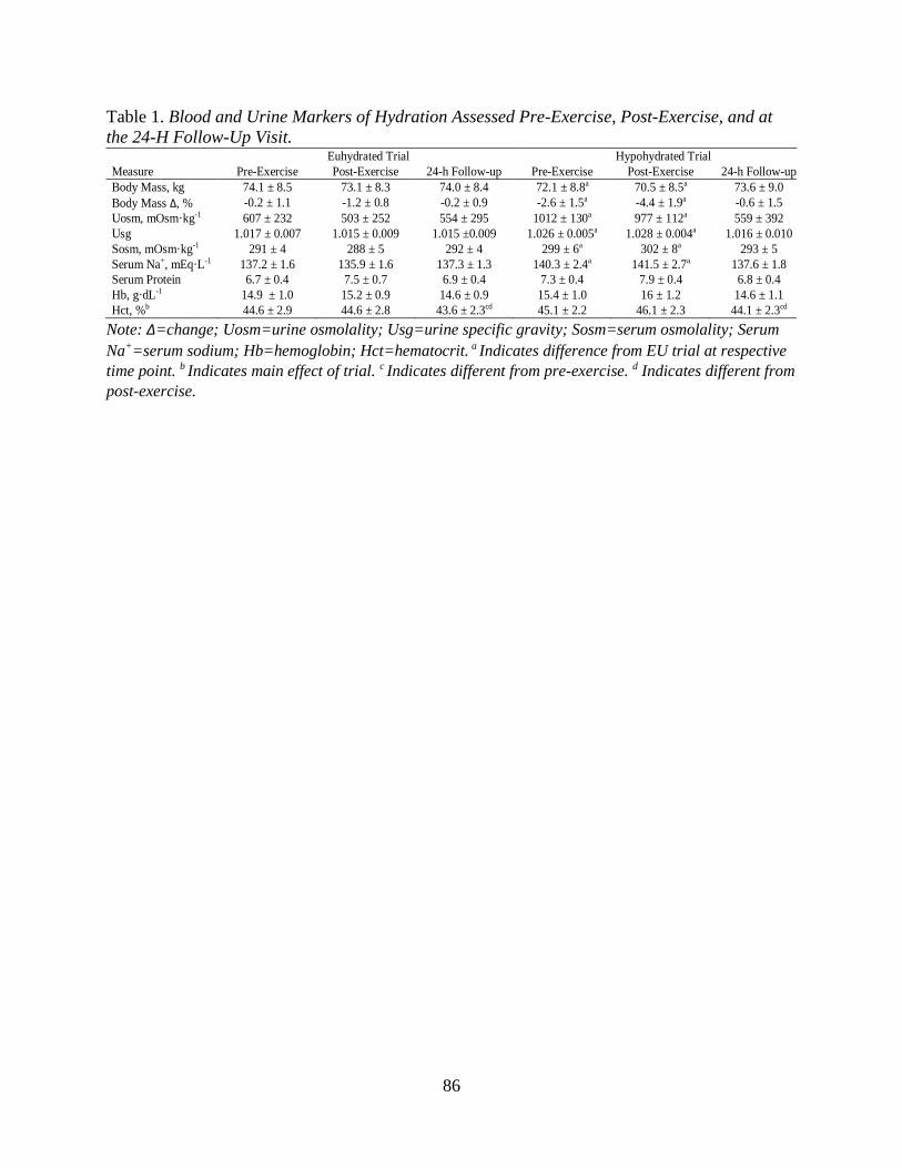
86
Table 1. Blood and Urine Markers of Hydration Assessed Pre-Exercise, Post-Exercise, and at
the 24-H Follow-Up Visit.
Note: Δ=change; Uosm=urine osmolality; Usg=urine specific gravity; Sosm=serum osmolality; Serum
Na+=serum sodium; Hb=hemoglobin; Hct=hematocrit. a Indicates difference from EU trial at respective
time point. b Indicates main effect of trial. c Indicates different from pre-exercise. d Indicates different from
post-exercise.
Euhydrated Trial Hypohydrated Trial
Measure Pre-Exercise Post-Exercise 24-h Follow-up Pre-Exercise Post-Exercise 24-h Follow-up
Body Mass, kg 74.1 ± 8.5 73.1 ± 8.3 74.0 ± 8.4 72.1 ± 8.8a 70.5 ± 8.5a 73.6 ± 9.0
Body Mass Δ, % -0.2 ± 1.1 -1.2 ± 0.8 -0.2 ± 0.9 -2.6 ± 1.5a -4.4 ± 1.9a -0.6 ± 1.5
Uosm, mOsm·kg-1 607 ± 232 503 ± 252 554 ± 295 1012 ± 130a 977 ± 112a 559 ± 392
Usg 1.017 ± 0.007 1.015 ± 0.009 1.015 ±0.009 1.026 ± 0.005a 1.028 ± 0.004a 1.016 ± 0.010
Sosm, mOsm·kg-1 291 ± 4 288 ± 5 292 ± 4 299 ± 6a 302 ± 8a 293 ± 5
Serum Na+, mEq·L-1 137.2 ± 1.6 135.9 ± 1.6 137.3 ± 1.3 140.3 ± 2.4a 141.5 ± 2.7a 137.6 ± 1.8
Serum Protein 6.7 ± 0.4 7.5 ± 0.7 6.9 ± 0.4 7.3 ± 0.4 7.9 ± 0.4 6.8 ± 0.4
Hb, g·dL-1 14.9 ± 1.0 15.2 ± 0.9 14.6 ± 0.9 15.4 ± 1.0 16 ± 1.2 14.6 ± 1.1
Hct, %b 44.6 ± 2.9 44.6 ± 2.8 43.6 ± 2.3cd 45.1 ± 2.2 46.1 ± 2.3 44.1 ± 2.3cd

87
Figure 1
ab
abcde
ab a
abcfg abcdefg
A
C
B
fg
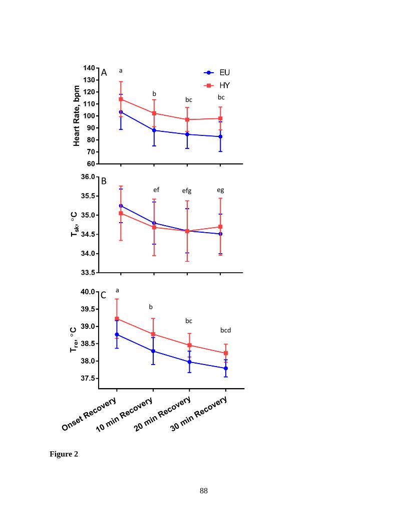
88
Figure 2
a
bcd
ef efg eg
a
b
bc
bc b bc
A
C
B
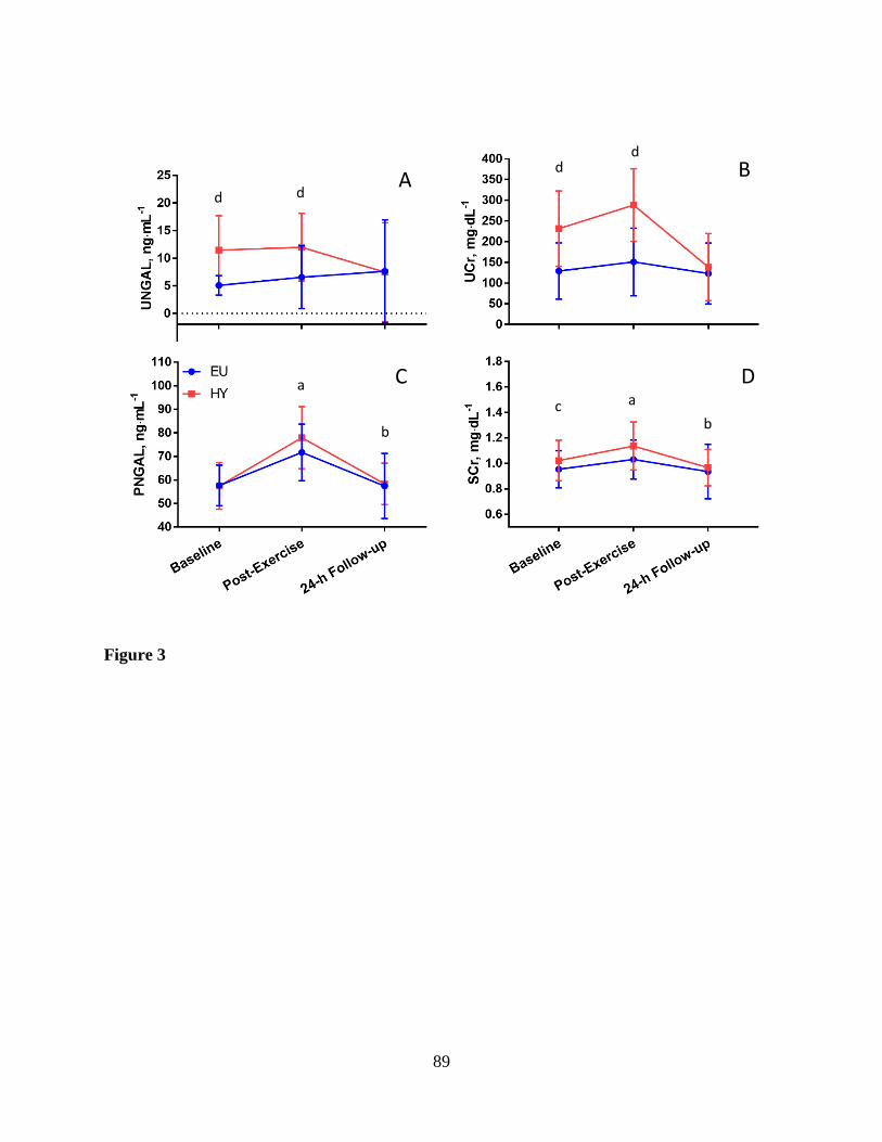
89
Figure 3
A
C
B
D a
b
a
b c
d d
d d
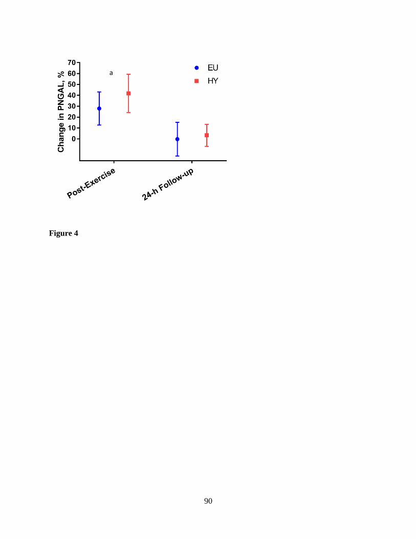
90
Figure 4
a
91
V. Manuscript #2: Influences of Hypohydration During Exercise-Induced Muscle Damage
on Recovery
Cory L. Butts, Aaron R. Caldwell, Matthew S. Ganio, Nicholas P. Greene, Stavros A. Kavouras,
Ronna C. Turner, Brendon P. McDermott
Exercise Science Research Center, University of Arkansas, Fayetteville, Arkansas, USA

92
Abstract
Purpose: Identify the effects of mild hypohydration on muscular strength recovery indices as
well as muscle damage biomarkers from eccentric knee flexion followed by exercise in the heat.
Methods: Recreationally active males (n = 18, age 24±5 y, mass 75.9±10.0 kg, bf 17.3±6.2%)
completed two experimental conditions consisting of either euhydration (EU; maintaining
hydration) or hypohydration (HY; restricting fluid consumption for 24 hours prior to and during
the trial) separated by ≥28 days. Participants completed a baseline 20-minute rest, muscle
damaging protocol, treadmill exercise in the heat, passive recovery, and a rehydrated 24-h
follow-up visit, respectively. The muscle damage was induced through contralateral (opposite
leg for each trial) eccentric knee flexion exercises (30°/sec) on an isokinetic dynamometer.
Isometric (70° and 90°) and isokinetic (60°/sec) strength was performed immediately before and
after damage as well as during the 24-h follow-up. Results: Fluid restriction induced -2.6±1.5%
reduction in body mass at the beginning of the trial, while body mass was maintained in the EU
trial (-0.2±1.1%, P < 0.001). Strength was reduced independent of trial for isometric strength at
70° (F2,32 = 12.54, P < 0.001), isometric strength at 90° (F2,32 = 8.96, P = 0.001), and isokinetic
strength at 60°·sec-1 (F2,32 = 8.11, P = 0.001). Serum creatine kinase increased regardless of trial
(F1.32,18.4 = 24.42, P < 0.001), with the 24-h follow-up greater (grand mean; 58.7±25.1 U/L) than
at baseline (grand mean; 35.7±23.1, P < 0.001) and post exercise (grand mean; 51.6±23.2 U/L,
P=0.009). Conclusions: We demonstrated no significant impact of hydration status when
performing muscle damaging exercise, followed by exercise in the heat, on indices of muscle
damage recovery. Further, the rehydration protocol successfully returned participants to a EU
state by the 24-h follow-up, which may have impacted the recovery from muscle damaging
protocol.

93
Introduction
A potential consequence of resistance exercise involves damaging muscle tissue,
particularly if the movements involve an eccentric component (1-3). Exercise induced muscle
damage may range from asymptomatic increases in biomarkers to exertional rhabdomyolysis
requiring medical attention. Subclinical rhabdomyolysis not requiring medical treatment can
easily be treated with rest and hydration (1). Muscle damaging exercise decreases the ability to
generate force and may have implications for subsequent performance (4-10). As such,
controlling factors that may affect muscle damage and recovery is essential for athletes to enable
expeditious returns in performance.
Athletes commonly arrive to exercise bouts, athletic events, and practices in a fluid
conservation state when evaluated by urinary indices (e.g. specific gravity, color, osmolality)
(11-14). Further, reductions in body weight have been shown across pre-season practices in
addition to concentrated urine production, suggesting potential losses in total body water (12).
As such, poor hydration practices during and following practices may lead to dehydration,
potentially impacting performance and altering recovery (2, 15, 16). This particularly applies in
settings where there is a reduced time between practices (i.e. two-a-days) or preseason where
individuals are undergoing rigorous workouts day after day (i.e. muscle damaging exercise).
The impact of suboptimal hydration on muscle damaging exercise has received relatively
little investigation. While hypohydration is consistently found to reduce endurance performance,
impacts on muscular strength or power are more controversial (2, 15). In a review by Judelson et
al. (2), authors concluded that the overall effect of dehydration on muscular strength and power
was negative. However, study limitations often prevent the clear interpretation of findings,
making it difficult to ascertain the effects of hypohydration on resistance and power performance

94
(2, 15). Regardless, increases in physiological and perceptual strain associated with
hypohydration merit the support for athletes to perform these exercises in a well hydrated state.
Hydration impacts resistance exercise performance, however, it has been shown to have
no impact on circulating markers of muscle damage (i.e. creatine kinase and myoglobin) often
seen following exercise (3). Moderate hypohydration (5%) was successful at inducing slight
increases in myoglobin one and two hours post-exercise, however, total work was not affected
compared to an euhydrated condition. Dehydration combined with hyperthermia may also
impact recovery from muscle damaging exercise (6). Cleary et al. (6) investigated the effects
exercise induced dehydration on delayed onset muscle soreness (DOMS) recovery from downhill
running in the heat. Perceptions of lower extremity pain were significantly elevated in the
dehydrated trial, albeit with no differences in muscular strength (6). Interestingly, when
downhill running was performed in a thermoneutral environment, the effects of dehydration on
DOMS were ameliorated (7). Therefore, the addition of hyperthermia with concomitant
dehydration may impact skeletal muscle recovery. In organized sport, when compared with a
normothermic soccer match, there was no impact of heat stress on markers of muscle damage
recovery, however perceptions were not assessed (17). Furthermore, these data were collected
post-soccer match therefore, the hydration and damage responses may have differed significantly
between individuals. Regardless, the impact of muscle damaging exercise with concomitant
dehydration may exacerbate symptoms of DOMS, potentially due to delayed recovery induced
via hyperthermia and dehydration.
Recovery from muscle damaging exercise performed while in a fluid conserving state has
not been extensively investigated. Further, muscle damaging exercise is likely associated with a
strength and conditioning session that would be completed in a normothermic environment

95
followed by exercise in the heat, rather than all in a heated environment. Therefore, the purpose
of this study was to identify the effects of performing muscle damaging exercise followed by
exercise in the heat while hypohydrated, on muscular strength indices (i.e. isometric and
isokinetic strength) as well as muscle damage biomarkers (creatine kinase), when full
rehydration was conducted in recovery. Based on previous investigations, it was hypothesized
that there would be no differences in muscle damage biomarkers with hypohydration, however,
muscle strength or recovery would be modestly impaired (i.e. slightly greater reductions) when
compared to a euhydrated state.
Methods
Eighteen healthy, recreationally active males (age 24 ± 5 y, wt 75.9 ± 10.0 kg, ht 1.79 ±
0.05m, bf 17.3 ± 6.2%) were recruited from the University and surrounding areas to participate
in this randomized, counterbalanced, crossover design study. All procedures were approved by
the University Institutional Review Board and written informed consent was acquired from all
individuals prior to participation. The study consisted of five visits including one familiarization
day and two experimental days (one euhydrated; EU and one hypohydrated; HY) each with 24-h
follow-ups. Experimental days were separated by ≥28 days (average; 41 ± 16 days), and muscle
damaging exercise was completed on contralateral legs to mitigate the repeated bout effect (18).
Familiarization Day:
During the initial familiarization visit, participants completed a medical history
questionnaire and signed an informed consent form. Exclusionary criteria included previous heat
exhaustion or heat stroke within the past 3 years, current musculoskeletal injury, hypertension
where vigorous exercise is contraindicated, diagnosed sickle cell trait, use of medications that
may alter thermoregulation or kidney function, current use of creatine supplementation, and a
96
history of kidney disease. Upon approval, baseline demographic information was collected and
body composition assessed via dual energy x-ray absorptiometry (DXA). Participants then
completed a five-minute warm-up on a cycle ergometer (~50W) and were fitted to the isokinetic
dynamometer (Biodex System 3, Biodex Medical Systems, New York) with seat and leg
positions recorded for future testing. Baseline isometric strength at 70° and 90°, and isokinetic
strength at 60°·sec-1 were competed in triplicate and future eccentric procedures were explained.
Because these were used as a familiarization, these measures were not included in analysis.
Experimental Days:
All participants were asked to refrain from alcohol use for 24-h, caffeine use for 12-h,
resistance training for 5-days and exercise for 24-h prior to each trial. For the HY trial, the
dehydration protocol consisted of 24-h fluid restriction with minimal fluid provided throughout
the trial. Prior to the EU trial, participants were instructed to consume fluids prior to arrival,
while water was provided during the trial to ensure less than 2% body mass loss.
Prior to arrival for both trials, participants recorded three-day baseline body masses on a
lab provided scale (BalanceFrom High Accuracy Bathroom Scale, BalanceFrom LLC, China) on
the days leading up to the trials. For the 24-h prior to arrival, participants recorded their diet
using a standard diet log and were asked to repeat a similar diet for the second trial.
Additionally, participants collected all urinations for the 24-h prior to the start of trial, which was
subsequently analyzed for 24-hr urine osmolality (freezing point depression, Model 3250,
Advanced Instruments Inc., Norwood, MA).
Upon arrival to the laboratory, participants completed a nude body mass and dressed in
shorts and a t-shirt, followed by a 20-minute semi-recumbent baseline rest in a thermoneutral
environment (~20°C). During this time, participants were informed of perceptual scales for

97
rating of perceived exertion (RPE) and muscle pain and baseline values were recorded. This was
followed by a blood draw via venipuncture to assess serum creatine kinase (SCK).
Participants moved to a cycle ergometer (Monark 828E, Monark Exercise AB, Sweden)
to complete a 5-minute warm-up at 50W before completing the muscle damaging protocol on an
isokinetic dynamometer (Biodex System 3, Biodex Medical Systems, New York) (18). The
muscle damaging procedure involved maximal effort unilateral eccentric knee extension
exercise, with the contralateral leg utilized during the second trial to minimize potential repeated
bout effects. The leg used during the first trial was randomized and counterbalanced for
hydration and dominance between participants. For each trial, participants completed 10 sets of
10 maximal effort eccentric knee flexion repetitions at a speed of 30°/s with sets separated by
one-minute (18). Prior to, and immediately following the eccentric protocol, isometric strength
(i.e. peak torque) measures were performed via three 5-second maximal voluntary concentric
knee extensor contractions at 70° and 90° knee flexion with one minutes of rest between
repetitions. Isokinetic concentric knee extensor strength (i.e. peak torque) was also completed in
triplicate at 60°·sec-1. Perceptions of muscle pain and RPE were recorded following the exercise
induced muscle damage. Decreases in strength, as well as elevations in SCK and muscle pain,
served as indices of muscle damage (5). As this was part of a larger study on renal biomarkers,
the purpose of the eccentric exercise protocol was to induce mild muscle damage similar to
athletic practices or labor-intensive occupational settings, thus physiological adaptations were
not expected to alter the findings given our counterbalanced, crossover study design. Also, the
participants were directed to avoid changes in their exercise regimen between trials to minimize
changes in fitness.

98
Upon completion of the exercise induced muscle damage, participants moved to an
environmental chamber set to a hot, humid environment (33°C, 50% relative humidity) to
complete treadmill running at 60% VO2max (1% grade) for 60 minutes with a five-minute
walking (3 mph) warm-up, break at 30 minutes, and cool-down at 60 minutes. This protocol was
designed to increase thermal stress and further dehydrate participants during the HY trial and
prevent >2% body mass loss in the EU trial. Immediately following exercise, participants
remained in the chamber to commence a 30-minute semi-recumbent recovery with a post-
exercise blood draw taken at 20 minutes.
After the recovery period, participants provided another body mass and urine sample, and
were provided with a rehydration protocol in both trials, such that the participant would replace
100% of losses in the initial four hours following the trial, ensuring to consume food to prevent
over-hydration. Additionally, participants were then encouraged to consume at least an
additional 2.5 liters to aid in the production of dilute urine samples.
Participants returned to the laboratory for a 24-h follow-up visit. Visits involved a nude
body mass followed by 20-minute semi-recumbent rest period, during which perceptual
measures, and a blood draw were collected. Participants then completed the five-minute warm-
up on a cycle ergometer at 50W before moving to the isokinetic dynamometer. Concentric knee
extension strength was once again recorded for isometric contractions at 70° and 90° of knee
flexion as well as isokinetic contractions at 60°·sec-1 knee extension. Participants were then
instructed to resume normal exercise routines and second visits were scheduled, if applicable.
Blood Analysis
After clotting, serum samples were centrifuged for 15 minutes at 1000 g and 4°C. Serum
was aliquoted and stored at -80°C for further analysis. Serum creatine kinase (SCK) was
99
assessed using a commercially available colorimetric assay (Bioassay Systems Inc, Hayward,
CA) with an average coefficient of variation of 2.1%.
Statistical Analysis
All statistical analyses were completed using SPSS version 24 (IBM Corporation,
Somers, NY). A repeated measures multivariate analysis of variance was used to assess absolute
peak torque (i.e. strength) across time (pre-muscle damage, post-muscle damage, and the 24-h
follow-up) and between hydration (EU and HY) for isometric peak torque at 70° and 90° knee
flexion and 60°·sec-1 isokinetic peak torque. Additional variables (body mass, creatine kinase)
were analyzed using two-way (time x hydration) repeated measures analysis of variance. When
sphericity was violated, Greenhouse-Geisser adjustments were used. Post-hoc analyses involved
pairwise comparisons with an appropriate Bonferroni corrected alpha to identify significant time
point differences. Dependent t-tests were used to assess differences in 24-h urine osmolality,
total eccentric work, average eccentric peak torque and ratings of perceived exertion between
trials. Data that failed normality tests (muscle pain) were analyzed with a Friedman test across
time and between hydration states. Follow-up pairwise analysis were conducted using a
Wilcoxon signed rank test for individual time point differences. All partial eta squared (ηp²) and
Hedge’s g values were calculated using a spreadsheet from Lakens (19). Alpha of 0.05 was set a
priori to determine significance at the omnibus level. Based on a .80 power calculation using the
primary outcome variable NGAL (20, 21) with a correlation between time points of 0.42, a 2-
standard deviation effect size, β of 0.20, and α of 0.05, it was determined 17 participants would
be sufficient to complete this study. An increased experiment-wise type I error rate is
acknowledged due to the multiple multivariate and univariate analyses conducted. Because the
experimental protocol is time, resource, and cost intensive, power estimates were calculated

100
based on singular analyses to provide initial experimental outcome indicators and guide future
research.
Results
Hydration
There was a significant interaction of body mass for time and trial (F2,32 = 24.51, P <
0.001, ηp² = 0.61), as three-day baseline body masses were not different between trials (EU 73.7
± 8.6, HY 73.8 ± 9.4 kg, P = 0.87), while body mass at the beginning of the trial was reduced
following fluid restriction (HY 72.1 ± 8.8kg, EU 74.1 ± 8.5kg, P < 0.001). By design, the body
masses were not different at the 24-h follow-up visit (EU 74.0 ± 8.4, HY 73.6 ± 9.0 kg, P =
0.29). There was also an interaction of time and trial (F1,15 = 103.50, P < 0.001, ηp² = 0.87) for
percent body mass change, with the fluid restriction inducing a -2.6±1.5% reduction in body
mass from the three-day baseline body mass at the beginning of the trial, while body mass was
maintained in the EU trial (-0.2±1.1%, P < 0.001). There were no differences between trials at
the 24-h follow-up for percent change from the three-day baseline (EU -0.2±0.9, HY -0.6±1.5%,
P=0.26). Urine collection for 24 h prior to the trial days showed greater urine osmolality
following fluid restriction (HY 775±180, EU 427±188 mOsm/kg, t17 = -6.71, P < 0.001, Hedge’s
g = -3.01).
Muscular Strength & Muscle Damage Markers
Total work completed during eccentric exercise was not different between trials (EU
8663 ± 2651, HY 8280 ± 2102 J, t17 = 0.90, P = 0.38, Hedge’s g = 0.40). Average peak eccentric
torque was also not different between trials (EU 196.1±62.5, HY 186.3±57.1 N·m, t17 = 1.22, P =
0.24, Hedge’s g = 0.55).

101
Muscular strength was assessed using a multivariate analysis including isometric peak
torque measured at 70° and 90° as well as peak isokinetic torque assessed at 60°·sec-1. At the
multivariate level, there was a main effect of time (Wilks Λ = 0.35, F6,11 = 3.49, P = 0.04),
however, there was no effect of hydration (Wilks Λ = 0.80, F3,14 = 1.14, P = 0.37) or interaction
of time and hydration (Wilks Λ = 0.51, F6,11 = 1.76, P = 0.20).
Univariate analysis of time was then conducted using an alpha of 0.017. Strength was
reduced independent of trial (i.e. main effect of time) for isometric strength at 70° (F2,32 = 12.54,
P < 0.001, ηp² = 0.44, Figure 1C), isometric strength at 90° (F2,32 = 8.96, P = 0.001, ηp² = 0.36,
Figure 1B), and isokinetic strength at 60°·sec-1 (F2,32 = 8.11, P = 0.001, ηp² = 0.34, Figure 1A).
Pairwise comparisons for each of the strength measures were then completed using an alpha of
0.006. Isometric strength at 70° decreased from baseline (grand mean; 203.2 ± 56.8 N·m) to
immediate-post damage (grand mean; 166.0±45.2 N·m, P = 0.001) and remained reduced at the
24 h follow-up (grand mean; 182.9 ± 50.8 N·m, P = 0.006). Isometric at 90° was also reduced
from baseline (grand mean; 227.2 ± 65.7 N·m) to immediate-post damage (grand mean; 188.9 ±
55.1 N·m, P < 0.001) and tended to be reduced at the 24 h follow-up (grand mean; 200.2 ± 58.6
N·m, P = 0.007). Isokinetic strength at 60°·sec-1 was reduced immediately post-damage (grand
mean; 143.2 ± 42.0 N·m) compared to baseline (grand mean; 168.0 ± 53.7 N·m, P =0.003),
however, there was no difference between baseline and the 24-h follow-up (grand mean; 156.5 ±
38.0 N·m, P = 0.10).
SCK analysis revealed increases regardless of hydration (F1.32,18.4 = 24.42, P < 0.001, ηp²
= 0.64) with no differences between trials (F1,14 = 0.36, P = 0.56, ηp² = 0.04) and no interaction
effects (F1.2,16.5 = 1.002, P = 0.35, ηp² = 0.07, Figure 2). The 24-h follow-up SCK was greater
(grand mean; 58.7 ± 25.1 U/L) than baseline (grand mean; 35.7 ± 23.1, P < 0.001) and post-
102
exercise (grand mean; 51.6 ± 23.2 U/L, P=0.009). In addition, the post-exercise SCK was
greater than at baseline (P < 0.001).
Perceptual Measures
Muscle pain was affected by time (χ22 = 30.10, P < 0.001) and trial (χ2
1 = 4.57, P = 0.03),
however, there were no differences between trials for muscle pain at any time (all P > 0.017).
Rather, pain was elevated above baseline (EU 0.1 ± 0.2, HY 0.1 ± 0.2) immediately post damage
(EU 2.7 ± 1.7, HY 3.5 ± 1.6, both P < 0.001) and at the 24-h follow-up (EU 1.3 ± 1.4, HY 1.4 ±
1.6, both P < 0.001). Ratings of Perceived Exertion at the end of the muscle damaging exercise
were lower in the EU trial (EU 15.8 ± 2.0, HY 16.6 ± 1.8, t16 = -2.75, P = 0.01, Hedge’s g = -
1.27).
Discussion
The purpose of this investigation was to evaluate the influence of performing muscle
damaging exercise while fluid restricted on indices of muscle damage recovery when muscle
damaging exercise was followed by exertional hyperthermia. Further, we removed the impact of
hypohydration in recovery by prescribing fluid replacement following both EU and HY trials,
thus isolating the impact of hypohydration during muscle damaging exercise. Contrary to our
hypothesis, our participants demonstrated no differences between trials in strength decreases, a
marker of muscle damage. As expected, however, there was no difference between trials for
SCK responses at any time point.
Maintaining a positive fluid balance with exercise can be difficult, particularly when
completing repeated bouts in hot, humid conditions (e.g. pre-season practice). This is
demonstrated by athletes regularly reporting to activities in a state of water conservation (i.e.
producing concentrated urine samples) (11, 12, 14, 22). This suboptimal hydration has many

103
implications on athlete safety and performance. For example, many sources have found
detriments in resistance exercise performance when dehydrated (2, 15). Poor hydration is also
implicated to alter physiological and perceptual recovery following exercise in the heat (23-26).
Therefore, proper rehydration strategies such as those recommended in consensus statements
(27, 28) are important to prevent delayed recovery and enhance the preparation for subsequent
bouts (24, 26).
The impacts of exercise induced dehydration on symptoms of DOMS have been
conducted in both hyperthermic (6) and normothermic males (7). Similar to the current study,
Cleary et al. (7) found no impact of hypohydration on measures of isometric strength or muscle
pain following muscle damaging exercise when individuals performed the exercise in a
normothermic state. The authors utilized 45 minutes of downhill running to induce muscle
damage presenting isometric strength decreases similar to the current study. In contrast,
dehydration was conducted using walking in a hot, humid environment while we utilized 24-h
fluid restriction to initiate dehydration. Regardless, the work by Cleary et al. (7) suggests that
dehydration alone does not impact the extent of muscle damage, perceptions, or recovery.
In a separate study, Cleary et al. found that performing muscle damaging exercise with
concomitant dehydration and hyperthermia led to increased perceived pain compared to the
euhydrated hyperthermic trial (6). The current study utilized muscle damaging exercise while in
a normothermic state, then participants experienced marked exertional hyperthermia, without
showing any differences between hydration states. Therefore, the presence of increased muscle
temperature during muscle damaging exercise may be required for hypohydration to have an
impact on perceived muscular soreness (6). Cleary et al. (6) suggested that the presence of
hypohydration augmented the intramuscular temperature due to thermoregulatory
104
compensations, which compromised the structural integrity of the tissue and led to increased
damage. However, the authors did not report any differences between hydration trials for
isometric quadriceps strength measures, therefore the extent of damage may have been related
purely to perceptual measures (6). Further, another study confirmed these strength findings by
elevating intramuscular temperature and completing muscle damaging exercise of the biceps
(29). The authors found no impact of higher muscle temperature on indices of muscle damage
compared to exercise performed in a normothermic state (29). This would suggest that the
presence of hypohydration is necessary to influence changes in perception when muscle damage
is performed in a hyperthermic condition and that these moderations are not purely temperature
driven. Rather, hypohydration may alter the inflammatory response to increase sensitivity and
subsequently drive pain (9). Fielding et al. showed that a 2% dehydration exercise protocol
followed by exercise induced muscle damage caused greater circulating neutrophil release
compared to the rest trial (30). Further, there were no differences in z-band damage nor SCK
between the trials (30). The lack of differences in muscle damage indices in the current study
further support that hyperthermia with concomitant hypohydration is necessary during the
damaging process to alter recovery.
Hypohydration has also been shown to have minimal impact on biomarkers of muscle
damage (3). Yamamoto et al. (3) evaluated the impact of hypohydration at 2.5% and 5% body
mass loss on muscle damage markers following an intense resistance exercise protocol. The
authors found no differences in SCK, but there were minor elevations in myoglobin with
dehydration (3). Although the present study did not investigate myoglobin, we found no
differences between hydration on the elevations in SCK, however, our damaging protocol
resulted in >1.5-fold elevations in SCK at 24-h. Peak SCK values are generally reported at ~3-4

105
days post damage, therefore it is likely there were greater elevations in these values (9).
Additionally, it is recognized that the values reported for the current study are within normal
range for healthy individuals (<175 U/L) (8). However, SCK as a biomarker of muscle damage
does not necessarily reflect the extent of disruption in the tissue with a great amount of inter-
individual variability (5, 8, 30). Regardless, the low SCK response may have been a function of
the methodology used for inducing muscle damage (i.e. unilateral knee extension) resulting in
low concentrations or possibly the colorimetric assay used to assess CK activity rather than true
concentration. Regardless, the SCK response was one that increased, thus confirming the
presence of muscle damage.
As this study was part of a larger investigation of biomarkers of acute kidney injury, there
were limitations in the methodology. We provided instructions to participants to rehydrate within
four hours of finishing the trial and continue recommended fluid consumption until the 24-h
follow-up. This rehydration was performed by the participant outside the laboratory, therefore
we relied on participants to complete the procedure and verbally confirmed compliance during
the 24-h follow-up. Additionally, we cannot comment on the impacts of poor hydration
following muscle damaging exercise on recovery indices. However, restricting fluids following
a muscle damaging exercise bout with exercise in the heat may place substantial strain on the
renal system and increase risk for exertional rhabdomyolysis. The presence of hypohydration,
heat stress, and unaccustomed exercise are thought to create the “perfect storm” and have been
reported in case studies to contribute to clinically significant cases of exertional rhabdomyolysis
(31, 32). As such, the goal of this study was to evaluate the implications for reporting to an
exercise session following poor fluid intake practices but finishing trials with proper rehydration
to evaluate the impacts on muscle damage recovery. To further ensure safety, we used a

106
unilateral knee extensor protocol for inducing muscle damage to mitigate the risk of
rhabdomyolysis in participants. As such, future studies may be necessary to evaluate the impacts
of delayed rehydration or partial rehydration on muscle damage severity and recovery.
In conclusion, the findings of this study apply particularly well in settings where teams
may undergo a strength training protocol (i.e. muscle damaging) followed by practice or
conditioning session in a hot, humid environment. We demonstrated no significant impact of
hydration status when performing muscle damaging exercise, followed by exercise in the heat,
on severity of muscle damage and pain, or indices of recovery. Further, the rehydration protocol
successfully returned participants to a euhydrated state by the 24-h follow-up, which may have
facilitated the recovery from our muscle damaging protocol. Therefore, when athletes report to
activities in a state of water conservation, it is unlikely that recovery form any muscle damaging
exercise will be impacted as long as the athlete rehydrates according to the National Athletic
Training Association and American College of Sport Medicine guidelines (27, 28). However,
hypohydration, heat stress and unaccustomed exercise may increase risk for exertional
rhabdomyolysis. As such, to improve overall safety, coaches, strength staff, and clinicians
should encourage athletes to report to practices to well prepared with proper hydration and diet.
References
1. Tietze DC, Borchers J. Exertional rhabdomyolysis in the athlete: a clinical review. Sports
Health. 2014;6(4):336-9. doi: 10.1177/1941738114523544. PubMed PMID: 24982707; PubMed
Central PMCID: PMCPMC4065559.
2. Judelson DA, Maresh CM, Anderson JM, Armstrong LE, Casa DJ, Kraemer WJ, et al.
Hydration and muscular performance: does fluid balance affect strength, power and high-
intensity endurance? Sports Med. 2007;37(10):907-21. PubMed PMID: 17887814.
3. Yamamoto LM, Judelson DA, Farrell MJ, Lee EC, Armstrong LE, Casa DJ, et al. Effects
of hydration state and resistance exercise on markers of muscle damage. J Strength Cond Res.
2008;22(5):1387-93. doi: 10.1519/JSC.0b013e3181739403. PubMed PMID: 18714253.

107
4. Doma K, Leicht A, Sinclair W, Schumann M, Damas F, Burt D, et al. The impact of
exercise-induced muscle damage on performance test outcomes in elite female basketball
players. J Strength Cond Res. 2017. Epub 2017/09/11. doi: 10.1519/JSC.0000000000002244.
PubMed PMID: 28930877.
5. Damas F, Nosaka K, Libardi CA, Chen TC, Ugrinowitsch C. Susceptibility to Exercise-
Induced Muscle Damage: a Cluster Analysis with a Large Sample. Int J Sports Med.
2016;37(8):633-40. Epub 2016/04/26. doi: 10.1055/s-0042-100281. PubMed PMID: 27116346.
6. Cleary MA, Sweeney LA, Kendrick ZV, Sitler MR. Dehydration and symptoms of
delayed-onset muscle soreness in hyperthermic males. J Athl Train. 2005;40(4):288-97. PubMed
PMID: 16404450; PubMed Central PMCID: PMCPMC1323290.
7. Cleary MA, Sitler MR, Kendrick ZV. Dehydration and symptoms of delayed-onset
muscle soreness in normothermic men. J Athl Train. 2006;41(1):36-45. PubMed PMID:
16619093; PubMed Central PMCID: PMCPMC1421497.
8. Baird MF, Graham SM, Baker JS, Bickerstaff GF. Creatine-kinase- and exercise-related
muscle damage implications for muscle performance and recovery. J Nutr Metab.
2012;2012:960363. Epub 2012/01/11. doi: 10.1155/2012/960363. PubMed PMID: 22288008;
PubMed Central PMCID: PMCPMC3263635.
9. Peake JM, Neubauer O, Della Gatta PA, Nosaka K. Muscle damage and inflammation
during recovery from exercise. J Appl Physiol (1985). 2017;122(3):559-70. Epub 2016/12/29.
doi: 10.1152/japplphysiol.00971.2016. PubMed PMID: 28035017.
10. Hubal MJ, Rubinstein SR, Clarkson PM. Mechanisms of variability in strength loss after
muscle-lengthening actions. Med Sci Sports Exerc. 2007;39(3):461-8. doi:
10.1249/01.mss.0000247007.19127.da. PubMed PMID: 17473772.
11. Osterberg KL, Horswill CA, Baker LB. Pregame urine specific gravity and fluid intake
by National Basketball Association players during competition. J Athl Train. 2009;44(1):53-7.
doi: 10.4085/1062-6050-44.1.53. PubMed PMID: 19180219; PubMed Central PMCID:
PMCPMC2629040.
12. Godek SF, Godek JJ, Bartolozzi AR. Hydration status in college football players during
consecutive days of twice-a-day preseason practices. Am J Sports Med. 2005;33(6):843-51. doi:
10.1177/0363546504270999. PubMed PMID: 15827364.
13. Yeargin SW, Casa DJ, Armstrong LE, Watson G, Judelson DA, Psathas E, et al. Heat
acclimatization and hydration status of American football players during initial summer
workouts. J Strength Cond Res. 2006;20(3):463-70. doi: 10.1519/20596.1. PubMed PMID:
16937956.
14. Yeargin SW, Casa DJ, Judelson DA, McDermott BP, Ganio MS, Lee EC, et al.
Thermoregulatory responses and hydration practices in heat-acclimatized adolescents during
preseason high school football. J Athl Train. 2010;45(2):136-46. doi: 10.4085/1062-6050-
45.2.136. PubMed PMID: 20210617; PubMed Central PMCID: PMCPMC2838465.

108
15. Cheuvront SN, Kenefick RW. Dehydration: physiology, assessment, and performance
effects. Compr Physiol. 2014;4(1):257-85. doi: 10.1002/cphy.c130017. PubMed PMID:
24692140.
16. Judelson DA, Maresh CM, Farrell MJ, Yamamoto LM, Armstrong LE, Kraemer WJ, et
al. Effect of hydration state on strength, power, and resistance exercise performance. Med Sci
Sports Exerc. 2007;39(10):1817-24. doi: 10.1249/mss.0b013e3180de5f22. PubMed PMID:
17909410.
17. Nybo L, Girard O, Mohr M, Knez W, Voss S, Racinais S. Markers of muscle damage and
performance recovery after exercise in the heat. Med Sci Sports Exerc. 2013;45(5):860-8. doi:
10.1249/MSS.0b013e31827ded04. PubMed PMID: 23247710.
18. Xin L, Hyldahl RD, Chipkin SR, Clarkson PM. A contralateral repeated bout effect
attenuates induction of NF-κB DNA binding following eccentric exercise. J Appl Physiol (1985).
2014;116(11):1473-80. doi: 10.1152/japplphysiol.00133.2013. PubMed PMID: 23950163.
19. Lakens D. Calculating and reporting effect sizes to facilitate cumulative science: a
practical primer for t-tests and ANOVAs. Front Psychol. 2013;4:863. Epub 2013/11/26. doi:
10.3389/fpsyg.2013.00863. PubMed PMID: 24324449; PubMed Central PMCID:
PMCPMC3840331.
20. Junglee NA, Di Felice U, Dolci A, Fortes MB, Jibani MM, Lemmey AB, et al. Exercising
in a hot environment with muscle damage: effects on acute kidney injury biomarkers and kidney
function. Am J Physiol Renal Physiol. 2013;305(6):F813-20. doi: 10.1152/ajprenal.00091.2013.
PubMed PMID: 23825078.
21. Melin B, Jimenez C, Savourey G, Bittel J, Cottet-Emard JM, Pequignot JM, et al. Effects
of hydration state on hormonal and renal responses during moderate exercise in the heat. Eur J
Appl Physiol Occup Physiol. 1997;76(4):320-7. doi: 10.1007/s004210050255. PubMed PMID:
9349646.
22. Godek SF, Bartolozzi AR, Burkholder R, Sugarman E, Dorshimer G. Core temperature
and percentage of dehydration in professional football linemen and backs during preseason
practices. J Athl Train. 2006;41(1):8-14; discussion -7. PubMed PMID: 16619090; PubMed
Central PMCID: PMCPMC1421487.
23. Gagnon D, Lynn AG, Binder K, Boushel RC, Kenny GP. Mean arterial pressure
following prolonged exercise in the heat: influence of training status and fluid replacement.
Scand J Med Sci Sports. 2012;22(5):e99-e107. doi: 10.1111/j.1600-0838.2012.01506.x. PubMed
PMID: 22830505.
24. McDermott BP, Casa DJ, Lee E, Yamamoto L, Beasley K, Emmanuel H, et al.
Thermoregulation and stress hormone recovery after exercise dehydration: comparison of
rehydration methods. J Athl Train. 2013;48(6):725-33. Epub 2013/10/23. doi: 10.4085/1062-
6050-48.6.01. PubMed PMID: 24143900; PubMed Central PMCID: PMCPMC3867082.
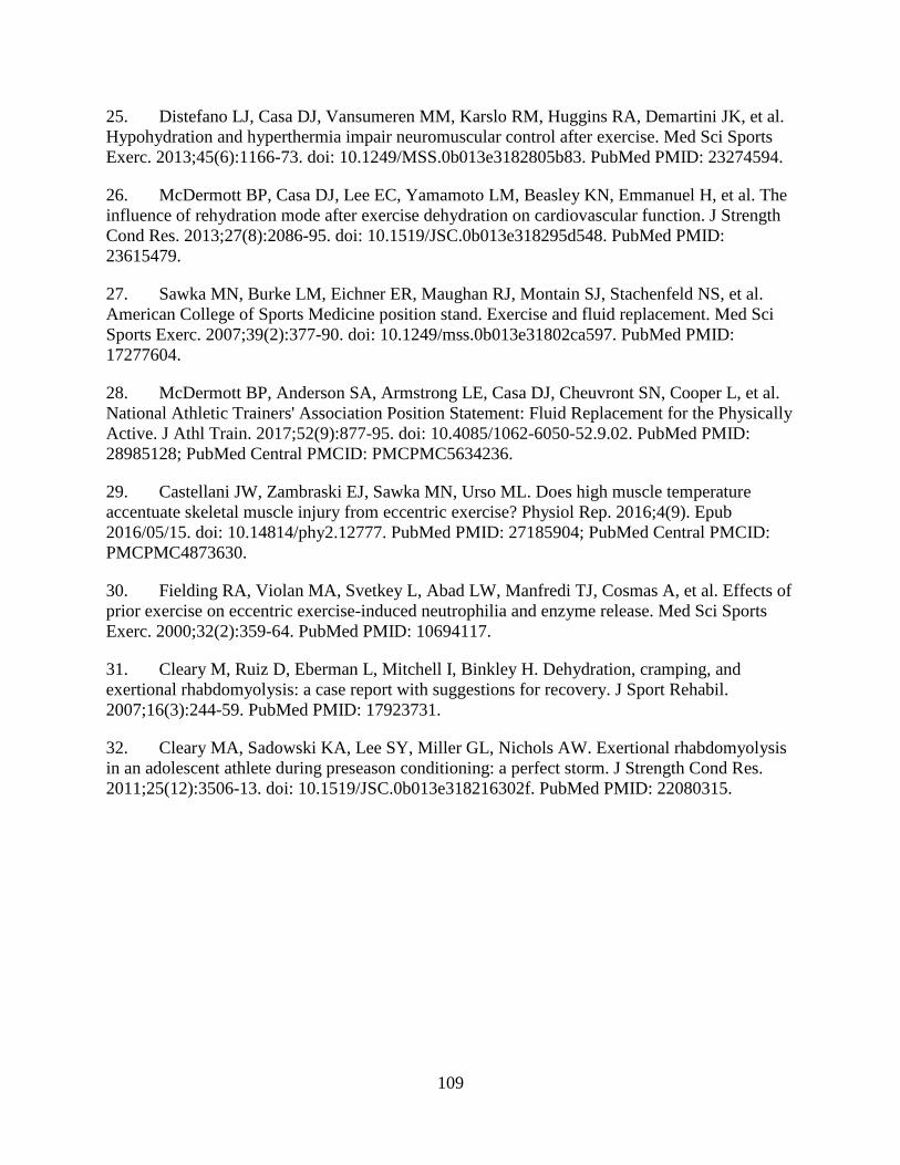
109
25. Distefano LJ, Casa DJ, Vansumeren MM, Karslo RM, Huggins RA, Demartini JK, et al.
Hypohydration and hyperthermia impair neuromuscular control after exercise. Med Sci Sports
Exerc. 2013;45(6):1166-73. doi: 10.1249/MSS.0b013e3182805b83. PubMed PMID: 23274594.
26. McDermott BP, Casa DJ, Lee EC, Yamamoto LM, Beasley KN, Emmanuel H, et al. The
influence of rehydration mode after exercise dehydration on cardiovascular function. J Strength
Cond Res. 2013;27(8):2086-95. doi: 10.1519/JSC.0b013e318295d548. PubMed PMID:
23615479.
27. Sawka MN, Burke LM, Eichner ER, Maughan RJ, Montain SJ, Stachenfeld NS, et al.
American College of Sports Medicine position stand. Exercise and fluid replacement. Med Sci
Sports Exerc. 2007;39(2):377-90. doi: 10.1249/mss.0b013e31802ca597. PubMed PMID:
17277604.
28. McDermott BP, Anderson SA, Armstrong LE, Casa DJ, Cheuvront SN, Cooper L, et al.
National Athletic Trainers' Association Position Statement: Fluid Replacement for the Physically
Active. J Athl Train. 2017;52(9):877-95. doi: 10.4085/1062-6050-52.9.02. PubMed PMID:
28985128; PubMed Central PMCID: PMCPMC5634236.
29. Castellani JW, Zambraski EJ, Sawka MN, Urso ML. Does high muscle temperature
accentuate skeletal muscle injury from eccentric exercise? Physiol Rep. 2016;4(9). Epub
2016/05/15. doi: 10.14814/phy2.12777. PubMed PMID: 27185904; PubMed Central PMCID:
PMCPMC4873630.
30. Fielding RA, Violan MA, Svetkey L, Abad LW, Manfredi TJ, Cosmas A, et al. Effects of
prior exercise on eccentric exercise-induced neutrophilia and enzyme release. Med Sci Sports
Exerc. 2000;32(2):359-64. PubMed PMID: 10694117.
31. Cleary M, Ruiz D, Eberman L, Mitchell I, Binkley H. Dehydration, cramping, and
exertional rhabdomyolysis: a case report with suggestions for recovery. J Sport Rehabil.
2007;16(3):244-59. PubMed PMID: 17923731.
32. Cleary MA, Sadowski KA, Lee SY, Miller GL, Nichols AW. Exertional rhabdomyolysis
in an adolescent athlete during preseason conditioning: a perfect storm. J Strength Cond Res.
2011;25(12):3506-13. doi: 10.1519/JSC.0b013e318216302f. PubMed PMID: 22080315.
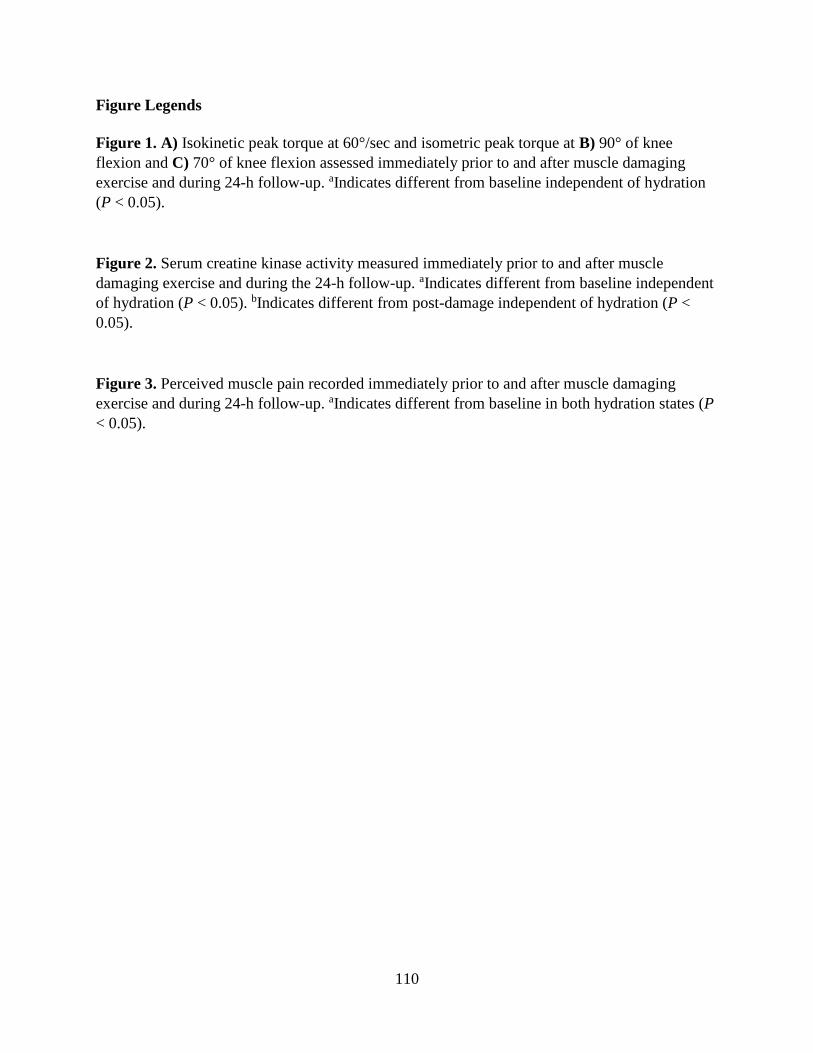
110
Figure Legends
Figure 1. A) Isokinetic peak torque at 60°/sec and isometric peak torque at B) 90° of knee
flexion and C) 70° of knee flexion assessed immediately prior to and after muscle damaging
exercise and during 24-h follow-up. aIndicates different from baseline independent of hydration
(P < 0.05).
Figure 2. Serum creatine kinase activity measured immediately prior to and after muscle
damaging exercise and during the 24-h follow-up. aIndicates different from baseline independent
of hydration (P < 0.05). bIndicates different from post-damage independent of hydration (P <
0.05).
Figure 3. Perceived muscle pain recorded immediately prior to and after muscle damaging
exercise and during 24-h follow-up. aIndicates different from baseline in both hydration states (P
< 0.05).
111
Figure 1
a
a
a
a
A
C
B

112
Figure 2.
a a b
113
Figure 3.
a
a

114
IV. Conclusions
These investigations were conducted to evaluate the influence of poor fluid intake
practices on renal stress and recovery from muscle damaging exercise. By using fluid restriction
prior to exercise, we mimicked conditions found in athletic, military, and occupational
populations when reporting for activity. Further, providing minimal fluid during exercise further
dehydrated individuals leading to greater physiological and perceptual strain. Therefore, our
dehydration protocol was effective in creating a state of hypohydration prior to and throughout
exercise. We followed this by providing fluid intake recommendations based on recent position
stands for rapid rehydration. As such, the rehydration protocol was successful at returning
individuals to a euhydrated state. By using these methodologies, we were able to evaluate the
effects of performing exercise while suboptimally hydrated on renal stress and muscle damage
recovery.
In Study 1, we demonstrated significantly greater increases in biomarkers of AKI when
participants were hypohydrated. However, the fluid replacement protocol in this study returned
biomarkers to baseline levels, indicating no lasting impairments in renal function. The
dehydration protocol also augmented cardiovascular, thermal strain, and perceptual strain during
exercise in the heat.
In Study 2, we demonstrated no impact of hydration status when performing muscle
damaging exercise, followed by exercise in the heat, on muscle damage and pain recovery. We
also demonstrated no influence of hydration on serum creatine kinase, a common biomarker of
muscle damage or on isokinetic or isometric muscular strength. The rehydration protocol used
may have aided in recovery to mitigate any detriments in muscular strength between hydration
trials.
115
There is overwhelming evidence to suggest a negative role of dehydration in renal
function, thermoregulation, performance, and cardiovascular stability. The concomitant
exposure to physiological (i.e. exercise, muscle damage) and environmental (high ambient
temperature and humidity) stressors commonly experienced by athletes, military, and
occupational populations augment the deleterious responses to dehydration. We demonstrated
that poor fluid intake practices leading into, and throughout, activity negatively impact renal,
physiological, and perceptual measures. However, proper fluid intake following this stressful
environment ameliorated the negative impacts of the previous hypohydration. Together, these
studies provide support for proper rehydration following dehydrating activity to prevent
deleterious impacts on renal and muscular recovery.
116
References
Agarwal, D., Elks, C. M., Reed, S. D., Mariappan, N., Majid, D. S., & Francis, J. (2012).
Chronic exercise preserves renal structure and hemodynamics in spontaneously
hypertensive rats. Antioxid Redox Signal, 16(2), 139-152. doi:10.1089/ars.2011.3967
Aizawa, H., Morita, K., Minami, H., Sasaki, N., & Tobise, K. (1995). Exertional rhabdomyolysis
as a result of strenuous military training. J Neurol Sci, 132(2), 239-240.
Alge, J. L., & Arthur, J. M. (2015). Biomarkers of AKI: a review of mechanistic relevance and
potential therapeutic implications. Clin J Am Soc Nephrol, 10(1), 147-155.
doi:10.2215/CJN.12191213
Anzalone, M. L., Green, V. S., Buja, M., Sanchez, L. A., Harrykissoon, R. I., & Eichner, E. R.
(2010). Sickle cell trait and fatal rhabdomyolysis in football training: a case study. Med
Sci Sports Exerc, 42(1), 3-7. doi:10.1249/MSS.0b013e3181ae0700
Aparicio, V. A., Tassi, M., Nebot, E., Camiletti-Moirón, D., Ortega, E., Porres, J. M., & Aranda,
P. (2014). High-intensity exercise may compromise renal morphology in rats. Int J Sports
Med, 35(8), 639-644. doi:10.1055/s-0033-1354383
Armstrong, L. E., Johnson, E. C., Casa, D. J., Ganio, M. S., McDermott, B. P., Yamamoto, L.
M., . . . Emmanuel, H. (2010). The American football uniform: uncompensable heat
stress and hyperthermic exhaustion. J Athl Train, 45(2), 117-127. doi:10.4085/1062-
6050-45.2.117
Bach, T. M., & Clement, D. B. (1980). Exercise induced acute renal failure in an athlete. Can
Fam Physician, 26, 591-595.
Baker, L. B., Stofan, J. R., Hamilton, A. A., & Horswill, C. A. (2009). Comparison of regional
patch collection vs. whole body washdown for measuring sweat sodium and potassium
loss during exercise. J Appl Physiol (1985), 107(3), 887-895.
doi:10.1152/japplphysiol.00197.2009
Banfi, G., Del Fabbro, M., d'Eril, G. M., & Melegati, G. (2009). Reliability of cystatin C in
estimating renal function in rugby players. Ann Clin Biochem, 46(Pt 5), 428.
doi:10.1258/acb.2009.009135
Banfi, G., Sloand, J., Shelly, M., Del Fabbro, M., Barassi, A., & Melzi d'Eril, G. V. (2012).
Limitations of Cockcroft-Gault and MDRD formulas in estimating GFR among top-level
rugby players. J Nephrol, 25(6), 1047-1053. doi:10.5301/jn.5000094
Bankir, L. (2013). Water and Kidney Physiology. In (pp. S13-S17). Nutrition Today
Bardis, C. N., Kavouras, S. A., Arnaoutis, G., Panagiotakos, D. B., & Sidossis, L. S. (2013).
Mild dehydration and cycling performance during 5-kilometer hill climbing. J Athl Train,
48(6), 741-747. doi:10.4085/1062-6050-48.5.01

117
Basile, D. P., Anderson, M. D., & Sutton, T. A. (2012). Pathophysiology of acute kidney injury.
Compr Physiol, 2(2), 1303-1353. doi:10.1002/cphy.c110041
Baylis, P. H., & Robertson, G. L. (1980). Plasma vasopressin response to hypertonic saline
infusion to assess posterior pituitary function. J R Soc Med, 73(4), 255-260.
Beierwaltes, W. H., Harrison-Bernard, L. M., Sullivan, J. C., & Mattson, D. L. (2013).
Assessment of renal function; clearance, the renal microcirculation, renal blood flow, and
metabolic balance. Compr Physiol, 3(1), 165-200. doi:10.1002/cphy.c120008
Bhalla, M. C., & Dick-Perez, R. (2014). Exercise induced rhabdomyolysis with compartment
syndrome and renal failure. Case Rep Emerg Med, 2014, 735820.
doi:10.1155/2014/735820
Bodin, T., García-Trabanino, R., Weiss, I., Jarquín, E., Glaser, J., Jakobsson, K., . . . Group, W.
P. W. (2016). Intervention to reduce heat stress and improve efficiency among sugarcane
workers in El Salvador: Phase 1. Occup Environ Med. doi:10.1136/oemed-2016-103555
Bongers, C. C. W. G., Alsady, M., Nijenhuis, T., Hartman, Y. A. W., Eijsvogels, T. M. H., Deen,
P. M. T., & Hopman, M. T. E. (2017). Impact of acute versus repetitive moderate
intensity endurance exercise on kidney injury markers. Physiol Rep, 5(24).
doi:10.14814/phy2.13544
Borg, G. (1970). Perceived exertion as an indicator of somatic stress. Scand J Rehabil Med, 2(2),
92-98.
Borkan, S. C., Emami, A., & Schwartz, J. H. (1993). Heat stress protein-associated
cytoprotection of inner medullary collecting duct cells from rat kidney. Am J Physiol,
265(3 Pt 2), F333-341.
Bosch, X., Poch, E., & Grau, J. M. (2009). Rhabdomyolysis and acute kidney injury. N Engl J
Med, 361(1), 62-72. doi:10.1056/NEJMra0801327
Bouby, N., Bachmann, S., Bichet, D., & Bankir, L. (1990). Effect of water intake on the
progression of chronic renal failure in the 5/6 nephrectomized rat. Am J Physiol, 258(4 Pt
2), F973-979.
Bouby, N., Hassler, C., & Bankir, L. (1999). Contribution of vasopressin to progression of
chronic renal failure: study in Brattleboro rats. Life Sci, 65(10), 991-1004.
Brophy, R. H., Gamradt, S. C., Barnes, R. P., Powell, J. W., DelPizzo, J. J., Rodeo, S. A., &
Warren, R. F. (2008). Kidney injuries in professional American football: implications for
management of an athlete with 1 functioning kidney. Am J Sports Med, 36(1), 85-90.
doi:10.1177/0363546507308940
Butts, C. L., Luhring, K. E., Smith, C. R., Tucker, M. A., Moyen, N. E., Ganio, M. S., &
McDermott, B. P. (2016). Effects of mild hypohydration on cooling during cold-water
118
immersion following exertional hyperthermia. Eur J Appl Physiol, 116(4), 687-695.
doi:10.1007/s00421-016-3329-7
Casa, D. J., DeMartini, J. K., Bergeron, M. F., Csillan, D., Eichner, E. R., Lopez, R. M., . . .
Yeargin, S. W. (2015a). National Athletic Trainers' Association Position Statement:
Exertional Heat Illnesses. J Athl Train, 50(9), 986-1000. doi:10.4085/1062-6050-50.9.07
Casa, D. J., DeMartini, J. K., Bergeron, M. F., Csillan, D., Eichner, E. R., Lopez, R. M., . . .
Yeargin, S. W. (2015b). National Athletic Trainers' Association Position Statement:
Exertional Heat Illnesses. J Athl Train. doi:10.4085/1062-6050-50-9-07
Casa, D. J., Stearns, R. L., Lopez, R. M., Ganio, M. S., McDermott, B. P., Walker Yeargin, S., . .
. Maresh, C. M. (2010). Influence of hydration on physiological function and
performance during trail running in the heat. J Athl Train, 45(2), 147-156.
doi:10.4085/1062-6050-45.2.147
Castellani, J. W., Zambraski, E. J., Sawka, M. N., & Urso, M. L. (2016). Does high muscle
temperature accentuate skeletal muscle injury from eccentric exercise? Physiol Rep, 4(9).
doi:10.14814/phy2.12777
Charkoudian, N. (2010). Mechanisms and modifiers of reflex induced cutaneous vasodilation
and vasoconstriction in humans. J Appl Physiol (1985), 109(4), 1221-1228.
doi:10.1152/japplphysiol.00298.2010
Charkoudian, N., Halliwill, J. R., Morgan, B. J., Eisenach, J. H., & Joyner, M. J. (2003).
Influences of hydration on post-exercise cardiovascular control in humans. J Physiol,
552(Pt 2), 635-644. doi:10.1113/jphysiol.2003.048629
Charlton, J. R., Portilla, D., & Okusa, M. D. (2014). A basic science view of acute kidney injury
biomarkers. Nephrol Dial Transplant, 29(7), 1301-1311. doi:10.1093/ndt/gft510
Cheema, B. S., Abas, H., Smith, B. C., O'Sullivan, A. J., Chan, M., Patwardhan, A., . . .
Fiatarone Singh, M. A. (2011). Effect of resistance training during hemodialysis on
circulating cytokines: a randomized controlled trial. Eur J Appl Physiol, 111(7), 1437-
1445. doi:10.1007/s00421-010-1763-5
Cheuvront, S. N., & Kenefick, R. W. (2014). Dehydration: physiology, assessment, and
performance effects. Compr Physiol, 4(1), 257-285. doi:10.1002/cphy.c130017
Cheuvront, S. N., Kenefick, R. W., Montain, S. J., & Sawka, M. N. (2010). Mechanisms of
aerobic performance impairment with heat stress and dehydration. J Appl Physiol (1985),
109(6), 1989-1995. doi:10.1152/japplphysiol.00367.2010
Chew-Harris, J. S., Florkowski, C. M., George, P. M., Elmslie, J. L., & Endre, Z. H. (2013). The
relative effects of fat versus muscle mass on cystatin C and estimates of renal function in
healthy young men. Ann Clin Biochem, 50(Pt 1), 39-46. doi:10.1258/acb.2012.011241

119
Clark, W. F., Sontrop, J. M., Huang, S. H., Moist, L., Bouby, N., & Bankir, L. (2016). Hydration
and Chronic Kidney Disease Progression: A Critical Review of the Evidence. Am J
Nephrol, 43(4), 281-292. doi:10.1159/000445959
Clark, W. F., Susan Huang, S. H., Garg, A. X., House, A., Moist, L. M., Weir, M., & Sontrop, J.
M. (2014). "Drink at least 8 glasses of water a day to be healthy???". Home Healthc
Nurse, 32(4), 237-242. doi:10.1097/NHH.0000000000000050
Clarkson, P. M. (2007). Exertional rhabdomyolysis and acute renal failure in marathon runners.
Sports Med, 37(4-5), 361-363.
Clarkson, P. M., & Eichner, E. R. (2006). Exertional rhabdomyolysis: does elevated blood
creatine kinase foretell renal failure? Curr Sports Med Rep, 5(2), 57-60.
Clarkson, P. M., Kearns, A. K., Rouzier, P., Rubin, R., & Thompson, P. D. (2006). Serum
creatine kinase levels and renal function measures in exertional muscle damage. Med Sci
Sports Exerc, 38(4), 623-627. doi:10.1249/01.mss.0000210192.49210.fc
Cleary, M., Ruiz, D., Eberman, L., Mitchell, I., & Binkley, H. (2007). Dehydration, cramping,
and exertional rhabdomyolysis: a case report with suggestions for recovery. J Sport
Rehabil, 16(3), 244-259.
Cleary, M. A., Sadowski, K. A., Lee, S. Y., Miller, G. L., & Nichols, A. W. (2011). Exertional
rhabdomyolysis in an adolescent athlete during preseason conditioning: a perfect storm. J
Strength Cond Res, 25(12), 3506-3513. doi:10.1519/JSC.0b013e318216302f
Cleary, M. A., Sitler, M. R., & Kendrick, Z. V. (2006). Dehydration and symptoms of delayed-
onset muscle soreness in normothermic men. J Athl Train, 41(1), 36-45.
Cleary, M. A., Sweeney, L. A., Kendrick, Z. V., & Sitler, M. R. (2005). Dehydration and
symptoms of delayed-onset muscle soreness in hyperthermic males. J Athl Train, 40(4),
288-297.
Colombini, A., Corsetti, R., Machado, M., Marco, M., Graziani, R., Lombardi, G., . . . Banfi, G.
(2012). Serum creatine kinase activity and its relationship with renal function indices in
professional cyclists during the Giro d'Italia 3-week stage race. Clin J Sport Med, 22(5),
408-413. doi:10.1097/JSM.0b013e31825e66cc
Colombini, A., Machado, M., Lombardi, G., Lanteri, P., & Banfi, G. (2014). Modifications of
biochemical parameters related to protein metabolism and renal function in male soccer
players after a match. J Sports Med Phys Fitness, 54(5), 658-664.
Crowe, J., Nilsson, M., Kjellstrom, T., & Wesseling, C. (2015). Heat-related symptoms in
sugarcane harvesters. Am J Ind Med, 58(5), 541-548. doi:10.1002/ajim.22450
Crowe, J., Wesseling, C., Solano, B. R., Umaña, M. P., Ramírez, A. R., Kjellstrom, T., . . .
Nilsson, M. (2013). Heat exposure in sugarcane harvesters in Costa Rica. Am J Ind Med,
56(10), 1157-1164. doi:10.1002/ajim.22204
120
Damas, F., Nosaka, K., Libardi, C. A., Chen, T. C., & Ugrinowitsch, C. (2016). Susceptibility to
Exercise-Induced Muscle Damage: a Cluster Analysis with a Large Sample. Int J Sports
Med, 37(8), 633-640. doi:10.1055/s-0042-100281
DE WARDENER, H. E., & HERXHEIMER, A. (1957). The effect of a high water intake on the
kidney's ability to concentrate the urine in man. J Physiol, 139(1), 42-52.
Distefano, L. J., Casa, D. J., Vansumeren, M. M., Karslo, R. M., Huggins, R. A., Demartini, J.
K., . . . Maresh, C. M. (2013). Hypohydration and hyperthermia impair neuromuscular
control after exercise. Med Sci Sports Exerc, 45(6), 1166-1173.
doi:10.1249/MSS.0b013e3182805b83
Ehlers, G. G., Ball, T. E., & Liston, L. (2002). Creatine Kinase Levels are Elevated During 2-A-
Day Practices in Collegiate Football Players. J Athl Train, 37(2), 151-156.
Ely, M. R., Cheuvront, S. N., Roberts, W. O., & Montain, S. J. (2007). Impact of weather on
marathon-running performance. Med Sci Sports Exerc, 39(3), 487-493.
doi:10.1249/mss.0b013e31802d3aba
Emami, A., Schwartz, J. H., & Borkan, S. C. (1991). Transient ischemia or heat stress induces a
cytoprotectant protein in rat kidney. Am J Physiol, 260(4 Pt 2), F479-485.
Endre, Z. H., Pickering, J. W., Walker, R. J., Devarajan, P., Edelstein, C. L., Bonventre, J. V., . .
. George, P. M. (2011). Improved performance of urinary biomarkers of acute kidney
injury in the critically ill by stratification for injury duration and baseline renal function.
Kidney Int, 79(10), 1119-1130. doi:10.1038/ki.2010.555
Engell, D. B., Maller, O., Sawka, M. N., Francesconi, R. N., Drolet, L., & Young, A. J. (1987).
Thirst and fluid intake following graded hypohydration levels in humans. Physiol Behav,
40(2), 229-236.
Epstein, Y., & Roberts, W. O. (2011). The pathopysiology of heat stroke: an integrative view of
the final common pathway. Scand J Med Sci Sports, 21(6), 742-748. doi:10.1111/j.1600-
0838.2011.01333.x
Ferguson, M. A., Vaidya, V. S., & Bonventre, J. V. (2008). Biomarkers of nephrotoxic acute
kidney injury. Toxicology, 245(3), 182-193. doi:10.1016/j.tox.2007.12.024
Fortes, M. B., Di Felice, U., Dolci, A., Junglee, N. A., Crockford, M. J., West, L., . . . Walsh, N.
P. (2013). Muscle-damaging exercise increases heat strain during subsequent exercise
heat stress. Med Sci Sports Exerc, 45(10), 1915-1924.
doi:10.1249/MSS.0b013e318294b0f8
Fortney, S. M., Wenger, C. B., Bove, J. R., & Nadel, E. R. (1984). Effect of hyperosmolality on
control of blood flow and sweating. J Appl Physiol Respir Environ Exerc Physiol, 57(6),
1688-1695.

121
Gagnon, D., Lynn, A. G., Binder, K., Boushel, R. C., & Kenny, G. P. (2012). Mean arterial
pressure following prolonged exercise in the heat: influence of training status and fluid
replacement. Scand J Med Sci Sports, 22(5), e99-e107. doi:10.1111/j.1600-
0838.2012.01506.x
Galvez, R., Stacy, J., & Howley, A. (2008). Exertional rhabdomyolysis in seven division-1
swimming athletes. Clin J Sport Med, 18(4), 366-368.
doi:10.1097/JSM.0b013e31817dd506
Garcia-Miranda, P., Peral, M. J., & Ilundain, A. A. (2010). Effect of antidiuresis on renal
creatine metabolism. J Physiol Pharmacol, 61(1), 83-88.
García-Trabanino, R., Jarquín, E., Wesseling, C., Johnson, R. J., González-Quiroz, M., Weiss, I.,
. . . Barregard, L. (2015). Heat stress, dehydration, and kidney function in sugarcane
cutters in El Salvador--A cross-shift study of workers at risk of Mesoamerican
nephropathy. Environ Res, 142, 746-755. doi:10.1016/j.envres.2015.07.007
Gellai, M., Edwards, B. R., & Valtin, H. (1979). Urinary concentrating ability during
dehydration in the absence of vasopressin. Am J Physiol, 237(2), F100-104.
Gerstenbluth, R. E., Spirnak, J. P., & Elder, J. S. (2002). Sports participation and high grade
renal injuries in children. J Urol, 168(6), 2575-2578.
doi:10.1097/01.ju.0000037535.88844.cc
Glaser, J., Lemery, J., Rajagopalan, B., Diaz, H. F., García-Trabanino, R., Taduri, G., . . .
Johnson, R. J. (2016). Climate Change and the Emergent Epidemic of CKD from Heat
Stress in Rural Communities: The Case for Heat Stress Nephropathy. Clin J Am Soc
Nephrol, 11(8), 1472-1483. doi:10.2215/CJN.13841215
Godek, S. F., Bartolozzi, A. R., Burkholder, R., Sugarman, E., & Dorshimer, G. (2006). Core
temperature and percentage of dehydration in professional football linemen and backs
during preseason practices. J Athl Train, 41(1), 8-14; discussion 14-17.
Godek, S. F., Godek, J. J., & Bartolozzi, A. R. (2005). Hydration status in college football
players during consecutive days of twice-a-day preseason practices. Am J Sports Med,
33(6), 843-851. doi:10.1177/0363546504270999
González-Alonso, J., Calbet, J. A., & Nielsen, B. (1998). Muscle blood flow is reduced with
dehydration during prolonged exercise in humans. J Physiol, 513 ( Pt 3), 895-905.
González-Alonso, J., Mora-Rodríguez, R., Below, P. R., & Coyle, E. F. (1995). Dehydration
reduces cardiac output and increases systemic and cutaneous vascular resistance during
exercise. J Appl Physiol (1985), 79(5), 1487-1496.
González-Alonso, J., Mora-Rodríguez, R., Below, P. R., & Coyle, E. F. (1997). Dehydration
markedly impairs cardiovascular function in hyperthermic endurance athletes during
exercise. J Appl Physiol (1985), 82(4), 1229-1236.

122
González-Alonso, J., Mora-Rodríguez, R., & Coyle, E. F. (2000). Stroke volume during exercise:
interaction of environment and hydration. Am J Physiol Heart Circ Physiol, 278(2),
H321-330.
González-Alonso, J., Teller, C., Andersen, S. L., Jensen, F. B., Hyldig, T., & Nielsen, B. (1999).
Influence of body temperature on the development of fatigue during prolonged exercise
in the heat. J Appl Physiol (1985), 86(3), 1032-1039.
Grimby, G. (1965). Renal clearances during prolonged supine exercise at different loads. In (Vol.
20, pp. 1294-1298). J Appl Physiol.
Grinsell, M. M., Butz, K., Gurka, M. J., Gurka, K. K., & Norwood, V. (2012). Sport-related
kidney injury among high school athletes. Pediatrics, 130(1), e40-45.
doi:10.1542/peds.2011-2082
Hales, J. R., Rowell, L. B., & King, R. B. (1979). Regional distribution of blood flow in awake
heat-stressed baboons. Am J Physiol, 237(6), H705-712.
Halliwill, J. R. (2001). Mechanisms and clinical implications of post-exercise hypotension in
humans. Exerc Sport Sci Rev, 29(2), 65-70.
Han, M., Li, Y., Liu, M., & Cong, B. (2012). Renal neutrophil gelatinase associated lipocalin
expression in lipopolysaccharide-induced acute kidney injury in the rat. BMC Nephrol,
13, 25. doi:10.1186/1471-2369-13-25
Hanatani, S., Izumiya, Y., Araki, S., Rokutanda, T., Kimura, Y., Walsh, K., & Ogawa, H. (2014).
Akt1-mediated fast/glycolytic skeletal muscle growth attenuates renal damage in
experimental kidney disease. J Am Soc Nephrol, 25(12), 2800-2811.
doi:10.1681/ASN.2013091025
Hansen, A. L., Bi, P., Ryan, P., Nitschke, M., Pisaniello, D., & Tucker, G. (2008). The effect of
heat waves on hospital admissions for renal disease in a temperate city of Australia. Int J
Epidemiol, 37(6), 1359-1365. doi:10.1093/ije/dyn165
Harduar Morano, L., Watkins, S., & Kintziger, K. (2016). A Comprehensive Evaluation of the
Burden of Heat-Related Illness and Death within the Florida Population. Int J Environ
Res Public Health, 13(6). doi:10.3390/ijerph13060551
Harrison, E. M., Sharpe, E., Bellamy, C. O., McNally, S. J., Devey, L., Garden, O. J., . . .
Wigmore, S. J. (2008). Heat shock protein 90-binding agents protect renal cells from
oxidative stress and reduce kidney ischemia-reperfusion injury. Am J Physiol Renal
Physiol, 295(2), F397-405. doi:10.1152/ajprenal.00361.2007
Herget-Rosenthal, S., Metzger, J., Albalat, A., Bitsika, V., & Mischak, H. (2012). Proteomic
biomarkers for the early detection of acute kidney injury. Prilozi, 33(1), 27-48.
Hilliard, L. M., Colafella, K. M., Bulmer, L. L., Puelles, V. G., Singh, R. R., Ow, C. P., . . .
Denton, K. M. (2016). Chronic recurrent dehydration associated with periodic water

123
intake exacerbates hypertension and promotes renal damage in male spontaneously
hypertensive rats. Sci Rep, 6, 33855. doi:10.1038/srep33855
Hiraki, K., Kamijo-Ikemori, A., Yasuda, T., Hotta, C., Izawa, K. P., Watanabe, S., . . . Kimura,
K. (2013). Moderate-intensity single exercise session does not induce renal damage. J
Clin Lab Anal, 27(3), 177-180. doi:10.1002/jcla.21579
Hoffman, J. R., Kang, J., Ratamess, N. A., & Faigenbaum, A. D. (2005). Biochemical and
hormonal responses during an intercollegiate football season. Med Sci Sports Exerc,
37(7), 1237-1241.
Hoffman, M. D., & Weiss, R. H. (2016). Does Acute Kidney Injury From an Ultramarathon
Increase the Risk for Greater Subsequent Injury? Clin J Sport Med, 26(5), 417-422.
doi:10.1097/JSM.0000000000000277
Hohimer, A. R., Hales, J. R., Rowell, L. B., & Smith, O. A. (1983). Regional distribution of
blood flow during mild dynamic leg exercise in the baboon. J Appl Physiol Respir
Environ Exerc Physiol, 55(4), 1173-1177.
Hohimer, A. R., & Smith, O. A. (1979). Decreased renal blood flow in the baboon during mild
dynamic leg exercise. Am J Physiol, 236(1), H141-150.
Huerta-Alardín, A. L., Varon, J., & Marik, P. E. (2005). Bench-to-bedside review:
Rhabdomyolysis -- an overview for clinicians. Crit Care, 9(2), 158-169.
doi:10.1186/cc2978
Hummel, K., Gregory, A., Desai, N., & Diamond, A. (2016). Rhabdomyolysis in adolescent
athletes: review of cases. Phys Sportsmed, 44(2), 195-199.
doi:10.1080/00913847.2016.1170582
Jay, O., Gagnon, D., DuCharme, M. B., Webb, P., Reardon, F. D., & Kenny, G. P. (2008).
Human heat balance during postexercise recovery: separating metabolic and nonthermal
effects. Am J Physiol Regul Integr Comp Physiol, 294(5), R1586-1592.
doi:10.1152/ajpregu.00717.2007
Johnson, R. J., Stenvinkel, P., Jensen, T., Lanaspa, M. A., Roncal, C., Song, Z., . . . Sánchez-
Lozada, L. G. (2016). Metabolic and Kidney Diseases in the Setting of Climate Change,
Water Shortage, and Survival Factors. J Am Soc Nephrol, 27(8), 2247-2256.
doi:10.1681/ASN.2015121314
Judelson, D. A., Maresh, C. M., Farrell, M. J., Yamamoto, L. M., Armstrong, L. E., Kraemer, W.
J., . . . Anderson, J. M. (2007). Effect of hydration state on strength, power, and
resistance exercise performance. Med Sci Sports Exerc, 39(10), 1817-1824.
doi:10.1249/mss.0b013e3180de5f22
Junglee, N. A., Di Felice, U., Dolci, A., Fortes, M. B., Jibani, M. M., Lemmey, A. B., . . .
Macdonald, J. H. (2013). Exercising in a hot environment with muscle damage: effects
124
on acute kidney injury biomarkers and kidney function. Am J Physiol Renal Physiol,
305(6), F813-820. doi:10.1152/ajprenal.00091.2013
Junglee, N. A., Lemmey, A. B., Burton, M., Searell, C., Jones, D., Lawley, J. S., . . . Macdonald,
J. H. (2012). Does proteinuria-inducing physical activity increase biomarkers of acute
kidney injury? Kidney Blood Press Res, 36(1), 278-289. doi:10.1159/000343417
Kahanov, L., Eberman, L. E., Wasik, M., & Alvey, T. (2012). Exertional rhabdomyolysis in a
collegiate american football player after preventive cold-water immersion: a case report.
J Athl Train, 47(2), 228-232.
Kamijo, Y., Ikegawa, S., Okada, Y., Masuki, S., Okazaki, K., Uchida, K., . . . Nose, H. (2012).
Enhanced renal Na+ reabsorption by carbohydrate in beverages during restitution from
thermal and exercise-induced dehydration in men. Am J Physiol Regul Integr Comp
Physiol, 303(8), R824-833. doi:10.1152/ajpregu.00588.2011
Kao, W. F., Hou, S. K., Chiu, Y. H., Chou, S. L., Kuo, F. C., Wang, S. H., & Chen, J. J. (2015).
Effects of 100-km ultramarathon on acute kidney injury. Clin J Sport Med, 25(1), 49-54.
doi:10.1097/JSM.0000000000000116
Kenny, G. P., Gagnon, D., Jay, O., McInnis, N. H., Journeay, W. S., & Reardon, F. D. (2008).
Can supine recovery mitigate the exercise intensity dependent attenuation of post-
exercise heat loss responses? Appl Physiol Nutr Metab, 33(4), 682-689. doi:10.1139/H08-
053
Kenny, G. P., Jay, O., & Journeay, W. S. (2007). Disturbance of thermal homeostasis following
dynamic exercise. Appl Physiol Nutr Metab, 32(4), 818-831. doi:10.1139/H07-044
Kenny, G. P., Jay, O., Zaleski, W. M., Reardon, M. L., Sigal, R. J., Journeay, W. S., & Reardon,
F. D. (2006). Postexercise hypotension causes a prolonged perturbation in esophageal and
active muscle temperature recovery. Am J Physiol Regul Integr Comp Physiol, 291(3),
R580-588. doi:10.1152/ajpregu.00918.2005
Kjellstrom, T., Butler, A. J., Lucas, R. M., & Bonita, R. (2010). Public health impact of global
heating due to climate change: potential effects on chronic non-communicable diseases.
Int J Public Health, 55(2), 97-103. doi:10.1007/s00038-009-0090-2
Knochel, J. P., Dotin, L. N., & Hamburger, R. J. (1974). Heat stress, exercise, and muscle injury:
effects on urate metabolism and renal function. Ann Intern Med, 81(3), 321-328.
Koba, S., Yoshida, T., & Hayashi, N. (2006). Renal sympathetic and circulatory responses to
activation of the exercise pressor reflex in rats. Exp Physiol, 91(1), 111-119.
doi:10.1113/expphysiol.2005.031666
Kodama, K., Ikeda, K., Kawamura, S., Oyama, T., Fujita, S., & Kobayashi, Y. (1985). A case of
severe dehydration with marked rhabdomyolysis. Jpn J Med, 24(2), 150-154.

125
Koshimizu, T. A., Nakamura, K., Egashira, N., Hiroyama, M., Nonoguchi, H., & Tanoue, A.
(2012). Vasopressin V1a and V1b receptors: from molecules to physiological systems.
Physiol Rev, 92(4), 1813-1864. doi:10.1152/physrev.00035.2011
Koçer, G., Kuru, O., Gündüz, F., Bayram, Z., Ozdem, S., Aksoy, D., & Sentürk, U. K. (2011).
The effect of exercise training on the responsiveness of renal resistance arteries in rats.
Ren Fail, 33(6), 587-592. doi:10.3109/0886022X.2011.585000
Kraemer, W. J., Looney, D. P., Martin, G. J., Ratamess, N. A., Vingren, J. L., French, D. N., . . .
Fleck, S. J. (2013). Changes in creatine kinase and cortisol in National Collegiate
Athletic Association Division I American football players during a season. J Strength
Cond Res, 27(2), 434-441. doi:10.1519/JSC.0b013e318281d1b0
Kraemer, W. J., Spiering, B. A., Volek, J. S., Martin, G. J., Howard, R. L., Ratamess, N. A., . . .
Maresh, C. M. (2009). Recovery from a national collegiate athletic association division I
football game: muscle damage and hormonal status. J Strength Cond Res, 23(1), 2-10.
doi:10.1519/JSC.0b013e31819306f2
Kratz, A., Lewandrowski, K. B., Siegel, A. J., Chun, K. Y., Flood, J. G., Van Cott, E. M., & Lee-
Lewandrowski, E. (2002). Effect of marathon running on hematologic and biochemical
laboratory parameters, including cardiac markers. Am J Clin Pathol, 118(6), 856-863.
doi:10.1309/14TY-2TDJ-1X0Y-1V6V
Kuipers, N. T., Sauder, C. L., Kearney, M. L., & Ray, C. A. (2007). Changes in forearm muscle
temperature alter renal vascular responses to isometric handgrip. Am J Physiol Heart Circ
Physiol, 293(6), H3432-3439. doi:10.1152/ajpheart.00822.2007
Kuwabara, M., Hisatome, I., Roncal-Jimenez, C. A., Niwa, K., Andres-Hernando, A., Jensen, T.,
. . . Johnson, R. J. (2017). Increased Serum Sodium and Serum Osmolarity Are
Independent Risk Factors for Developing Chronic Kidney Disease; 5 Year Cohort Study.
PLoS One, 12(1), e0169137. doi:10.1371/journal.pone.0169137
Lakens, D. (2013). Calculating and reporting effect sizes to facilitate cumulative science: a
practical primer for t-tests and ANOVAs. Front Psychol, 4, 863.
doi:10.3389/fpsyg.2013.00863
Laukkanen, T., Khan, H., Zaccardi, F., & Laukkanen, J. A. (2015). Association between sauna
bathing and fatal cardiovascular and all-cause mortality events. JAMA Intern Med,
175(4), 542-548. doi:10.1001/jamainternmed.2014.8187
Laws, R. L., Brooks, D. R., Amador, J. J., Weiner, D. E., Kaufman, J. S., Ramírez-Rubio, O., . . .
McClean, M. D. (2015). Changes in kidney function among Nicaraguan sugarcane
workers. Int J Occup Environ Health, 21(3), 241-250.
doi:10.1179/2049396714Y.0000000102
Laws, R. L., Brooks, D. R., Amador, J. J., Weiner, D. E., Kaufman, J. S., Ramírez-Rubio, O., . . .
McClean, M. D. (2016). Biomarkers of Kidney Injury Among Nicaraguan
Sugarcane Workers. Am J Kidney Dis, 67(2), 209-217. doi:10.1053/j.ajkd.2015.08.022

126
Leon, L. R., & Bouchama, A. (2015). Heat stroke. Compr Physiol, 5(2), 611-647.
doi:10.1002/cphy.c140017
Leon, L. R., & Helwig, B. G. (2010). Heat stroke: role of the systemic inflammatory response. J
Appl Physiol (1985), 109(6), 1980-1988. doi:10.1152/japplphysiol.00301.2010
Lin, H., Chie, W., & Lien, H. (2006). Epidemiological analysis of factors influencing an episode
of exertional rhabdomyolysis in high school students. Am J Sports Med, 34(3), 481-486.
doi:10.1177/0363546505281243
Lippi, G., Sanchis-Gomar, F., Salvagno, G. L., Aloe, R., Schena, F., & Guidi, G. C. (2012).
Variation of serum and urinary neutrophil gelatinase associated lipocalin (NGAL) after
strenuous physical exercise. Clin Chem Lab Med, 50(9), 1585-1589. doi:10.1515/cclm-
2011-0954
Lopez, R. M., Casa, D. J., Jensen, K. A., DeMartini, J. K., Pagnotta, K. D., Ruiz, R. C., . . .
Maresh, C. M. (2011). Examining the influence of hydration status on physiological
responses and running speed during trail running in the heat with controlled exercise
intensity. J Strength Cond Res, 25(11), 2944-2954. doi:10.1519/JSC.0b013e318231a6c8
Lorenzo, S., Halliwill, J. R., Sawka, M. N., & Minson, C. T. (2010). Heat acclimation improves
exercise performance. J Appl Physiol (1985), 109(4), 1140-1147.
doi:10.1152/japplphysiol.00495.2010
Lorenzo, S., & Minson, C. T. (2010). Heat acclimation improves cutaneous vascular function
and sweating in trained cyclists. J Appl Physiol (1985), 109(6), 1736-1743.
doi:10.1152/japplphysiol.00725.2010
Lynn, B. M., Minson, C. T., & Halliwill, J. R. (2009). Fluid replacement and heat stress during
exercise alter post-exercise cardiac haemodynamics in endurance exercise-trained men. J
Physiol, 587(Pt 14), 3605-3617. doi:10.1113/jphysiol.2009.171199
Machado, M., Zini, E. N., Valadão, S. D., Amorim, M. Z., Barroso, T. Z., & de Oliveira, W.
(2012). Relationship of glomerular filtration rate and serum CK activity after resistance
exercise in women. Int Urol Nephrol, 44(2), 515-521. doi:10.1007/s11255-011-9963-4
Mansour, S. G., Verma, G., Pata, R. W., Martin, T. G., Perazella, M. A., & Parikh, C. R. (2017).
Kidney Injury and Repair Biomarkers in Marathon Runners. Am J Kidney Dis.
doi:10.1053/j.ajkd.2017.01.045
Massett, M. P., Johnson, D. G., & Kregel, K. C. (1996). Cardiovascular and sympathoadrenal
responses to heat stress following water deprivation in rats. Am J Physiol, 270(3 Pt 2),
R652-659.
McAllister, R. M. (1998). Adaptations in control of blood flow with training: splanchnic and
renal blood flows. Med Sci Sports Exerc, 30(3), 375-381.
127
McCullough, P. A., Chinnaiyan, K. M., Gallagher, M. J., Colar, J. M., Geddes, T., Gold, J. M., &
Trivax, J. E. (2011). Changes in renal markers and acute kidney injury after marathon
running. Nephrology (Carlton), 16(2), 194-199. doi:10.1111/j.1440-1797.2010.01354.x
McDermott, B. P., Casa, D. J., Lee, E., Yamamoto, L., Beasley, K., Emmanuel, H., . . . Maresh,
C. (2013). Thermoregulation and stress hormone recovery after exercise dehydration:
comparison of rehydration methods. J Athl Train, 48(6), 725-733. doi:10.4085/1062-
6050-48.6.01
McDermott, B. P., Casa, D. J., Lee, E. C., Yamamoto, L. M., Beasley, K. N., Emmanuel, H., . . .
Maresh, C. M. (2013). The influence of rehydration mode after exercise dehydration on
cardiovascular function. J Strength Cond Res, 27(8), 2086-2095.
doi:10.1519/JSC.0b013e318295d548
Meade, R. D., Lauzon, M., Poirier, M. P., Flouris, A. D., & Kenny, G. P. (2015). An Evaluation
of the Physiological Strain Experienced by Electrical Utility Workers in North America.
J Occup Environ Hyg, 12(10), 708-720. doi:10.1080/15459624.2015.1043054
Mehta, R. L., Kellum, J. A., Shah, S. V., Molitoris, B. A., Ronco, C., Warnock, D. G., . . .
Network, A. K. I. (2007). Acute Kidney Injury Network: report of an initiative to
improve outcomes in acute kidney injury. Crit Care, 11(2), R31. doi:10.1186/cc5713
Melin, B., Jimenez, C., Savourey, G., Bittel, J., Cottet-Emard, J. M., Pequignot, J. M., . . .
Gharib, C. (1997). Effects of hydration state on hormonal and renal responses during
moderate exercise in the heat. Eur J Appl Physiol Occup Physiol, 76(4), 320-327.
doi:10.1007/s004210050255
Melin, B., Koulmann, N., Jimenez, C., Savourey, G., Launay, J. C., Cottet-Emard, J. M., . . .
Gharib, C. (2001). Comparison of passive heat or exercise-induced dehydration on renal
water and electrolyte excretion: the hormonal involvement. Eur J Appl Physiol, 85(3-4),
250-258.
Mellor, A., Boos, C., Stacey, M., Hooper, T., Smith, C., Begley, J., . . . Woods, D. R. (2013).
Neutrophil gelatinase-associated lipocalin: its response to hypoxia and association with
acute mountain sickness. Dis Markers, 35(5), 537-542. doi:10.1155/2013/601214
Mingels, A., Jacobs, L., Kleijnen, V., Wodzig, W., & Dieijen-Visser, M. (2009). Cystatin C a
marker for renal function after exercise. Int J Sports Med, 30(9), 668-671. doi:10.1055/s-
0029-1220733
Minson, C. T., Wladkowski, S. L., Cardell, A. F., Pawelczyk, J. A., & Kenney, W. L. (1998).
Age alters the cardiovascular response to direct passive heating. J Appl Physiol (1985),
84(4), 1323-1332.
Mishra, J., Ma, Q., Prada, A., Mitsnefes, M., Zahedi, K., Yang, J., . . . Devarajan, P. (2003).
Identification of neutrophil gelatinase-associated lipocalin as a novel early urinary
biomarker for ischemic renal injury. J Am Soc Nephrol, 14(10), 2534-2543.

128
Momen, A., Bower, D., Leuenberger, U. A., Boehmer, J., Lerner, S., Alfrey, E. J., . . . Sinoway,
L. I. (2005). Renal vascular response to static handgrip exercise: sympathetic vs.
autoregulatory control. Am J Physiol Heart Circ Physiol, 289(4), H1770-1776.
doi:10.1152/ajpheart.01213.2004
Momen, A., Handly, B., Kunselman, A., Leuenberger, U. A., & Sinoway, L. I. (2006). Influence
of sex and active muscle mass on renal vascular responses during static exercise. Am J
Physiol Heart Circ Physiol, 291(1), H121-126. doi:10.1152/ajpheart.00931.2005
Momen, A., Thomas, K., Blaha, C., Gahremanpour, A., Mansoor, A., Leuenberger, U. A., &
Sinoway, L. I. (2006). Renal vasoconstrictor responses to static exercise during
orthostatic stress in humans: effects of the muscle mechano- and the baroreflexes. J
Physiol, 573(Pt 3), 819-825. doi:10.1113/jphysiol.2005.104612
Montain, S. J., & Coyle, E. F. (1992). Influence of graded dehydration on hyperthermia and
cardiovascular drift during exercise. J Appl Physiol (1985), 73(4), 1340-1350.
Montain, S. J., Latzka, W. A., & Sawka, M. N. (1995). Control of thermoregulatory sweating is
altered by hydration level and exercise intensity. J Appl Physiol (1985), 79(5), 1434-
1439.
Montain, S. J., Sawka, M. N., Latzka, W. A., & Valeri, C. R. (1998). Thermal and cardiovascular
strain from hypohydration: influence of exercise intensity. Int J Sports Med, 19(2), 87-91.
doi:10.1055/s-2007-971887
Moyce, S., Joseph, J., Tancredi, D., Mitchell, D., & Schenker, M. (2016). Cumulative Incidence
of Acute Kidney Injury in California's Agricultural Workers. J Occup Environ Med,
58(4), 391-397. doi:10.1097/JOM.0000000000000668
Mårtensson, J., Martling, C. R., & Bell, M. (2012). Novel biomarkers of acute kidney injury and
failure: clinical applicability. Br J Anaesth, 109(6), 843-850. doi:10.1093/bja/aes357
Nadel, E. R. (1979). Control of sweating rate while exercising in the heat. Med Sci Sports, 11(1),
31-35.
Nadel, E. R., Bullard, R. W., & Stolwijk, J. A. (1971). Importance of skin temperature in the
regulation of sweating. J Appl Physiol, 31(1), 80-87.
Nadel, E. R., Pandolf, K. B., Roberts, M. F., & Stolwijk, J. A. (1974). Mechanisms of thermal
acclimation to exercise and heat. J Appl Physiol, 37(4), 515-520.
Nagashima, K., Wu, J., Kavouras, S. A., & Mack, G. W. (2001). Increased renal tubular sodium
reabsorption during exercise-induced hypervolemia in humans. J Appl Physiol (1985),
91(3), 1229-1236.
Nejat, M., Pickering, J. W., Devarajan, P., Bonventre, J. V., Edelstein, C. L., Walker, R. J., &
Endre, Z. H. (2012). Some biomarkers of acute kidney injury are increased in pre-renal
acute injury. Kidney Int, 81(12), 1254-1262. doi:10.1038/ki.2012.23

129
Nolin, A. C., Mulhern, R. M., Panchenko, M. V., Pisarek-Horowitz, A., Wang, Z., Shirihai, O., .
. . Havasi, A. (2016). Proteinuria causes dysfunctional autophagy in the proximal tubule.
Am J Physiol Renal Physiol, 311(6), F1271-F1279. doi:10.1152/ajprenal.00125.2016
Nybo, L., Girard, O., Mohr, M., Knez, W., Voss, S., & Racinais, S. (2013). Markers of muscle
damage and performance recovery after exercise in the heat. Med Sci Sports Exerc, 45(5),
860-868. doi:10.1249/MSS.0b013e31827ded04
O'Connor, F. G., Bergeron, M. F., Cantrell, J., Connes, P., Harmon, K. G., Ivy, E., . . . Deuster,
P. A. (2012). ACSM and CHAMP summit on sickle cell trait: mitigating risks for
warfighters and athletes. Med Sci Sports Exerc, 44(11), 2045-2056.
doi:10.1249/MSS.0b013e31826851c2
Ozkan, I., & Ibrahim, C. H. (2016). Dehydration, skeletal muscle damage and inflammation
before the competitions among the elite wrestlers. J Phys Ther Sci, 28(1), 162-168.
doi:10.1589/jpts.28.162
Palatini, P. (2012). Glomerular hyperfiltration: a marker of early renal damage in pre-diabetes
and pre-hypertension. Nephrol Dial Transplant, 27(5), 1708-1714.
doi:10.1093/ndt/gfs037
Patterson, M. J., Stocks, J. M., & Taylor, N. A. (2004). Sustained and generalized extracellular
fluid expansion following heat acclimation. J Physiol, 559(Pt 1), 327-334.
doi:10.1113/jphysiol.2004.063289
Paula Santos, U., Zanetta, D. M., Terra-Filho, M., & Burdmann, E. A. (2015). Burnt sugarcane
harvesting is associated with acute renal dysfunction. Kidney Int, 87(4), 792-799.
doi:10.1038/ki.2014.306
Paull, G., Dervis, S., Barrera-Ramirez, J., McGinn, R., Haqani, B., Flouris, A. D., & Kenny, G.
P. (2016). The effect of plasma osmolality and baroreceptor loading status on
postexercise heat loss responses. Am J Physiol Regul Integr Comp Physiol, 310(6), R522-
531. doi:10.1152/ajpregu.00435.2015
Peraza, S., Wesseling, C., Aragon, A., Leiva, R., García-Trabanino, R. A., Torres, C., . . .
Hogstedt, C. (2012). Decreased kidney function among agricultural workers in El
Salvador. Am J Kidney Dis, 59(4), 531-540. doi:10.1053/j.ajkd.2011.11.039
Phillips, S. M., Sykes, D., & Gibson, N. (2014). Hydration Status and Fluid Balance of Elite
European Youth Soccer Players during Consecutive Training Sessions. J Sports Sci Med,
13(4), 817-822.
Podhorska-Okolow, M., Dziegiel, P., Murawska-Cialowicz, E., Saczko, J., Kulbacka, J.,
Gomulkiewicz, A., . . . Zabel, M. (2007). Effects of adaptive exercise on apoptosis in
cells of rat renal tubuli. Eur J Appl Physiol, 99(3), 217-226. doi:10.1007/s00421-006-
0335-1

130
Poirier, M. P., Meade, R. D., McGinn, R., Friesen, B. J., Hardcastle, S. G., Flouris, A. D., &
Kenny, G. P. (2015). The Influence of Arc-Flash and Fire-Resistant Clothing on
Thermoregulation during Exercise in the Heat. J Occup Environ Hyg, 12(9), 654-667.
doi:10.1080/15459624.2015.1029615
Ponte, B., Pruijm, M., Ackermann, D., Vuistiner, P., Guessous, I., Ehret, G., . . . Bochud, M.
(2015). Copeptin is associated with kidney length, renal function, and prevalence of
simple cysts in a population-based study. J Am Soc Nephrol, 26(6), 1415-1425.
doi:10.1681/ASN.2014030260
Poortmans, J. R. (1977). Exercise and renal function. Exerc Sport Sci Rev, 5, 255-294.
Poortmans, J. R. (1984). Exercise and renal function. Sports Med, 1(2), 125-153.
Poortmans, J. R. (1985). Postexercise proteinuria in humans. Facts and mechanisms. JAMA,
253(2), 236-240.
Poortmans, J. R. (1995). [Renal response to exercise in healthy and diseased patients].
Nephrologie, 16(4), 317-324.
Poortmans, J. R., Auquier, H., Renaut, V., Durussel, A., Saugy, M., & Brisson, G. R. (1997).
Effect of short-term creatine supplementation on renal responses in men. Eur J Appl
Physiol Occup Physiol, 76(6), 566-567. doi:10.1007/s004210050291
Poortmans, J. R., Blommaert, E., Baptista, M., De Broe, M. E., & Nouwen, E. J. (1997).
Evidence of differential renal dysfunctions during exercise in men. Eur J Appl Physiol
Occup Physiol, 76(1), 88-91.
Poortmans, J. R., Brauman, H., Staroukine, M., Verniory, A., Decaestecker, C., & Leclercq, R.
(1988). Indirect evidence of glomerular/tubular mixed-type postexercise proteinuria in
healthy humans. Am J Physiol, 254(2 Pt 2), F277-283.
Poortmans, J. R., Gulbis, B., De Bruyn, E., Baudry, S., & Carpentier, A. (2013). Limitations of
serum values to estimate glomerular filtration rate during exercise. Br J Sports Med,
47(18), 1166-1170. doi:10.1136/bjsports-2012-090976
Poortmans, J. R., & Haralambie, G. (1979). Biochemical changes in a 100 km run: proteins in
serum and urine. Eur J Appl Physiol Occup Physiol, 40(4), 245-254.
Poortmans, J. R., Jeannaud, F., Baudry, S., & Carpentier, A. (2015). Changes in Kidney
Functions during Middle-distance Triathlon in Male Athletes. Int J Sports Med, 36(12),
979-983. doi:10.1055/s-0034-1398681
Poortmans, J. R., & Labilloy, D. (1988). The influence of work intensity on postexercise
proteinuria. Eur J Appl Physiol Occup Physiol, 57(2), 260-263.
131
Poortmans, J. R., Mathieu, N., & De Plaen, P. (1996). Influence of running different distances on
renal glomerular and tubular impairment in humans. Eur J Appl Physiol Occup Physiol,
72(5-6), 522-527.
Poortmans, J. R., Rampaer, L., & Wolfs, J. C. (1989). Renal protein excretion after exercise in
man. Eur J Appl Physiol Occup Physiol, 58(5), 476-480.
Poortmans, J. R., & Vancalck, B. (1978). Renal glomerular and tubular impairment during
strenuous exercise in young women. Eur J Clin Invest, 8(3), 175-178.
Poortmans, J. R., & Vanderstraeten, J. (1994). Kidney function during exercise in healthy and
diseased humans. An update. Sports Med, 18(6), 419-437.
Pricher, M. P., Holowatz, L. A., Williams, J. T., Lockwood, J. M., & Halliwill, J. R. (2004).
Regional hemodynamics during postexercise hypotension. I. Splanchnic and renal
circulations. J Appl Physiol (1985), 97(6), 2065-2070.
doi:10.1152/japplphysiol.00465.2004
Radigan, L. R., & Robinson, S. (1949). Effects of environmental heat stress and exercise on renal
blood flow and filtration rate. J Appl Physiol, 2(4), 185-191.
Ramanathan, N. L. (1964). A New Weighting System For Mean Surface Temperature Of The
Human Body. J Appl Physiol, 19, 531-533.
Ricci, Z., Cruz, D. N., & Ronco, C. (2011). Classification and staging of acute kidney injury:
beyond the RIFLE and AKIN criteria. Nat Rev Nephrol, 7(4), 201-208.
doi:10.1038/nrneph.2011.14
Rihal, C. S., & Kashani, K. B. (2011). Intravascular volume expansion before primary
angioplasty for prevention of acute kidney injury: hydration or dilution? Circ Cardiovasc
Interv, 4(5), 405-406. doi:10.1161/CIRCINTERVENTIONS.111.964304
Robertson, G. L. (1984). Abnormalities of thirst regulation. Kidney Int, 25(2), 460-469.
Robertson, G. L., & Athar, S. (1976). The interaction of blood osmolality and blood volume in
regulating plasma vasopressin in man. J Clin Endocrinol Metab, 42(4), 613-620.
doi:10.1210/jcem-42-4-613
Robertson, G. L., Shelton, R. L., & Athar, S. (1976). The osmoregulation of vasopressin. Kidney
Int, 10(1), 25-37.
Roncal-Jimenez, C., García-Trabanino, R., Barregard, L., Lanaspa, M. A., Wesseling, C., Harra,
T., . . . Johnson, R. J. (2016). Heat Stress Nephropathy From Exercise-Induced Uric Acid
Crystalluria: A Perspective on Mesoamerican Nephropathy. Am J Kidney Dis, 67(1), 20-
30. doi:10.1053/j.ajkd.2015.08.021
132
Rosinger, A. Y., Lawman, H. G., Akinbami, L. J., & Ogden, C. L. (2016). The role of obesity in
the relation between total water intake and urine osmolality in US adults, 2009-2012. Am
J Clin Nutr, 104(6), 1554-1561. doi:10.3945/ajcn.116.137414
Roussel, R., Fezeu, L., Marre, M., Velho, G., Fumeron, F., Jungers, P., . . . Bichet, D. G. (2014).
Comparison between copeptin and vasopressin in a population from the community and
in people with chronic kidney disease. J Clin Endocrinol Metab, 99(12), 4656-4663.
doi:10.1210/jc.2014-2295
Rowell, L. B. (1974). Human cardiovascular adjustments to exercise and thermal stress. Physiol
Rev, 54(1), 75-159.
Rowell, L. B., Brengelmann, G. L., Blackmon, J. R., & Murray, J. A. (1970). Redistribution of
blood flow during sustained high skin temperature in resting man. J Appl Physiol, 28(4),
415-420.
Sawka, M. N., Cheuvront, S. N., & Kenefick, R. W. (2012). High skin temperature and
hypohydration impair aerobic performance. Exp Physiol, 97(3), 327-332.
doi:10.1113/expphysiol.2011.061002
Sawka, M. N., Leon, L. R., Montain, S. J., & Sonna, L. A. (2011). Integrated physiological
mechanisms of exercise performance, adaptation, and maladaptation to heat stress.
Compr Physiol, 1(4), 1883-1928. doi:10.1002/cphy.c100082
Schiffl, H., & Lang, S. M. (2012). Update on biomarkers of acute kidney injury: moving closer
to clinical impact? Mol Diagn Ther, 16(4), 199-207. doi:10.2165/11634310-000000000-
00000
Schlader, Z. J., Chapman, C. L., Sarker, S., Russo, L., Rideout, T. C., Parker, M. D., . . . Hostler,
D. (2017). Firefighter Work Duration Influences the Extent of Acute Kidney Injury. Med
Sci Sports Exerc. doi:10.1249/MSS.0000000000001254
Schrier, R. W., Hano, J., Keller, H. I., Finkel, R. M., Gilliland, P. F., Cirksena, W. J., & Teschan,
P. E. (1970). Renal, metabolic, and circulatory responses to heat and exercise. Studies in
military recruits during summer training, with implications for acute renal failure. Ann
Intern Med, 73(2), 213-223.
Share, L. (1996). Control of Vasopressin Release: An Old But Continuing Story In (Vol. 11,
pp. 7-13). Physiology
Shibasaki, M., Aoki, K., Morimoto, K., Johnson, J. M., & Takamata, A. (2009). Plasma
hyperosmolality elevates the internal temperature threshold for active thermoregulatory
vasodilation during heat stress in humans. Am J Physiol Regul Integr Comp Physiol,
297(6), R1706-1712. doi:10.1152/ajpregu.00242.2009
Skenderi, K. P., Kavouras, S. A., Anastasiou, C. A., Yiannakouris, N., & Matalas, A. L. (2006).
Exertional Rhabdomyolysis during a 246-km continuous running race. Med Sci Sports
Exerc, 38(6), 1054-1057. doi:10.1249/01.mss.0000222831.35897.5f
133
Smith, J. H., Robinson, S., & Pearcy, M. (1952). Renal responses to exercise, heat and
dehydration. J Appl Physiol, 4(8), 659-665.
Smoot, M. K., Amendola, A., Cramer, E., Doyle, C., Kregel, K. C., Chiang, H. Y., . . . Herwaldt,
L. A. (2013). A cluster of exertional rhabdomyolysis affecting a Division I Football team.
Clin J Sport Med, 23(5), 365-372. doi:10.1097/JSM.0b013e3182914fe2
Smoot, M. K., Cavanaugh, J. E., Amendola, A., West, D. R., & Herwaldt, L. A. (2014). Creatine
kinase levels during preseason camp in National Collegiate Athletic Association Division
I football athletes. Clin J Sport Med, 24(5), 438-440.
doi:10.1097/JSM.0000000000000057
Sparks, M. A., Crowley, S. D., Gurley, S. B., Mirotsou, M., & Coffman, T. M. (2014). Classical
Renin-Angiotensin system in kidney physiology. Compr Physiol, 4(3), 1201-1228.
doi:10.1002/cphy.c130040
Stachenfeld, N. S., Gleim, G. W., Zabetakis, P. M., & Nicholas, J. A. (1996). Fluid balance and
renal response following dehydrating exercise in well-trained men and women. Eur J
Appl Physiol Occup Physiol, 72(5-6), 468-477.
Stover, E. A., Zachwieja, J., Stofan, J., Murray, R., & Horswill, C. A. (2006). Consistently high
urine specific gravity in adolescent American football players and the impact of an acute
drinking strategy. Int J Sports Med, 27(4), 330-335. doi:10.1055/s-2005-865667
Suzuki, M., Sudoh, M., Matsubara, S., Kawakami, K., Shiota, M., & Ikawa, S. (1996). Changes
in renal blood flow measured by radionuclide angiography following exhausting exercise
in humans. Eur J Appl Physiol Occup Physiol, 74(1-2), 1-7.
Tawatsupa, B., Lim, L. L., Kjellstrom, T., Seubsman, S. A., Sleigh, A., & Team, T. C. S. (2012).
Association between occupational heat stress and kidney disease among 37,816 workers
in the Thai Cohort Study (TCS). J Epidemiol, 22(3), 251-260.
Thornton, S. N. (2010). Thirst and hydration: physiology and consequences of dysfunction.
Physiol Behav, 100(1), 15-21. doi:10.1016/j.physbeh.2010.02.026
Tidgren, B., Hjemdahl, P., Theodorsson, E., & Nussberger, J. (1991). Renal neurohormonal and
vascular responses to dynamic exercise in humans. J Appl Physiol (1985), 70(5), 2279-
2286.
Tietze, D. C., & Borchers, J. (2014). Exertional rhabdomyolysis in the athlete: a clinical review.
Sports Health, 6(4), 336-339. doi:10.1177/1941738114523544
Toner, M. M., Drolet, L. L., & Pandolf, K. B. (1986). Perceptual and physiological responses
during exercise in cool and cold water. Percept Mot Skills, 62(1), 211-220.
doi:10.2466/pms.1986.62.1.211
Update: Exertional rhabdomyolysis, active component, U.S. Armed Forces, 2011. (2012).
MSMR, 19(3), 17-19.

134
Vaidya, V. S., Ferguson, M. A., & Bonventre, J. V. (2008). Biomarkers of acute kidney injury.
Annu Rev Pharmacol Toxicol, 48, 463-493.
doi:10.1146/annurev.pharmtox.48.113006.094615
Vaidya, V. S., Ozer, J. S., Dieterle, F., Collings, F. B., Ramirez, V., Troth, S., . . . Bonventre, J.
V. (2010). Kidney injury molecule-1 outperforms traditional biomarkers of kidney injury
in preclinical biomarker qualification studies. Nat Biotechnol, 28(5), 478-485.
doi:10.1038/nbt.1623
Vanmassenhove, J., Vanholder, R., Nagler, E., & Van Biesen, W. (2013). Urinary and serum
biomarkers for the diagnosis of acute kidney injury: an in-depth review of the literature.
Nephrol Dial Transplant, 28(2), 254-273. doi:10.1093/ndt/gfs380
Wang, C. J., Grantham, J. J., & Wetmore, J. B. (2013). The medicinal use of water in renal
disease. Kidney Int, 84(1), 45-53. doi:10.1038/ki.2013.23
Wesseling, C., Aragón, A., González, M., Weiss, I., Glaser, J., Bobadilla, N. A., . . . Barregard,
L. (2016). Kidney function in sugarcane cutters in Nicaragua - A longitudinal study of
workers at risk of Mesoamerican nephropathy. Environ Res, 147, 125-132.
doi:10.1016/j.envres.2016.02.002
Wilson, T. E. (2017). Renal sympathetic nerve, blood flow, and epithelial transport responses to
thermal stress. Auton Neurosci, 204, 25-34. doi:10.1016/j.autneu.2016.12.007
Xiang, J., Hansen, A., Pisaniello, D., & Bi, P. (2015). Extreme heat and occupational heat
illnesses in South Australia, 2001-2010. Occup Environ Med, 72(8), 580-586.
doi:10.1136/oemed-2014-102706
Xin, L., Hyldahl, R. D., Chipkin, S. R., & Clarkson, P. M. (2014). A contralateral repeated bout
effect attenuates induction of NF-κB DNA binding following eccentric exercise. J Appl
Physiol (1985), 116(11), 1473-1480. doi:10.1152/japplphysiol.00133.2013
Yamamoto, L. M., Judelson, D. A., Farrell, M. J., Lee, E. C., Armstrong, L. E., Casa, D. J., . . .
Maresh, C. M. (2008). Effects of hydration state and resistance exercise on markers of
muscle damage. J Strength Cond Res, 22(5), 1387-1393.
doi:10.1519/JSC.0b013e3181739403
Yeargin, S. W., Casa, D. J., Judelson, D. A., McDermott, B. P., Ganio, M. S., Lee, E. C., . . .
Maresh, C. M. (2010). Thermoregulatory responses and hydration practices in heat-
acclimatized adolescents during preseason high school football. J Athl Train, 45(2), 136-
146. doi:10.4085/1062-6050-45.2.136
135
Appendix