descriptive epidemiology of holoprosencephaly and arhinencephaly in metropolitan atlanta,...
TRANSCRIPT

American Journal of Medical Genetics 66:320433 (1996)
Descriptive Epidemiology of Holoprosencephaly and Arhinencephaly in Metropolitan Atlanta, 1968-1992
Sonja A. Rasmussen, Cynthia A. Moore, Muin J. Khoury, and Jose F. Cordero Division of Genetics, Department of Pediatrics, University of Florida College of Medicine, Gainesuille, Florida (S.A.R.); Birth Defects and Genetic Diseases Branch, Division of Birth Defects and Developmental Disabilities, National Center for Environmental Health (C.A.M., M.J.K.), Office of the Director, National Immunization Program (J.F.C.), Centers for Disease Control and Prevention (C.D.C.), Atlanta, Georgia
We report the descriptive epidemiology of holoprosencephaly and arhinencephaly us- ing data from the Metropolitan Atlanta Con- genital Defects Program, a population-based birth defects surveillance system with multiple sources of ascertainment. From 1968-1992, we ascertained 63 cases of holo- prosencephaly and arhinencephaly from ap- proximately 734,000 births, for a birth prev- alence of 0.86 per 10,000. Thirteen case infants with holoprosencephaly and four case infants with arhinencephaly were cate- gorized as having syndromes. Of the case infants with non-syndromic holoprosenceph- aly, 55% had malformations not attributable to the underlying brain defect. The rate of holoprosencephaly and arhinencephaly increased from 0.58 per 10,000 during 1968-1972 to 1.2 per 10,000 during 1988-1992 (P for trend = 0.016). Rates were higher for females than for males (risk ratio = 1.45,95% C.I. 0.88-2.41) and higher for nonwhites than for whites (risk ratio = 1.74, 95% C.I. 1.06-2.86). There was a U-shaped distribu- tion of risk associated with maternal age with a slightly increased risk for younger women (risk ratio for maternal age < 20 years, compared with age 25-29 years = 1.68, 95% C.I. 0.77-3.62) and older women (risk ra- tio for maternal age > 34 years, compared with age 25-29 years = 2.30,95% C.I. 0.93-5.71, but this was not statistically significant. The increased risk in the older age group could be largely explained by the presence of cases with autosomal trisomies. Neonatal mortality
Received for publication December 22, 1995; revision received March 28,1996.
Address reprint requests to Dr. Cynthia Moore, Birth Defects and Genetic Diseases Branch (F45), National Center for Environ- mental Health, Centers for Disease Control and Prevention, 4700 Buford Highway, NE, Atlanta, GA 30341-3724.
0 1996 Wiley-Liss, Inc.
was higher for infants with malformations that were not attributable to the underlying brain defect and for infants with syndromes than for infants with isolated holoprosen- cephaly. This analysis is the first population- based study with long-term data on this rare defect. Further epidemiologic studies will be necessary to assess the risk factors for holo- prosencephaly and arhinencephaly. 0 1996 Wiley-Liss, Inc.
KEY WORDS: holoprosencephaly, arhinen- cephaly, epidemiology, sur- veillance
INTRODUCTION Holoprosencephaly (HPE) is a developmental field
defect that results from incomplete cleavage of the em- bryonic forebrain. This brain malformation has been classified into three types (alobar, semilobar, and lobar) [DeMyer and Zeman, 19631 and is usually accompanied by abnormalities of the face, which vary in severity from cyclopia to a single central incisor [DeMyer et al., 1964; Cohen, 19821. The term arhinencephaly (AR) strictly refers to the absence of the olfactory tract and bulbs and may be seen as an isolated malformation or as part of the HPE spectrum. However, in the past this term has been used synonymously with HPE [Fitz, 1983; Leech and Shuman, 1986; Kobori et al., 19871. These conditions are causally heterogeneous and have been associated with various chromosomal abnormali- ties [Munke et al., 19881, single-gene disorders [Cohen, 1989a1, and exposure to certain teratogenic agents [Cohen, 1989bI. Recent examination of families with autosomal dominant HPE has demonstrated linkage to a gene in the 7q36 region, and evidence for genetic heterogeneity in autosomal dominant HPE has been shown [Muenke et al., 19941. Information on the epi- demiology of HPE and AR is limited [Cohen, 1989a1, and available studies are based primarily on hospital or

Epidemiology of Holoprosencephaly 321
ties and cases with a presumed single-gene cause that was determined on the basis of family history.
For the analyses of survival, birth weight, and small- for-gestational age (SGA) status, we evaluated cases of HPE and excluded stillbirths and pregnancy termina- tions. The birth weight of case infants was compared with the birth weight of infants of the same race (white vs. nonwhite) and sex who were born in Atlanta from 1982 through 1986. SGA was defined as a birth weight less than the 10th centile for gestational age, and pre- maturity was defined as gestational age less than 37 weeks. Chi squares, risk ratios, and 95% confidence in- tervals (C.I.s), and the Mantel test for trend were per- formed to determine statistical significance by using a statistical analysis package [James, 19901. Statistical significance was assumed to be a P value less than 0.05.
RESULTS We ascertained 53 cases of HPE and 10 cases of
AR among the 734,272 births during the period 1968-1992. These numbers give a birth prevalence of 0.72 cases per 10,000 births for HPE and an overall birth prevalence of HPE/AR of 0.86 per 10,000, We ob- served an upward secular trend in the prevalence of HPE/AR (Mantel test for trend; xz = 5.78, P = 0.016) and this trend persisted when cases of AR were ex- cluded (Table I).
Chromosome analyses were available for 37 (59%) of the case infants, and the availability of chromosome analyses increased over time. For example, during the first 12 years of the study, chromosome analysis was available for 5 of 19 (26%) case infants, but in the last 13 years (1980-1992), chromosome studies were done on 32 of 44 (73%).
We categorized 13 case infants with HPE (25%) and 4 case infants with AR (40%) as having syndromes (Table 11). When syndromes were excluded, the preva- lence rate of HPE/AR was 0.63 cases per 10,000 births. When we divided non-syndromic cases of HPE into iso- lated and multiple malformation categories, the multi- ple category accounted for 55% of the non-syndromic cases. Of these 22 case infants with HPE and multiple malformations, 14 (64%) had skeletal or limb malfor- mations, 11 (50%) had cardiac malformations, 10 (45%) had malformations of the genital or reproductive sys- tem, 7 (32%) had malformations of the gastrointestinal tract, and 6 (27%) had malformations of the renal and urinary tract (see Appendix A). Nine case infants (41% of those with multiple malformations) had polydactyly.
In Table 111, we show the number of cases and rates of HPE/AR by race, sex, and maternal age. Nonwhites were more likely than whites to have HPE/AR (risk ratio = 1.74; 95% C.I., 1.06-2.86). More females than males had HPE/AR (risk ratio = 1.45; 95% C.I., 0.88-2.41), and this female predominance was seen pri- marily in the cases with multiple malformations. How- ever, this association was not statistically significant. These relationships were also seen among case infants with HPE, but the difference , ween whites and non- whites was not statistically sigrificant. We observed a U-shaped distribution for maternal age, with women younger than 20 years (risk ratio = 1.68; 95% C.I.,
clinic populations. We present data on the descriptive epidemiology of HPE and AR in a well-defined popula- tion over a 25-year period.
METHODS For this study we used data from the Metropolitan
Atlanta Congenital Defects Program (MACDP). This program, operated by the Centers for Disease Control and Prevention, is an ongqing birth defects surveillance registry that covers approximately 40,000 live births and stillbirths per year in the five-county metropolitan Atlanta area. Case subjects include all infants in whom serious structural defects had been diagnosed among stillbirths more than 20 weeks’ gestation or weighing more than 500 g and liveborn infants in the first year of life. Cases are ascertained by hospital record review of newborns and their mothers, vital records, and cytoge- netic studies. Records of hospitalized children are also periodically reviewed to ascertain other defects diag- nosed in the first year of life and to modify, on the basis of results of further studies, such as genetics consulta- tions, cytogenetic studies, radiologic examinations, and autopsies, the diagnoses for babies who had previously been ascertained as having a defect. Collection of death certificates provides mortality data on babies identified with birth defects [Edmonds et al., 19811. Information is also recorded about significant maternal diseases, drug exposures, and family history data that are noted in hospital medical records. Each defect is coded into a six-digit code, modified from the International Classifi- cation of Diseases (ICD-9 CM) and the British Paedi- atric Association coding systems.
To ascertain cases of HPE and AR, we used the six- digit codes for HPE, AR, cyclopia, proboscis or tubular nose, and “other specified brain defect.” We reviewed available information to confirm the presence of HPE or AR. Case infants with a severe facial defect known to be associated with HPE, including cyclopia, cebocephaly, ethmocephaly, proboscis, and single nostril, were as- sumed to have HPE unless brain pathologic examina- tion or radiologic imaging demonstrated otherwise. When necessary, we reviewed the original medical records to confirm the diagnosis.
Because of concern that cases with only AR may be different from those with HPE, the analyses were done for all cases (HPE/AR) and for HPE with or without doc- umented AR (HPE), excluding cases of AR only (AR). We analyzed the rates of this defect by sex, race, maternal age, and year of birth. The denominators used were ob- tained from vital records of the Maternal and Child Health Division, Georgia Department of Human Re- sources, for live births occurring to residents of the MACDP surveillance area for the period 1968-1992.
Cases of HPE were divided into isolated, multiple, and syndrome categories. Cases were considered iso- lated if they included additional minor or unspecified anomalies or anomalies that have been shown to be a part of the HPE sequence. Cases were considered. mul- tiple if the infants had a t least one other unrelated ma- jor congenital anomaly but did not have a recognized ge- netic syndrome. Cases classified as syndromes included known single-gene conditions, chromosomal abnormali-

322 Rasmussen et al.
TABLE I. Number of Cases of Holoprosencephaly and Arhinencephaly and Rates Per 10,000 Births, Metropolitan Atlanta, 1968-1992
Year
Holoprosencephaly and arhinencephaly
HPE/AR
Cases Rate"
Holoprosencephaly HPE
Cases Rateb
1968-1972 1973-1977 1978-1982 1983-1987 1988-1992 Total
8 0.58 5 0.42 11 0.83 17 1.09 22 1.16 63 0.86
6 0.44 5 0.42 8 0.61 15 0.96 19 1.00 53 0.72
Total births
137,202 119,136 132,000 155,966 189,968 734,272
"Mantel test for trend x! = 5.78, P = 0.016. bMantel test for trend x 2 = 5.89, P = 0.015.
0.77-3.62), and older than 34 years (risk ratio = 2.30; 95% C.I., 0.93-5.7) being at higher risk of having an in- fant with HPE/AFt, using the age group of 25-29 years as the referent, but this association was also not statis- tically significant. Of the seven case infants born to women older than 35 years of age, four had autosomal trisomies. When isolated cases were evaluated sepa- rately, this association with maternal age was not observed.
Most case infants with HPE/AR (52 of 63 cases, or 83%) were liveborn. Eight (13%) case infants were still- born, and three cases (5%) were pregnancy termina- tions. Similar findings were observed for case infants with HPE with 85% being liveborn, 11% stillborn, and 4% being pregnancy terminations. Of the liveborn infants with HPE, 34 (76%) died during the neonatal period, 5 (11%) died during the first year (but after the first month), and 6 died at more than a year of age or were believed to still be alive (no death certificate lo- cated-the child was either still alive or had moved from the MACDP area) (Table IV). Infants with isolated HPE were less likely than infants with multiple mal- formations or syndromes to die during the neonatal period (65% for infants with isolated HPE, compared with 82% for infants with multiple malformations or syndromes), although this difference was not statisti- cally significant. The only case with a syndrome still alive when older than 1 year of age was a child with no facial or extrafacial malformations whose sib was also
affected; this child was presumed to have an autosomal recessive syndrome.
Family histories were significant for congenital mal- formations in nine case infants. These include two in- fants in our series who were born to the same parents. After HPE was diagnosed in her 7-month-old child, the mother in this family underwent a prenatal ultrasound in a subsequent pregnancy, and HPE was identified in the 22-week-old fetus. According to the medical records, neither the infant nor the fetus had any asso- ciated malformations or an identified syndrome. In addition, neither parent had any manifestations sug- gestive of HPE, such as single central incisor or hy- potelorism, and no teratogenic exposures were noted.
Family histories were remarkable in seven other in- fants with non-syndromic HPE/AR. In the cases of HPE, these include a sib with a heart defect (Case 1, see Ap- pendix A); a mother with an unspecified heart defect (Case 6); a sib with Potter's sequence (Case 14); a mother with unspecified congenital heart and renal disease (Case 13); a great uncle with multiple congenital anom- alies, including an imperforate anus (Case 15); and a first cousin with spina bifida (Case 10). In one case of AR (Case 55), the mother was mentally retarded and had complete duplication of the uterus, cervix, and vagina. None of these case infants with remarkable family his- tories had similar defects to those in their relatives.
Regarding teratogenic exposures, mothers of four case infants with HPE (one with an inherited unbalanced
TABLE 11. Syndromes Identified in Cases of Holoprosencephaly and Arhinencephaly, Metropolitan Atlanta, 1968-1992
Syndromes identified in cases with holoprosencephaly (HPE) Chromosome abnormalities
Trisomy 13 (eight cases) Trisomy 18 (one case) Duplication 3p/deletion 18q (46,XY,- 18,+der(l8)t(3;18)(p21.1;q23)mat (one case)
Short rib polydactyly-Saldino-Noonan (one case) Presumed autosomal recessive (two cases-sibs)
Single-gene conditions
Syndromes identified in cases with arhinencephaly only (AR) Chromosome abnormalities
Trisomy 13 (two cases) Trisomy 18 (one case)
Meckel syndrome (one case) Single-gene conditions

Epidemiology of Holoprosencephaly 323
TABLE 111. Number of Cases and Rates of Holoprosencephaly and Arhinencephaly by Race, Sex,
and Maternal Age, Metropolitan Atlanta, 1968-1992 Holoprosencephaly and arhinencephaly Holoprosencephaly only
(HPEIAR) (HPE)
No. Cased Risk No. Cased Risk Variable cases 10,000 ratio 95% (2.1. cases 10,000 ratio 95% C.I.
-
Race White 31 0.67 1.00 Referent 27 0.59 1.00 Referent Other 32 1.17 1.74 1.06-2.86" 26 0.95 1.63 0.95-2.79
Sexb Male 26 0.69 1.00 Referent 22 0.58 1.00 Referent Female 36 1.01 1.45 0.8 8-2.4 1 30 0.84 1.43 0.83-2.48
< 20 years 12 1.06 1.68 0.77-3.62 10 0.88 1.50 0.66-3.43 0.61-2.63
30-34 years 10 0.76 1.19 0.53-2.69 9 0.68 1.16 0.49-2.71 0.63-5.00
Maternal age
20-24 years 20 0.93 1.47 0.74-2.9 1 16 0.74 1.27 25-29 years 14 0.63 1.00 Referent 13 0.59 1.00 Referent
2 35 years 7 1.46 2.30 0.93-5.70 5 1.04 1.77
'Xy = 4.42, P = 0.036. case with sex undetermined.
chromosome translocation, the others with nonsyn- dromic HPE) and one case with AR had diabetes melli- tus. In one case infant with HPE and Trisomy 13, evi- dence of cytomegalovirus ( C M V ) and herpes simplex virus were noted at birth, and in one infant with non- syndromic HPE, evidence for in utero CMV infection was noted. An infant with Trisomy 18 and HPE was noted to have congenital syphilis; this infant's mother was also noted to have abused alcohol and other sub- stances during her pregnancy. Another mother of an in- fant with non-syndromic HPE was noted to have con- sumed alcohol heavily during pregnancy. One mother of an infant with non-syndromic HPE took lithium during her pregnancy.
All of the case infants were singletons, except for one twin gestation. Both twins were girls; the case infant had short-rib polydactyly (Saldino-Noonan syndrome) and HPE and the cotwin was unaffected.
Thirteen infants with HPE/AR had been thought to have an abnormality prenatally. Most of these were recognized in recent years; before 1989 only two cases were suspected prenatally. In fact, all six case infants born in 1992 were suspected prenatally. All infants, ex- cept one, were thought to have an abnormality on the basis of prenatal ultrasonography, whereas one case infant was found to have Trisomy 13 after amniocente- sis. About half of the cases (7 of 13) were recognized be- fore 27 weeks' gestation. However, the specific brain defect was not always appreciated on prenatal evalua- tion. In one early case (1978), the initial diagnosis on
ultrasonography was anencephaly; after pregnancy termination at 30 weeks' gestation, the final diagnosis on pathologic examination was AR, microcephaly, and occipital encephalocele. For another case infant, ven- triculomegaly was noted on ultrasound a t 30 weeks' gestation, but HPE was not recognized.
We analyzed cases of HPE among liveborn infants for prematurity and SGA status. Of 42 infants, 20 (48%) for whom gestational age was known were delivered a t less than 37 weeks' gestation, and 26 (62%) were con- sidered SGA. Infants with multiple malformations and syndromes were more likely to be SGA and premature than isolated cases, but this relationship was not sta- tistically significant.
DISCUSSION The prevalence a t birth of HPE/AR in this population
was 0.86 cases in 10,000 births, and the prevalence of HPE was 0.72 per 10,000 births. These results are similar to those in previous hospital-based studies in Indiana (0.63 per 10,000) [Roach et al., 19751, Spain (0.75 per 10,000) [Martinez-Frias e t al., 19941, and Italy (0.769 per 10,000) [Mastroiacovo et al., 19921. The Indiana study considered only nonchromosomal HPE; if chromosome abnormalities are excluded from our study, the prevalence of HPE/AR would be 0.68 per 10,000, and the prevalence of HPE would be 0.59 per 10,000. A higher estimate of 1.9 per 10,000 was ob- served in Bristol, United Kingdom, from 1979-1982 [Saunders et al., 19843, but this was based on six cases,
TABLE IV. Survival Among Liveborn Infants With Holoprosencephaly (HPE) by Type
Age a t death Isolated (%) malformations (%) Syndromes (%) Total (%)
Younger than 1 month 11 (65%) 16 (84%) 7 (78%) 34 (76%) 1 month-1 year 3 (18%) 1 (5%) 1(11%) 5 (11%) Older than 1 year 3 (18%) 2 (11%) l(ll%) 6 (13%)
Multiple

324 Rasmussen et al.
and the rate for the same population from 1976-1979 was 0.69 per 10,000 [Saunders et al., 19841.
An upward secular trend was observed for HPE/AR in the Atlanta population. Results of the study by Saunders et al. [1984] mentioned above also suggested an increasing rate of HPE in their population. How- ever, i t is possible that our increase is due to improved ascertainment of the abnormality with the advent of better radiographic techniques. Ascertainment of early cases depended more on facial abnormalities, whereas in later years, specific information about the brain was more often available.
The risk for HPE/AR was higher among nonwhite than white infants in Atlanta. This association has not been evaluated in other epidemiologic studies of HPE. Rates of congenital malformations have been previ- ously shown to differ among minority groups in the United States; for example, rates for microcephalus, patent ductus arteriosus, and pulmonary artery steno- sis were shown to be highest among blacks, and club- foot, hip dislocation, and hypospadias were more fre- quent among whites [Chavez et al., 19881. In the Atlanta population, an increased rate of small intesti- nal atresia was noted among nonwhite infants [Cragan et al., 19931. The reasons for these differences are unclear, but genetic and environmental factors and socioeconomic differences have been proposed [Chavez et al., 19881.
Previous reports have also found more females than males with HPE [Roach et al., 1975; Mastroiacovo et al., 19921. Roach et al. [1975] reported the female predominance only among those patients with alobar HPE (at the more severe end of the spectrum). An epi- demiologic study of cyclopia, representing the severe end of the holoprosencephalic spectrum, also showed a female predominance [Kallen et al., 19921. However, Mastroiacovo et al. [1992] reported the opposite rela- tionship between severity and sex. Specific information regarding the severity of the brain defect was not avail- able on all our cases to allow for this analysis.
One possible explanation for the female predomi- nance is that males are more likely to be lost through spontaneous abortion. Previous studies of congenital malformations in embryos have demonstrated a much higher rate of HPE in this population [Nishimura et al., 1966; Matsunaga and Shiota, 19771, suggesting that a large proportion of embryos with this condition are lost prenatally. About half of 38 fetuses with HPE identified prenatally a t 16-36 weeks’ gestation were female [Berry et al., 19901, possibly suggesting an equal sex ra- tio earlier in pregnancy.
HPE/AR was seen more frequently among the off- spring of women younger than 20 years and women 35 years and older when compared with women 25-29 years of age, although this difference was not statisti- cally significant. The increased risk in the older age group was due to the increased frequency of nondis- junction in older mothers, with four of seven case in- fants born to women 35 years and older having autoso- ma1 trisomies. The relationship to increasing maternal age was also seen in the epidemiologic study of cyclopia [Kallen et al., 19921; however, their study was also
plagued with a low proportion of karyotyped patients (less than 10% in their study). In Mastroiacovo’s analy- sis [1992], the increased rate among older mothers was only observed in the chromosomal cases, but not in cases of isolated HPE or multiple malformations. A relationship between HPE and younger maternal age was not seen in the previous two studies of HPE [Kallen et al., 1992; Mastroiacovo et al., 19921 but has been observed in another study of congenital malfor- mations, with a relative risk of greater than 3 for ner- vous system anomalies [Croen and Shaw, 19951.
Family history in most cases of HPE/AR was unre- markable. Two case infants with HPE in our series were siblings and were presumed to have an autosomal recessive form of the condition. The remaining cases had family histories of other conditions (not structural brain defects), and no specific relationship was dis- cernible between these conditions and HPE/AR in the case infant.
With regard to teratogenic exposures, maternal dia- betes appears to be associated with a significant num- ber of cases of HPE [Barr et al., 19831, and was noted in the five cases of HPEIAR presented here. Maternal di- abetes was noted in 4 of 103 patients with cyclopia [Kallen et al., 19921. Other exposures noted in our cases that may be significant were CMV (two cases) and excessive maternal alcohol consumption (two cases); both these exposures have been associated previous- ly with HPE [Byrne et al., 1987; Barr et al., 1983; Jellinger et al., 1981; Majewski, 1981; Pfeiffer et al., 1979; Ronen and Andrews, 19911.
In our study, a significant proportion of cases of HPE (66%) was associated with other malformations (cate- gorized as multiples) or considered to be syndromes, similar to the results of several other studies [Berry et al., 1990; Emanuel e t al., 1972; Mastroiacovo et al., 1992; Matsunaga and Shiota, 19773. The association between chromosome abnormalities and HPE has long been recognized in the literature and was reviewed by Munke et al. [19881. Chromosome abnormalities were observed in ten case infants with HPE and three case infants with AR. Most case infants with chromosome abnormalities had trisomy 13; there were two case in- fants with trisomy 18 and one infant with an unbal- anced translocation involving duplication of 3p and deletion of 18q. Trisomy 13 is frequently associated with HPE, trisomy 18 has been observed in a few pa- tients with HPE or AR, and duplication of 3p has been observed in several cases of HPE [Munke et al., 19881. Unfortunately, chromosome analyses were not avail- able on all our case subjects. Although we identified no infants with HPE associated with trisomy 21 as others have reported [Pi e t al., 1980; Urioste et al., 1988; Epstein et al., 19881, the unavailability of cytogenetic information on all case infants does not allow us to pro- vide any evidence of whether or not an association ex- ists [Martinez-Frias, 19891.
We categorized four case infants as having single- gene conditions. One case infant with AR had Meckel syndrome; this condition has previously been associ- ated with HPEIAR [Opitz and Howe, 1969; Hsia et al., 1971; Hori et al., 1980; Leech and Shuman, 1986;

Ahdab-Barmada and Claassen, 19901. One infant with HPE had short-rib polydactyly of the Saldino-Noonan type. Although this specific relationship has not been previously recognized, short-rib polydactyly of the Majewski type has been noted in one infant [Nivelon- Chevallier, 19821. Two of our case infants were sibs (both male), and reportedly, the parents had no signs of HPE; therefore, HPE in them was classified as proba- bly autosomal recessive. Autosomal recessive HPE, as evidenced by affected sibships and consanguinity in some families, has been previously described [Cohen, 1989a1.
I t has been hypothesized that midline field defects such as HPE may be causally related to monozygotic twinning [Opitz, 1982; Suslak et al., 19871, and an in- creased likelihood of twinning has been observed in previous studies of HPE [Roach et al., 1975; Mastroiacovo et al., 19921 and cyclopia [Kallen et al., 19921. We are unable to support this hypothesis since all our case in- fants, except one (Case 48), were singletons. This case infant, with HPE and Saldino-Noonan syndrome, was most likely dizygotic, since the cotwin was unaffected with the single-gene condition, and therefore would not be relevant to this hypothesis.
Both premature and SGA cases were frequent among our series. Low birthweight was seen in 9 of 32 infants with normal karyotypes studied by Roach et al. [19751. On the basis of our study, i t appears that low birth weight observed among infants with HPE is related both to growth deficiency and prematurity.
Most case infants with HPE in MACDP died in the first month of life. This finding is similar to that in the study by Mastroiacovo et al. [ 19921 in which about 63% died in the first week of life. I t appears that case infants with multiple malformations and syndromes are more likely to die in the neonatal period. However, six infants were believed to have survived beyond a year of life. I t is possible that some of these infants died after moving from the MACDP area, and therefore were not recorded in MACDP. However, several infants with alobar HPE have been documented as surviving past a year of life [Barr and Cohen, 19921.
Several problems exist that could potentially limit the conclusions of our study. First, our prevalence rate should be viewed as an underestimate of the actual prevalence, since very mildly affected infants with HPE or AR [Hattori et al., 19871 may not be ascertained by our surveillance system. A bias toward more severe cases would modify both the survival information and epidemiologic characteristics presented here. The find- ing of an increased risk for HPE in females may actu- ally be due to females being more severely affected and thus more likely ascertained in our population. In ad- dition, ascertainment of prenatally diagnosed cases for which pregnancy termination is performed may not be complete.
Another potential problem is the broad case defini- tion of HPE/AR. We included infants with AR in our study because the defect has been thought to represent one end of the HPE spectrum [Cohen, 19821. In addi- tion, this term previously (including during the early years of our study) had been used synonymously with
Epidemiology of Holoprosencephaly 325
the HPE spectrum [Fitz, 1983; Leech and Shuman, 1986; Kobori e t al., 19871. However, Cohen [1989b] has suggested that this defect may not always be part of the HPE spectrum, and although AR is frequently associated with HPE, this association is not consistent [Delezoide et al., 19901. Therefore, in the future, AR should be considered separately from HPE in patho- logic descriptions of the brain. However, because of pre- vious confusion on this issue, we chose to include cases of AR in the epidemiologic analyses but also analyzed the cases of HPE excluding AR alone. In addition, we included infants with facial defects consistent with HPE (cyclopia, single nostril, proboscis, or tubular nose) alone in the absence of radiographic or autopsy evaluation of the brain, since we believed these infants had a high likelihood of having HPE. However, facial features of HPE have been associated with other brain defects [Akimoto et al., 1986; Lurie e t al., 19921. In a study of 82 cases of cerebral midline malformations, the facial features of cyclopia or cebocephaly always ac- companied HPE [Delezoide et al., 19901, so the inad- vertent inclusion of other brain defects with our broad definition, although possible, would appear to be infre- quent.
Another potential problem with our study exists in the division of HPE cases into isolated, multiples and syndromes. One could argue that some cases catego- rized as isolated actually may more appropriately be la- beled multiples. For example, infants with severe ear malformations such as anotia or microtia were included as having isolated HPE, since several infants with HPE have been shown to have this abnormality [Mastroiacovo et al., 1992; Mieden, 19821 and these malformations have been shown to be associated in epidemiologic studies [Stevens et al., 19951. Another example is that of two infants with HPE who also had evidence of am- niotic bands. This association has been seen in at least one case previously [Hunter and Carpenter, 19861, but could have been a chance occurrence; if so, these cases may be more appropriately included in the isolated category.
Further, it is likely that some infants with multiple malformations actually had chromosomal syndromes, because of the low rate of cytogenetic data in the early years of our series. We did not attempt to identify case infants with probable chromosome defects (such as tri- somy 13) based on their accompanying malformations because of concern for misclassification. For example, patients with features of trisomy 13 (HPE and postaxial polydactyly) may actually have a normal karyotype as has been previously reported [Cohen, 1989a; Hennekam et al., 19911; the term “pseudotrisomy 13” has been used to describe these infants [Cohen and Gorlin, 1991; Lurie and Wulfsberg, 19931. However, a recent paper by Berry et al. [1990] supports our attempt to divide in- fants into isolated and multiple categories. None of their 17 case infants with HPE but without extrafacial malformations had cytogenetic abnormalities, whereas about half of the fetuses with extrafacial malforma- tions had abnormal karyotypes. Therefore, this study by Berry et al. 119901 suggests that the majority of the cases in our isolated group were chromosomally normal.

326 Rasmussen et al.
Only four single-gene conditions were recognized among our case infants; however, our study is limited in this regard since parents were not routinely evaluated for mild manifestations of the condition, a process that may have resulted in identifying more patients with fa- milial HPE. In addition, a problem may have occurred with our labelling 2 affected sibs as having a single- gene condition (presumed to be autosomal recessive). An alternative explanation is that the mother of these case infants was exposed to a teratogenic agent in both pregnancies, resulting in HPE in her two offspring.
Despite these limitations, our study represents the first population-based epidemiologic study with long- term data on HPE and AR and provides useful preva- lence and epidemiologic data regarding this malforma- tion. Continued evaluation of the Atlanta population as well as epidemiologic studies in other populations will be helpful to determine the significance of our findings.
ACKNOWLEDGMENTS We thank Dr. Michael M. Cohen for his review of our
cases and helpful discussions. We also thank Ms. Anne B. McClearn for her assistance in computer programming, Mr. Lee M. James for use of his statistical analysis pack- age (SABER), and Dr. Charles A. Williams for his help- ful comments on the manuscript. Dr. Rasmussen re- ceived funding from NIH grant T35 HL07489 for part of this study. The authors acknowledge the Metropolitan Atlanta Congenital Defects Program abstractors, Charlie Mae Peters, Connie Thompson, Debbie Nurmi, Joan Garcia, Joann Donaldson, and Jo-Anne Croghan, whose constant data collection efforts provide the foundation upon which Metropolitan Atlanta Congenital Defects Program is built.
REFERENCES Ahdab-Barmada M, Claassen D (1990): A distinctive triad of malfor-
mations of the central nervous system in the Meckel-Gruber syn- drome. J Neuropathol Exp Neurol49:610-620.
Akimoto N, Ikeda T, Satow Y, Lee JY, Okamoto N (1986): Craniofacial and oral malformations in an autopsy population of Japanese hu- man fetuses and newborns. J Craniofac Genet Dev Biol Suppl 2: 213-233.
Barr M Jr, Cohen MM Jr (1992): Alobar holoprosencephaly: When is “lethal” lethal? (abstract) Teratology 45459.
Barr M Jr , Hanson JW, Currey K, Sharp S, Toriello H, Schmickel RD, Wilson GN (1983): Holoprosencephaly in infants of diabetic moth- ers. J Pediatr 102:565-568.
Berry SM, Gosden C, Snijders RJM, Nicolaides KH (1990): Fetal holo- prosencephaly: Associated malformations and chromosomal de- fects. Fetal Diagn Ther 5:92-99.
Byrne PJ, Silver MM, Gilbert JM, Cadera W, Tanswell AK (1987): Cy- clopia and congenital cytomegalovirus infection. Am J Med Genet 28:61-65.
Chavez GF, Corder0 JF, Becerra JE (1988): Leading major congenital malformations among minority groups in the United States, 1981-1986. MMWR CDC Surveil1 Summ 37:17-24.
Cohen MM Jr , Gorlin FLJ (1991): Pseudo-trisomy 13 syndrome. Am J Med Genet 39:332-335.
Cohen MM J r (1982): An update on the holoprosencephalic disorders. J Pediatr 101:865-869.
Cohen MM Jr (1989a): Perspectives on holoprosencephaly: Part I. Epi- demiology, genetics, and syndromology. Teratology 40:211-235.
Cohen MM J r (198913): Perspectives on holoprosencephaly: Part 111. Spectra, distinctions, continuities, and discontinuities. Am J Med Genet 34:271-288
Cragan JD, Martin ML, Moore CA, Khoury MJ (1993): Descriptive epidemiology of small intestinal atresia, Atlanta, Georgia. Tera- tology 48:441450.
Croen LA, Shaw GM (1995): Young maternal age and congenital mal- formations: A population-based study. Am J Public Health 85: 7 10-7 13.
Delezoide AL, Narcy F, Larroche JC (1990): Cerebral midline devel- opmental anomalies: Spectrum and associated features. Genet Couns 1:197-210.
DeMyer W, Zeman W (1963): Alobar holoprosencephaly (arhinen- cephaly) with median cleft lip and palate: Clinical, electroen- cephalographic, and nosologic considerations. Confin Neurol 23:
DeMyer WE, Zeman W, Palmer CG (1964) The face predicts the brain: Diagnostic significance of median facial anomalies for holoprosen- cephaly (arhinencephaly). Pediatrics 34:256-263.
Edmonds LD, Layde PM, James LM, Flynt JW, Erickson JD, Oakley GP Jr (1981): Congenital malformations surveillance: Two American systems. Int J Epidemiol 10:247-252.
Emanuel I, Huang SW, Gutman LT, Yui FC, Lin CC (1972): The incidence of congenital malformations in a Chinese population. Teratology 5159-169.
Epstein CJ, Seto S, Golabi M (1988): Chance vs. causality in the asso- ciation of Down syndrome and holoprosencephaly. Am J Med Genet 30:939-942.
Fitz CR (1983): Holoprosencephaly and related entities. Neuroradi- ology 25:225-238.
Hattori H, Okuno T, Momoi T, Kataoka K, Mikawa H, Shiota K (1987): Single central maxillary incisor and holoprosencephaly. Am J Med Genet 28:483487.
Hennekam RCM, van Noort G, de la Fuente AA (1991): Familial holo- prosencephaly, heart defects, and polydactyly. Am J Med Genet 41:258-262.
Hori A, Orthner H, Kohlschiitter A, Schott KM, Hirabayashi K, Shimokawa K (1980): CNS dysplasia in dysencephalia splanchno- cystica (Gruber’s syndrome): A case report. Acta Neuropathol Berl 51:93-97.
Hsia YE, Bratu M, Herbordt A (1971): Genetics of the Meckel syn- drome (dysencephalia splanchnocystica). Pediatrics 48:237-247.
Hunter AGW, Carpenter BF (1986): Implications of malformations not due to amniotic bands in the amniotic band sequence. Am J Med Genet 24:691-700.
James LM (1990): “Statistical Analysis for Epidemiologic Research (SABER).” Atlanta, GA: Centers for Disease Control.
Jellinger K, Gross H, Kaltenback E, Grisold W (1981): Holoprosen- cephaly and agenesis of the corpus callosum: Frequency of associ- ated malformations. Acta Neuropathol Berl 551-10.
Kallen B, Castilla EE, Lancaster PAL, Mutchinick 0, Knudsen LB, Martinez-Frias ML, Mastroiacovo P, Robert E (1992): The cyclops and the mermaid: An epidemiological study of two types of rare malformation. J Med Genet 29:30-35.
Kobori JA, Herrick MK, Urich H (1987): Arhinencephaly: The spec- trum of associated malformations. Brain 110:237-260.
Leech RW, Shuman RM (1986): Holoprosencephaly and related mid- line cerebral anomalies: A review. J Child Neur 1:3-18.
Lurk IW, Kirillova IA, Nedzved MK, Krapiva GA (1992): Which brain defects accompany cyclopia? Genet Couns 3:127-132.
Lurie IW, Wulfsberg EA (1993): “Holoprosencephaly-polydactyly” (pseudotrisomy 13) syndrome: Expansion of the phenotypic spec- trum. Am J Med Genet 47:405-409.
Majewski F (1981): Alcohol embryopathy: Some facts and speculations about pathogenesis. Neurobehav Toxicol Teratol3: 129-144.
Martinez-Frias ML (1989): Association of holoprosencephaly and Down syndrome. Am J Med Genet 32:435.
Martinez-Frias ML, Bermejo E, Garcia A, Galan E, Prieto L (1994): Holoprosencephaly associated with caudal dysgenesis: A clinical- epidemiological analysis. Am J Med Genet 53:46-51.
Mastroiacovo P, Botto LD, Cavalcanti DP, Zampino G, Serafini MA (1992): Epidemiological and genetic study of holoprosencephaly in 106 cases observed in the Italian multicentric registry 1978-1989. In “Proceedings of the First International Meeting of the Genetic and Reproductive Epidemiology Research Society,” pp 71-82.
1-36.

Epidemiology of Holoprosencephaly 327
Pfeiffer J, Majewski F, Fishbach H, Bierich JR, Volk B (1979): Alcohol embryo- and fetopathy. Neuropathology of 3 children and 3 fe- tuses. J Neurol Sci 41:125-137.
Pi SY, Fineman RM, Wing SD, Grunnet M, Chan G (1980): Holopros- encephaly in a Down syndrome child. Am J Med Genet 5:201-206.
Roach E, DeMyer W, Conneally PM, Palmer C, Merritt AD (1975): Holoprosencephaly: Birth data, genetic and demographic analyses in 30 families. New York Alan R. Liss, Inc., for the National Foun- dation-March of Dimes. BD:OAS XI(2):294-313.
Ronen GM, Andrews WL (1991): Holoprosencephaly as a possible em- bryonic alcohol effect. Am J Med Genet 40:151-154.
Saunders ES, Shortland D, Dunn PM (1984): What is the incidence of holoprosencephaly? J Med Genet 21:21-26.
Stevens CA, Moore CA, Mastroiacovo P, Botto LD, Li Z, Khoury MJ (1995): Severe ear malformations and holoprosencephaly. A non- random association. Presented at the David W. Smith meeting, Big Sky, Montana, July 1995.
Suslak L, Mimms GM, Desposito F (1987): Monozygosity and holo- prosencephaly: Cleavage disorders of the “midline field.” Am J Med Genet 28:99-102.
Urioste M, Valcarcel E, Gomez MA, Pine1 I, Garcia de Leon R, Diaz de Bustamante A, Tebar R, Martinez-Frfas ML (1988): Holo- prosencephaly and trisomy 21 in a child born to a nondiabetic mother. Am J Med Genet 30:925-928.
Matsunaga E, Shiota K (1977): Holoprosencephaly in human embryos: Epidemiologic studies of 150 cases. Teratology 16:261-272.
Mieden GD (1982): An anatomical study of three cases of alobar holo- prosencephaly. Teratology 26:123-133.
Miinke M, Emanuel BS, Zackai EH (1988): Holoprosencephaly: Asso- ciation with interstitial deletion of 2p and review of the cytogenetic literature. Am J Med Genet 30:929-938.
Muenke M, Gurrieri F, Bay C, Yi DH, Collins AL, Johnson VP, Hennekam RC, Schaefer GB, Weik L, Lubinsky MS, et al. (1994): Linkage of a human brain malformation, familial holoprosen- cephaly, to chromosome 7 and evidence for genetic heterogeneity. Proc Natl Acad Sci USA 91:8102-8106.
Nishimura H, Takano K, Tanimura T, Yasuda M, Uchida T (1966): High incidence of several malformations in the early human em- bryos as compared with infants. Biol Neonat 10:93-107.
Nivelon-Chevallier A (1982): Chondrodysplasie letale a cotes courtes type Majewski. Diagnostic in utero. Pediatric 37:453460.
Opitz JM (1982): The developmental field concept in clinical genetics. J Pediatr 101:805-809.
Opitz JM, Howe JJ (1969): The Meckel syndrome (Dysencephalia splanchnocystica, the Gruber syndrome). New York: Alan R. Liss, Inc., for the National Foundation-March of Dimes. BD:OAS VI:167-179.

APP
EN
DIX
A.
Sum
mar
y of
Hol
opro
senc
epha
ly C
ases
Isol
ated
cas
es
Cas
e D
ate
of
num
ber
birt
h Se
x"
Bra
in d
efec
ts
Kar
yoty
pel
Cra
niof
acia
l ano
mal
ies
Oth
er a
bnor
mal
itie
s sy
ndro
me
1
2 3 4 5 6 7 8 9 10
11
12
13
14
15
10/0
6/69
1212
5/69
05/1
3/70
1010
3/74
0 1
/26/
79
01/0
8/81
06
/17/
81
0311
7/82
07/1
9/83
04/3
0/84
05/0
7/84
09/1
0/84
07/2
3/85
11/2
2/86
09/0
3/87
F M
F
F
F
F M
M
F F M
F F
M
M
Arh
inen
ceph
aly,
sin
gle
cere
bral
hem
isph
ere,
Arh
inen
ceph
aly,
sin
gle
cere
bral
hem
isph
ere,
Not
sta
ted
abse
nt p
itui
tary
gla
nd
abse
nt p
itui
tary
gla
nd
Abs
ence
of p
itui
tary
gla
nd
Hol
opro
senc
epha
ly, c
ereb
ral h
ypop
lasi
a,
abse
nt c
orpu
s ca
llosu
m
Hol
opro
senc
epha
ly
Hol
opro
senc
epha
ly
Hyd
roce
phal
us
Hol
opro
senc
epha
ly, c
ortic
al h
ypop
lasi
a,
Hol
opro
senc
epha
ly
abse
nt p
itui
tary
gla
nd
Sing
le h
orse
shoe
-sha
ped
cere
bral
mas
s w
ith
sing
le v
entr
icle
, abs
ent s
eptu
m p
ellu
cidu
m,
hydr
ocep
haly
H
olop
rose
ncep
haly
, abs
ent c
orpu
s ca
llosu
m
Hol
opro
senc
epha
ly
Hol
opro
senc
epha
ly, a
bsen
t sep
tum
pel
luci
-
Hol
opro
senc
epha
ly w
ith
sing
le v
entr
icle
du
m, h
ypop
last
ic c
orpu
s ca
llosu
m
16
01/3
0/89
M
H
olop
rose
ncep
haly
17
07/3
0/90
M
H
olop
rose
ncep
haly
, ab
senc
e of
cor
tex
18
11/0
9/92
F
A
loba
r ho
lopr
osen
ceph
aly
Ano
phth
alm
ia, s
ingl
e ce
ntra
l nos
tril
Tub
ular
nos
e, c
left
lipl
pala
te,
Mic
roph
thal
mia
, sin
gle
nost
ril,
low
un
ilat
eral
ano
tia
set e
ars,
cle
ft p
alat
e, m
icro
mat
hia
- C
yclo
pia
Ceb
ocep
haly
, mic
roph
thal
mia
, lo
w
set e
ars,
cle
ft p
alat
e M
icro
ceph
aly
Uns
peci
fied
col
obom
a, cl
eft l
ip
Prob
osci
s, h
ypot
elor
ism
, bil
ater
al
Ceb
ocep
haly
Mic
roce
phal
y, h
ypot
elor
ism
, sin
gle
nost
ril,
clef
t lip
lpal
ate,
low
set
and
po
ster
ior
rota
ted
ears
C
eboc
epha
ly, c
orne
al o
paci
ty, l
ow s
et
ears
Mic
roce
phal
y, s
ever
e m
idfa
cial
hy
popl
asia
, cle
ft li
plpa
late
M
icro
ceph
aly,
hyp
otel
oris
m,
mid
faci
al
hypo
plas
ia, s
ingl
e no
stri
l, cl
eft l
ip,
faci
al p
alsy
, lam
bdoi
dal
cran
iosy
nost
osis
anot
ia
Mic
roce
phal
y, h
ypot
elor
ism
, sm
all
nare
s M
icro
ceph
aly,
ano
phth
alm
ia, c
left
li
plpa
late
, low
set
ear
s
Mic
roce
phal
y, a
bsen
t lef
t pin
na,
hypo
plas
tic r
ight
pin
na
Mic
roce
phal
y, s
ingl
e no
stri
l, cl
eft p
alat
e
Mid
line
clef
t lip
, not
ched
sup
erio
r al
veol
ar r
idge
, pos
sibl
e bi
fid u
vula
, po
ssib
le lo
w s
et e
ars
Non
e
Smal
l pen
is, t
alip
es
Flex
ion
cont
ract
ures
of d
igits
Abs
ence
of a
dren
al g
land
s N
one
equi
nova
rus
Non
e Sm
all p
enis
Si
ngle
um
bilic
al a
rter
y
Hyp
opla
stic
adr
enal
gla
nds
Non
e
Non
e
Non
e
Non
e
Non
e
Smal
l pen
is, f
acia
l hem
angi
- om
a, s
hort
nec
k, s
hiel
d ch
est,
umbi
lical
her
nia,
po
ster
ior
prom
inen
ce o
f he
els,
nar
row
hyp
erco
nvex
fi
nger
nails
N
one
Mic
rope
nis,
und
esce
nded
Shor
t ste
rnum
, mild
test
es, b
ilat
eral
foot
abn
or-
mal
ity
clin
odac
tyly
, clu
bbed
1st
toe
bila
tera
lly,
ant
erio
rly
dis-
pl
aced
anu
s
Unk
now
n
Unk
now
n
Unk
now
n
Unk
now
n N
orm
al
Unk
now
n N
orm
al
Nor
mal
Unk
now
n
Nor
mal
Unk
now
n
Nor
mal
Unk
now
n
Nor
mal
Nor
mal
Nor
mal
Nor
mal
Nor
mal
(con
tinue
d)

Cas
es w
ith
mul
tipl
e m
alfo
rmat
ions
Cas
e D
ate
of
Kar
yoty
pel
num
ber
birt
h Se
x"
Bra
in d
efec
ts
Cra
niof
acia
l an
omal
ies
Oth
er a
bnor
mal
itie
s sy
ndro
me
19
02/1
6/68
F
Fuse
d ce
rebr
al h
emis
pher
es
Mic
roce
phal
y, a
noph
thal
mia
, abs
ent
nare
s w
ith
mid
line
pore
20
02/2
8/69
F
Fu
sed
cere
bral
hem
isph
eres
, abs
ence
of
Cyc
lopi
a
21
11/0
2/72
F
Si
ngle
fro
ntal
lobe
, hyd
roce
phal
y M
icro
ceph
aly,
ceb
ocep
haly
po
ster
ior
infe
rior
cer
ebra
l hem
isph
eres
22
02/0
5/74
23
06/2
1/74
24
09/0
7/76
F H
olop
rose
ncep
haly
wit
h si
ngle
ven
tric
le a
nd
Mic
roce
phal
y, a
bsen
ce o
f nos
e, c
left
fu
sed
fron
tal l
obes
li
plpa
late
F N
ot s
tate
d C
yclo
pia,
mis
shap
en e
ar
F A
rhin
ence
phal
y, f
used
fro
ntal
lobe
, sin
gle
Mic
roce
phal
y, f
acia
l and
sku
ll an
teri
or v
entr
icle
as
ymm
etry
, bil
ater
al ir
is c
olob
omas
, ri
ght r
etin
al c
olob
oma,
bro
ad n
ose,
hy
pert
elor
ism
, hi
ghly
-arc
hed
asym
met
ric
pala
te, l
ow s
et a
nd
sim
ple
ears
, mic
rogn
athi
a, a
lveo
lar
cyst
s w
ith
abno
rmal
den
titi
on
Mic
roce
phal
y, a
noph
thal
mia
, tub
ular
no
se w
ith
sing
le o
peni
ng
Hyp
otel
oris
m, m
icro
phth
alm
ia,
sing
le
nost
ril,
clef
t pal
ate,
low
set
ear
s
25
02/0
6/77
M
N
ot s
tate
d
26
04/2
4/79
F
H
olop
rose
ncep
haly
27
0 113
1/80
28
12/0
8/80
29
06/2
3/84
M
Lob
ar h
olop
rose
ncep
haly
, an
omal
ous
Not
sta
ted
M
Mon
oven
tric
ular
hyp
opla
stic
cer
ebru
m,
Mic
roce
phal
us,
anop
htha
lmia
, abs
ent
deve
lopm
ent o
f lat
eral
cer
ebra
l ve
ntri
cles
abse
nt o
ptic
ner
ve
nose
F N
ot s
tate
d C
yclo
pia,
pro
bosc
is, l
ow s
et e
ars
Pers
iste
nt tr
uncu
s ar
teri
osus
, co
mpl
ete
dupl
icat
ion
of
uter
us a
nd v
agin
a,
unsp
ecif
ied
poly
dact
yly,
cl
ubfe
et, a
tres
ia o
f com
mon
bi
le d
uct
Dup
licat
ion
of l
eft u
rete
r
Hyp
opla
stic
thym
us, v
entr
icu-
la
r se
ptal
def
ect,
pers
iste
nt
trun
cus
arte
rios
us, p
osta
xial
po
lyda
ctyl
y, w
ebbe
d fi
nger
s,
acce
ssor
y sp
leni
c ti
ssue
in
tail
of p
ancr
eas,
mild
left
hy
dron
ephr
osis
H
ypop
last
ic le
ft a
dren
al g
land
, ab
sent
righ
t adr
enal
gla
nd,
hem
iver
tebr
ae, l
eft c
lubf
oot
Ven
tric
ular
sep
tal d
efec
t, he
pati
c cy
st, p
olyd
acty
ly le
ft
hand
and
foot
R
ight
hyd
rone
phro
sis,
web
bed
neck
, shi
eld
ches
t, hy
po-
plas
tic l
abia
, hyp
opla
stic
na
ils
Impe
rfor
ate
anus
, hyp
opla
stic
ri
ght l
ung
Tra
nspo
siti
on of
gre
at v
esse
ls,
pate
nt fo
ram
en o
vale
, en
larg
ed lo
bula
ted
kidn
ey,
bila
tera
l pos
taxi
al p
oly-
da
ctyl
y of
han
ds
Hyp
ospa
dias
with
cho
rdee
, un
desc
ende
d te
stes
Bil
ater
al h
ydro
neph
rosi
s,
poly
dact
yly
of h
ands
and
fe
et, s
mal
l pen
is, b
ifid
scro
tum
A
tria
l sep
tal d
efec
t, si
ngle
ve
ntri
cle,
pul
mon
ary
atre
sia,
bi
late
rial
pos
taxi
al p
oly-
da
ctyl
y of
han
ds
Unk
now
n
Nor
mal
Unk
now
n
Unk
now
n
Unk
now
n
Nor
mal
Unk
now
n
Unk
now
n
Nor
mal
Unk
now
n
Unk
now
n

APP
EN
DIX
A.
(con
tinue
d)
Cas
es w
ith
mul
tipl
e m
alfo
rmat
ions
C
ase
Dat
e of
K
aryo
type
/ nu
mbe
r bi
rth
Sex"
B
rain
def
ects
C
rani
ofac
ial
anom
alie
s O
ther
abn
orm
alit
ies
synd
rom
e
30
06/3
0/84
F
Hol
opro
senc
epha
ly, a
bsen
t pit
uita
ry
Ceb
ocep
haly
, ano
phth
alm
ia
glan
d, a
bsen
t opt
ic n
erve
31
03/1
1/86
U
H
olop
rose
ncep
haly
, hyd
roce
phal
us
32
08/0
7/87
M
H
olop
rose
ncep
haly
33
11/2
9/87
F
Hol
opro
senc
epha
ly, h
ydro
ceph
aly
34
09/0
1/88
F
Hol
opro
senc
epha
ly
35
01/1
5/89
F
Hol
opro
senc
epha
ly, h
ydro
ceph
aly
36
03/1
5/89
F
Hol
opro
senc
epha
ly
Mic
roph
thal
mia
, cat
arac
ts, l
ow s
et
Low
-set
ear
s, c
left
lip/
pala
te,
defo
rmed
ear
s
defe
ct o
f max
illa
ry b
one
wit
h ru
dim
enta
ry ja
w
Mic
roph
thal
mia
, sin
gle
nost
ril,
mal
rota
ted
low
set
ear
s
Mic
roce
phal
y, c
left
lip,
mic
roph
thal
- m
ia, h
ypot
elor
ism
, pr
obos
cis,
m
alfo
rmed
ear
s
Mic
roce
phal
y
Hyp
otel
oris
m, c
left
lipl
pala
te,
mic
roph
thal
mia
Pulm
onar
y hy
popl
asia
, bi
late
ral h
ydro
neph
rosi
s an
d hy
drou
rete
r, h
ypo-
pl
asti
c ov
arie
s an
d ut
erus
, bi
late
ral p
osta
xial
pol
y-
dact
yly
of h
ands
A
rthr
ogry
posi
s at w
rist
, w
ebbe
d ne
ck, a
mbi
guou
s ge
nita
lia,
om
phal
ocel
e L
eft t
empo
ral s
calp
def
ect,
mul
tipl
e am
niot
ic b
ands
on
hand
s an
d fe
et w
ith
ampu
- ta
tion
of l
eft m
iddl
e fi
nger
an
d ri
ght h
allu
x, re
mai
nder
of
fin
gers
fuse
d
situ
s in
vers
us, i
mpe
rfor
ate
anus
, am
bigu
ous g
enit
alia
w
ith
hypo
plas
tic v
agin
a
bila
tera
l pol
ydac
tyly
, lef
t tr
ansv
erse
pal
mar
cre
ase,
th
in p
oste
rior
rib
s, b
icor
nate
ut
erus
, mit
ral v
alve
hyp
o-
plas
ia, p
ersi
sten
t tru
ncus
ar
teri
osus
, red
unda
nt n
eck
skin
A
tria
l sep
tal d
efec
t, tr
icus
pid
insu
ffic
ienc
y, m
itra
l in
suff
icie
ncy,
pat
ent d
uctu
s ar
teri
osus
, art
hrog
rypo
sis
mul
tipl
ex c
onge
nita
P
aten
t for
amen
ova
le, p
aten
t du
ctus
art
erio
sus,
ve
ntri
cula
r sep
tal d
efec
t
Dex
troc
ardi
a w
ith
com
plet
e
Atr
ial s
epta
l def
ect,
Unk
now
n
Unk
now
n
Nor
mal
Unk
now
n
Unk
now
n
Nor
mal
Nor
mal
(con
tinue
d)
37
09/1
7/90
F
Alo
bar
holo
pros
ence
phal
y, D
andy
-Wal
ker
Mic
rogn
athi
a, a
noph
thal
mia
, low
set
m
alfo
rmat
ion
ears
38
12/2
9/90
F
Hol
opro
senc
epha
ly,
hydr
ocep
haly
M
idfa
cial
hyp
opla
sia,
hyp
otel
oris
m
39
01/1
1/91
F
Hol
opro
senc
epha
ly
Cle
ft li
p/pa
late
, ano
phth
alm
ia,
abse
nt fr
onta
l bon
e, a
bsen
t nas
al
sept
um
and
post
erio
rly
rota
ted
ears
40
01
/18/
92
M
Hol
opro
senc
epha
ly,
hydr
ocep
haly
A
noph
thal
mia
, si
ngle
nos
tril
, low
set
Tet
ralo
gy o
f Fal
lot,
anor
ecta
l N
orm
al
atre
sia,
dou
ble
uter
us,
cerv
icot
hora
cic
vert
ebra
l an
omal
ies,
hyp
opla
stic
ad
rena
l gla
nds,
hyp
opla
stic
an
d w
idel
y-sp
aced
nip
ples
duct
us a
rter
iosu
s, a
cces
sory
sp
leen
amni
otic
ban
d am
puta
tion
of
fin
gers
and
toes
Po
lyda
ctyl
y of
han
ds a
nd fe
et,
abse
nt te
stes
, mic
rope
nis
Pat
ent f
oram
en o
vale
, pat
ent
Nor
mal
Om
phal
ocel
e, b
ilat
eral
N
orm
al
Nor
mal
~
Cas
es w
ith
synd
rom
es
Cas
e D
ate
of
num
ber
birt
h Se
x"
Bra
in d
efec
ts
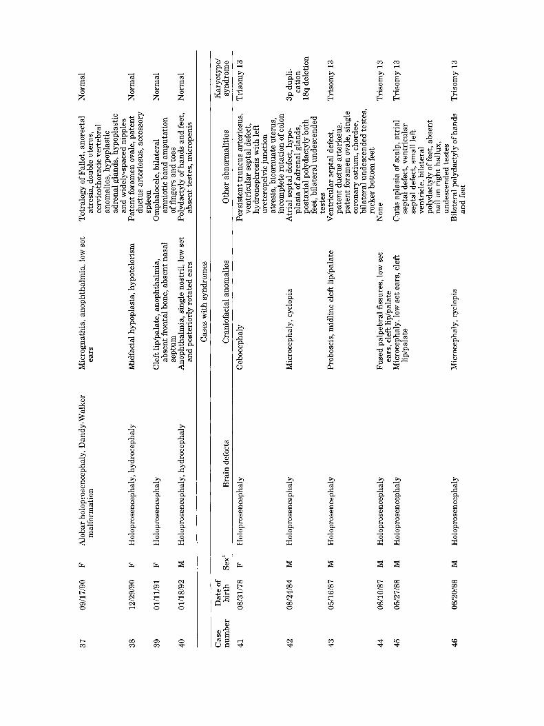
41
08/3
1/78
F
H
olop
rose
ncep
haly
42
08/2
4/84
M
H
olop
rose
ncep
haly
43
05/1
6/87
M
H
olop
rose
ncep
haly
44
08/1
0/87
M
H
olop
rose
ncep
haly
45
05/2
7/88
M
H
olop
rose
ncep
haly
46
08/2
0/88
M
H
olop
rose
ncep
haly
Cra
niof
acia
l ano
mal
ies
Ceb
ocep
haly
Mic
roce
phal
y, c
yclo
pia
Prob
osci
s, m
idlin
e cl
eft l
iplp
alat
e
Fuse
d pa
lpeb
ral
fiss
ures
, low
set
Mic
roce
phal
y, lo
w s
et e
ars,
cle
ft
ears
, cle
ft li
p/pa
late
lip/
pala
te
Mic
roce
phal
y, c
yclo
pia
Oth
er a
bnor
mal
itie
s
Pers
iste
nt tr
uncu
s ar
teri
osus
, ve
ntri
cula
r se
ptal
def
ect,
hydr
onep
hros
is w
ith
left
ur
eter
opel
vic
junc
tion
at
resi
a, b
icor
nuat
e ut
erus
, in
com
plet
e ro
tati
on o
f col
on
plas
ia o
f adr
enal
gla
nds,
po
stax
ial
poly
dact
yly
both
fe
et, b
ilat
eral
und
esce
nded
te
stes
pate
nt d
uctu
s ar
teri
osus
, pa
tent
for
amen
ova
le, s
ingl
e co
rona
ry o
stiu
m, c
hord
ee,
bila
tera
l un
desc
ende
d te
stes
, ro
cker
bot
tom
fee
t
Atr
ial s
epta
l def
ect,
hypo
-
Ven
tric
ular
sep
tal d
efec
t,
Non
e
Cut
is a
plas
ia o
f sca
lp, a
tria
l se
ptal
def
ect,
vent
ricu
lar
sept
al d
efec
t, sm
all l
eft
vent
ricl
e, b
ilat
eral
po
lyda
ctyl
y of
fee
t, ab
sent
na
il o
n ri
ght h
allu
x,
unde
scen
ded
test
es
and
feet
B
ilat
eral
pol
ydac
tyly
of h
ands
Kar
yoty
pe/
synd
rom
e
Tri
som
y 13
3p d
upli
- ca
tion
18q
dele
tion
Tri
som
y 13
Tri
som
y 13
Tri
som
y 13
Tri
som
y 13

APP
EN
DIX
A.
(con
tinue
d)
Cas
es w
ith
synd
rom
es
Cas
e D
ate
of
Kar
yoty
pel
num
ber
birt
h Se
x”
Bra
in d
efec
ts
Cra
niof
acia
l ano
mal
ies
Oth
er a
bnor
mal
itie
s sy
ndro
me
47
10/0
9/90
M
L
obar
hol
opro
senc
epha
ly, a
bsen
t cor
pus
Non
e st
ated
ca
llosu
m
48
12/1
8/90
F
Hol
opro
senc
epha
ly, a
bsen
t cor
pus
callo
sum
, pol
ymic
rogy
ria,
hy
droc
epha
lus,
bra
inst
em m
alfo
rmat
ion
49
08/2
1/91
M
H
olop
rose
ncep
haly
50
01/2
3/92
M
Fu
sed
port
ion
of c
ereb
ral v
entr
icle
s
51
03/3
0/92
F
H
olop
rose
ncep
haly
, abs
ent c
orpu
s ca
llosu
m, s
ingl
e ce
rebr
al v
entr
icle
52
05/1
5/92
F
Alo
bar
holo
pros
ence
phal
y
53
07/2
0/92
M
A
rhin
ence
phal
y w
ith
cent
ral b
rain
def
ect
Mac
roce
phal
y, m
icro
phth
alm
ia,
clef
t lip
/pal
ate,
low
set
ear
s
Not
sta
ted
Sing
le n
ostr
il, h
ypot
elor
ism
Non
e
Smal
l che
st, v
ery
shor
t ex
trem
itie
s, m
icro
gnat
hia,
bi
late
ral b
ifid
thum
bs,
bila
tera
l bif
id h
allu
ces,
po
lyda
ctyl
y of
rig
ht h
and
and
left
foo
t N
one
Abs
ence
rig
ht e
xter
nal e
ar a
nd c
anal
Bil
ater
al c
left
lipl
pala
te,
Ven
tric
ular
sep
tal d
efec
t, cr
osse
d re
nal e
ctop
ia
Secu
ndum
atr
ial s
epta
l de
fect
, pol
ydac
tyly
, rig
ht
tran
sver
se p
alm
ar c
reas
e,
sing
le u
mbi
lical
art
ery,
sy
ndac
tyly
mid
dle
fing
ers
of
left
han
d Po
lyda
ctyl
y of
han
ds a
nd le
ft
foot
, ven
tric
ular
sep
tal
defe
ct, i
ncom
plet
e lo
batio
n of
rig
ht lu
ng
prob
osci
s, m
icro
phth
alm
ia v
ersu
s ro
cker
bot
tom
fee
t wit
h an
opht
halm
ia
valg
us d
efor
mity
, hyp
o-
plas
tic
nail
s, p
ossi
ble
tran
sver
se p
alm
ar c
reas
e ri
ght h
and
mic
roph
thal
mia
, bil
ater
al c
atar
acts
Mid
faci
al h
ypop
lasi
a, p
arti
al
Uns
peci
fied
pol
ydac
tyly
,
APP
EN
DIX
A.
(con
tinu
ed) S
umm
ary
of A
rhin
ence
phal
y ca
ses
Nor
mal
/ pr
esum
ed
auto
som
al
rece
ssiv
e (s
ib of
Cas
e 49
) U
nkno
wn/
Sa
ldin
o-
Noo
nan
synd
rom
e
Nor
mal
/ pr
esum
ed
auto
som
al
rece
ssiv
e (s
ib of
Cas
e 47
) T
riso
my
18
Tri
som
y 13
Tri
som
y 13
Tri
som
y 13
Cas
e D
ate
of
Kar
yoty
pel
num
ber
birt
h Se
x“
Bra
in d
efec
ts
Cra
niof
acia
l an
omal
ies
Oth
er a
bnor
mal
itie
s sy
ndro
me
54
12/0
7/72
F
A
rhin
ence
phal
y M
icro
ceph
aly,
ano
phth
alm
ia
Dia
phra
gmat
ic h
erni
a,
Nor
mal
re
nal a
gene
sis,
abs
ent
thum
bs, p
ulm
onar
y hy
popl
asia
defo
rmitv
’. ab
senc
e of
55
08
/04/
72
F A
rhin
ence
phal
y, h
ydro
ceph
alus
N
ot s
tate
d O
mph
aloc
ele,
‘spi
nal
Unk
now
n
56
01/2
0/78
M
A
rhin
ence
phal
y, o
ccip
ital e
ncep
halo
cele
M
icro
ceph
aly
left
arm
N
one
Unk
now
n (c
onti
nued
)

APP
EN
DIX
A.
(con
tinue
d)
Cas
e D
ate
of
num
ber
birt
h Se
x"
Bra
in d
efec
ts
Cra
niof
acia
l an
omal
ies
57
05/1
0/78
F
Arh
inen
ceph
aly,
abs
ence
of c
orpu
s ca
llosu
m,
Ano
phth
alm
ia,
low
set
ear
s, c
left
hy
droc
epha
lus
pala
te
58
08/2
0/78
F
A
rhin
ence
phal
y
59
03/1
0/86
F
Arh
inen
ceph
aly
60
10/2
7/87
F
Arh
inen
ceph
aly
61
05/1
7/88
F
A
rhin
ence
phal
y, a
gene
sis o
f ce
rebe
llum
, pol
ymic
rogy
ria,
oc
cipt
al e
ncep
halo
cele
62
07/0
1/88
M
A
rhin
ence
phal
y, a
bsen
t cor
pus
callo
sum
63
08/2
3/90
M
A
rhin
ence
phal
y
Cor
neal
opa
city
, low
set
ear
s, c
left
pa
late
Mic
roce
phal
y, b
ilat
eral
cle
ft li
p/
pala
te, r
ight
iris
col
obom
a,
mic
roph
thal
mia
Not
sta
ted
Mid
line
clef
t pal
ate,
mic
roph
thal
mia
Eth
moc
epha
ly, m
icro
phth
alm
ia,
low
se
t ear
s, u
nspe
cifi
ed c
olob
omas
, hy
popl
astic
man
dibl
e
Mic
roce
phal
y, b
ilat
eral
cle
ft li
plpa
late
m
icro
phth
alm
ia,
low
set
ear
s
Oth
er a
bnor
mal
itie
s
pulm
onar
y hy
polo
batio
n,
pate
nt d
uctu
s ar
teri
osus
, in
test
inal
mal
rota
tion
, ch
olec
ystic
age
nesi
s,
impe
rfor
ate
anus
, abs
ent
digi
t of h
ands
and
feet
, tr
ansv
erse
pal
mar
cre
ases
, m
ale
pseu
dohe
rmap
hro-
di
tism
dupl
icat
ion
of r
enal
ar
teri
es, h
epat
omeg
aly,
di
sloc
ated
elb
ow,
hype
rext
ende
d 3r
d an
d 4t
h fi
nger
s, tr
ansv
erse
pa
lmar
cre
ases
Po
lyda
ctyl
y of
fee
t, ve
ntri
cula
r se
ptal
def
ect,
atri
al s
epta
l def
ect,
bico
rnua
te u
teru
s, c
ervi
x an
d va
gina
, pol
ycys
tic
hors
esho
e ki
dney
, Mec
kel's
di
vert
icul
um
Lef
t pul
mon
ary
arte
ry
hypo
plas
ia, p
aten
t fo
ram
en o
vale
, pat
ent
duct
us a
rter
iosu
s,
vent
ricu
lar
sept
al d
efec
t, le
ft d
iaph
ragm
atic
her
nia
Hyp
opla
stic
lung
s, p
olyc
ystic
ki
dney
s, p
osta
xial
pol
y-
dact
yly
of h
ands
and
fee
t, pa
tent
for
amen
ova
le,
pate
nt d
uctu
s ar
teri
osus
, bi
late
ral t
rans
vers
e pa
lmar
cr
ease
s
tali
pes
equi
nova
rus,
m
yelo
men
ingo
cele
, rig
ht
foot
with
sin
gle
digi
t an
d ro
cker
bot
tom
, sy
ndac
tyly
of f
inge
rs
14
on r
ight
and
1-2
, 4-
5 on
left
Po
stax
ial
poly
dact
yly
of f
eet,
sing
le u
mbi
lical
art
ery,
m
icro
peni
s w
ith
shaw
l sc
rotu
m, M
ecke
l's d
iver
ticu-
lu
m, i
rreg
ular
fusi
on o
f tai
l
Men
ingo
cele
, bil
ater
al
Shie
ld c
hest
,
Art
hrog
rypo
sis,
lef
t
Kar
yoty
pel
synd
rom
e
Unk
now
n
Unk
now
n
Tri
som
y 13
Tri
som
y 18
Unk
now
n/
Mec
kel
synd
rom
e
Nor
mal
Tri
som
y 13
of p
ancr
eas
to s
plee
n, b
icus
pid
aort
ic v
alve
, pat
ent
fora
men
ov
ale,
om
phal
ocel
e "M
, mal
e; F
, fem
ale;
U, u
ndet
erm
ined
.