designer nanoparticle: nanobiotechnology tool for cell biology · pdf filedesigner...
TRANSCRIPT
Thimiri Govinda Raj and Khan Nano Convergence (2016) 3:22 DOI 10.1186/s40580-016-0082-x
REVIEW
Designer nanoparticle: nanobiotechnology tool for cell biologyDeepak B. Thimiri Govinda Raj1,2,3* and Niamat Ali Khan4
Abstract
This article discusses the use of nanotechnology for subcellular compartment isolation and its application towards subcellular omics. This technology review significantly contributes to our understanding on use of nanotechnology for subcellular systems biology. Here we elaborate nanobiotechnology approach of using superparamagnetic nano-particles (SPMNPs) optimized with different surface coatings for subcellular organelle isolation. Using pulse-chase approach, we review that SPMNPs interacted differently with the cell depending on its surface functionalization. The article focuses on the use of functionalized-SPMNPs as a nanobiotechnology tool to isolate high quality (both purity and yield) plasma membranes and endosomes or lysosomes. Such nanobiotechnology tool can be applied in gen-erating subcellular compartment inventories. As a future perspective, this strategy could be applied in areas such as immunology, cancer and stem cell research.
Keywords: Nanoparticles, Subcellular proteomics, Nanobiotechnology, Systems biology
© 2016 The Author(s). This article is distributed under the terms of the Creative Commons Attribution 4.0 International License (http://creativecommons.org/licenses/by/4.0/), which permits unrestricted use, distribution, and reproduction in any medium, provided you give appropriate credit to the original author(s) and the source, provide a link to the Creative Commons license, and indicate if changes were made.
1 BackgroundNanotechnology is defined as molecular engineering of functional systems for generating high performance tech-nologies for both research and industry [1, 2]. Specifi-cally, use of molecular engineering of biological systems resulted in emergence of Nanobiotechnology [3]. Some of the nanobiotechnology applications include (a) nanos-copy for bio imaging [4–7]; (b) nanoparticle for subcel-lular fractionation [8]; (c) nanoparticle for drug delivery, vaccine [9]; (d) nanoparticle for cancer therapy by hyper-thermia [10]; (e) nanomaterials for tissue engineering and artificial/synthetic organ generation [11]; (f ) nanotech-nology for cell tracking [12, 13]; and (g) nanotechnology for large-scale data generation [14]. Particularly, use of nanotechnology in generating subcellular omics is less studied and understood. This article focuses on nanobio-technology strategy for organelle isolation and also deci-phers innovative approaches for omics analysis. Using physical properties, its nanoparticle-cell interaction and endocytosis machinery, we propose the nanobiotech-nology strategy that has robust advantages in isolating
subcellular compartments [15]. Due to such advantages, it is possible to isolate subcellular compartments in native and physiological conditions. In this article, we also case study the impact of nano-biotechnology tool for subcellular omics analysis.
2 Subcellular omicsTo generate subcellular omics datasets, it is essential to understand the locality and functional activity of pro-teins in given eukaryotic cell [16]. It is well-known that proteins are spatially distributed and localized function [17]. It has been reported that majority of the mature glycosylated protein (for example: Nicastrin) are present in post-Golgi compartments like plasma membrane, endosomes or lysosome and immature glycosylated pro-tein are present in the pre-Golgi compartments [18]. Similarly, cholesterol is predominately present in the cell membrane at the level of 90 % of the total cholesterol level in cell extract [19]. While performing proteom-ics or lipidomics in total cell extract or single cell omics, there is a high possibility of reduced spatial and localized distribution of proteins and lipids in any given cell. This has led to a major interest for subcellular omics such as plasma membrane and endosomal compartments like endosomes and lysosomes [20]. This is due to several
Open Access
*Correspondence: [email protected] 2 Envirotransgene Bio-solutions Global, Chennai, IndiaFull list of author information is available at the end of the article

Page 2 of 12Thimiri Govinda Raj and Khan Nano Convergence (2016) 3:22
scientific findings which confirm that the majority of pro-tein functional activity and substrate cleavage occurs at cell surface and endosomal compartments (Table 1) [21]. Similarly by isolating plasma membrane or endosomes, it is possible to generate proteomics, glycomics and lipi-domics for these organelles [22]. By compiling obtained omics (proteomics, glycomics, and lipidomics), for dif-ferent organelles, comprehensive whole cell omics can be generated both under native and altered conditions [23–26]. The key factor for generating comprehensive omics datasets s is to isolate subcellular compartments with high purity and yield. Several types of fractionation methodologies have been applied for organelle isolation for subcellular omics analysis. In the technology review, we elaborate advantages among different subcellular compartmental isolation and how nanobiotechnology strategy is superior in isolating plasma membrane and endosomes as in Tables 2 and 3.
3 Technology review3.1 Organelle fractionation and subcellular
compartmental isolationThe governing factor for organelle fractionation is high yield and high purity (Fig. 1). Most commonly used methodology is density-gradient centrifugation (sucrose-based fractionation). This method is based on principle of differential (density) equilibrium or non-equilibrium based centrifugation for organelle separa-tion [27, 28]. Other commonly used fractionation is antibody based pulldown assay. This assay makes use of magnetic beads that are tagged with antibodies selec-tively targeting the subcellular compartments [29]. An example is the use of TOM22 antibodies conjugated with magnetic beads for mitochondria isolation [30, 31]. This principle is also used for post-nanoparticle labeling based fractionation. Here, organelle-specific antibody conjugated nanoparticles are used to target fractionated
Table 1 List of subcellular organelles and their functions
Subcellular Organelles Isolation techniques
Cell wall Gradient centrifugation
Chloroplast Gradient centrifugation antibody based pull-down assay, SPMNPS: Tag-anti-tag; Antibody conjugated; Biotin-streptavidin
Cilia and flagella Gradient centrifugation Antibody based pull-down assay; Tag-anti-tag; Antibody conjugated; Biotin-streptavidin
Cytoplasm Gradient centrifugation antibody based pull-down assay; Tag-anti-tagged; Antibody conjugated; Biotin-streptavidin tagged SPMNPs
Cytoskeleton Gradient centrifugation antibody based pull-down assay; Tag-anti-tagged; Antibody conjugated; Biotin-streptavidin SPM-NPs
Early endosomes Gradient centrifugation SPMNP isolation assay antibody based pull-down assay; Biotin-streptavidin tagged SPMNPs; antibody-SPMNPs; Negatively charged lipid-SPMNPs
Endoplasmic reticulum (ER)—rough or smooth
Gradient centrifugation antibody based pull-down assay; Tag-anti-tag; Antibody conjugated; Biotin-streptavidin tagged SPMNPs
Golgi apparatus Gradient centrifugation antibody based pull-down assay; Tag-anti-tagged; Antibody conjugated; Biotin-streptavidin tagged SPMNPs
Late endosomes Gradient centrifugation SPMNP isolation assay; Biotin-streptavidin; antibody-SPMNPs; Negatively charged SPMNPs
Multi-vesicular bodies Gradient centrifugation SPMNP isolation assay; antibody based pull-down assay; Biotin-streptavidin; antibody-SPMNPs
Nucleus Gradient centrifugation antibody based pull-down assay; Tag-anti-tagged; Antibody conjugated; Biotin-streptavidin
Peroxisomes Gradient centrifugation antibody based pull-down assay; Tag-anti-tagged; Antibody conjugated; Biotin-streptavidin tagged SPMNPs
Phagosomes Gradient centrifugation SPMNP isolation assay Antibody based pull-down assay; Biotin-streptavidin; antibody SPMNPs
Lysosomes Gradient centrifugation SPMNP isolation assay Antibody based pull-down assay; Biotin-streptavidin tagged SPMNPs; antibody
Plasma Membrane or Cell Membrane
Gradient centrifugation; Cationic silica beads; SPMNP isolation; antibody conjugated; Biotin-Streptavidin; lectin–SPMNPs
Ribosomes Gradient centrifugation; pull -down assay; anti-S10/anti–EF-Tu-SPMNPs; Biotin-streptavidin; antibody- SPMNPs
Lipid rafts Gradient centrifugation antibody based pull-down assay; Tag-antitagged-SPMNPs; Biotin-streptavidin; protein conjugated magnetic isolation
Secretory granules or vesicles
Gradient centrifugation antibody based pull-down assay; Tag-antitagged SPMNPs; Biotin-streptavidin-SPMNPs
Synaptosomes Gradient centrifugation antibody based pull-down assay; Tag-anti-tag SPMNPs; Antibody conjugated; Biotin-streptavidin SPMNPs
Vacuoles Gradient centrifugation; antibody based pull-down assay; antibody conjugated magnetic nanoparticles
Mitochondria Gradient centrifugation; pull-down assay; antibody conjugated SPMNPs; Tag-anti-tagged; Antibody conjugated; Biotin-streptavidin; Anti-TOM22 antibody tagged SPMNPs

Page 3 of 12Thimiri Govinda Raj and Khan Nano Convergence (2016) 3:22
subcellular compartment. This technique is largely used in isolating compartments that are larger in size and less dynamic or more static such as ER, Golgi, nucleus,
mitochondria and lysosomes [32]. However, the applica-bility of this technique is limited as it cannot be used to isolate intact cell membrane. Many methods have used
Table 2 Comparison of existing technologies for plasma membrane isolation
Isolation methodologies Advantages Disadvantages
Density Gradient Centrifugation Conventional method that can be used to isolate plasma mem-brane along with other subcellular compartments
Effective method to isolate lysosomes from tissue or in vivo cell fractions
Simple procedure that can be performed using an ultracentri-fuge
Low yield and low purity Cannot isolate intact membrane layersNot efficient for isolating cell membrane lipids
and for performing functional studies
Cationic silica based isolation Classical method used to isolate cell membrane layers with high purity
Generic method used to isolate cell membrane from tissue, in vitro and in vivo
Formation of matrix by the use of polylysine crosslinker helps in the isolation of membrane layers without any breakage
Low yield Isolates only available membrane layer i:e only
50 % of cell surface of adherent cells grown on a petri dish
Not robust to perform lipidomics, glycomics and native condition experiments
Cell Surface biotinylation based pull-down assay
Generic method that targets cell surface lysine residueCan be used in combination with magnetic or non-magnetic
beadsCan be used in combination to isolate endosomal compart-
ments
Can isolate only available membrane layer i:e only 50 % of cell surface of adherent cells growing on a petri dish
Technology has not been established to isolate cell membrane from cell suspension
Requires detergent in fractionations
Antibody conjugated magnetic nanoparticle based pulldown assay
Can be used to pull down proteins after post-fractionationCan also be used to target selective micro-domains Can be used in combination with biotin-streptavidin assay
Isolates only available membrane layer (only 50 % surface of adherent cells on a petri dish
Technology has not been established to isolate cell membrane from cell suspension
requires detergent in fractionations
SPMNPs based plasma mem-brane isolation;
A novel strategy that is generic for any kind of cell systems Method does not involve use of detergent or antibody that
affects the nativity of membranes Method can also use targeted plasma membrane micro-domain
by using ligand-taggingCan be used to isolate protein under native conditions which
can hence be used for functional studiesCan isolate cell membrane lipidsCan perform, first of its kind, cell membrane glycosylationHigh yield and high purity Can be used in combination with endosomal isolations
Technology has not be established for tissue cell membrane isolation and in vivo experiments
Isolates only available membrane layer (only 50 % surface of adherent cells on a petri dish
Technology has not been established to isolate cell membrane from cell suspension. Possibility exists to use this technology for cells in suspen-sion culture
Table 3 Comparison of existing technologies for endosomes and lysosome isolation
Isolation meth-odologies
Advantages Disadvantages
Density gradient centrifugation;
Conventional method that can be used to isolate endosomes and lys-osomes along other subcellular compartments
Effective method to isolate lysosomes from tissue or in vivo cell fractions Simple procedure that can be performed using an ultracentrifuge
Low yieldDifficulties in separating endosomes from lysosome
vesiclesDifficulties in separating different endosomal and its
associated vesicles
Antibody based pull-down assay
Can be used to pull down proteins after post-fractionationCan also be used to target selective endocytic uptakesCan be used in combination with biotin-streptavidin assay
Limited applicability for certain endocytosis uptake Limited yield and low purity Cannot isolate vesicles under native conditions
SPMNPs based isolation;
A novel strategy that is generic for any kind of cell systems with reason-able purity and yield
Method does not involve the use of detergent or antibody that affects native conditions
Method can also use targeted endosomal uptake pathway by using ligand tagged nanoparticle
Can be used to isolate protein under native conditions and all endoso-mal uptake
Technology has not be established for isolation of vesicles form tissue cells and in vivo experiments
Technology has not been established to isolate vesi-cles from cell suspension. Possibility exists to use this technology for cells in suspension culture

Page 4 of 12Thimiri Govinda Raj and Khan Nano Convergence (2016) 3:22
charge based affinity to isolate eukaryotic cell mem-brane [33]. The cell membrane is negatively charged due to presence of anionic components such as proteins, lipids and carbohydrates. Hence, conventional method such as cationic latex or silica beads is used for isolat-ing cell membrane. By using poly-lysine for crosslinking silica beads, cell membranes are isolated as cross-linked membrane layers which are disadvantage for performing functional studies [34]. An alternative method that has been used is biotin-streptavidin affinity fractionation. The main principle behind biotin-streptavidin affin-ity assay is based on selective binding of available lysine
residues on cell surface protein by biotin molecule which is further captured by streptavidin [35]. These streptavi-din tagged micro beads or magnetic beads are used to pull down cell membrane protein or protein complexes from cell fractionation. By using pulse-chase method (elaborated in the later part of the paper), biotin-strepta-vidin affinity can be used to isolate early, intermediate and late endosomal compartments [36]. Recently, nano-particle based fractionation has emerged as the strategy to isolate dynamic subcellular compartments like cell membrane, endosomes and lysosome by using endocy-tosis machinery.
Fig. 1 Subcellular compartments and specific purification methodology

Page 5 of 12Thimiri Govinda Raj and Khan Nano Convergence (2016) 3:22
There are different categories of endocytosis (Fig. 2). One of the main subsets is phagocytosis that mainly involves cellular uptake of beads of size 1–100 microns using phagosome [37, 38]. Another strategy is use of pinocytosis which is further classified into (1) micro-pinocytosis (<1 micron) [39]; (2) clathrin-mediated pinocytosis: size ~120 nm by clathrin mediated recep-tor-ligand induced cellular uptake [40]; (3) caveolin-mediated pinocytosis: is cellular uptake that uses caveolin and lipid rafts in endosomal assembly with size of 20–60 nm [41]; and (4) caveolin and clathrin independent endocytosis (<90 nm) that is independ-ent of clathrin and caveolin but includes ARF6 [42]. In macropinocytosis, there is non-specific cellular uptake, which is mainly utilized by several nanoparticles–cell interactions [43]. In clathrin mediated endocytosis, ligand coupled nanoparticle endocytosis through recep-tor mediated uptake mechanism [44, 45]. While for caveolin mediated endocytosis, targeting caveolin1/2 or lipid raft associated protein flotilin-1 with the specific antibody tagged nanoparticle is the commonly used approach [46]. However, when clathrin mediated endo-cytosis was blocked using sucrose, nanoparticle uptake was not limited, and thereby showing that there is size dependent cellular uptake of nanoparticle via caveo-lin mediated endocytosis [47]. Nevertheless it is clear that surface coating and size of the nanoparticle are
governing factors for selective endocytosis and cellular uptake.
4 Nanoparticle synthesisThe surface functionalization, size, physical properties and endocytosis machinery of nanoparticle are key fac-tors for nanobiotechnology strategy. The physical proper-ties are dependent on the type of core–shell material [48, 49]. It is possible to govern the magnetic properties of the nanoparticle by using iron oxide or cobalt-iron oxide as a core. Here superparamagnetic properties can be achieved when nanomaterial is of size <30 nm [50]. Shell material (surface coating) which acts as an interface between core and biological environment governs the use of nanopar-ticle for different biological applications. Nanoparticles that are synthesized in organic phase tend to be water insoluble. They require an additional step of exchange with water soluble ligands for biological applications. Commonly used methods for synthesizing nanoparticle are (a) chemical precipitation method and (b) thermal decomposition method [51]. Thermal decomposition method is preferred for its high monodispersity (in terms of its size) and high quality yield. However, it requires additional step for water-soluble ligand exchange or addi-tion [52, 53] (Fig. 3). Further, functionalized nanoparticle can be used for ligand coupling and bioconjugation using the free end-groups like NH2, COOH and -SH.
Fig. 2 Endocytosis pathway

Page 6 of 12Thimiri Govinda Raj and Khan Nano Convergence (2016) 3:22
5 Bioconjugation strategy for subcellular interaction
Surface functionalization of nanoparticle determines the kinetics behind nanoparticle-cellular uptake. Depending on the surface coating, nanoparticle can be ± charged which is determined by the presence of NH2 and COOH respectively. Addition of PEG group in supports bio-compatibility [54] and the nanoparticle can be further functionalized by coupling with the endgroup (Fig. 4). By tagging fluorescent ligand, it is possible to perform live cell imaging and nanoparticle tracking for studying
the receptor-ligand and nanoparticle-cell interaction. Depending on the target, an appropriate ligand can be selected and conjugated to the nanoparticle [55]. Ligand size and shape determines the surface area to volume ratio which is the governing factor for nanoparticle func-tionalization [56]. Another key factor is ligand selection. Ligand selection depends—(a) receptor that can be well internalized (for cellular trafficking); b) 1:1 ratio of ligand: nanoparticle to avoid nanoparticle crosslinking. Here we illustrate three strategies for bioconjugation of SPMNPs that are functionalized with lipids, DMSA, TMAOH (Fig. 5). First approach is to use monovalent avidin and target biotinylated protein. Second approach proposes the conjugation of FIAsH-EDT2 with SPMNPs. FIAsH-EDT2 coupled SPMNPs are used to couple tetra-cysteine containing motif proteins [57]. Third approach is to con-jugate SPMNPs with DOGS-NTA-Ni (II) in order to anchor histidine-tagged protein. However, major limita-tion for bioconjugation is nanoparticle aggregation due to coupling reagent like glutaraldehyde. Although glu-taraldehyde works very well for protein conjugation and crosslinking, there are tendency for reagent to result in multiple layer crosslinking among nanoparticles due to non-specific interaction and competitive affinity. Such multilayer crosslinking results in increase of size and change in physical properties, thereby affecting organelle isolation. Hence some key guidelines to be considered during bioconjugation of nanoparticles are (1) retain-ing size and stability of nanoparticle; (2) performing sequential bioconjugation; (3) implementing biocompat-ible surface functionalization; and (4) finally, a strategy that monitors protein association/disassociation with the nanoparticle.
6 Pulse‑chase methodologyPulse-chase methodology is a commonly used approach to study the mechanism of endocytosis. Generally, pulse-chase strategy for omics analysis includes five stages or phases wherein Phase-I: includes generation of water-soluble nanoparticle by existing (thermal decomposi-tion or chemical precipitation) synthesis and quality control using characterization; Phase II: includes selec-tive bioconjugation of nanoparticle for a selective path-way-specific cellular uptake. For such pathway-specific cellular uptake, protein/ligand/synthetic peptide is used for receptor mediated endocytosis and charge depend-ent shell uptake is used for receptor independent endo-cytosis. Phase III: Pulse-Chase methodology is used to optimize pulse and chase period to selectively localize nanoparticle in vesicle. Phase IV: Magnetic separation strategy is used for subcellular compartmental enrich-ment along with ultracentrifugation. Phase V: Endo-somal proteome using Mass Spectrometry analysis.
Fig. 3 Manufacturing of water-soluble superparamagnetic nanopar-ticles
Fig. 4 Nanoparticle-cell interaction

Page 7 of 12Thimiri Govinda Raj and Khan Nano Convergence (2016) 3:22
This pulse-chase strategy was commonly used in radio-active labeling in the cell and this technique has now been extended to nanoparticle based subcellular com-partmental isolation (Fig. 6). Briefly, pulse-chase strat-egy is used to govern receptor-mediated endocytosis of nanoparticle-ligand complex and has recently been extended to other endocytosis mechanisms [58]. Dur-ing pulse period, nanoparticle is incubated with cells at 37 °C or at 4 °C for a certain period of time (0–1 h.) in the presence of medium. Depending on the application, nanoparticle with appropriate concentration is incu-bated in PBS or culture medium at 4 °C (for non-chase conditions) and 37 °C (for chase conditions). This time frame allows nanoparticle to interact with the cell sur-face and its protein. Depending on the dynamics and kinetics of nanoparticle-ligand interaction, pulse incuba-tion involves a time period in the range of 10 min to 1 h. For example, if it is for cell membrane or plasma mem-brane isolation, the nanoparticle is incubated at 4 °C for 15–20 min in PBS with the adherent cells. Depending on whether the cells are adherent or in suspension, or it is receptor mediated or charge mediated, there is variation in the pulse time period required for cellular uptake [59]. After pulse period is performed, the chase is incubated at
appropriate time period depending on the compartmen-tal isolation. Chase period represents the time where the nanoparticle containing medium is replaced with fresh medium without nanoparticle. This supports stream-lining nanoparticle internalization in the cell and accu-mulation of nanoparticle into a certain compartment of interest depending on the timeframe. For endosomal isolation, chase period is generally for a timeframe of 10–15 min. For late endosomes, chase period is generally for 15–20 min and for lysosomes it is more than 30 min. However since endocytosis is dynamic in mechanism, it is relatively difficult to isolate highly pure early and late endosomes (Fig. 7). At the same time it is possible to iso-late highly pure lysosome by performing a chase period of more than 3 h and up to 24 h. This is mainly because lysosome is the endpoint for most of the endocytosis [60]. For targeting, phagosome or autophagosome, chase period is adjusted accordingly for 30–60 min before phagosome fuses to lysosome. Nanoparticle can be con-centrated in lysosome after 60 min of chase period. An advantage of using chase period is that it provides use-ful information for nanoparticle tracking. For this reason, fluorescence tagged nanoparticle is used for pulse–chase methodology and live-cell imaging [61]. By incubating
Fig. 5 Bioconjugation strategy for nanoparticle

Page 8 of 12Thimiri Govinda Raj and Khan Nano Convergence (2016) 3:22
with endocytic inhibitors for endosomal or lysosomal fusion, it is possible to limit the nanoparticle-cellular internalization and subcellular trafficking. For example,
by limiting the endosome-lysosomal fusion using Latrun-culin-A, it is possible to concentrate the nanoparticle in early or late endosomes [62]. It is also reported that the
Fig. 6 Step by step approach toward subcellular compartmental proteomics
Fig. 7 Pulse-chase methodology
Page 9 of 12Thimiri Govinda Raj and Khan Nano Convergence (2016) 3:22
nanoparticle coupled ligand does not mimic the ligand cellular uptake and subcellular compartment localization. Few examples in the later part of this article elaborate on the deviation in cellular uptake. These examples are confirmed by using pulse-chase methodology, magnetic organelle isolation and live cell imaging using fluores-cence tag [63]. For optimal use of pulse-chase method, it is important to establish a methodology for specific sub-cellular compartments. Here, we describe two interest-ing nanoparticle based methodologies to isolate plasma membrane and endosomal compartments using affinity purification.
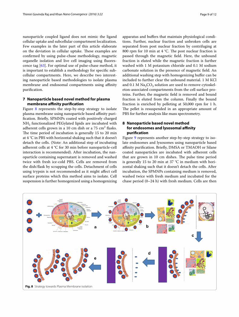
7 Nanoparticle based novel method for plasma membrane affinity purification
Figure 8 represents the step-by-step strategy to isolate plasma membrane using nanoparticle based affinity puri-fication. Briefly, SPMNPs coated with positively charged NH2 functionalized PEGylated lipids are incubated with adherent cells grown in a 10 cm dish or a 75 cm2 flasks. The time period of incubation is generally 15 to 20 min at 4 °C in PBS with horizontal shaking such that it doesn’t detach the cells. (Note: An additional step of incubating adherent cells at 4 °C for 30 min before nanoparticle-cell interaction is recommended). After incubation, the nan-oparticle containing supernatant is removed and washed twice with fresh ice-cold PBS. Cells are removed from the dish/flask by scrapping the cells. Detachment of cells using trypsin is not recommended as it might affect cell surface proteins which this method aims to isolate. Cell suspension is further homogenized using a homogenizing
apparatus and buffers that maintain physiological condi-tions. Further, nuclear fraction and unbroken cells are separated from post nuclear fraction by centrifuging at 800 rpm for 10 min at 4 °C. The post nuclear fraction is passed through the magnetic field. Here, the unbound fraction is eluted while the magnetic fraction is further washed with 1 M potassium chloride and 0.1 M sodium carbonate solution in the presence of magnetic field. An additional washing step with homogenizing buffer can be included to further clear the unbound material. 1 M KCl and 0.1 M Na2CO3 solution are used to remove cytoskel-eton-associated compartments from the cell surface pro-teins. Further, the magnetic field is removed and bound fraction is eluted from the column. Finally the bound fraction is enriched by pelleting at 50,000 rpm for 1 h. The pellet is resuspended in an appropriate amount of PBS for further analysis like mass spectrometry.
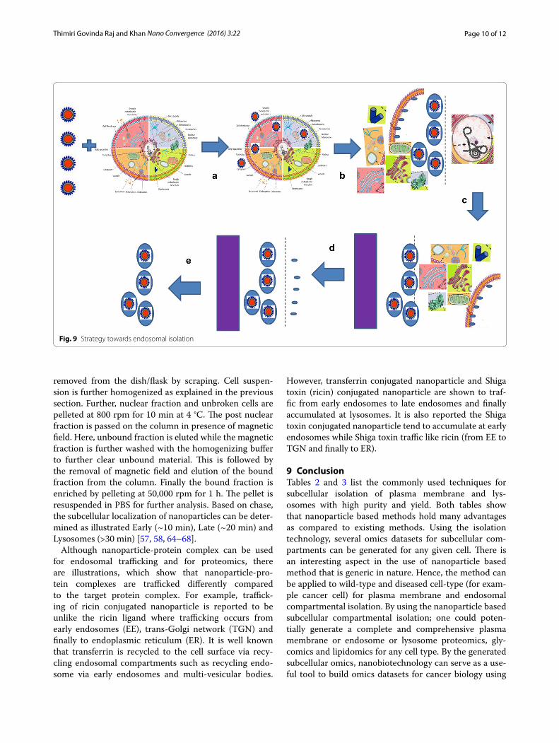
8 Nanoparticle based novel method for endosomes and lysosomal affinity purification
Figure 9 represents another step-by-step strategy to iso-late endosomes and lysosomes using nanoparticle based affinity purification. Briefly, DMSA or TMAOH or Silane coated nanoparticles are incubated with adherent cells that are grown in 10 cm dishes. The pulse time period is generally 15 to 20 min at 37 °C in medium with hori-zontal shaking such that it doesn’t detach the cells. After incubation, the SPMNPs containing medium is removed, washed twice with fresh medium and incubated for the chase period (0–24 h) with fresh medium. Cells are then
Fig. 8 Strategy towards Plasma Membrane isolation
Page 10 of 12Thimiri Govinda Raj and Khan Nano Convergence (2016) 3:22
removed from the dish/flask by scraping. Cell suspen-sion is further homogenized as explained in the previous section. Further, nuclear fraction and unbroken cells are pelleted at 800 rpm for 10 min at 4 °C. The post nuclear fraction is passed on the column in presence of magnetic field. Here, unbound fraction is eluted while the magnetic fraction is further washed with the homogenizing buffer to further clear unbound material. This is followed by the removal of magnetic field and elution of the bound fraction from the column. Finally the bound fraction is enriched by pelleting at 50,000 rpm for 1 h. The pellet is resuspended in PBS for further analysis. Based on chase, the subcellular localization of nanoparticles can be deter-mined as illustrated Early (~10 min), Late (~20 min) and Lysosomes (>30 min) [57, 58, 64–68].
Although nanoparticle-protein complex can be used for endosomal trafficking and for proteomics, there are illustrations, which show that nanoparticle-pro-tein complexes are trafficked differently compared to the target protein complex. For example, traffick-ing of ricin conjugated nanoparticle is reported to be unlike the ricin ligand where trafficking occurs from early endosomes (EE), trans-Golgi network (TGN) and finally to endoplasmic reticulum (ER). It is well known that transferrin is recycled to the cell surface via recy-cling endosomal compartments such as recycling endo-some via early endosomes and multi-vesicular bodies.
However, transferrin conjugated nanoparticle and Shiga toxin (ricin) conjugated nanoparticle are shown to traf-fic from early endosomes to late endosomes and finally accumulated at lysosomes. It is also reported the Shiga toxin conjugated nanoparticle tend to accumulate at early endosomes while Shiga toxin traffic like ricin (from EE to TGN and finally to ER).
9 ConclusionTables 2 and 3 list the commonly used techniques for subcellular isolation of plasma membrane and lys-osomes with high purity and yield. Both tables show that nanoparticle based methods hold many advantages as compared to existing methods. Using the isolation technology, several omics datasets for subcellular com-partments can be generated for any given cell. There is an interesting aspect in the use of nanoparticle based method that is generic in nature. Hence, the method can be applied to wild-type and diseased cell-type (for exam-ple cancer cell) for plasma membrane and endosomal compartmental isolation. By using the nanoparticle based subcellular compartmental isolation; one could poten-tially generate a complete and comprehensive plasma membrane or endosome or lysosome proteomics, gly-comics and lipidomics for any cell type. By the generated subcellular omics, nanobiotechnology can serve as a use-ful tool to build omics datasets for cancer biology using
Fig. 9 Strategy towards endosomal isolation

Page 11 of 12Thimiri Govinda Raj and Khan Nano Convergence (2016) 3:22
the bottom-up pyramid approach. Using the bottom-up pyramid approach in omics analysis, subcellular omics datasets (including genomics, proteomics, lipidomics and glycomics) can be compiled together and compared with the whole cell omics analysis. This approach can also be used to generate several omics datasets in cancer biology that can enable the researchers to revisit the subcellular omics in order to understand the biological significance and functional relevance. For all such omics analysis stud-ies, it is necessary to have an efficient, robust and high precision technology for subcellular compartmental iso-lation. The technology also needs optimization and fine tuning depending on its applicability with host cell sys-tem. Using different nanobiotechnology tools for subcel-lular compartmental isolation, several high-throughput functional omics dataset like fluxomics, metabolomics, interactomics and localizomics can be generated. Using all these datasets, comprehensive Phenome and subcellu-lar omics are generated that can by analyzed using nano-technology for data storage studies. Further dataset thus gets larger for different diseases such as cancer, diabetes, infectious diseases, ageing related diseases, and neurode-generative diseases. These dataset and nanotechnology based analytics can be used in drug development, pre-clinical studies, patent analytics, and other applications. As a future perspective, the use of nanoparticle as nano-biotechnology tool is all set to be a game changer in the generation of Datasets for systems biology.
Authors’ contributionsDBTGR and NAK designed, and wrote the manuscript. Both authors read and approved the final manuscript.
Author details1 European Molecular Biology Laboratory (EMBL), Grenoble Outstation and Unit of Virus Host-Cell Interactions (UVHCI), UJF-EMBL-CNRS, UMR 5233 Grenoble, France. 2 Envirotransgene Bio-solutions Global, Chennai, India. 3 Biotechnology Centre for Oslo, Centre for Molecular Medicine Norway (NCMM), P.O. Box 1137, Blindern, 0318 Oslo, Norway. 4 Laboratory of Lipid Metabolism and Cancer, O&N I, Herestraat 49, Box 902, 3000 Louvain, Belgium.
AcknowledgementsFigures are made through Servier Medical Art (http://www.servier.com). The author thanks funding from the Not for Profit firms Envirotransgene™ Biosolu-tions Global and Deepak Thimiri Consulting Group (DTCG™) Grenoble, France/Chennai, India.
Competing interestsThe authors declare that they have no competing interests. Deepak B. Thimiri Govinda Raj is an inventor of Plasma membrane isolation patent and consult-ant for Envirotransgene™ Biosolutions.
Received: 26 April 2016 Accepted: 29 August 2016
References 1. Robert Lee, Elen Stokes, Twenty-first century novel: regulating nanotech-
nologies. J. Environ. Law. 21(3), 469–482 (2009)
2. R. Chau, S. Datta et al., Benchmarking nanotechnology for high-perfor-mance and low-power logic transistor applications. IEEE Trans. Nanotech-nol. 4(2), 153–158 (2005)
3. Ehud Gazit, Plenty of room for biology at the bottom: an introduction to bionanotechnology (Aviv University, Israel, 2007). ISBN: 978-1-84816-930-2
4. Y. Zhang et al., Nanobiotechnology: quantum dots in bioimaging. Expert Rev Proteomics 4(4), 565–572 (2007)
5. K. Bonroy et al. Nanotechnology towards Biomedical Applications. Nano-bioEurope Congress (2007)
6. J. Trekker, DBTG. Raj et al. Effect of the core size of monodisperse super-paramagnetic nanoparticles on their relaxometric enhancing properties for MRI. 8th International Conference on Scientific and Clinical Applica-tions of Magnetic Carriers, Rostock, 2010
7. J. Trekker, DBTG. Raj et al. The synthesis of superparamagnetic nanoparti-cles towards MRI contrasts agents, DIMI/EMl meeting (2008) Abstract
8. D.B. Thimiri Govinda Raj et al., A novel strategy for the comprehensive analysis of the biomolecular composition of isolated plasma membranes. Mol. Syst. Biol. 7, 541 (2011)
9. H.M. Mansour et al., Nanomedicine in pulmonary delivery. Int. J. Nanomed. 4, 299–319 (2009)
10. M.E. Davis, Z. Chen, Nanoparticle therapeutics: an emerging treatment modality for cancer. Nat. Rev. 7, 771–782 (2008)
11. J.L. Brown et al., Composite scaffolds: bridging nanofiber and micro-sphere architectures to improve bioactivity of mechanically competent constructs. J. Biomed. Mater. Res. Part A 95, 1150–1158 (2010)
12. Ivo Iavicoli et al., Opportunities and challenges of nanotechnology in the green economy. Environ. Health 13, 78 (2014)
13. J. Trekker, DBTG. Raj et al. Synthesis of superparamagnetic nanoparticles for stem cell tracking, Nanobioeurope (2008) Abstract
14. H. John, Phan et al., Convergence of biomarkers, bioinformatics and nanotechnology for individualized cancer treatment. Trends. Biotechnol. 27(6), 350–358 (2009)
15. H.-S. Li, D.B. Stolz, G. Romero, Characterization of endocytic vesicles using magnetic microbeads coated with signalling ligands. Traffic 6, 324–334 (2005)
16. H. Lodish. The architecture of cells—molecular cell biology (2000) 17. Arseny S. Kaprelyants, Dynamic spatial distribution of proteins in the cell.
Trends. Biochem. Sci. 13(2), 43–46 (1988) 18. M. Sperandio et al., Glycosylation in immune cell trafficking. Immunol.
Rev. 230(1), 97–113 (2009) 19. P. Goluszko, B. Nowicki, Membrane cholesterol: a crucial molecule affect-
ing interactions of microbial pathogens with mammalian cells. Infect. Immun. 73(12), 7791–7796 (2005)
20. L. Pelkmans et al., Genome-wide analysis of human kinases in clathrin- and caveolae/raft-mediated endocytosis. Nature 436, 78–86 (2005)
21. W.M. Rideout III et al., Nuclear cloning and epigenetic reprogramming of the genome. Science 293(5532), 1093–1098 (2001)
22. D.B. Thimiri Govinda Raj et al. Plasma membrane Isolation (2011) US Pat-ent 8, 936,935
23. G.R. Rosania et al., Visualizing chemical structure-subcellular localization relationships using fluorescent small molecules as probes of cellular transport. J. Cheminform 5, 44 (2013)
24. V. Collura, G. Boissy, From protein-protein complexes to interactomics. Subcell. Biochem. 43, 135–183 (2007)
25. A.R. Joyce, B.Ø. Palsson, The model organism as a system: integrating ‘omics’ data sets. Nat. Rev. Mol. Cell Biol. 7, 198–210 (2006)
26. Md Altaf-Ul-Amin, F.M. Afendi, S.K. Kiboi, S. Kanaya, Systems biology in the context of big data and networks. BioMed Res Int. 2014, 11 (2014). doi:10.1155/2014/428570
27. G. Raposo et al., B lymphocytes secrete antigen-presenting vesicles. J. Exp. Med. 183(3), 1161–1172 (1996)
28. J.Graham and J. R. Harris. Isolation and Functional Analysis of Organelles, in Cell Biology Protocols In: J. R. Harris, J. Graham and D. Rickwood eds, John Wiley & Sons, Ltd, Chichester, UK (2006)
29. Ankur Jain et al., Single-molecule pull-down for studying protein interac-tions. Nat. Protoc. 7, 445–452 (2012)
30. T.H. Taylor et al., Analysis of individual mitochondria via fluorescent immunolabeling with anti-TOM22 antibodies. Anal. Bioanal. Chem. 406(6), 1683–1691 (2014)
31. C.P. Satori et al., Bioanalysis of eukaryotic organelles. Chem. Rev 113(4), 2733–2811 (2013)

Page 12 of 12Thimiri Govinda Raj and Khan Nano Convergence (2016) 3:22
32. Nowacek A et al. Methods for Isolation and Identification of Nanoparticle-Containing Subcellular Compartments. Methods. Mol. Biol. (Clifton NJ 991: 47–55) (2013)
33. D. Josic, J.G. Clifton, Mammalian plasma membrane proteomics. Proteom-ics 7, 3010–3029 (2007)
34. Kim Y et al. Use of colloidal silica-beads for the isolation of cell-surface proteins for mass spectrometry-based proteomics. Methods. Mol. Biol. (Clifton, N.J.) 748: 227–241 (2011).
35. Yingxin Zhao et al., Proteomic analysis of integral plasma membrane proteins. Anal. Chem. 76(7), 1817–1823 (2004)
36. J. Fahrer et al., C2-streptavidin mediates the delivery of biotin-conjugated tumor suppressor protein p53 into tumor cells. Bioconjug Chem. 24(4), 595–603 (2013)
37. Jason M. Kinchen, Kodi S. Ravichandran, Phagosome maturation: going through the acid test. Nat. Rev. Mol. Cell Biol. 9, 781–795 (2008)
38. J.P. Luzio et al., Lysosomes: fusion and function. Nat. Rev. Mol. Cell Biol. 8, 622–632 (2007)
39. L. Jet Phey, A. Gleeson, Macropinocytosis: an endocytic pathway for internalizing large gulps. Immunol. Cell Biol. 89, 836–843 (2011)
40. H.T. McMahon, Emmanuel Boucrot, Molecular mechanism and physi-ological functions of clathrin-mediated endocytosis. Nat. Rev. Mol. Cell Biol. 12, 517–533 (2011)
41. R. Ivan, Nabi and Phuong U. Le., Caveolae/raft-dependent endocytosis. J. Cell Biol. 161(4), 673–677 (2003)
42. J.K. Schweitzer, A.E. Sedgwick, C. D’Souza-Schorey, ARF6-mediated endo-cytic recycling impacts cell movement, cell division and lipid homeosta-sis. Semin. Cell Dev. Biol. 22(1), 39–47 (2011)
43. T.G. Iversen, N. Frerker, K. Sandvig, Uptake of ricinB-quantum dot nano-particles by a macropinocytosis-like mechanism. J. Nanobiotechnol 10, 33 (2012)
44. H. Li, Z.M. Qian, Transferrin/transferrin receptor-mediated drug delivery. Med. Res. Rev. 22, 225–250 (2002)
45. Li H1, Qian ZM. Transferrin/transferrin receptor-mediated drug delivery. Med. Res. Rev. 22(3):225–50 (2002)
46. Carcea, Ioana et al. Flotillin-Mediated Endocytic Events Dictate Cell-Type Specific Responses to Semaphorin 3A. J. Neurosci. 30.45: 15317–15329 (2010)
47. Emily M. Plummer, Marianne Manchester, Endocytic uptake pathways utilized by CPMV nanoparticles. Mol. Pharm. 10(1), 26–32 (2013)
48. A. K. Haghi et al. Nanomaterials: Synthesis, Characterization, and Applica-tions. Series: Advances in Nanoscience and Nanotechnology (2013) Apple Academic Press 296: 167 ISBN 9781926895192
49. Alkilany, Alaaldin M. et al. Toxicity and Cellular Uptake of Gold Nanopar-ticles: What We Have Learned so Far? J. Nanoparticle Res. (2010) 12.7 2313–2333
50. Yu, Faquan et al. “The Magnetophoretic Mobility and Superparamagnet-ism of Core-Shell Iron Oxide Nanoparticles with Dual Targeting and Imaging Functionality.” Biomaterials 31.22: 5842–5848 (2010)
51. Barakat, Assem et al. One step synthesis of NiO Nanoparticles via solid-state thermal decomposition at low-temperature of novel aqua (2,9-Dimethyl-1,10-phenanthroline)NiCl2 complex. Int. J. Mol. Sci. 14.12 23941–23954 (2013)
52. Puri, Anu et al., Lipid-based nanoparticles as pharmaceutical drug carriers: from concepts to clinic. Crit. Rev. Ther. Drug. Carrier. Syst. 26(6), 523–580 (2009)
53. C. Xu, S. Shouheng, Superparamagnetic Nanoparticles as Targeted Probes for Diagnostic and Therapeutic Applications. Dalton Trans. 29, 5583–5591 (2009)
54. Boylan, Nicholas J. et al. Highly Compacted DNA Nanoparticles with Low MW PEG Coatings: in Vitro, ex Vivo and in Vivo Evaluation. Journal of Controlled Release 157.1 72–79 (2012)
55. Adam D. Friedman, Sarah E. Claypool, Rihe Liu, The Smart Targeting of Nanoparticles. Curr. Pharm. Des. 19(35), 6315–6329 (2013)
56. Yu, Mi Kyung, Jinho Park, and Sangyong Jon. Targeting Strategies for Mul-tifunctional Nanoparticles in Cancer Imaging and Therapy. Theranostics 2.1 3–44 (2012)
57. Thimiri Govinda Raj DB, Fluorescent Nano-switches for diagnostic appli-cation. Protoc. Exch. (2016). doi:10.1038/protex.2015.125
58. Deepak Balaji TG et al., Biarsenical nanoparticles for in vivo labeling, WO2013076159 (2013)
59. Prijic, Sara et al. “Increased Cellular Uptake of Biocompatible Superpara-magnetic Iron Oxide Nanoparticles into Malignant Cells by an External Magnetic Field.” The Journal of Membrane Biology 236.1 167–179 (2010)
60. Baltazar, Gabriel C. et al. “Acidic Nanoparticles Are Trafficked to Lysosomes and Restore an Acidic Lysosomal pH and Degradative Function to Com-promised ARPE-19 Cells.” PLoS. One. 7(12) e49635 (2012)
61. Walling, Maureen A, Jennifer A Novak, and Jason R. E Shepard. “Quantum Dots for Live Cell and In Vivo Imaging.” Int. J. Mol. Sci. 10(2) 441–491 (2009)
62. D.A. Richards et al., Effects of Wortmannin and Latrunculin A on slow endocytosis at the frog neuromuscular junction. J. Physiol. 557(Pt 1), 77–91 (2004)
63. Thimiri Govinda Raj, B. Deepak et al., A novel strategy for the compre-hensive analysis of the biomolecular composition of isolated plasma membranes. Mol. Syst. Biol. 7, 541 (2011)
64. R. Sannerud et al., Restricted location of PSEN2/g-secretase determines substrate specificity and generates an intracellular ab pool. Cell 166, 1–16 (2016)
65. Raghunath AT et al. Novel surface functionalized superparamagnetic nanoparticles with engineered cellular uptake as a means for intracellular omics. Mol. Biol. Cell. (2014), 25 2p
66. DBTG Raj et al., Cellular uptake of superparamagnetic nanoparticles as a strategy to isolate and characterize endosomal compartments. Frontiers Annual meeting Leuven (2007) Abstract
67. DBTG Raj et al., Development and application of SPMNPs for subcellular proteomics. International Conference for Micro and Nanotechnologies for BioScience (2008)
68. DBTG Raj et al., Surface coating dependent selection of Superparamag-netic Nanoparticles for whole cell isolation. 8th International Conference on the Scientific and Clinical Applications of Magnetic carrier, Rostock 2010