detection of ergot alkaloids from claviceps species in agricultural products by competitive elisa...
TRANSCRIPT

I090 J. Agric. Food Chem. 1992, 40, 1090-1092
Detection of Ergot Alkaloids from Claviceps Species in Agricultural Products by Competitive ELISA Using a Monoclonal Antibody+
Richard A. Shelby* and Virginia C. Kelley
Departments of Plant Pathology and Botany and Microbiology and A l a b ~ a Agricultural Experiment Station, Auburn University, Auburn University, Alabama 36849
A competitive inhibition enzyme-linked immunosorbent assay (CI-ELISA) was developed which was able to detect ergot alkaloids in seed and flour a t the picograms per gram level. A monoclonal antibody (MAb) directed against ergonovine was found to be sensitive to the secondary metabolites of Claviceps spp. having an intact ergoline moiety in wheat, bahiagrass, bluegrass, and tall fescue seeds. The assay could detect the alkaloids of Claviceps purpurea when sclerotia were diluted 10-5 by weight in whole wheat flour, or approximately one sclerotium in 20 kg of wheat. Ergonovine added to ergot-free wheat flour was detected at 10 ng/g. Total elapsed time for the assay is 1.5 h for 96 samples in the microplate format.
INTRODUCTION
Clauiceps is a cosmopolitan genus represented by at least 10 species parasitic on over 200 species of grass and sedge hosts (Farr e t al., 1989). As phytopathogens, they cause losses to crops that can be substantial (Craig and Hignight, 1991), but their impact is probably greatest as producers of mycotoxins. Vining (1973) listed 46 ergot alkaloids as having been identified in Claviceps species, including ergoline and lysergic acid derivatives. Since that time, the number of ergot alkaloids reported has grown as new members of the group have been added (Flieger et al., 1984, 1989). The ergot alkaloids include many with profound pharmacological activities which can be bene- ficial when administered in the proper context or deriva- tized or which can be toxic to humans and livestock when ingested in agricultural commodities (Floss et al., 1973). Clauiceps purpurea, the most common ergot fungus, can cause gangrene, loss of extremities (Floss et al., 1973), and other animal health problems (Coppock et al., 1989; Riet- Correa et al., 1988). Clauiceps paspali, the ergot fungus of paspalum species, causes symptoms known as “paspal- um staggers” (Yamazaki, 1980) when livestock are allowed to graze infested fields.
Current chromatographic separation methods for these alkaloids are based on TLC or electrophoresis on silica gel plates (Agurell, 1965) and HPLC in reversed-phase columns (Scott and Lawrence, 1980; Ware et al., 1986; Rottinghaus et al., 1990). Detection is by fluorescence or, in the case of nonfluorescing ergot alkaloids on TLC, spraying with a chromagen such as p-(dimethylamino)- benzaldehyde. Mass spectrometry (Plattner et al., 1983; Yates et al., 1985; Porter et al., 1987) has also proven to be useful.
An earlier competitive inhibition enzyme-linked immu- nosorbent assay (CI-ELISA) based on a rabbit polyclonal antibody developed against ergotamine (Shelby and Kelley, 1990) proved to be sensitive when the target was ergopeptine alkaloids having a phenylalanine moiety as part of the cyclol peptide but was unreactive to most other ergot alkaloids. This resulted in an assay which was insensitive to some potentially toxic ergot alkaloids, such as ergonovine, clavines, and lysergic acid derivatives.
t Journal Article 18-913104 of the Alabama Agricultural Experiment Station.
002 1-856 1 1921 1440-1090$03.0010
Our recently developed monoclonal antibody (MAb) directed against ergonovine (Shelby and Kelley, 1991) has proven to be useful in detection of the ergot alkaloids produced by Acremonium coenophialum in tall fescue seed and forage. This MAb, directed against ergonovine, also recognizes a broad spectrum of ergopeptines, clavines, and lysergic acid derivatives which can be produced by ergot fungi on various hosts. The present paper describes the use of this antibody, EN9F10, in the detection of ergot alkaloids produced by Claviceps spp. in various seeds and grain products.
MATERIALS AND METHODS
Synthetic Blending of Flours. Wheat ergot sclerotia were collected from infected fields or were isolated from lots of seed sent to us or our colleagues for routine analysis. Whole wheat flour was purchased from a local grocer and tested by the HPLC method of Rottinghaus et al. (1990,1992) and ELISA to conf i i the absence of ergot alkaloids for spiking studies. Ergot scle- rotia were ground in a cyclone mill to pass a 1-mm screen and blended with flour to obtain mixtures which would approximate ergot sclerotial contamination at ratios of from to lo4 by weight.
Spiking Studies. Ergot-free whole wheat flour was spiked with ergonovine (maleate salt, SigmaE6500) by making dilutions in 10 mL of methanol and adding each to 10 g of flour. Methanol was evaporated in ambient atmosphere in darkness in a fume hood.
Other Grass Seed. Dallisgrass (Paspalum dilitatum) and bahiagrass (Paspalum notatum) seeds were identified as con- taminated by the Alabama Department of Agriculture Seed Laboratory or in our laboratory from commercial seed lots. In these grasses, the predominant infection was the conidial or “honeydew” stage of the Clauiceps life cycle. Contaminated seed were identified by macroscopic visual observation and confirmed by microscopic observation of conidia on the seed (Luttrell, 1977). Sclerotia were not observed in these Paspalum seeds but may have been present in small amounts. Fescue and bluegrass seeds were represented in this study by sclerotia isolated from infected seed. All seeds and sclerotia were milled as above prior to analysis.
Competitive Inhibition ELISA. The CI-ELISA protocol was essentially as reported previously (Shelby and Kelley, 1991), except as noted below. All operations were carried out using disposable glass- or plasticware to avoid sample carryover and contamination. For extraction of alkaloids, milled samples (1.0 g) were weighed into sample cups, and 10 mL of phosphate- buffered saline (pH 7.4) plus 0.05 % Tween 20 (PBST) was added. The mixture was stirred briefly and incubated at room temper- ature for 30 min. To test the effectiveneea of prolonged extraction,
0 1992 American Chemical Soclety
Ergot Alkaloids from Ckvtceps
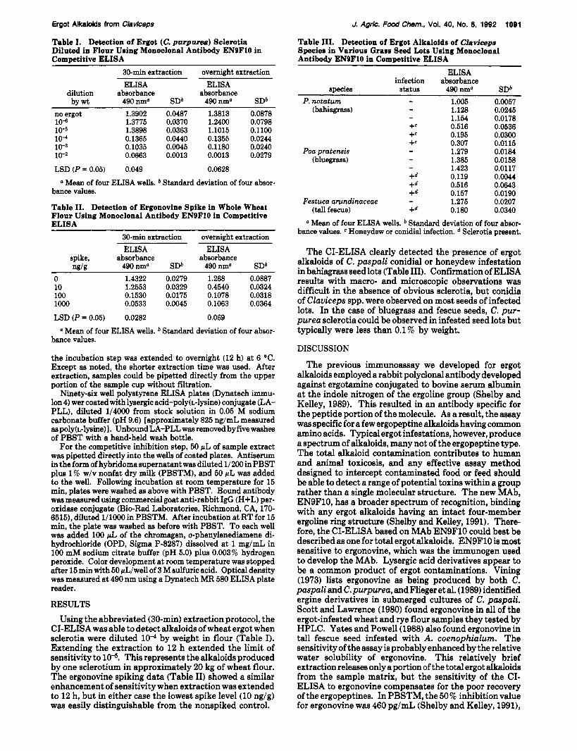
Table I. Detection of Ergot (C. purpurea) Sclerotia Diluted in Flour Using Monoclonal Antibody ENSF10 in Commtitive ELISA
J. Agrlc. Food Chem., Vol. 40, No. 0, lQ92 1001
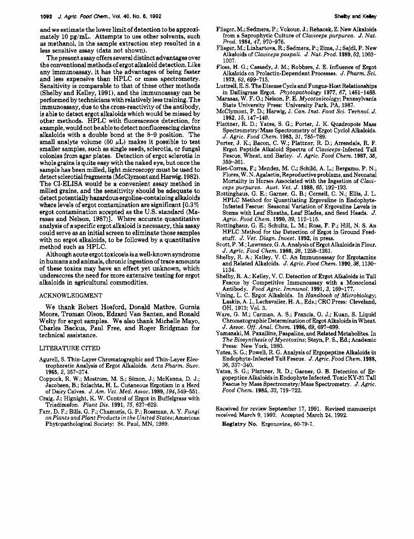
Table 111. Detection of Ergot Alkaloide of Claviceps Species in Various Grass Seed Lots Using Monoclonal Antibody ENSF10 in Competitive ELISA
ELISA infection absorbance
species status 490 nma SDb
30-min extraction overnight extraction ELISA ELISA
dilution absorbance absorbance by wt 490nma SDb 490nm' SDb
no ergot 1.3902 0.0487 1.3813 0.0878 10-6 1.3775 0.0370 1.2400 0.0798 10-6 1.3898 0.0363 1.1015 0.1100 10-4 0.1365 0.0440 0.1355 0.0244 10-3 0.1035 0.0045 0.1180 0.0240 10-2 0.0663 0.0013 0.0013 0.0279
LSD (P = 0.05) 0.049 0.0628
bance values.
Table 11. Detection of Ergonovine Spike in Whole Wheat Flour Using Monoclonal Antibody ENSFlO in Competitive ELISA
a Mean of four ELISA wells. Standard deviation of four absor-
30-min extraction overnight extraction
- P. notatum 1.005 0.0057 (bahiagrass) 1.128 0.0245
1.154 0.0178 +e 0.516 0.0536 +e 0.195 0.0300 +e 0.307 0.0115
(bluegrass) 1.385 0.0158 1.423 0.0117
+d 0.119 0.0044 +d 0.516 0.0643 +d 0.157 0.0190
Festuca arundinaceae 1.275 0.0207 (tall fescue) +d 0.180 0.0340 Mean of four ELISA wells. b Standard deviation of four absor-
bance values. e Honeydew or conidial infection. Sclerotia present.
- -
- Poa pratensis 1.279 0.0184 - -
-
ELISA spike, absorbance nglg 490nma SDb
0 1.4322 0.0279 10 1.2553 0.0329 100 0.1530 0.0175 lo00 0.0533 0.0045
ELISA absorbance
490nma SDb 1.268 0.0887 0.4540 0.0324 0.1078 0.0318 0.1063 0.0364
LSD (P = 0.05) 0.0282 0.069
a Mean of four ELISA wells. Standard deviation of four absor- bance values.
the incubation step was extended to overnight (12 h) at 6 "C. Except as noted, the shorter extraction time was used. After extraction, samples could be pipetted directly from the upper portion of the sample cup without filtration.
Ninety-six well polystyrene ELISA plates (Dynatech immu- lon 4) wer coated with lysergic acid-poly(L-lysine) conjugate (LA- PLL), diluted 1/4000 from stock solution in 0.05 M sodium carbonate buffer (pH 9.6) [approximately 825 ng/mL measured as poly(~-lysine)]. UnboundLA-PLL was removed by five washes of PBST with a hand-held wash bottle.
For the competitive inhibition step, 50 pL of sample extract was pipetted directly into the wells of coated plates. Antiserum in the form of hybridoma supernatant was diluted 1/200 in PBST plus 1 % w/v nonfat dry milk (PBSTM), and 50 pL was added to the well. Following incubation at room temperature for 15 min, plates were washed as above with PBST. Bound antibody was measured using commercial goat anti-rabbit IgG (H+L) per- oxidase conjugate (Bio-Rad Laboratories, Richmond, CA, 170- 6515), diluted 1/1OOO in PBSTM. After incubation at RT for 15 min, the plate was washed as before with PBST. To each well was added 100 pL of the chromagen, o-phenylenediamene di- hydrochloride (OPD, Sigma P-8287) dissolved at 1 mg/mL in 100 mM sodium citrate buffer (pH 5.0) plus 0.003 % hydrogen peroxide. Color development at room temperature was stopped after 15 min with 50 pLlwell of 3 M sulfuric acid. Optical density was measured at 490 nm using a Dynatech MR 580 ELISA plate reader.
RESULTS Using the abbreviated (30-min) extraction protocol, the
CI-ELISA was able to detect alkaloids of wheat ergot when sclerotia were diluted lo4 by weight in flour (Table I). Extending the extraction to 12 h extended the limit of sensitivity to This represents the alkaloids produced by one sclerotium in approximately 20 kg of wheat flour. The ergonovine spiking data (Table 11) showed a similar enhancement of sensitivity when extraction was extended to 12 h, but in either case the lowest spike level (10 ng/g) was easily distinguishable from the nonspiked control.
The CI-ELISA clearly detected the presence of ergot alkaloids of C. paspali conidial or honeydew infestation in bahiagrass seed lots (Table III). Confirmation of ELISA results with macro- and microscopic observations was difficult in the absence of obvious sclerotia, but conidia of Claviceps spp. were observed on most seeds of infected lots. In the case of bluegrass and fescue seeds, C. pur- purea sclerotia could be observed in infested seed lots but typically were less than 0.1 % by weight.
DISCUSSION
The previous immunoassay we developed for ergot alkaloids employed a rabbit polyclonal antibody developed against ergotamine conjugated to bovine serum albumin at the indole nitrogen of the ergoline group (Shelby and Kelley, 1989). This resulted in an antibody specific for the peptide portion of the molecule. As a result, the assay was specific for a few ergopeptine alkaloids having common amino acids. Typical ergot infestations, however, produce a spectrum of alkaloids, many not of the ergopeptine type. The total alkaloid contamination contributes to human and animal toxicosis, and any effective assay method designed to intercept contaminated food or feed should be able to detect a range of potential toxins within a group rather than a single molecular structure. The new MAb, ENSF10, has a broader spectrum of recognition, binding with any ergot alkaloids having an intact four-member ergoline ring structure (Shelby and Kelley, 1991). There- fore, the CI-ELISA based on MAb ENSF10 could best be described as one for total ergot alkaloids. ENSF10 is most sensitive to ergonovine, which was the immunogen used to develop the MAb. Lysergic acid derivatives appear to be a common product of ergot contaminations. Vining (1973) lists ergonovine as being produced by both C. paspali and C. purpurea, and Flieger et al. (1989) identified ergine derivatives in submerged cultures of C. paspali. Scott and Lawrence (1980) found ergonovine in all of the ergot-infested wheat and rye flour samples they tested by HPLC. Yates and Powell (1988) also found ergonovine in tall fescue seed infested with A. coenophialum. The sensitivity of the assay is probably enhanced by the relative water solubility of ergonovine. This relatively brief extraction releases only a portion of the total ergot alkaloids from the sample matrix, but the sensitivity of the CI- ELISA to ergonovine compensates for the poor recovery of the ergopeptines. In PBSTM, the 50% inhibition value for ergonovine was 460 pg/mL (Shelby and Kelley, 1991),
1092 J. Agrlc. Food Chem., Vol. 40, No. 6, 1992
and we estimate the lower limit of detection to be approxi- mately 10 pg/mL. Attempts to use other solvents, such as methanol, in the sample extraction step resulted in a less sensitive assay (data not shown).
The present assay offers several distinct advantages over the conventional methods of ergot alkaloid detection. Like any immunoassay, it has the advantages of being faster and less expensive than HPLC or mass spectrometry. Sensitivity is comparable to that of these other methods (Shelby and Kelley, 1991), and the immunoassay can be performed by technicians with relatively less training. The immunoassay, due to the cross-reactivity of the antibody, is able to detect ergot alkaloids which would be missed by other methods. HPLC with fluorescence detection, for example, would not be able to detect nonfluorescing clavine alkaloids with a double bond at the 8-9 position. The small analyte volume (50 pL) makes it possible to test smaller samples, such as single seeds, sclerotia, or fungal colonies from agar plates. Detection of ergot sclerotia in whole grains is quite easy with the naked eye, but once the sample has been milled, light microscopy must be used to detect sclerotial fragments (McClymont and Harwig, 1982). The CI-ELISA would be a convenient assay method in milled grains, and the sensitivity should be adequate to detect potentially hazardous ergoline-containing alkaloids where levels of ergot contamination are significant [0.3 % ergot contamination accepted as the US. standard (Ma- rasas and Nelson, 198711. Where accurate quantitative analysis of a specific ergot alkaloid is necessary, this assay could serve as an initial screen to eliminate those samples with no ergot alkaloids, to be followed by a quantitative method such as HPLC.
Although acute ergot toxicosis is a well-known syndrome in humans and animals, chronic ingestion of trace amounts of these toxins may have an effect yet unknown, which underscores the need for more extensive testing for ergot alkaloids in agricultural commodities.
ACKNOWLEDGMENT
We thank Robert Hosford, Donald Mathre, Gurnia Moore, Truman Olson, Edzard Van Santen, and Ronald Welty for ergot samples. We also thank Michelle Mayo, Charles Backus, Paul Free, and Roger Bridgman for technical assistance.
Shelby and Kelley
Flieger, M.; Sedmera, P.; Vokoun, J.; Rehacek, Z. New Alkaloids from a Saprophytic Culture of Claviceps purpurea. J. Nut. Prod. 1984, 47, 970-976.
Flieger, M.; Linhartova, R.; Sedmera, P.; Zima, J.; Sajdl, P. New Alkaloids of Claviceps paspali. J. Nut. Prod. 1989,52,1003- 1007.
Floss, H. G.; Cassady, J. M.; Robbers, J. E. Influence of Ergot Alkaloids on Prolactin-Dependent Processes. J. Pharm. Sci. 1973, 62, 699-715.
Luttrell, E. S. The Disease Cycle and Fungus-Host Relationships in Dallisgrass Ergot. Phytopathology 1977, 67, 1461-1468.
Marasas, W. F. 0.; Nelson, P. E. Mycotoxicology; Pennsylvania State University Press: University Park, PA, 1987.
McClymont, P. D.; Harwig, J. Can. Inst. Food Sci. Technol. J .
Plattner, R. D.; Yates, S. G.; Porter, J. K. Quadropole Mass Spectrometry/Mass Spectrometry of Ergot Cyclol Alkaloids. J. Agric. Food Chem. 1983,31,785-789.
Porter, J. K.; Bacon, C. W.; Plattner, R. D.; Arrendale, R. F. Ergot Peptide Alkaloid Spectra of Claviceps-Infected Tall Fescue, Wheat, and Barley. J. Agric. Food Chem. 1987, 35, 359-361.
Riet-Correa, F.; Mendez, M. C.; Schild, A. L.; Bergamo, P. N.; Flores, W. N. Agalactia, Reproductive problems, and Neonatal Mortality in Horses Associated with the Ingestion of Clavi- ceps purpurea. Aust. Vet. J . 1988,65, 192-193.
Rottinghaus, G. E.; Garner, G. B.; Cornell, C. N.; Ellis, J. L. HPLC Method for Quantitating Ergovaline in Endophyte- Infested Fescue: Seasonal Variation of Ergovaline Levels in Stems with Leaf Sheaths, Leaf Blades, and Seed Heads. J. Agric. Food Chem. 1990,39, 112-115.
Rottinghaus, G. E.; Schultz, L. M.; Ross, F. P.; Hill, N. S. An HPLC Method for the Detection of Ergot in Ground Feed- stuff. J. Vet. Diagn. Invest. 1992, in press.
Scott, P. M.; Lawrence, G. A. Analysis of Ergot Alkaloids in Flour. J. Agric. Food Chem. 1980,243, 1258-1261.
Shelby, R. A.; Kelley, V. C. An Immunoassay for Ergotamine and Related Alkaloids. J. Agric. Food Chem. 1990,38,113& 1134.
Shelby, R. A.; Kelley, V. C. Detection of Ergot Alkaloids in Tall Fescue by Competitive Immunoassay with a Monoclonal Antibody. Food Agric. Immunol. 1991, 3, 169-177.
Vining, L. C. Ergot Alkaloids. In Handbook of Microbiology; Laskin, A. I., Lechevalier, H. A., Eds.; CRC Press: Cleveland, OH, 1973; Vol. 3.
Ware, G. M.; Carman, A. S.; Francis, 0. J.; Kuan, S. Liquid Chromatographic Determination of Ergot Alkaloids in Wheat. J. Assoc. Of f . Anal. Chem. 1986, 69, 697-699.
Yamazaki, M. Paxalline, Paspaline, and Related Metabolites. In The Biosynthesis of Mycotoxins; Steyn, P. S., Ed.; Academic Press: New York, 1980.
Yates, S. G.; Powell, R. G. Analysis of Ergopeptine Alkaloids in Endophyte-Infected Tall Fescue. J. Agric. Food Chem. 1988,
Yates, S. G.; Plattner, R. D.; Garner, G. B. Detection of Er- gopeptine Alkaloids in Endophyte Infected, Toxic KY-31 Tall Fescue by Mass SpectrometryIMass Spectrometry. J. Agric. Food Chem. 1985,33, 719-722.
1982,15, 147-149.
36, 337-340.
LITERATURE CITED
Agurell, S. Thin-Layer Chromatographic and Thin-Layer Elec- trophoretic Analysis of Ergot Alkaloids. Acta Pharm. Suec.
Coppock, R. W.; Mostrom, M. S.; Simon, J.; McKenna, D. J.; Jacobsen, B.; Szlachta, H. L. Cutaneous Ergotism in a Herd of Dairy Calves. J. Am. Vet. Med. Assoc. 1989,194,549-551.
Craig, J.; Hignight, K. W. Control of Ergot in Buffelgrass with Triadimefon. Plant Dis. 1991, 75, 627-629.
Farr, D. F.; Bills, G. F.; Chamuris, G. P.; Rossman, A. Y. Fungi on Plants and Plant Products in the United States; American Phytopathological Society: St. Paul, MN, 1989.
1965, 2, 357-374.
Received for review September 17, 1991. Revised manuscript received March 9, 1992. Accepted March 24, 1992.
Registry No. Ergonovine, 60-79-7.