detection of promoter hypermethylation of gstp1 and...
TRANSCRIPT

Advances in Breast Cancer Research, 2018, 7, 91-106 http://www.scirp.org/journal/abcr
ISSN Online: 2168-1597 ISSN Print: 2168-1589
DOI: 10.4236/abcr.2018.72006 Mar. 15, 2018 91 Advances in Breast Cancer Research
Detection of Promoter Hypermethylation of GSTP1 and CDH1 Genes and the Relationship of Histopathological Parameters of the Breast
Onur Eroglu1,2*, Mine Erci Baysak3, Beyhan Durak Aras3, Oguz Cilingir3, Sevilhan Artan3
1Department of Molecular Biology and Genetics, Faculty of Art and Science, Bilecik Seyh Edebali University, Bilecik, Turkey 2Biotechnology Research and Application Center, Bilecik Seyh Edebali University, Bilecik, Turkey 3Department of Medical Genetics, Faculty of Medicine, Osmangazi University, Eskisehir, Turkey
Abstract
Background: Breast cancer is the most common cancer in women. Histopa-thology plays an important part in determining the treatment strategy for women with breast cancer. GSTP1 plays an important role in protecting cells from cytotoxic and carcinogenic agents and it is expressed in normal tissues at variable levels in different cell types. CDH1 plays a critical role for establish-ment and maintenance of polarity and differentiation of epithelium during the development period. Also, it plays an important role in signal transduc-tion, differentiation, gene expression, cellmotility and inflammations. Me-thods: In this study the promoter methylation levels of GSTP1 and CDH1 gene which are associated with breast cancer were investigated by technique of Methylation Sensitive High Resolution Melting Analysis (MS-HRM). We analysed primary tumor core biopsies from 80 high-risk primary breast cancer patients (tumors ≥ 2 cm and/or lymphatic metastase and/or distant metastases and/or under 40 years). Also the patients’ histopathologic types were asso-ciated with the methylation levels. Results: In our study the promoter hyper-methylation status was observed at different rates; GSTP1 and CDH1 hyper-methylation frequencies were 82% and 95% respectively. The promoter hypermethylation levels of the genes were found to be significant with lymph node positivity, ER positivity and HER2/neu negativity. Conclusion: Our study is important as being the first study that analyzes association between histopathologic type and GSTP1 and CDH1 gene promotor methylation status in Turkish population. Keywords
Breast Cancer, Methylation, GSTP1, CDH1, Histopathological Types
How to cite this paper: Eroglu, O., Baysak, M.E., Aras, B.D., Cilingir, O. and Artan, S. (2018) Detection of Promoter Hyperme-thylation of GSTP1 and CDH1 Genes and the Relationship of Histopathological Pa-rameters of the Breast. Advances in Breast Cancer Research, 7, 91-106. https://doi.org/10.4236/abcr.2018.72006 Received: December 23, 2014 Accepted: March 12, 2018 Published: March 15, 2018 Copyright © 2018 by authors and Scientific Research Publishing Inc. This work is licensed under the Creative Commons Attribution International License (CC BY 4.0). http://creativecommons.org/licenses/by/4.0/
Open Access

O. Eroglu et al.
DOI: 10.4236/abcr.2018.72006 92 Advances in Breast Cancer Research
1. Introduction
Cancer is a process fuelled both by genetic alterations and epigenetic mechanisms. Epigenetics refer to changes in gene expression that can be mitotically inherited, but are not associated with the changes in the coding sequence of the affected genes [1]. In other words, epigenetics refer to the inheritance of information based on gene expression levels, in contrast to genetics that refer to transmission of information based on gene sequence [2].
Hypermethylation is an epigenetic change that blocks the promoter region of a gene and results in gene silencing. In breast cancer, tumor-related genes may be silenced by hypermethylation; many hypermethylated genes have been re-ported, and silencing of these genes plays important roles in carcinogenesis and tumor progression [3]. Identification of epigenetic changes and their correlation with other clinical factors could lead to improvements in cancer diagnosis and treatment.
Pi-class glutathione-S-transferase (GSTP1) located on chromosome 11q13 encodes a phase II metabolic enzyme that detoxifies reactive electrophilic inter-mediates. GSTP1 plays an important role in protecting cells from cytotoxic and carcinogenic agents and it is expressed in normal tissues at variable levels in dif-ferent cell types. Altered GSTP1 activity and expression have been reported in many tumors and this is largely due to GSTP1 DNA hypermethylation at the CpG island in the promoter-5’. Glutathione S-transferase P1 (GSTP1) is a bio-transformation enzyme expressedin normal breast epithelial cells which can be epigenetically inactivated in breast cancer [4].
CDH1 (E-cadherin) acts as a transmembrane glycoprotein that is important in epithelial cell-cell interactions, and also interacts with α and β catenin together with actin in cytoskeleton [5] [6]. CDH1 plays a critical role for establishment and maintenance of polarity and differentiation of epithelium during the devel-opment period [7]. Also, it plays an important role in signal transduction, diffe-rentiation, gene expression, cellmotility and inflammations [8]. However, so-matic loss of CDH1 expression is associated with lobular breast cancer [9]. Be-sides the mutational changes observed in gene expression, there are evidences for epigenetic silencing of CDH1 gene including promoter region methylation in CpG sites [10] [11]. Loss of CDH1 expression in cancer cells leads to a diffused growth pattern and metastasis potential in tumors; thus, CDH1 generally acts as a tumor suppressor gene [12].
Promoter methylation of cancer-related genes has been demonstrated to be one of the most important molecular abnormalities in cancer [13]. Aberrant DNA methylation at the cytosines of CpG dinucleotide-rich islands presents in the promoter region of genes; it is associated with the formation of repressive chromatin structure and leads to the silencing of gene expression. CpG island hypermethylation leads to the loss of expression of various genes involved in tumorigenesis such as tumour suppressor genes, DNA repair genes and metasta-sis suppressor genes. DNA methylation changes are believed to be an alternative

O. Eroglu et al.
DOI: 10.4236/abcr.2018.72006 93 Advances in Breast Cancer Research
pathway to cancer and therefore have the potential to be diagnostic markers that can be used in the clinical context [13]. Since DNA methylation differs in dif-ferent cancer types and changes at different stages of tumour progression we have assessed whether DNA methylation profiling may be used clinically to dis-tinguish theearly diagnosis of cancer, tumorclassification, prognosis, the regula-tion of treatmentprotocols, response to treatment and cancer prevention may belight onways tocontrol [14].
Thus, the objective of the present study was to evaluate 2 tumor suppressor genes (CDH1, GSTP1) frequently found to be methylated in breast cancers but not in normal breast tissue, when combined with the methylation-sensitive high resolution meltinganalysis methods to assess promoter methylation status, would lead to a sufficiently sensitive and specific methylation marker.
We analyzed promoter methylation in DNA from Formalin-fixed, paraf-fin-embedded samples collected at the time of diagnosis from 80 breast cancer cases and their normal tissue samples as controls who donated tissues from 2010 to 2011 to the Oncology Clinic in Eskişehir Osmnagazi University. Also we in-vestigate the relation between the methylation levels and the histopathological parameters.
2. Material and Methods
2.1. Sample Collection and DNA Preparation
Formalin-fixed, paraffin-embedded samples from 80 primary breast tumors tis-sue samples and their normal tissue samples were collected from Eskisehir Os-mangazi University, Medical Faculty, Oncology Clinic were included. Being in-side eppendorf tubes; samples embedded in parafin tissue blocks as 10 sections of 10 microns thick, were taken from Eskisehir Osmangazi University of medical faculty of Pathology department. All tumor specimens have at least 70% of tu-morous tissue. After deparafinization genomic DNA was extracted using Mag-NA Pure Compact Nucleic Acid Isolation Kit I (Roche) according to the manu-facturer’s instructions.
2.2. Bisulphite Modification
Approximately 250 - 500 nanogram of genomic DNA was bisulphite modified using the EpiTect Bisulphite kit (Qiagen) according to the manufacturer’s in-structions. The modified DNA was eluted three times in 50 µl buffer EB. CpGe-nome TM Universal Methylated DNA (Chemicon/Millipore, Billerica, MA) and CpGenome Universal Unmethylated DNA were used as fully methylated and unmethylated controls, respectively. Methylation standards (under 25%, 25%, 50%, 75% and 100%) made by diluting the fully methylated control in the un-methylated DNA were used as controls. Whole-genome amplification (WGA) was also used to make a fully unmethylated control and performed as previously described [15].

O. Eroglu et al.
DOI: 10.4236/abcr.2018.72006 94 Advances in Breast Cancer Research
2.3. Methylation-Sensitive High Resolution Melting (MS-HRM)
Methylation analysis was performed using MS-HRM in which the approximate level of methylation and presence of heterogeneous methylation can be deter-mined in bisulphite modified DNA [16]. Since methylated samples have more cytosines compared to unmethylated samples after bisulphite modification, the methylated samples melt later than unmethylated samples. Methylated samples with different methylation status will therefore have distinct methylated and unmethylated peaks depending on the levels of methylation. Complex melting profiles were seen in heterogeneously methylated samples as multiple hetero-duplexes may be formed [17].
PCR amplification of the DNA was carried out using a Roche LightCycler 480 (Roche Applied Science, Laval, PQ, Canada) equipped with the Gene Scanning software (Version 1.5.0). MS-HRM primer details are listed in Table 1. Promo-ter CpG islands in the promoter were identified in each gene for designing the primers. The primers were designed according to the principles outlined pre-viously [17]. Amplicons were generally less than 100 base pairs in size so that amplification from the majority of FFPE DNAs waspossible.
PCR was performed in a 19.5 μl reaction volume and 10 μl of BSC DNA tem-plates were added to each well which contained 10 μl LightCycler 480 High Res-olution Melting (HRM) Master Mix® (Roche), 2.5 μl MgCl2 and 3.0 μl of each primer. The cycling protocol conditions included a single enzyme activation step of 10 min at 95˚C followed by 50 cycles of the following steps: denaturation 95˚C, 10 s, annealing 65˚C, 15 s, and extension 72˚C, 25 s.
It is important to note that a touchdown PCR protocol as recommended by the LC480-HRM Master Mix was not adopted. Inclusion of a touchdown PCR protocol would have favoured the PCR bias towards the higher melting and thermodynamically more stable methylated DNA product.
The high resolution melting (HRM) step was performed after 50 cycles of am-plification. The HRM analysis was initiated by denaturing all products at 95˚C for 1 min, followed by annealing at 40˚C for 1 min. Samples were quickly
Table 1. MS-HRM primer details (M: Methylated, U: Unmethylated, F: Forward, R: Re-verse).
Genes Primers
GSTP1
M-F: 5’-TTCGGG GTG TAGCGGTCGTC-3’
M-R: 5’-GCC CCA ATACTA AATCACGACG-3’
U-F: 5’-GATGTT TGG GGTGTAGTGGTT GTT-3’
U-R: 5’-CCA CCC CAATACTAA ATCACA ACA-3’
CDH1
M-F: 5’-TTAGGTTAGAGG GTT ATCGCCT-3’
M-R: 5’-TAA CTAAAAATTCAC CTACCGAC-3’
U-F: 5’-TAATTTTAG GTTAGAGGGTTATTG T-3’
U-R: 5’-CAC AAC CAATCA ACA ACACA-3’

O. Eroglu et al.
DOI: 10.4236/abcr.2018.72006 95 Advances in Breast Cancer Research
warmed to 65˚C and then slowly warmed to 95˚C at 0.1˚C per second. Fluores-cence data were collected at 25 acquisitions per second. The LC480-HRM Master Mix® employed a saturating dye (ResolLight™, Roche) which facilitated the pre-cise measurement of the melt curves of the amplicons. The Roche Gene Scan-ning software was employed for end product analysis. This algorithm allowed the raw melt curves to be normalized for fluorescence intensity, and a tempera-ture shift was applied to align the normalized melt curves, which facilitated the analysis of samples with varying crossing point (Cp) values. A difference curve was then derived from the first derivative of the melt curves. Data for the differ-ence melt curves were exported to Excel (Office 2010; Microsoft Corp., Red-mond, WA, USA) for further analysis after the graphs were plotted and inverted vertically. Both peak-height and area-under-the-curve from the normalized, temperature-shifted, difference curves were used to generate a standard curve and determine the degree of methylation of each DNA sample.
2.4. Statistical Analysis
Methylation levels of genes have been identified by comparing the peaks of con-trols and the patients’ samples. Initially the patient’s samples were grouped based on the prevelent prognostic factors such as age, disease stage, tumortype, lymphnode metastasis, ER, PR and HER2/neu. Then the methylation levels of these groups were compared to each other.
The v2 test and binary logistic regression were used to analyse the relation-ship of methylation with the clinicopathological findings. The analyses were carried out with SPSS version 17.0 (SPSS Inc., Chicago, IL). A two tailed P-value of less than 0.05 was considered to be statistically significant for each comparison.
3. Results
3.1. Patient Characteristics
Patient characteristics included in this study are summarised in Table 2. Patients with primary breast tumors were aged between 32 and 83 (median = 58.44 ± 11.09). Respectively 19/80 cases of them (24%) was ≤50 years of age, 61/80’ sin (76%) > 50 years of age. The selection criteria of patients are as follows.The tu-mors should be 2 cm or higher, and/or the patients have lymphatic metastase, and/or distant metastases, and/or the patients should be under 40 years old.
3.2. Methylation Status in Primary Breast Tumor Samples
A detail summary of the promoter hypermethylation frequencies of 2 genes in 80 primary breast tumours are shown in Table 3 and Table 4. Examples of the MS-HRM results are shown in Figures 1-8. Two genes showed detectable pro-moter methylation in primary breast tumours. GSTP1 and CDH1 were methy-lated in 82% and 95% of primary breast tumors, respectively.

O. Eroglu et al.
DOI: 10.4236/abcr.2018.72006 96 Advances in Breast Cancer Research
Table 2. Methylation frequecies of GSTP1.
Methylation ratio (%) Patient ratio (%)
100% Methylated 21%
75% Methylated 3%
50% Methylated 17%
25% Methylated 16%
↓25% Methylated 25%
Unmethylated 18%
Table 3. Methylation frequecies of CDH1.
Methylation ratio (%) Patient ratio (%)
100% Methylated 19%
75% Methylated 40%
50% Methylated 26%
25% Methylated 10%
Unmethylated 5%
3.3. Correlation between DNA Methylation and Clinicopathological Parameters
When the only methylated groups were compared, there is no correlation be-tween the GSTP1 and CDH1 genes promoter hypermethylation and tumor stage (p > 0.05) and age (p > 0.05). Both CDH1 and GSTP1 genes promoter hyperme-thylation was found to be significant for lymph node positivity [CDH1 = (p < 0.05), GSTP1 = (p < 0.001)], ER positivity (p < 0.05), and HER2/NEU negativity (p < 0.05). The correlation details are listed in Table 5 and Table 6.
4. Discussion
Nowadays many studies were conducted examining several genes for epigenetic mechanisms. Methylation has been the most important field of the epigenetic changes. Most of the researchers interested in understanding the role of methy-lation of cancer genetics in recent years because of the methylation profiles of different genes in cancer and focused on results.
Researchers examining the changes in the genome methylation profiles have used many different techniques. With the addition of these techniques with each passing day, new methods in this research area has reached large proportions, and many clearly defined changes in gene methylation.
In recent years Methylation-sensitive high resolution melting (MS-HRM) analysis method is newly used in throughout, as well as in our country. An im-portant feature of our study is that breast cancer cases in the methylation analy-sis of this method are used for the first time.

O. Eroglu et al.
DOI: 10.4236/abcr.2018.72006 97 Advances in Breast Cancer Research
Table 4. Associations between GSTP1 gene promoter hypermethylation and clinicopa-thological features of breast cancer.
AGE Methylated Unmethylated Total P value
AGE ≤ 50 15 (42.1) 4 19
AGE > 50 51 (19.7) 10 61
STAGE p > 0.05
STAGEII 25 (%38) 5 30
STAGEIII 37 (%56) 7 44
STAGEIV 4 (%6) 2 6
LYMPH NODE
+ 57 (86.4) 11 68 P < 0.001
- 9 (13.6) 3 12
ER
+ 44 (66.7)
10 54 P < 0.05
- 22 (33.3) 4 26
PR Nonevaluated
+ 33 (%50) 9 42
- 33 (%50) 5 38
HER2/neu
+ 26 (39.4) 4 30
- 40 (60.6) 10 50 P < 0.05
Figure 1. Melting curve shows the methylation standards (25%, 50%, 75% and 100%) made by diluting the fully methylated control into the unmethylated DNA were used as controls.
A recent study examined the methylation of two genes in primary breast tu-
mours. It is difficult to compare our results to theirs because the methodologies used are so different. Nested MSP is non-quantitative and even low amounts of background methylation will be scored whereas MS-HRM can be used for semi-quantitative estimation of methylation [17].
Breast cancer shows different clinical symptoms after being diagnosed. There are some biomarkers that are needed in order to calculate the risk of recurrence

O. Eroglu et al.
DOI: 10.4236/abcr.2018.72006 98 Advances in Breast Cancer Research
Figure 2. Melting curves of all samples.
Figure 3. Melting curves of all samples of the GSTP1 gene methylated and unmethylated samples.
of the disease, identify the subgroups with poor prognosis and determine the treatment to be given. In this way, it is possible to identify groups of patients whose prognosis is very good but who can’t see any advantage of systematic treatment or whose prognosis is very bad and who need to have an aggressive treatment. The standard breast cancer prognostic factors are as it follows; tumor size, lymph node involvement, histologic grad, ER and PR status. Lymph node involvement is the most powerful known prognostic factor in patients with breast cancer. The lymph node involvement has clinically been detected 50% of breast cancer patients. Lymph node involvement is associated with tumor size but ER and PR have been found to be independent. For many years now, Steroid receptors have been used for breast cancer treatment decisions. The positivity of ER (37% - 80%) and PR (45% - 69%) rates have been reported in invasive can-cers. The rate of ER-positive cells is associated with the degree of differentiation and the tumor’s response to hormonal therapy. The ER-positive tumor cells ra-tio is associated with response of hormonal therapy and differentiation degree.
O. Eroglu et al.
DOI: 10.4236/abcr.2018.72006 99 Advances in Breast Cancer Research
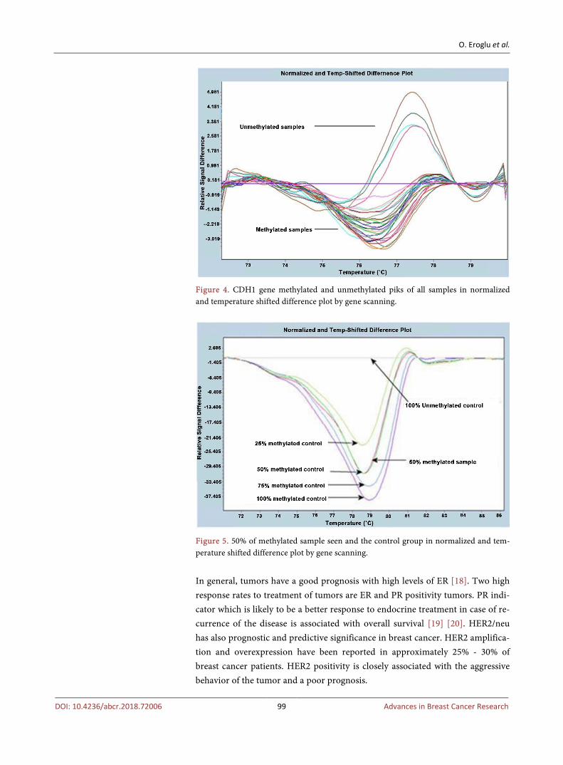
Figure 4. CDH1 gene methylated and unmethylated piks of all samples in normalized and temperature shifted difference plot by gene scanning.
Figure 5. 50% of methylated sample seen and the control group in normalized and tem-perature shifted difference plot by gene scanning.
In general, tumors have a good prognosis with high levels of ER [18]. Two high response rates to treatment of tumors are ER and PR positivity tumors. PR indi-cator which is likely to be a better response to endocrine treatment in case of re-currence of the disease is associated with overall survival [19] [20]. HER2/neu has also prognostic and predictive significance in breast cancer. HER2 amplifica-tion and overexpression have been reported in approximately 25% - 30% of breast cancer patients. HER2 positivity is closely associated with the aggressive behavior of the tumor and a poor prognosis.
O. Eroglu et al.
DOI: 10.4236/abcr.2018.72006 100 Advances in Breast Cancer Research
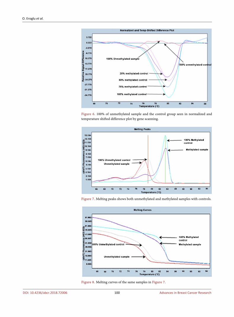
Figure 6. 100% of unmethylated sample and the control group seen in normalized and temperature shifted difference plot by gene scanning.
Figure 7. Melting peaks shows both unmethylated and methylated samples with controls.
Figure 8. Melting curves of the same samples in Figure 7.

O. Eroglu et al.
DOI: 10.4236/abcr.2018.72006 101 Advances in Breast Cancer Research
Table 5. Associations between CDH1 gene promoter hypermethylation and clinicopa-thological features of breast cancer.
AGE Methylated Unmethylated Total P value
AGE ≤ 50 17 (89.5) 2 19
AGE > 50 59 (88.5) 2 61
STAGE p > 0.05
STAGEII 28 (%37) 2 30
STAGEIII 42 (%55) 2 44
STAGEIV 6 (%8) 0
6
LYMPH NODE
+ 65 (%85.5) 3 68 P < 0.05
- 11 (%14.5) 1 12
ER
+ 51 (%67.1) 3 54 P < 0.05
- 25 (%32.9) 1
26
PR Nonevaluated
+ 39 (%51) 3 42
- 37 (%49) 1 38
HER2/neu
+ 29 (%38.2) 1 30
- 47 (%61.8) 3 50 P < 0.05
According to a study, there are no correlation between histopathologic types
and GSTP1 promoter hypermethylation. They found GSTP1 hypermethylation 36 out of 124 (29%) with nested PCR method. Also they showed the methylation ratios of the patients in invazive ductal hyperplasia, in situ ductal carninomas and invazive ductal carcinomas were 4/24 (16.7%), 18/49 (36.7%), 14/36 (38.9%) respectively. They showed no hypermethylation in normal breast tissues which were used for control group. The promoter hypermethylation of GSTP1 were seen in early stages of breast cancer progression. Also the frequecies of methyla-tion increases from normal tissues to invazive ductal carcinomas [21].
In our study we found higher methylation level than as Lee et al. found [21]. We found GSTP1 hypermethylation 66 out of 80 (82%). Our results show an in-creased level than comparing with the other research caused by the different technique. Nested MSP is non-quantitative and even low amounts of back-ground methylation will not be scored whereas MS-HRM can be used for semi-quantitative estimation of methylation and it’smore sensitivity and speci-ficity than the MSP. Also we found different rates of methylation. When we compare the histopathologic types, there is no correlation between age and tu-mor stage but we found GSTP1 gene promoter hypermethylation in ER(+) 44

O. Eroglu et al.
DOI: 10.4236/abcr.2018.72006 102 Advances in Breast Cancer Research
Table 6. Associations between gene promoter hypermethylation and clinicopathological features of breast cancer.
Variables Patients GSTP1 methylation CDH1 methylation
(n) (%) M U M U
n (%) n (%) n (%) n (%)
Totally: 80 patients 66 (82) 14 (18) 76 (95) 4 (5)
Age
≤50 19 (24) 15 (42.1) 4 17 (89.5) 2
>50 61 (76) 51 (19.7) 10 59 (88.5) 2
Stage
II 30 (37.5) 25 (38) 5 28 (37) 2
III 44 (55) 37 (56) 7 42 (55) 2
IV 6 (7.5) 4 (6) 2 6 (8) 0
Type of Tumor
IDC 67 (83.75) 58 (86.5) 9(13.5) 64 (95.5) 3(4.5)
ILC 13 (16.25) 8 (61.5) 5(38.5) 12 (92.3) 1(7.7)
Lymph Node
Positive 68 (85) 57 (86.4) 11 65(85.5) 3
Negative 12 (15) 9 (13.6) 3 11(14.5) 1
Estrogen Receptor
Positive 54 (67.5) 44 (66.7) 10 51 (67.1) 3
Negative 26 (32.5) 22 (33.3) 4 25 (32.9) 1
Progesterone Receptor
Positive 42 (52.5) 33 (50) 9 39 (51) 3
Negative 38 (47.5) 33 (50) 5 37 (49) 1
HER2/NEU
Positive 30 (37.5) 26 (39.4) 4 29 (38.2) 1
Negative 50 (62.5) 40 (60.6) 10 47 (61.8) 3
out of 66 (66.7%) cases. Estrogen receptor (ER)-positive breast cancers are con-sidered prognostically more favorable than ER-negative tumors.
Another study showed a correlation between 19 out of 24 (79%) ER(+) and 13 out of 24 (54%) lymph node(+) with GSTP1 promoter hypermethylation. They found GSTP1 hypermethylation 24 out of 174 (14%) with methylation spesific PCR method. They indicated that the status of CpG islands hypermethylation of the GSTP1 gene is statistically significant prognostic factor in breast cancer [22].
In our study we found GSTP1 hypermethylation 66 out of 80 (82%). Our re-sults show an increased level than the other research caused bythe different technique and the tumor heterogeneity. We found ER positivity in 44 out of 66 (66.7%) and lymph node positivity 57 out of 66 (86.4%) in samples. Both results

O. Eroglu et al.
DOI: 10.4236/abcr.2018.72006 103 Advances in Breast Cancer Research
were similar with the Park and his colleagues. According to Sunami et al. (2008) they analyzed 8 (RASSF1A, CCND2,
GSTP1, TWIST, APC, NES1, RARβ2 and CDH1) different genes promoter hypermethylation in 130 [(65 out of 130 ER (+), 65 out of 130 ER(−)] primary breast tumors by Methylation Specif PCR (MSP) [23].
For RASSF1A, CCND2, GSTP1, TWIST, and APC genes, the proportion of methylated genes was significantly higher in the ER-positive than in the ER-negative tumor group. However, no significant differences in methylation status were detected in NES1, RARβ2, and CDH1. They were analyzed in terms of the relation between ER status and HER2/neu status. Using matched samples, no difference was found in the frequency of HER2/neu-positive tumors between the ER-negative and ER-positive groups. Then, the differences in methylation status of all eight genes were analyzed between the HER2/neu-positive and HER2/neu-negative tumor groups [23].
The proportion of methylated RASSF1A, GSTP1 and APCgenes was signifi-cantly greater in the HER2/neu-positive than in the HER2/neu-negative tumor group; no significant differences in methylation status were detected for TWIST, NES1, RARβ2, and CCND2. Among the eight biomarkers studied, CDH1 showed predominance of methylation status in HER2/neu-negative tumors by univariate analysis [23].
Shinozaki and his colleagues (2005) analyzed 6 (RASSF1A, APC, TWIST, CDH1, GSTP1 and RARβ2) genes promoter hypermethylation in 151 primary breast tumors by MSP [24]. They found CDH1 and GSTP1 promoter hyperme-thylation 53% and 21% respectively. When they were compared the histopatho-logic types (age, stage, menopausal status, tumor size, lymph node metastasis, ER, PR) and the promoter hypermethylation of the genes, they found the associ-ation between GSTP1 hypermethylation and lymph node metastasis; also CDH1 promoter hypermethylation and lymphovascular invasion and ER negativity were associated [24].
In our study we found CDH1 promoter hypermethylation 76 out of 80 (95%). This is an expected result due to the fact that the CDH1 gene is an invasion gene. When we compare the histopathologic types of tumor and the methylation le-vels, GSTP1 promoter hypermethylation correlates with ER positivity [(51 out of 76 (67.1%)], CDH1 gene methylation correlates with HER2/neu negativity [(47 out of 76 (61.8%)]. This is an expected result that among large tumor size, excess number of cases with stage III and lymph node metastasis related with the HER2/neu negativity and CDH1 promoter hypermethylation.
5. Conclusions
Epigenetic mutation changes gene expression without any difference in DNA sequence and is effective as genetic change is in cancer formation. Promotor re-gion hypermethylation causes gene silencing and especially important significant in neutralization of the tumour supressor genes. Determining the epigenetic changes observed in cancer cells and increasing data related to the issue is grad-

O. Eroglu et al.
DOI: 10.4236/abcr.2018.72006 104 Advances in Breast Cancer Research
ually becoming important in terms of prevention of cancer, determination of prognosis and development of therapeutic approaches. This situation highlights the importance of methylation as a tumor marker.
Examining the methylation profiles of CpG islands in cancer studies in this direction, with data provided from the early diagnosis of cancer, tumor classifi-cation, prognosis, treatment protocols, regulation, response to treatment may lead the way for controlling and preventing the cancer. For personalized treat-ment the quantitive results of the methylation levels of the genes are important for the use of demethylation agent for each patient. Also the promoter hyper-methylation of CDH1 and GSTP1 genes can be used as a biomarker for detection of early stages of breast cancer progression.
Acknowledgements
This work was financially supported by Eskisehir Osmangazi University Re-search projects (grant number: 201011037) and the study was conducted in ac-cordance with the Declaration of Helsinki, and the protocol was approved by the Ethics Committee of Eskisehir Osmangazi University (number: 2010/173). The authors wish to thank Prof. Dr. Serap Isiksoy, Assistant Prof. Dr. Evrim Ciftci and the staff for prepairing formalin-fixed parrafin embeded tissues at Eskisehir Osmangazi University medical faculty of pathology department.
Author Contributions
O.E. identified cases; prepared samples; performed methylation assays; inter-preted data and analyzed data; and wrote the manuscript. M.E.B. prepared sam-ples; deparafinization and isolation of the samples; performed methylation as-says; interpreted and analyzed data. B.D.A. identified cases; interpreted and analyzed data; wrote and edited the manuscript. O.C. optimazed and performed methylation assays; conceptualized project; provided analysis; wrote and edited the manuscript. S.A. provided project oversight and coordination and analysis; wrote and edited the manuscript. All authors read and approved the final ma-nuscript.
Conflict of Interest
We declare that there is no conflict of interest in this work.
References
[1] Etseller, M. and Herman, J.G. (2002) Cancer as an Epigenetic Disease: DNA Me-thylation and Chromatin Alterations in Human Tumours. The Journal of Patholo-gy, 196, 1-7. https://doi.org/10.1002/path.1024
[2] Esteller, M. (2005) Dormant Hypermethylated Tumor Suppressor Genes: Questions and Answers. The Journal of Pathology, 205, 172-180. https://doi.org/10.1002/path.1707
[3] Bae, Y.K., Brown, A., Garrett, E., Bornman, D., Fackler, M.J., Sukumar, S., Herman, J.G. and Gabrielson, E. (2004) Hypermethylation in Histologically Distinct Classes

O. Eroglu et al.
DOI: 10.4236/abcr.2018.72006 105 Advances in Breast Cancer Research
of Breast Cancer. Clinical Cancer Research, 10, 5998-6005. https://doi.org/10.1158/1078-0432.CCR-04-0667
[4] Isabelle, M. (2010) Glutathione S-Transferase pi (GSTP1). Atlas of Genetics and Cytogenetics in Oncology and Haematology, 14, 1181-1185.
[5] Kim, E.K. and Sahin, A. (2005) E-Cadherin Expression Loss in T1 İnvasive Ductal Carcinoma of Breast as a Predictive Marker for Lymph Node Metastasis. The Ko-rean Journal of Pathology, 39, 187-191.
[6] Lombaerts, M., Wezel, T.V., Philippo, K., Dierssen, J.W.F., Zimmerman, R.M.E., Oosting, J., Eijek, R.V., Eilers, P.H., Water, B.V.D., Cornelisse, C.J., et al. (2006) E-Cadherin Transcriptional Downregulation by Promoter Methylation But Not Mutation İs Related to Epithelial-To-Mesenchymal Transition in Breast Cancer Cell Line. British Journal of Cancer, 94, 661-671. https://doi.org/10.1038/sj.bjc.6602996
[7] Wilson, B.A.R., Kaurah, P., Suriano, G., Leach, S., Senz, J., Grehan, N., Butterfield, Y.S.N., Jeyes, J., Schinas, J., Bacani, J., et al. (2004) Germline E-Cadherin Mutations in Hereditary Diffuse Gastric Cancer: Assessment of 42 New Families and Review of Genetic Screening Criteria. Journal of Medical Genetics, 41, 508-517. https://doi.org/10.1136/jmg.2004.018275
[8] Slaus, N.P. (2003) Tumor Suppressor Gene E-Cadherin and İt’s Role in Normal and Malignant Cells. Cancer Cell International, 3, 1-7. https://doi.org/10.1186/1475-2867-3-1
[9] Goldstein, N.S., Bassi, D., Watts, J.C., Layfield, L.J., Yaziji, H. and Gown, A.M. (2001) E-Cadherin Reactivity of 95 Noninvasive Ductal and Lobular Lesions of the Breast. Implications for the İnterpretation of Problematic Lesions. American Jour-nal of Clinical Pathology, 115, 534-542. https://doi.org/10.1309/B0DD-4M7H-GJG1-7KCW
[10] Mastracci, T.L., Tjan, S., Bane, A.L., OMalley, P.F. and Andrulis, I.L. (2005) Ecad-herin Alterations in Atypical Lobular Hyperplasia and Lobular Carcinoma İnsitu of the Breast. Modern Pathology, 18, 741-751. https://doi.org/10.1038/modpathol.3800362
[11] Tischoff, I. and Tannapfel, A. (2008) DNA Methylation in Hepatocellular Carcino-ma. World Journal of Gastroenterology, 14, 1741-1748. https://doi.org/10.3748/wjg.14.1741
[12] Maruyama, R, Toyooka, S., Toyooka, O.K., Harda, K. and Virmani, A.K. (2001) Aberrant Promoter Methylation Profile of Bladder Cancer and İt’s Relationship to Clinicopathological Features. Cancer Research, 61, 8659-8663.
[13] Yang, X., Yan, L. and Davidson, N.L. (2001) DNA Methylation in Breast Cancer. Endocrine Related Cancer, 8, 115-127. https://doi.org/10.1677/erc.0.0080115
[14] Colot, V. and Rossignol, J.L. (1999) Eukaryotic DNA Methylation as an Evolutio-nary Device. BioEssays, 21, 402-411. https://doi.org/10.1002/(SICI)1521-1878(199905)21:5<402::AID-BIES7>3.0.CO;2-B
[15] Epstein, A.H., Conolly, J.L. and Gelman, R. (1989) The Predictors of Distant Re-lapse Following Conservative Surgery and Radiotherapy for Early Breast Cancer Are Similar to Those Following Mastectomy. International Journal Radiation Oncology Biology Physics, 17, 755-760. https://doi.org/10.1016/0360-3016(89)90062-X
[16] Wojdacz, T.K. and Dobrovic, A. (2007) Methylation-Sensitive High Resolution Melting (MS-HRM): A New Approach for Sensitive and High-Throughput Assess-ment of Methylation. Nucleic Acids Research, 35, 6-41. https://doi.org/10.1093/nar/gkm013
[17] Wojdacz, T.K., Moller, T.H., Thestrup, B.B., Kristensen, L.S. and Hansen, L.L.

O. Eroglu et al.
DOI: 10.4236/abcr.2018.72006 106 Advances in Breast Cancer Research
(2010) Limitations and Advantages of MS-HRM and Bisulfite Sequencing for Single Locus Methylation Studies. Expert Review of Molecular Diagnostics, 10, 575-580. https://doi.org/10.1586/erm.10.46
[18] Francis, G., Beadlet, G., Thomas, S. and Mengersen, K. (2206) Evalution of Estrogen and Progesterone Receptor Status in HER-2 Positive Breast Carcinomas and Corre-lation with Outcome. Pathology, 38, 391-398. https://doi.org/10.1080/00313020600922488
[19] Putti, T.C., Abd El-Rehim, D.M. and Rakha, E. (2005) Etsrogen Receptor-Negative Breast Carcinomas: A Review of Morphology and Immunophenotypical Analysis. Modern Pathology, 18, 26-35. https://doi.org/10.1038/modpathol.3800255
[20] Rakha, A.E., El Sayed, E.A., Green, R.A. and Lee, H.S.A. (2007) Prognostic Markers in Triple-Negative Breast Cancers. Cancer, 1, 109 p.
[21] Lee, J.S. (2007) GSTP1 Promotor Hypermetylation Is an Early Event in Breast Car-cinogenesis. Virchows Archiv, 450, 637-642. https://doi.org/10.1007/s00428-007-0421-8
[22] Park, S.Y., Kim, B.H., Kim, J.H., Cho, N.Y., Choi, M. and Yu, E.J. (2007) Methyla-tion Profiles of CpG İsland Loci in Major Types of Human Cancers. Journal of Ko-rean Medical Science, 22, 311-317. https://doi.org/10.3346/jkms.2007.22.2.311
[23] Sunami, E., Shinozaki, M., Sim, M.S., Nguyen, S.L., Vu, A.T., Giuliano, A.E. and Hoon, D.S.B. (2008) Estrogen Receptor and HER2/Neu Satatus Affect Epigenetic Differences of Tumor-Related Genes in Primary Breast Tumors. Breast Cancer Re-search, 10, R46. https://doi.org/10.1186/bcr2098
[24] Shinozaki, M., Hoon, H.S., Giuliano, A.E., Hansen, N.M., Wang, H.J., Turner, R. and Taback, B. (2005) Distinct Hypermethylation Profile of Primary Breast Cancer Is Associated with Sentinel Lymph Node Metastasis. Clinical Cancer Research, 15, 2156-2162. https://doi.org/10.1158/1078-0432.CCR-04-1810.
Abbreviations
ER: Estrogen receptor; PR: Progesterone receptor; M: Methylation; UM: Unmethylation; MS-HRM: Methylation-sensitive high resolution melting; ER: Estrogen receptor; PR: Progesterone receptor; PCR: Polymerase chain reaction; CDH1: Cadherin-1; GSTP1: Glutathione S-transferase P; HER2/neu: Human epidermal growth factor receptor 2; F: Forward; R: Reverse.