determination of polyphenolic content and antioxidant
TRANSCRIPT

Journal of Applied Pharmaceutical Science Vol. 10(07), pp 073-081, July, 2020Available online at http://www.japsonline.comDOI: 10.7324/JAPS.2020.10710 ISSN 2231-3354
Determination of polyphenolic content and antioxidant potential of callus extract obtained from Celastrus paniculatus Willd. and their free radical estimation by electron spin resonance spectroscopy
Anil Kumar Moola*, Senthil Kumar Thiruppathi, Ranjitha Kumari Bollipo DianaDepartment of Botany, Bharathidasan University, Tiruchirappalli, India.
ARTICLE INFOReceived on: 12/09/2019Accepted on: 23/05/2020Available online: 04/07/2020
Key words:Celastrus paniculatus Willd, callus induction, free radicals scavenging, HPTLC profiling, electron spin resonance spectroscopy.
ABSTRACT Celastrus paniculatus Willd. belongs to the family Celastraceae, and it is an important medicinal plant distributed all over India. Since the antioxidative polyphenols in C. paniculatus have received an increase in attention for health-promoting properties by scavenging the free radicals, the objective of this study is aimed at understanding the antioxidant potential of calli cultures generated from C. paniculatus. To establish the calli cultures, leaf explants derived from direct organogenesis of C. paniculatus have been cultured on the Murashige and Skoog medium (MS). The culture medium is supplemented with different concentrations of 6-Benzylaminopurine (0.5 mg l−1) along with 2,4-D and naphthalene acetic acid (NAA) (0.1–0.7 mg l−1). The MS medium containing 0.5 mg l−1 [Benzylaminopurine (BAP) + NAA] and 0.5 mg l−1 of BAP + 0.3 mg l−1 2,4-D showed to be the best medium for the formation of calli. The calli cultures were harvested and lyophilized for methanolic extraction and estimated the total phenolic and flavonoid contents in the calli cultures by using the spectroscopic method technique and also analyzed by high-performance thin-layer chromatography (HPTLC) profiling and high-performance liquid chromatography (HPLC) to assess their antioxidant potential. The histological findings supported the result of HPTLC and HPLC by displaying a clear deposition of polyphenols in the vacuoles. Additionally, free radicals generated from the biological system were detected and the ‘g’ value was identified by the electron spin resonance spectrum and understood their radical scavenging activity by several nonenzymatic methods, which include 2,2-Diphenyl-1-picrylhydrazyl assay, reducing power activity, 2, 2′-Azino-bis(3-ethylbenzothiazoline-6-sulphonic acid) radical scavenging assay, and ferric reducing antioxidant power assay. The research results showed that 0.5 mg l−1 of BAP along with NAA is an optimal hormone concentration for developing friable calli which in turn yields higher phenolic and flavonoid content.
INTRODUCTIONThe important candidates responsible for the generation
of the oxidative stress is free radical (Cantuti-Castelvetri et al., 2000; Maxwell, 1995). When the concentration of free radicals is above the normal level, they become toxic to the mammalian cells and are responsible for inactivating enzymes, and they damage cell organelles, DNA, protein, and membrane (Ferreira et al., 2007; Karuppanapandian et al., 2011). The human body is not deprived of its defense against damage. It creates different types
of molecules called antioxidants to fight against these free radicals and defends the cells from attack by oxygen. It is well known that the free radicals, such as O2
•- (superoxide anion), •OH (hydroxyl radical), •NO (nitric oxide),O2 (superoxide anion) and hydrogen peroxide (H2O2), are closely linked to various diseases, such as cancer, alzheimer’s, inflammation, and various other disorders (Squadrito and Pryor, 1998; Subhasree et al., 2009). Antioxidants carefully interact with free radicals and halt the chain of damaging reactions before damage is done to the cells. However, important antioxidants were difficult to extract from field-grown plants, but in vitro plant cultures offer a substitute for the manufacture of secondary metabolites (Sasheva et al., 2013).
Ayurveda is a traditional Indian medicinal system in which Celastrus paniculatus Willd. is a very important medicinal
*Corresponding AuthorAnil Kumar Moola, Department of Botany, Bharathidasan University, Tiruchirappalli, India. E-mail: [email protected]
© 2020 Anil Kumar Moola et al. This is an open access article distributed under the terms of the Creative Commons Attribution 4.0 International License (https://creativecommons.org/licenses/by/4.0/).

Moola et al. / Journal of Applied Pharmaceutical Science 10 (07); 2020: 073-081 074
plant; it is a deciduous, climbing, or scrambling shrub which is grown mainly in the hilly regions of Northern India, at an altitude of 1,250 m (Phulwaria et al., 2013; Sharada et al., 2003) and it is vulnerable in the Western Ghats of South India (Rajasekharan et al., 2002). Phytochemical analysis revealed that the root extracts contain celastrol and the seed oil contains sesquiterpene alkaloids, such as celapagine, celapanigine, and celapanine (Krishnamurthi, 1969). This is one of the reasons which suggests that the plant is medicinally highly powerful (Parimala et al., 2009). It was reported that methanolic extract of C. paniculatus plant material has a high radical scavenging activity and it is competent of reducing hydrogen peroxide (H2O2)-induced cytotoxicity and DNA damage in human nonimmortal fibroblast (Godkar et al., 2006). Recent investigations by Anusha et al. (2016) concluded that callus formation from leaf explants contains many potential bioactive constituents, such as reducing sugars, phenols, flavonoids, tannins, steroids, and terpenoids. Researches are focusing on how to enhance the medicinally critical bioactive compounds in medicinal plants because of its antioxidant potent activity.
The main rationale of the present study is to report the related antioxidant potential of Benzylaminopurine (BAP) with naphthalene acetic acid (NAA) and 2, 4-D hormone-derived callus.
MATERIALS AND METHODS
ChemicalsThe Murashige and Skoog Medium (MS) medium
and solvents of high-performance liquid chromatography (HPLC) grade were purchased from Hi-Media, Mumbai. The standards of Butylated Hydroxytoluene (BHA) (2-tert-butyl-4-methoxyphenol), gallic acid, and ascorbic acid were obtained from Sigma Aldrich Chemicals.
Culture initiation, medium composition, and tissue culture condition
For callus initiation, leaf segments (0.25 × 0.25 cm) derived from direct regeneration (Moola and Kumari, 2019) were placed upside down in the MS medium Murashige and Skoog (1962), supplemented with sucrose (3.0%) and agar (0.7%) containing BAP along with NAA and 2,4-D in the range from 0.1 to 0.7 mg l−1. The pH of the medium was adjusted to 5.6–5.8 with 0.1 N NaOH or HCl before adding 0.7% agar and then autoclaved for 15 minutes at 121°C. The cultures were maintained at 24°C ± 2°C under a 12-hours photoperiod under fluorescent lamps (Philips, India). The percentages of callus induction and morphology were also recorded as
All the experiments were repeated in three different time periods as three different treatments and each treatment had 10 replications with a single explant per test tube, and all the results are expressed as mean ± standard deviation for the entire analysis. The culture period for callus induction is 4 weeks.
Light microscopyBAP + NAA and BAP + 2,4-D-derived calli were
collected from culture tubes and fixed according to Jang et al.’s (2016) study, with slight modifications. The callus sample was kept in a solution containing 10% formaldehyde buffer (0.05 M phosphate buffer solution) for 24 hours at room temperature (RT) and was subsequently dried in an alcohol series, and the later sections were prepared with tungsten knives on an automatic rotary microtome (Yarco YSI122). The sections were stained with eosin and hematoxylin for 5 minutes and examined using a Leica DMR light microscope.
Extract preparationThe callus obtained from the leaf explant, about 2 g,
was collected and dried in the dark condition. Subsequently, the samples were powdered with the help of an electronic blender and lyophilized. To prepare the extract, lyophilized samples were diluted with 10 ml of 80% methanol and stirred for 6 hours at RT. The mixture was centrifuged at 5,000 × g for 5 minutes at 25°C and the supernatant was collected and was allowed to evaporate. The dried extract obtained from the methanolic solvent was weighed and the percentage of yield was expressed in terms of the air-dried weight of plant materials. The samples were stored at –20°C in an airtight container for further analysis.
Quantification of total phenolics and flavonoidsPhenolics were assessed by using a method specified in
Ainsworth and Gillespie’s (2007) study. An aliquot of 0.5 ml was mixed with 1 ml of Folin–Ciocalteu’s phenol reagent and 2 ml of 700 mM sodium carbonate solution was added. The reaction was conducted in the dark for 90 minutes and the absorbance was noted at 765 nm by using spectrophotometer. The results were expressed as mean ± standard deviations with mg of gallic acid equivalents/g of extract (GAEs).
Flavonoids were estimated by the method mentioned in Park et al.’s (2015) study, with small modifications. An aliquot of 1 ml of extract was mixed individually with 4 ml of ultrapure water, followed by the addition of 0.3 ml of 5% (w/v) sodium nitrate (NaNO2) solution and the mixture was vortexed and incubated for 5 minutes at RT. After 5 minutes of incubation, 0.3 ml of 10% aluminum chloride (AlCl3) solution was added, subsequently 2 ml of 1M NaOH (sodium hydroxide) was added and the absorbance was measured at 510 nm by using spectrophotometer. The concentration of the flavonoids in the test samples was determined by the extrapolation of the catechol standard curve, and the results were expressed as mg of the catechol equivalents per gram of dry extract by using the formulae:
where F = total flavonoid content in mg/g DW, C = concentration of catechol established from the calibration curve in mg/ml, V = volume of the extract solution in ml, and M = weight of the extract in g
Moola et al. / Journal of Applied Pharmaceutical Science 10 (07); 2020: 073-081 075
High-performance thin-layer chromatography (HPTLC) profiling
HPTLC profiling was carried out on HPTLC-Camag, Switzerland system. Chromatographic separation was carried out on Merck Thin Layer Chromatography (TLC) plates which were precoated with silica gel 60 F254. The standard solution of the plant extract was prepared and 5 µl were spotted on the TLC plate as a narrow band (8 mm) using 100 µl sample syringe through the Camag linomat-5 applicator system. Furthermore, linear ascending development was carried out in a TLC chamber (20 × 10 cm) with mobile phase (toluene: ethyl acetate, 9:1). The chamber was already saturated with mobile phase vapor for 25 minutes at RT (25°C ± 20°C) and the plates were developed at a distance of approximately 80 mm from the point of application. Scanning was carried out on a Camag TLC scanner 3 (at 254 and 366 nm) through fluorescence mode and was operated by the winCATS software (version 1.4.1, Camag). The patterns of TLC of extracts were visualized under ultraviolet (254 and 366 nm) and visible light.
HPLC analysis of the extractsThe sample solution of 1 mg was dissolved in 1 ml
methanol and used for HPLC analysis. The experiment was carried out with HPLC analysis with Waters 2,695 equipped with an autosampler (injection volume 20 µl) with C18 column Zorbax, 5 mm, 4.6 × 250 mm. The mobile phase is a mixture of solvent A (methanol) and solvent B (850 ml of 10 mM acetic acid and 150 ml of acetonitrile) according to a linear gradient, lasting for 35 minutes, changing from 90% B to 0% B in 30 minutes, at a flow rate of 1 ml/minute. The detection was carried out by using the Waters 2,487 dual absorbance UV detector. Signals at a wavelength of 330 nm were stored and collected by the Chrome Perfect Data Management Software (Justice Laboratory Software, UK).
Antioxidant assays
2,2-Diphenyl-1-picrylhydrazyl (DPPH) radical scavenging activity assay
DPPH assay was carried out by the method mentioned in Brand-Williams et al.’s (1995) study, with small modifications. The extarct used for the total polyphenol assay is about 0.1 ml which was added to 3.9 ml of the freshly prepared methanolic solution containing DPPH (0.24 g l−1) and it was kept in the dark for 30 minutes at RT. The absorbance was measured at 515 nm by using a UV–visible spectrophotometer. The percentage of inhibition was calculated by using the following formulae:
where Ac(O) = absorbance of the blank control at the beginning of the assay and As(t) = absorption sample after 30 minutes.
2, 2′-Azino-bis(3-ethylbenzothiazoline-6-sulphonic acid) (ABTS) radical scavenging assay
ABTS radicals are formed by the reduction in potassium persulfate with ABTS under the dark conditions and
the scavenging effect was determined by Re et al.’s (1999) study, with slight modifications. In detail, 7 mM of ABTS solution had reacted with 2.4 mM of potassium persulfate and the reaction was kept in the dark for 12–16 hours to produce a dark-colored solution containing ABTS radical cation. Before being used in the assay, the reaction mixture’s initial absorption rate was about 0.30 at 734 nm. A drastic scavenging effect was observed by using 3 ml ABTS solution with 0.5 ml of the sample with different concentrations (20, 40, 60, 80, and 100 μg/ml), and the inhibition percentage was calculated based on the following formulae:
Ferric-reducing antioxidant power (FRAP) assayThe antioxidant activity of the callus methanolic
extract was determined by Benzie and Strain’s (1996) method. Stock solutions for FRAP reagent were freshly prepared by dissolving 10 mM of 2, 4, 6 (tripyridyl)-1, 3, 5-triazine (TPTZ) in 40 mM HCl, 20 mM FeCl3 in H2O, and 0.3 mM acetate buffer (pH 3.6) in the ratio of 1:1:10. The FRAP reagent contained 5 ml TPTZ solution, 5 ml FeCl3 solution, and 40 ml acetate buffer. It was freshly prepared and warmed at 37°C. Subsequently, 900 μl FRAP reagent was added to 0.1 ml of the extract with different concentrations (20, 40, 60, 80, and 100 μg/ml). The ferric-reducing ability of the sample was measured at 595 nm and known concentrations of ferrous sulfate in the range of the same concentrations were dissolved in the same solvent used for the extraction. Results were expressed as mg of Fe2+ of Dry Matter.
Reducing power activityThe reducing power of the methanolic extract was
determined by Oyaizu’s (1986) method. Various concentrations of the callus methanolic extract (1.5 ml) were mixed with 1.5 ml of 0.2 M phosphate buffer solution (pH 6.6) and 1.5 ml of 1% potassium ferricyanide. The reaction mixture was incubated in a water bath for 15 minutes at 50°C and allowed to cool. Subsequently, 1.5 ml Trichloroacetic acid (TCA) (10%) was added to the mixture and the sample was centrifuged at 3,000 rpm for 10 minutes. Volume of 2 ml supernatant was collected from each concentration and 2 ml of distilled water was added, followed by 0.5 ml of 0.1% of ferric chloride and kept in standing position for 10 minutes at RT. The absorbance was measured at 700 nm. The extract concentration providing EC50 was calculated from the graph against extract concentration, and butylated hydroxytoluene (BHT) was used as the standard. The increased absorbance of the reaction based on the color intensity suggested the reducing power strength.
Electron spin resonance spectroscopy (ESPR)The spectra were noted with an Electron Spin Resonance
(ESR) spectrometer, which operated at the X-band (8.75–9.65 GHz) equipped with 2.35(micro)T resolution, Japan, having an electrochemical cell. With the help of this, it is possible to keep the values of Q constant (quality factor of the resonator) during
Moola et al. / Journal of Applied Pharmaceutical Science 10 (07); 2020: 073-081 076
the sample measurements, thus allowing quantitative comparisons of the intensity of Electron paramagnetic resonance (EPR) signals to be made. The entire analysis was compared to a blank sample measured under the same conditions.
The ESPR instrument was set under the following conditions: modulation frequency, 100 kHz and field center 336.349 mT with a microwave power 20 mw. The EPR spectra were recorded at RT, immediately after the preparation of the reaction mixture. The number of the radicals in each sample (heterogeneous powder) gained from the double integration of the EPR signal from a computer program modeled on the known triplet of doublets of the NO radical. Methanol was scanned in each study to ensure that it was devoid of any EPR signal. For this experiment, without calli extracts, three independent replicates were carried out and data were reported.
Statistical analysisThe experimental results are expressed as mean ± SD.
One-way analysis of variance (ANOVA) was used to determine the significant difference in groups. The data were analyzed by an ANOVA, p ≤ 0.05 (Duncan, 1955), and the difference between the mean values was compared by Duncan’s Multiple Range Test by using the SPSS software 14.01. The IC50 values were calculated from a linear regression analysis.
RESULTS AND DISCUSSIONIt is well known that the methanolic extract of C.
paniculatus seed, bark, and leaf contains potent antioxidants, such as phenolic compounds, flavonoids, and alkaloids (Misra et al., 2012), and compounds showing an extensive range of health benefits, such as antiinflammatory effects, that can be used in leprosy medicines, treatment of skin diseases and headache, memory-enhancing effects, and can cure a wide range of disorders (Debnath et al., 2012). Because of its positive effect on memory and intellect, the oil of C. paniculatus seeds are used as a brain tonic (Nadkarni, 2014), but indiscriminate collection and usage of this plant has posed a severe threat to its existence and seed germination has become as low as 11.5% (Rekha et al., 2005). Owing to difficulty of obtaining phytochemicals from natural resources continuously, it is the need of the hour for the renewable method of producing these phytochemicals that are environmental friendly in nature, through which the need of pharmaceutical industries is fulfilled. But the selection of the source is tough
and it is a lengthy process. Therefore, with this knowledge, this study aims at finding a more reliable callus source for obtaining bioactive compounds with antioxidant potential.
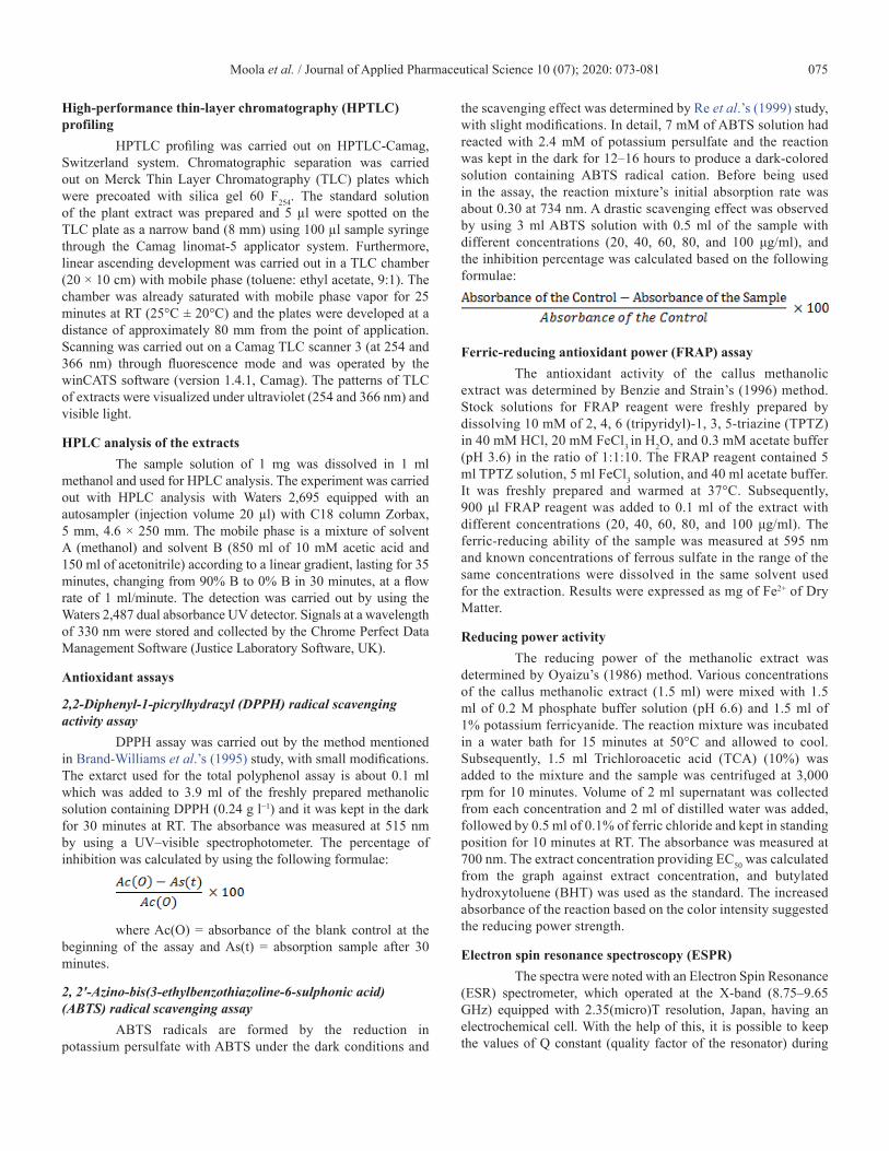
Callus cultures and light microscopyCallus from C. paniculatus was established by using
the leaf explants on the MS medium along with the diverse concentrations of BAP, NAA, and 2,4-D. The callus induction percentage was 91.66 ± 0.69 for BAP + NAA and 89.33 ± 1.11 for BAP + 2,4-D. The experimental results showed a high callus induction rate for BAP (0.5 mg/l) along with 2,4-D (0.3 mg/l) and 0.5 mg/l NAA (Table 1 and Fig. 1).
Histological analysis showed the accumulation of polyphenols in the fragile and compact callus with variying concentrations (Fig. 2), It was observed that BAP+NAA-derived fragile callus had higher accumulation than the compact calli. These results strongly suggest that the nature of the callus and its differentiation influence the accumulation of polyphenols and in concurrence with the previous report (Pandino et al., 2017) on Cynara scolymus L.
Total Phenolics and flavonoidsTo assess the antioxidant potential of the extract, the
total phenolic and flavonoid contents were analyzed (Fig. 3).
Table 1. Effect of different Concentration of BAP along with NAA and 2,4-D on Callus formation from leaf explant of C. paniculatus Willd.
BAP + NAA
0.0 – –
0.5 + 0.1 80.66 ± 0.78d Cream friable
0.5 + 0.3 86.33 ± 0.96bc Friable compact
0.5 + 0.5 91.66 ± 0.69a Friable callus
0.5 + 0.7 83.33 ± 1.34cd Necrotic friable
BAP + 2,4-D
0.0 – –
0.5 + 0.1 84.33 ± 1.20cd Necrotic white compact
0.5 + 0.3 89.33 ± 1.11ab Hard compact
0.5 + 0.5 85.00 ± 1.43c White green compact
0.5 + 0.7 80.66 ± 0.99d White green compact
Mean in each column followed by the same superscript letters are not significantly different according to DMRT AT p < 0.05
Figure 1. Callus induction from C. paniculatus Willd. (a) Leaf Explant (b) BAP + NAA derived fragile callus and (c) BAP + 2, 4-D compact callus.

Moola et al. / Journal of Applied Pharmaceutical Science 10 (07); 2020: 073-081 077
Antioxidant potential in the extract was observed owing to the presence of polyphenols and flavonoids (Ebrahimzadeh et al., 2010; Krishnan et al., 2015). Total phenolic contents of the extracts, expressed as gallic acid equivalents, varied from 36.11 ± 1.15 nmol of GAE/g for methanolic extract of BAP+NAA to 17.78 ± 1.56 mg GAE/g for methanolic extract of BAP+2,4-D. The total flavonoid content is expressed as milligram of catechol equivalents per gram of dried samples ranged from 20.16 ± 0.25 to 19.20 ± 0.10. The research findings were similar to the studies of Huang et al. (2005), Jakaria et al. (2018), Jakaria et al. (2019a), Jakaria et al. (2019b), Khorasani Esmaeili et al. (2015), and Sharififar et al. (2009), where the polyphenols are a good antioxidant source that can significantly increase the antioxidant activity in agreement with the earlier studies.
HPTLC analysisIn this study, the HPTLC showed 9 and 10 polyvalent
phytoconstituents in the extracts of BAP in combination with NAA and 2,4-D. Among the nine polyvalent phytocomponents, the Rf values varied from 0.03 to 0.96 as shown in Figures 4 and 5. The components with Rf values of 0.03, 0.30, and 0.67 are predominant
with an area of 7,824.4, 889.0, and 1,103.8 AU, respectively. The polyvalent compounds were 62.53%, 7.10%, and 8.82% of the NAA extract. Similarly, the 2,4-D extract showed the Rf values as 0.05 and 0.68, which is more prominent with an area percent of 65.20% and 10.24%, respectively.
HPLC analysisPeak differentiation and intensity analyses for
methanolic extract derived from both BAP + NAA- and BAP +2,4-D calli were conducted by HPLC based on their RT. The results are summarized in Table 2 and Figure 6. As expected, the intensity and height of the peak were higher in NAA than in 2,4-D. In medicinal plants, previous studies have showed a higher level of phenolic compounds in in vitro than in in vivo samples (Giri et al., 2012). HPLC analysis of soluble polyphenolics from friable calli culture showed enhanced accumulation of phenolic acids and flavonoids in comparsion with the compact calli.
Figure 2. Histological analysis of embryogenic calli on the MS medium containing BAP along with NAA (Left Image) and 2,4-D (Right Image) in C. paniculatus Willd. after 4 weeks of culture. (Polyphenols and polysaccharides in vacuoles.) Scale bar = 50 µm.
Figure 3. Content and productivity of total phenolics and flavonoids from BAP along with NAA and 2, 4-D callus extract from C. paniculatus Willd. (after 4 weeks of culture). Results are expressed as mean ± SD.
Figure 4. Chromatogram and TLC plate of BAP along with NAA-derived callus extracts of C. paniculatus.

Moola et al. / Journal of Applied Pharmaceutical Science 10 (07); 2020: 073-081 078
Antioxidant efficiency of the sampleAccording to Köksal et al.’s (2017) and Prior et al.’s
(2005) studies, the measurements of antioxidant activity are affected by specific conditions because the plant extract comprises different secondary metabolites. Therefore, it is difficult to determine the antioxidant potential by using single method. In this investigation, we have used four methods – DPPH, ABTS radical cation decolorization assay, reducing power activity, and FRAP assay with BHT – for detecting the radical scavenging activity. The stable free radical DPPH was reduced to yellow DPPH by the methanolic extract from the callus. The IC50 value of the DPPH scavenging ability of BAP + NAA was 66 µg/ml, whereas that of BA+2,4-D was 75 µg/ml. A lower IC50 value indicates maximum DPPH activity. Results of DPPH and ABTS assays are almost similar: the IC50 values of the ABTS radical scavenging activity for BA + NAA is 56 µg/ml and that for BA + 2,4-D is 66 µg/ml (Table 3). However, the antioxidant activities of these two extracts were lesser than those of the standard compounds. The results are summarized in Table 2. Our results are in concurrence with recent reports (Jacob et al., 2008) on Thymus vulgaris.
It is well known that polyphenols or electron-rich compounds can donate electrons by reacting with oxidizing agents to form stable species (Anjum et al., 2017), so that the oxidation process of the different molecules is delayed and indirectly avoids radical quenching and free radical formation. With this information, we studied the reducing potential of the methanolic extract by FRAP and reducing power assays. The yellow colored Fe3+ complex test solution was reduced to a blue colored Fe2+ complex, indicating the reducing activity of the extract (Figure 7).
Table 2. Retention Time (RT) and peak area of HPLC chromatogram identified in C. paniculatus calli extract.
BAP + NAA BAP + 2,4-D
RT Area RT Area
1.575 1,284 5.8 20,663
2.206 42,969 29.712 51,092
29.422 43,408 33.238 8,954
32.999 28,615 35.129 17,101
34.904 5,416 Nill Nill
Figure 5. Chromatogram and TLC plate of BAP along with 2,4-D-derived callus extracts of C. paniculatus.
Figure 6. Typical HPLC chromatogram of the calli of BAP along with NAA and 2,4-D from C. paniculatus Willd. as response (milliVolts) versus time (minutes).

Moola et al. / Journal of Applied Pharmaceutical Science 10 (07); 2020: 073-081 079
The reducing power of the antioxidants increases with increasing concentration. The sequence for this reducing power is BHT ≥ BAP + NAA ≥ BAP + 2,4-D.
Additionally, for more clarification regarding antioxidant potential, we carry out the FRAP assay (Table 3), for BAP + NAA (18.48 ± 0.16 µg/ml), followed by BAP + 2,4-D (10.22 ± 0.10), so that we can conclude that C. paniculatus extract can act as a suitable electron donor by converting free radicals to more stable products. Polyphenolic and flavonoid compounds were found to possess the antioxidant potential and NAA-derived callus expressed better activity in all four assays. Our results are equivalent to the recent research reports (Traverse et al., 2006) on flax, where it was found that BAP + NAA differentially increases the production of lignins and neolignins.
Electron spin resonance spectroscopyIn this study, with the objective of understanding the
radical scavenging potential in an extract, that is heterogeneous powder, we recorded a robust EPR signal in both BAP + NAA- and BAP + 2,4-D-derived calli extracts for scavenging potency. The spectrum consisted of three symmetric ESR peaks signal in both BAP + NAA- and BAP + 2,4-D- derived calli extracts (Figure 8). We labeled them A, B, and C (going from left to right), and we see that the spacing between A and B, and B and C is nearly equal, that is, AB = BC. For example, in the case of NAA
considering A, B, and C as hyperfine structure components, we see that the g-value of the central peak B can be expressed as follows:
AB = BC = 15.2 GTotal width of the chart = 267 mmMagnetic field spread = 360.971 mT – 311.079 Mt =
50.892 mT Scale factor is I mm = 0.19 mT, Resonance field of B =
336.349 Mt = BBFrequency nu = 9,437.485 MHzBased on the values in the spectrum conditions, the
g-value is given by the resonance condition g = h nu/Βbwhere ih = 6.626 × 10⁻³⁴ Js, β = 9.274 × 10⁻²⁸ J/G, Bb =
336.349 mT g = 2.0048This study reveals that BAP + NAA has the most potent
and prominent no-scavenging activity with a g–value of 2.0048, whereas for the BAP + 2,4-D heterogenous powder, even though
Table 3. Concentration required for IC50 of DPPH radical scavenging activity, ABTS scavenging activity of NAA and 2,4-D derived extract, including BHT.
CompoundsDPPH radical scavenging ABTS radical scavenging
IC50 R2 IC50 R2
BHT 59 0.986 53 0.953
BAP + NAA 66 0.987 56 0.964
BAP + 2,4-D 75 0.995 66 0.991
The values are expressed in µg/ml. Lower IC50 indicated in elevated radical scavenging activity.
Figure 7. Reducing the power of methanolic extract from C. paniculatus (BHT – Butyl hydroxytoluene is used as a standard.). Results are expressed as mean ± SD.
Figure 8. EPR spectra of the spin adduct of callus methanolic extract from C. paniculatus. (A) BAP along with NAA (B) BAP along with 2, 4- D-derived calli spin resonance.
Moola et al. / Journal of Applied Pharmaceutical Science 10 (07); 2020: 073-081 080
we obtained three peak signals, it has a little weaker prominent peak and lesser g-value. In agreement with Traverse et al.’s (2006) study, the results are equivalent to our data regarding the three-line spectra for no radical scavenging activity. These observations are confirmed and supportive of the nonenzymatic assays and conclude that NAA-derived friable callus has a great antioxidant potential.
CONCLUSIONIn conclusion, the present findings conclud that the MS
medium along with BAP and NAA is an ideal medium for solid antioxidant activity by effectively scavenge free radicals. We have also established a strong correlation between scavenging activity, phenolics, flavonoids content, and g value. This may be because the compact callus, i.e., BAP + 2,4-D-derived cells are densely aggregated, while in the friable callus derived from BAP + NAA the connection is looser. The friable callus is soft and breaks apart easily, so that polyphenols may be easily extract with methanolic solution and can be used for further isolation of active bioactive compounds. On the other hand, the present exprerimental results insist on the help of a substrate or callus for further studies, in order to enhance the secondary metabolites by using precursor-mediated, hairy root metabolism, and biotic and abiotic elicitors. However, more experimental studies are needed to elucidate the structure of the particular molecules responsible for the antioxidant potential of C. paniculatus Willd. If appropriate strategies are developed, then definitely this compound can be used in the mitigation of free radical-related disease. Moreover, the selection of explants particularly indicated that the choice of the solvent extract has a significant influence on the extraction yield.
ACKNOWLEDGMENTSAnil Kumar Moola thanks CSIR for providing
Senior Research Fellowship with the grant-in-aid number 09/475(0201)/2018-EMR –1. The authors are thankful to IIT Bombay, SAIF for their assistance with the ESPR.
CONFLICT OF INTERESTThe authors declare that there is no conflict of interest.
LIST OF ABBREVIATIONSNAA naphthalene acetic acid2,4-D 2, 4-Dichlorophenoxy acetic acidMS Murashige and Skoog MediumHPLC High-Performance Liquid ChromatographyHPTLC High-Performance Thin-Layer
ChromatographyDPPH 2,2-Diphenyl-1-picrylhydrazylFRAP Ferric-reducing antioxidant powerABTS 2, 2’-Azino-bis(3-ethylbenzothiazoline-6-
sulphonic acid)ESPR Electron Spin Resonance Spectroscopy
REFERENCESAinsworth EA, Gillespie KM. Estimation of total phenolic
content and other oxidation substrates in plant tissues using Folin–Ciocalteu reagent. Nat Protoc, 2007; 2(4):875–7.
Anjum S, Abbasi BH, Hano C. Trends in accumulation of pharmacologically important antioxidant-secondary metabolites in callus
cultures of Linum usitatissimum L. Plant Cell Tissue Org Cult, 2017; 129(1):73–87.
Anusha T, Joseph M, Elyas K. Callus induction and elicitation of total phenolics in callus cell suspension culture of Celastrus paniculatus–Willd, an endangered medicinal plant in India. Pharmacogn J, 2016; 8(5):471–5.
Benzie IF, Strain J. The ferric reducing ability of plasma (FRAP) as a measure of “antioxidant power”: the FRAP assay. Anal Biochem, 1996; 239(1):70–6.
Brand-Williams W, Cuvelier ME, Berset C. Use of a free radical method to evaluate antioxidant activity. Food Sci Technol, 1995; 28(1):25–30.
Cantuti-Castelvetri I, Shukitt-Hale B, Joseph JA. Neurobehavioral aspects of antioxidants in aging. Int J Dev Neurosci, 2000; 18(4–5):367–81.
Debnath M, Pushpan R, Kumari H, Nishteswar K. Medico ethnobotanical perspectives of jyotismati (Celastrus paniculatus. Willd)-a herbal tranquilizer. J Drug Deliv Sci Technol, 2012; 2(3):14.
Duncan DB. Multiple range and multiple F tests. Biom, 1955; 11(1):1–4.
Ebrahimzadeh MA, Nabavi SM, Nabavi SF, Bahramian F, Bekhradnia AR. Antioxidant and free radical scavenging activity of H. officinalis L. var. angustifolius, V. odorata, B. hyrcana and C. speciosum. Pak J Pharm Sci, 2010; 23(1):29–34.
Ferreira IC, Baptista P, Vilas-Boas M, Barros L. Free-radical scavenging capacity and reducing the power of wild edible mushrooms from northeast Portugal: Individual cap and stipe activity. Food Chem, 2007; 100(4):1511–6.
Giri L, Dhyani P, Rawat S, Bhatt ID, Nandi SK, Rawal RS, Pande V. In vitro production of phenolic compounds and antioxidant activity in callus suspension cultures of Habenaria edgeworthii: a rare Himalayan medicinal orchid. Ind Crops Prod, 2012; 39:1–6.
Godkar P, Gordon R, Ravindran A, Doctor B. Celastrus paniculatus seed oil and organic extracts attenuate hydrogen peroxide-and glutamate-induced injury in embryonic rat forebrain neuronal cells Phytomedicine. Int J Phytother Phytopharmacol, 2006; 13(1–2):29–36.
Huang D, Ou B, Prior RL. The chemistry behind antioxidant capacity assays. J Agric Food Chem, 2005; 53(6):1841–56.
Jacob JK, Hakimuddin F, Paliyath G, Fisher H. Antioxidant and antiproliferative activity of polyphenols in novel high-polyphenol grape lines. Food Res Int, 2008; 41(4):419–28.
Jakaria M, Azam S, Cho DY, Haque M, Kim IS, Choi DK. The methanol extract of allium cepa L. protects inflammatory markers in LPS-induced BV-2 microglial cells and upregulates the antiapoptotic gene and antioxidant enzymes in N27-A cells. Antioxidants, 2019a; 8(9):348.
Jakaria M, Azam S, Haque ME, Jo SH, Uddin MS, Kim IS, Choi DK. Taurine and its analogs in neurological disorders: focus on therapeutic potential and molecular mechanisms. Redox Biol, 2019b; 21(24):101223.
Jakaria M, Cho DY, Haque E, Karthivashan G, Kim IS, Ganesan P, Choi DK. Neuropharmacological potential and delivery prospects of thymoquinone for neurological disorders. Oxid Med Cell Longev, 2018; 2018:1209801.
Jang HR, Lee HJ, Park BJ, Pee OJ, Paek KY, Park SY. Establishment of embryogenic cultures and determination of their bioactive properties in Rosa rugosa. Hortic Environ Biotechnol, 2016; 57(3):291–8.
Karuppanapandian T, Moon JC, Kim C, Manoharan K, Kim W. Reactive oxygen species in plants: their generation, signal transduction and scavenging mechanisms. Aust J Crop Sci, 2011; 5(6):709.
Khorasani Esmaeili A, Mat Taha R, Mohajer S, Banisalam B. Antioxidant activity and total phenolic and flavonoid content of various solvent extracts from in vivo and in vitro grown Trifolium pratense L.(red clover). Biomed Res Int, 2015; 2015:11.
Köksal E, Bursal E, Gülçin İ, Korkmaz M, Çağlayan C, Gören AC, Alwasel SH. Antioxidant activity and polyphenol content of Turkish thyme (Thymus vulgaris) monitored by liquid chromatography and tandem mass spectrometry. Int J Food Properti, 2017; 20(3):514–25.
Moola et al. / Journal of Applied Pharmaceutical Science 10 (07); 2020: 073-081 081
Krishnamurthi A. The wealth of India: raw materials: Vol. VIII. Ph-Re the wealth of India: raw materials: Vol VIII Ph-Re. 1969.
Krishnan V, Ahmad S, Mahmood M. Antioxidant potential in different parts and callus of Gynura procumbens and different parts of Gynura bicolor. Biomed Res Int, 2015; 2015:7.
Maxwell SR. Prospects for the use of antioxidant therapies. Drugs, 1995; 49:345–61.
Misra R, Kumar S, Pani D, Bhandari D. Empirical tribal claims and correlation with bioactive compounds: a study on Celastrus paniculatus Willd., a vulnerable medicinal plant of Odisha. Indian J Tradit Knowl, 2012; 11(4):615–22.
Moola AK, Kumari BR. Direct regeneration of plantlets from shoot tip explants of a vulnerable medicinal plant–Celastrus paniculatus Willd. J Appl Hortic, 2019; 21(3):189–94.
Murashige T, Skoog F. A revised medium for rapid growth and bio assays with tobacco tissue cultures. Physiol Plant, 1962; 15(3):473–97.
Nadkarni A. Indian materia medica 3Rd Ed volume two. Popular Book Depot, Bombay, India, 2014.
Oyaizu M. Studies on products of browning reaction: antioxidative activity of products of browning reaction. Jpn J Nutr, 1986; 44(6):307–15.
Pandino G, Meneghini M, Tavazza R, Lombardo S, Mauromicale G. Phytochemicals accumulation and antioxidant activity in callus and suspension cultures of Cynara scolymus L. Plant Cell Tissue Org Cult, 2017; 128(1):223–30.
Parimala S, Shashidhar G, Sridevi C, Jyothi V, Suthakaran R. Anti-inflammatory activity of Celastrus paniculatus seeds. Int J Pharm Technol Res, 2009; 1:1326–9.
Park JA, Park BJ, Kim AH, Park SY, Paek KY. Airlift bioreactor system and nitrogen sources for biomass and antioxidant compound production from in vitro culture of Vitis flexuosa. Hortic Environ Biotechnol, 2015; 56(3):358–65.
Phulwaria M, Rai MK, Patel AK, Kataria V, Shekhawat N. A genetically stable rooting protocol for propagating a threatened medicinal plant—Celastrus paniculatus. AoB Plants, 2013; 5:054.
Prior RL, Wu X, Schaich K. Standardized methods for the determination of antioxidant capacity and phenolics in foods and dietary supplements. J Agric Food Chem, 2005; 53(10):4290–302.
Rajasekharan P, Ganeshan S. Conservation of medicinal plant biodiversity: an Indian perspective. J Trop Med Plants, 2002; 3(1):125–40.
Re R, Pellegrini N, Proteggente A, Pannala A, Yang M, Rice-Evans C. Antioxidant activity applying an improved ABTS radical cation decolorization assay. Free Radic Biol Med, 1999; 26(9–10):1231–7.
Rekha K, Bhan M, Balyan S, Dhar A. Cultivation prospects of endangered species Celastrus paniculatus Willd. Regional Research Laboratory, Jammu, India, 2005.
Sasheva P, Letkarska G, Ionkova I. Biotechnological production of podophyllotoxin and podophyllotoxin-related lignans in cultures of Linum thracicum Degen. C R Acad Bulg Sci, 2013; 66(10):1445–50.
Sharada M, Ahuja A, Kaul M. Regeneration of plantlets via callus cultures in Celastrus paniculatus Willd-A rare endangered medicinal plant. J Plant Biochem Biotechnol, 2003; 12(1):65–9.
Sharififar F, Dehghn-Nudeh G, Mirtajaldini M. Major flavonoids with antioxidant activity from Teucrium polium L. Food Chem, 2009; 112(4):885–8.
Squadrito GL, Pryor WA. Oxidative chemistry of nitric oxide: the roles of superoxide, peroxynitrite and carbon dioxide. Free Radic Biol Med, 1998; 25(4–5):392–403.
Subhasree B, Baskar R, Keerthana RL, Susan RL, Rajasekaran P. Evaluation of antioxidant potential in selected green leafy vegetables. Food Chem, 2009; 115(4):1213–20.
Traverse JH, Nesmelov YE, Crampton M, Lindstrom P, Thomas DD, Bache RJ. Measurement of myocardial free radical production during exercise using EPR spectroscopy. Am J Physiol-Heart Circ Physiol, 2006; 290(6):H2453–8.
How to cite this article: Moola AK, Thiruppathi SK, Diana RKB. Determination of polyphenolic content and antioxidant potential of callus extract obtained from Celastrus paniculatus Willd. and their free radical estimation by electron spin resonance spectroscopy. J Appl Pharm Sci, 2020; 10(07):073–081.