development of the digestive tube -...
TRANSCRIPT

Development of
the digestive tube
© David Kachlík 30.9.2015

© David Kachlík 30.9.2015

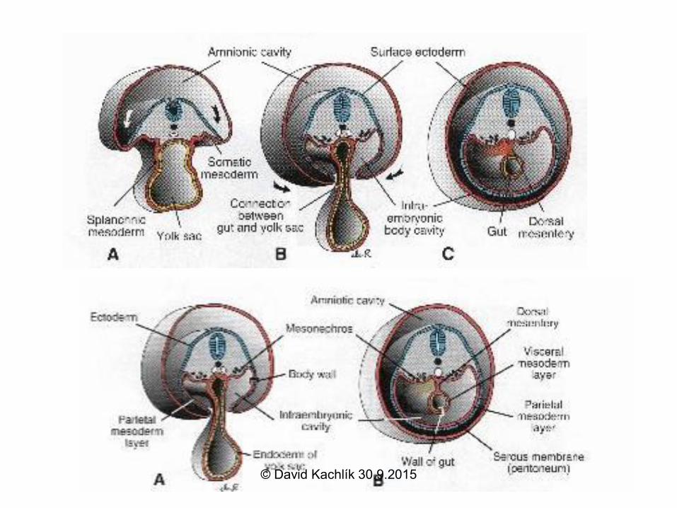
Development of digestive tubePrimitive gut
Week 4: folding of embryo causes partial incorporation of yolk sac
© David Kachlík 30.9.2015

3rd week4th week
© David Kachlík 30.9.2015

Development
Endoderm of primordial gut
Stomodeum Proctodeum
Ectoderm of stomodeum
Epithelium and glands of digestive tract
Ectoderm of proctodeum
Epithelium in part of oral cavity
Epithelium in anal canal© David Kachlík 30.9.2015

DevelopmentForegut Hindgut
Oropharyngeal membrane Cloacal
membrane
Foregut
Midgut
From oral cavity to duodenum (below papilla duodeni
major)
Celiac trunk
Hindgut
From duodenum to transverse colon
Superior mesentericartery
Inferior mesentericartery The rest of colon© David Kachlík 30.9.2015

© David Kachlík 30.9.2015

© David Kachlík 30.9.2015

Foregut (preentereon)
• separated by mebrana oropharyngea from stomodeum (ruptures on 26th day)
• evangination of:
– submandibular and sublingual glands
– thyroid gland
– evangination of primitive pharynx
– diverticle for lower respiratory tract
– diverticle for liver, gallbladder and ventral part of pancreas
• artery: truncus coealicus
• nerve: n.X (parasympathetic)© David Kachlík 30.9.2015

Midgut (mesentereon)
• distally to hepatic diverticle towards ductus
omphaloentericus
• further aborally as far as Cannon-Böhm‘s
point (at flexura coli sinistra)
• evangination:
– appendix vermiformis
• artery: a. mesenterica superior
• nerve: n.X (parasympathetic)
© David Kachlík 30.9.2015

Hindgut (metentereon)
• further aborally from Cannon-Böhm‘s point
at flexura coli sinistra
• as far as linea pectinata (anal canal)
• from proctodeum separated by membrana
cloacalis (ruptures at the end of 8th week)
• artery: a. mesenterica inferior
• nerve: plexus hypogastricus inferior
(parasympathetic)
© David Kachlík 30.9.2015

Hinge of primitive gut
• mesenterium ventrale primordiale
– only till the level of the end of foregut
– only a thin serous duplicature
→ omentum minus
→ peritoneum viscerale of liver
→ ligamentum falciforme
© David Kachlík 30.9.2015

© David Kachlík 30.9.2015

Hinge of primitive gut• mesenterium dorsale primordiale
– whole length of intestine, fixation to posterior abdominal wall
– contains connective tissue and vessels
– spleen originates within it
→ ligamentum gastrocolicum
→ omentum majus
→ peritoneum viscerale of the spleen
→ peritoneum viscerale of the transverse and sigmoid
colon
→ mesocolon transversum, mesoappendix,
mesogimoideum
• mesogastrium dorsale© David Kachlík 30.9.2015

Oesophageal development
• rotation → asymmetric course of n.X
• temporary obliteration of caliber
• upper third – skeletal muscle from lower
pharyngeal arches
• developmental defects:
– atresia
– stenosis
– fistula tracheooesophagealis
© David Kachlík 30.9.2015

Stomach development
• enlargement of aboral part of foregut
• faster growth of posterior wall
• subsequent rotation to the left by 90along the horizontal axis
• evagination of dorsal mesogastrium
• origin of bursa omentalis
• developmental defect: congenital hypertrophic pylorostenosis– 1:150 males x 1:750 females
– projectile vomiting© David Kachlík 30.9.2015
© David Kachlík 30.9.2015

© David Kachlík 30.9.2015

© David Kachlík 30.9.2015

© David Kachlík 30.9.2015

Development of greater omentum
• dorsal mesogastrium growth to the length
• extends ventrally to small intestine
• adhesions appear
– mesocolon + mesogastrium dorsale
– both layers of mesogastrium dorsale merge
→ recessus inferior bursae omentalis
© David Kachlík 30.9.2015

© David Kachlík 30.9.2015

© David Kachlík 30.9.2015

Animation
http://www.youtube.com/watch?v=s2cNCUL
1r3A&feature=BFa&list=PL9A2D6BB7F13
1CA12
© David Kachlík 30.9.2015

Intestine development
© David Kachlík 30.9.2015

Midgut development
• subsequent formation of
tubular gut
• ductus
omphaloentericus
(vitellio-intestinalis)
remains (6)
• midgut enlarges loop
around the axis of
arteria mesenterica
superior (9)
© David Kachlík 30.9.2015

Development of the midgut
• further enlargement of cranial limb of intestinal loop
• rotation 90° anti-clockwise along the axis of a. mesenterica sup.
• physiological herniation (6th-10th week)– due to large liver and two
pairs of kidneys
© David Kachlík 30.9.2015

Midgut development
• further 90° rotation
• colon (19) appears in
front of duodenum
(10)
• subsequent return of
loops (3rd month)
• caecal bud
© David Kachlík 30.9.2015

Midgut development
• terminal rotation by
90° (totally 270°)
• caecal bud (16)
located on the right
side under liver
© David Kachlík 30.9.2015

Midgut development
• caecum descends
caudally
• formation of colon
ascendens
• appendix
vermiformis
evaginates
– firstly it grows slowly,
then rotates medially
due to irregular
growth of caecum© David Kachlík 30.9.2015

Change of gut position and peritoneal cover
• ascending and descending colon
• duodenum
© David Kachlík 30.9.2015

© David Kachlík 30.9.2015

Animation
http://www.youtube.com/watch?v=j7OG4wS
pDqI&NR=1&feature=endscreen
http://www.youtube.com/watch?v=rs44cXvjb
MA&list=PL9A2D6BB7F131CA12
© David Kachlík 30.9.2015

Development of duodenum
• week 5-6: delumination of duodenum
happens (caliber fades out)
• week 8: subsequently the lumen
reappears
© David Kachlík 30.9.2015

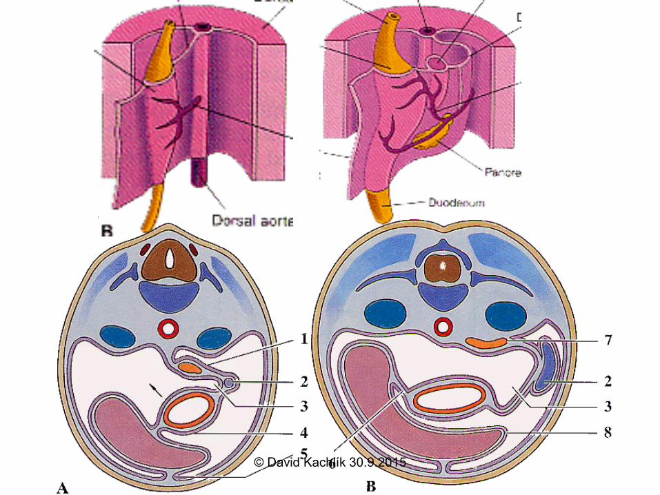
Development of liver, gallbladder and bile duct
• ventral endoderm of caudal part of foregut
(preenteron distale)
hepatic evagination (4th week)
– ingrowth into ventral mesenterium
Liver
diverticle
© David Kachlík 30.9.2015

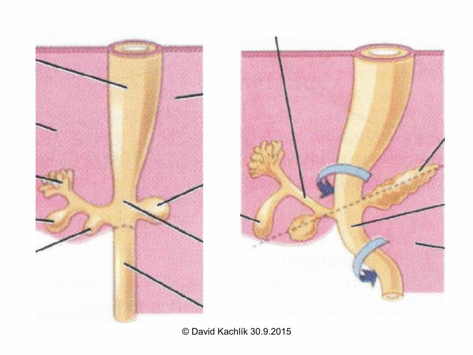
Development of liver, gall bladder and bile duct
• gemma hepatopancreatica
→ division into:
• diverticulum hepatis
– cranial larger part → liver diverticle
– caudal smaller part → gallbladder diverticle
• formation of ductus cysticus
• ductus choledochus forms by narrowing of
connection of liver with foregut– opens together with ductus pancreaticus Wirsungi at papilla
duodeni major Vateri
© David Kachlík 30.9.2015

© David Kachlík 30.9.2015
© David Kachlík 30.9.2015

Liver development
• 2 origins: foregut and septum transversum
• epitelial cells grow into mesenchyme of
septum transversum → hepatocyte columns
• mesoderm →
– tissue of interlobular spaces
– hematopoetic cells
– Ito cells
– Kupffer cells (migrated from bone marrow)
© David Kachlík 30.9.2015

Liver development
• blood production since 6th week
– subsequently ceases last 2 months of intrauterine
development
• mesogastrium ventrale:
– mesohepaticum lig. falciforme hepatis +
peritoneum viscerale of liver + omentum minus
• liver covered by peritoneum except for area nuda
• bile production since 12th week
developmental defects:
– extrahepatal bile ducts atresia
– pseudocysts© David Kachlík 30.9.2015

Atresia of bile ducts
© David Kachlík 30.9.2015

Pseudocysts of bile ducts
© David Kachlík 30.9.2015

21t. day
26th day
Diaphragm
development
© David Kachlík 30.9.2015

Septum transversum
30th
day
© David Kachlík 30.9.2015
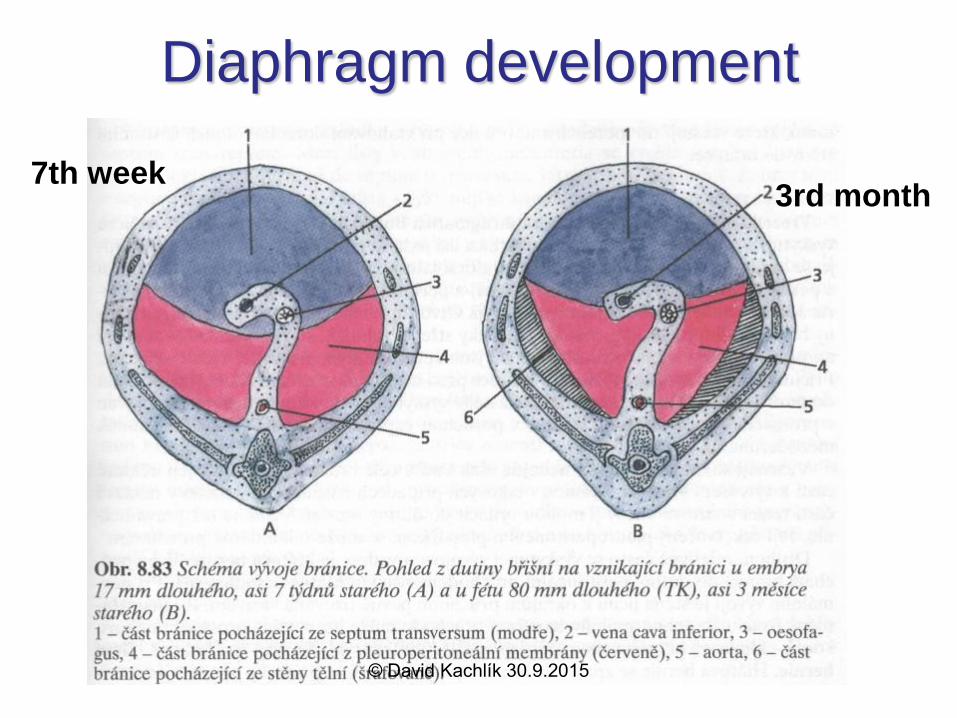
7th week3rd month
Diaphragm development
© David Kachlík 30.9.2015

Development of pancreas
• 4th week
• aboral part of foregut
• 2 pancreatic
evagination (11) from
endoderm
– dorsal (larger)
– ventral – from liver
diverticle
© David Kachlík 30.9.2015

5th-8th week
© David Kachlík 30.9.2015

© David Kachlík 30.9.2015

© David Kachlík 30.9.2015

• dorsal pancreas (7) grows between
layers of dorsal mesentery
(mesoduodenum)
• ventral pancreas (5) grows into
ventral mesoduodenum• future processus uncinatus and part of
pancreatic head
• their connection can happens due to
duodenal rotation• ductus pancreaticus Wirsungi papilla
duodeni major Vateri
• proximal part of duct of dorsal pancreas
remains as ductus pancreaticus
accessorius Santorini papilla duodeni
minor Santorini © David Kachlík 30.9.2015

Pancreatic development
• pancreas with duodenum and
mesoduodenum flops and fuses with
parietal peritoneum (Treitz‘s
retropancreatic membrane)
© David Kachlík 30.9.2015

Pancreatic development
• tissue cover and interlobular septa originate from surrounding splanchnic mesenchyme
– insuline production• begins in approx. 10th week, hypertrophy of beta-
cells in case of maternal diabetes mellitus
• developmental defects of pancreas:
– accessory pancreatic tissue• stomach wall, duodenal wall, Meckel‘s diverticle
– pancreas anulare (1:20.000)→ duodenal stenosis
– pancreas divisum (5-8 %)© David Kachlík 30.9.2015

PANCREAS DIVISUM
PANCREAS ANULARE
© David Kachlík 30.9.2015

Developmental defects of foregut
• stenosis or atresia of duodenum and gut
• diverticulum ilei Meckeli
• Inborn omphalocele
– umbilical hernia
• gastroschisis – defect of anterior abdominal wall
• defects of rotation
– situs viscerum inversus
• volvulus
• internal herniae
• gut duplication – defect of vacuolisation© David Kachlík 30.9.2015

STENOSIS
ATRESIA
© David Kachlík 30.9.2015

Diverticulum ilei
• ductus omphaloentericus
may persists
→ diverticulum ilei Meckeli
– 2%
– 0-100 cm from ostium
ileocaecale
© David Kachlík 30.9.2015

http://www.nejm.org/doi/full/10.1056/NEJMicm1001158
http://www.surgical-tutor.org.uk/default-home.htm?tutorials/meckels.htm~right
http://www.surgical-tutor.org.uk/default-home.htm?tutorials/meckels.htm~right
http://www.learningradiology.com/archives2009/COW%20378-
Meckels%20Tic/caseoftheweek378page.htm
Diverticulum
ilei
Meckeli
© David Kachlík 30.9.2015

Developmental defects of gut
• herniae
– omphalocele = defect of intestinal oops return
• covered only by amion
– inborn umbilical hernia
• normal return, later herniation
• covered by peritoneum and amnion
• abnormal rotation• lesser rotation
• reversal rotation
© David Kachlík 30.9.2015

Developmental defects of digestive
tube
• defects of tube luminization
– atresiae and stenoses
• duodenum, oesophagus, anus
– duplication
• gut
– cysts
© David Kachlík 30.9.2015

Bochdalek posterolateral hernia
© David Kachlík 30.9.2015

Gut malrotation
© David Kachlík 30.9.2015

Inborn umbilical hernia
© David Kachlík 30.9.2015

Omphalocele
© David Kachlík 30.9.2015

Gastroschisis
© David Kachlík 30.9.2015

Situs viscerum inversus
© David Kachlík 30.9.2015

Development of hindgut• proctodeum – invagination of ectoderm
• septum urorectale grows into cloaca
→ differentiation into anorectal and urogenital part
→ differentiation of membrana cloacalis into
membrana urogenitalis et membrana analis
© David Kachlík 30.9.2015

Proctodeum
• invagination of ectoderm
• from linea dentata/pectinata onward
• artery: a. pudenda interna
• innervation: n. pudendus
• disappears at the end of 8th week by
rupture of membrana analis
© David Kachlík 30.9.2015

Developmental defects of hindgut
• anus imperforatus
• agenesis of anus
• stenosis / atresia of anus
• rectal fistula
• intestinal agangliosis (megacolon
congenitum; Hirschsprung disease)
© David Kachlík 30.9.2015

Development of peritoneum
• intraembryonal coelom
• coelomic „epithelium“ = mesothel
• classificatiob of organs according to the
peritoneal cover (tunica serosa):
– intraperitoneal
• visceral and parietal peritoneum
• duplicatures
– retroperitoneal
• primary x secondary
– subperitoneal
• mesos and folds (bursa omentalis)© David Kachlík 30.9.2015
© David Kachlík 30.9.2015

© David Kachlík 30.9.2015
© David Kachlík 30.9.2015

© David Kachlík 30.9.2015

Development of bursa omentalis
© David Kachlík 30.9.2015

© David Kachlík 30.9.2015

Development of bursa omentalis
© David Kachlík 30.9.2015

© David Kachlík 30.9.2015

© David Kachlík 30.9.2015

Extent of bursa omentalis
© David Kachlík 30.9.2015

© David Kachlík 30.9.2015

Development of bursa omentalis
© David Kachlík 30.9.2015

© David Kachlík 30.9.2015

© David Kachlík 30.9.2015

© David Kachlík 30.9.2015