developmental dysplasia of the hip: a case study
TRANSCRIPT
University of North DakotaUND Scholarly Commons
Physical Therapy Scholarly Projects Department of Physical Therapy
2016
Developmental Dysplasia of the Hip: A Case StudyJoseph D. TaylorUniversity of North Dakota
Follow this and additional works at: https://commons.und.edu/pt-grad
Part of the Physical Therapy Commons
This Scholarly Project is brought to you for free and open access by the Department of Physical Therapy at UND Scholarly Commons. It has beenaccepted for inclusion in Physical Therapy Scholarly Projects by an authorized administrator of UND Scholarly Commons. For more information,please contact [email protected].
Recommended CitationTaylor, Joseph D., "Developmental Dysplasia of the Hip: A Case Study" (2016). Physical Therapy Scholarly Projects. 575.https://commons.und.edu/pt-grad/575

Developmental Dysplasia of the Hip; A Case Study
by
Joseph D Taylor Bachelor of Science in Biology, Human Biology Emphasis 2013
A Scholarly Project Submitted to the Graduate Faculty of the
Department of Physical Therapy
School of Medicine and Health Sciences
University of North Dakota
In partial fulfillment of the requirements for the degree of
Doctor of Physical Therapy
Grand Forks, North Dakota May, 2016
This Scholarly Project, submitted by Joseph D Taylor in partial fulfillment of the requirements for the Degree of Doctor of Physical Therapy from the University of North Dakota, has been read by the Advisor and Chairperson of Physical Therapy under whom the work has been done and is hereby approved.
(Chairperson, P sical Therapy)
2

PERMISSION
Title Developmental Dysplasia of the Hip; A Case Study
Department Physical Therapy
Degree Doctor of Physical Therapy
In presenting this Scholarly Project in partial fulfillment of the requirements for a graduate degree from the University of North Dakota, I agree that the Department of Physical Therapy shall make it freely available for inspection. I further agree that permission for extensive copying for scholarly purposes may be granted by the professor who supervised my work or, in her absence, by the Chairperson of the department. It is understood that any copying or publication or other use of this Scholarly Project or part thereof for financial gain shall not be allowed without my written permission. It is also understood that due recognition shall be given to me and the University of North Dakota in any scholarly use which may be made of any material in this Scholarly Project.
Signature
Date
3
TABLE OF CONTENTS
LIST OF FIGURES .............. "' .................. "' ..................................... 5
LIST OF TABLES ......................................................................... 6
ACKNOWLEDGEMENTS ................................................................ 7
ABSTRACT ................................................................................... 8
CHAPTER I.
II.
BACKGROUND AND PURPOSE ........................ 10
CASE DESCRIPTION ........................................ 13
Examination, Evaluation and Diagnosis .................. 15
Prognosis and Plan of Care .................................. 19
Intervention ..................................................... 22
Outcomes ....................................................... 29
III. DiSCUSSiON .................................................... 32
Reflective Practice ............................................. 34
APPEND~A ................................................................................ 38
APPENDIX B ................................................................................ 39
REFERENCES ............................................................................. 40
4

LIST OF FIGURES
1. Figure 1 ................................................................................. 21
2. Figure 2 .................................................................................. 38
2. Figure 3 .................................................................................. 39
5

LIST OF TABLES
1. Table 1 .................................................................................... 16
2. Table 2 ..................................................................................... 16
3. Table3 ................................................................................... 17
4. Table 4 .................................................................................. 24
6

ACKNOWLEDGEMENTS
I would like to thank my clinical instructor and faculty advisor for all of their
help in the creation of the case report. I would also like to thank my peers for
their continued help in the review process.
7
ABSTRACT
PURPOSE: The primary purpose of this case study was to describe the
implementation of a conservative treatment protocol for dysplasia of the hip in a
young girl in an attempt to prevent surgical intervention. A secondary objective
of reducing femoral acetabular impingement was subsequently undertaken in the
course of the case study.
BACKGROUND: The patient was a 16 year old female with a diagnosis of
bilateral developmental dysplasia of the hip (DOH), bilateral femoral acetabular
impingement (FAI), anterior superior labral tear to left (L) hip, elevated
antinuclear antibody (ANA) screening and a history of depression.
PLAN OF CARE INTERVENTiONS: An 8 week course of conservative treatment
was pursued following a specific non-operative protocol provided by an
orthopedic physician. The primary focus of the protocol was to decrease pain in
intensity and duration and to normalize range of motion and strength.
RESULTS: The patient was seen for follow-up appointment after 8 weeks of
physical therapy. Gains were made in strength and ROM. Pain had decreased in
frequency, intensity, and duration. However, the patient was still unable to
increase her activity level due to pain. At the conclusion of this case study, the
patient was referred to an orthopedic specialist for consultation about performing
surgical intervention.
CONCLUSIONS: While the protocol in use was successful in decreasing pain
and increasing ROM and strength, measurement of quality of life for the patient
8

was not implemented. The lack of monitoring of the patient's QOl may have
contributed to the necessity of surgical intervention ..
9

CHAPTER I
BACKGROUND AND PURPOSE
Developmental dysplasia of the Hip, or DOH, is a relatively rare congenital
defect of the hip joint in humans, occurring on average in only 1-10 percent of
live births.1 In cases of DOH the acetabulum (or "socket" portion of the joint) is
shallower than in typically developing children, allowing for easy dislocation and
instability of the hip joint.1 The incidence of DOH is greater in countries such as
Finland, Canada, and Croatia while being vastly less in African countries.1 There
is a possible link between newborns raised in cold weather climates and the
development of DOH and an increase in DOH incidence in children born during
winter months versus those born in summer months2 Swaddling has been shown
to be strongly associated with DDH1 and breech presentation, positive family
history, and female gender are considered predicting factors.1 Prematurity, low
birth weights, or multi-fetal pregnancies are have been identified as protective
factors for DDH.1DDH has also been shown to be a major contributing factor in
the development of osteoarthritis in the hip knee and ankle joints in younger aged
children.3
DOH is commonly accompanied by femoral acetabular impingement
(FAI).3,4 In FAI, the malalignment ofthe hip joint causes a boney overgrowth of
either the acetabulum or of the femoral head, which in turn causes an
impingement of the hip labrum.4 When accompanied by one another, DOH and
FAI, can become a debilitating syndrome and restrict individuals' participation in
10

activities such as physical exercise, education tasks, occupational duties, and
family, social and community events. Pain is often the limiting factor in most
cases of DDH and FAI, leading to the lack of participation. As a result it is not
uncommon to see a reduction in the quality of life in individuals with DDH and
FAI, if the conditions occur separately or simultaneously4
The patient in this case study was a 16-year-old female with a diagnosis of
bilateral DDH and FAI. The patient had been limited in educational, vocational
and familial involvement secondary to her diagnoses. The patient had
requested the opinion of an orthopedic physician regarding possible surgical
intervention options. However, the orthopedic surgeon suggested a course of
conservative treatment via physical therapy in order to mitigate the patient's
current symptoms and improve her quality of life. The primary purpose of this
case study is to describe the implementation of a conservative treatment
protocol for DDH for this patient in an attempt to reduce femoral acetabular
impingement and prevent surgical intervention.
Conservative intervention included, but was not limited to; therapeutic
exercise, passive range of motion (PROM), active assistive range of motion
(AAROM), active range of motion (AROM), stretching, strengthening, manual
therapy, soft tissue mobilization, joint mobilization, moist heat, cold packs,
ultrasound, iontophoresis, phonophoresis, electrical stimulation, home exercise
programing, neuromuscular education, posture training, body mechanics,
therapeutic activities, and activity modification. With a course of conservative
treatment in patients with DDH and FAI there may not be gains in strength and
11

range of motion (ROM), but there may be vast improvements in quality of life
(QOL).4,5,6 Current li!erature4.5,6 strongly suggests that the success of the
conservative treatment be based on QOL improvements rather than strength,
ROM, and pain reduction.
12

CHAPTER II
CASE DESCRIPTION
The patient in this case report was a 16 year old English speaking female.
The patient lived at home with her mother, stepfather, and half-brother. The
patient was a full time student in the 10th grade and worked part-time at a
convenience stofe (15-20 hours per week). The patient's mother reported a
normal course of pregnancy and delivery without complications. The patient's
medical record indicated she appeared well developed and well-nourished upon
presentation to her primary care provider (PCP). The patient did not use any
assistive devices at this time and lived at home with her family in a second floor
apartment without an elevator. The patient was in good overall health, 5' 6"
(1.6764 m) tall, and weighed 132 Ibs. (59.87 kg) with a BMI of 20.99. A review of
systems at the patient's PCP office revealed no obvious concems. The patient
reported that she was "not very" physically active, not involved with sports or
other extracurricular activities other than working at the convenience store. The
patient was previously diagnosed with depression and had been subsequently
taking sertraline HCl (Zoloft) for this condition. The patient was not a smoker, or
exposed to secondhand smoke, and did not report drinking alcohol. The patient
had a familial history of arthritis on her maternal side (grandmother diagnosed in
her 30's) and reported other family members had hip replacements in their 50's.
The patient had no previous history of surgeries or other medical comorbidities.
The patient presented to her PCP 3 months prior to reporting to physical therapy
13

with a primary complaint of bilateral hip pain. She reported having had this pain
for 1 month prior to seeking the advice from her PCP. The pain began suddenly
and did not have any clear mechanism of injury. Pain was present in both hips,
but slightly worse in the R hip. Physical activity seemed to aggravate her
symptoms. The patient did not report a change in gait at the time of initial visit;
however, on follow-up visits she reported that she felt like she "waddles like a
penguin." The patient also reported audible popping in both hips from "time to
time." After this first visit with the PCP, an bilateral X-ray of the hips, complete
blood count (CBC), erythrocyte sedimentation rate (ESR), Rh factor, and
antinuclear antibodies (ANA) were ordered. The CBC document high levels of
monocytes (15). The hips were not fractured or dislocated and the pelvic ring
was intact. An ANA test was reported to be positive (low titer), possibly indicative
of juvenile arthritis. The patient had a follow up appointment with her PCP 3
weeks later and was referred to a rheumatologist to address her hip pain. The
patient never saw the rheumatologist and was again seen approximately 1 month
later by her PCP. At this time, she reported that she felt like her hip pain was
getting worse and sometimes she felt as if her hips were going to, "pop out of
place." The patient's pain in her hips increased with activity and nothing (neither
rest, ice, nor meds) seemed to relieve the pain. The patient's PCP decided to
order an MRI and then referred the patient to orthopedist. The patient's MRI was
shown to exhibit "tiny nonspecific bilateral joint effusions, without evidence of
painful bone marrow edema or evidence of avascular necrosis. No evidence of
trochanteric bursitis nor evidence of gluteal insertional tendonopathy. Mild
14

bilateral developmental hip dysplasia, without definite labral tear identified." The
patient had a follow up with an orthopedic physician who, upon examination,
reported severely limited hip intemal rotation (IR) ROM and excessive hip
external rotation (ER) ROM. The orthopedic physician concluded there was an
anterior-superior labral tear in the l hip and possible labral involvement which
was in contrast to the initial MRI interpretation. This physician also reported
significant femoral retroversion in both hips and probable femoral acetabular
impingement. A course of physical therapy was recommended and the patient
was referred to physical therapy.
Examination, Evaluation and Diagnosis
Approximately 3.5 months after her visit with her PCP, the patient
experienced an acute exacerbation of bilateral hip pain. The patient sought
evaluation and treatment from a different physician and was referred to an
outpatient PT clinic. The patient presented to PT with her mother on the day of
initial evaluation. A comprehensive review of this patient's electronic medical was
completed prior to the physical examination.
Observations. The patient sat on the observation table with slumped
posture and a posteriorly rotated pelvis. During observation of her arrival to PT,
no great deviations in gait other than slight ER bilaterally during toe off and initial
swing phase of gait were noted.
Physical Examination. Range of motion (ROM) measurements were obtained
of bilateral lower extremities and the results can be viewed in Table 1.
15

L hip flexion
L hip abduction
L hip ER
L hip IR
R hip flexion
R hip abduction
R hip ER
R hip IR
Motion
Table1
Range of Motion Measurements
ROM (measured in degrees)
108°
60°
80°
7°
110°
63°
90°
15°
Strength testing was completed via manual muscle test (MMT) to bilateral
lower extremities as well and the results can be found in Table 2.
Table 2
Manual Muscle Testing
Motion Left Right
Flexion 3+/5 3+/5
Extension 2/5 2+/5
Abduction 3+/5 3+/5
Adduction 4/5 4/5
IR 2/5 2/5
ER 3/5 3/5
16

Upon palpation, the patient was tender to the touch over the anterior,
lateral, and posterior aspects of her hips bilaterally, with her primary complaint
presenting anteriorly. The patient was alert, oriented to person, place, time, and
situation, and followed directions appropriately.
To more accurately determine the diagnosis and develop the plan of care,
multiple special tests were performed. The results can be seen in table 3.
Faber's Test
Ober's Test
Hip Scour Test
Test
Table 3
Diagnostic Special Test Results
Result
+ bilaterally
- bilaterally
+ bilaterally
Faber's test was included to determine if hip pathology was present.
Faber's test is a reliable, sensitive, and specific test for hip pathology
(sensitivity=.89, specificity= .99).7 Ober's test was included to examine if the
patient had tight or pathological iliotibial band/tensor fasciae latae (sensitivity=.88
specificity=.83)? The hip scour test was included to test for hip labral
involvement, hip arthritis or acetabular defects (sensitivity=.91 specificity=.75).7
Results. It was determined that this patient presented with strength and
ROM limitations affecting the hips secondary to pain. Special tests indicated that
the patient exhibited pathology located in the hip joints which could result from
17

arthritis, impingement, retro/anteversion, or labral tear. The patient demonstrated
limitations in activities at school (pain when walking from one class to another
and during gym class), work/occupation (pain with prolonged standing or carrying
items), and recreation (pain with walking with friends, standing at a concert,
participating in activities with friends). It was determined, to prevent further
limitations, the patient should begin a course of conservative PT treatment
focusing on strengthening the proximal hip musculature and working on proper
gait mechanics.
According to the Guide to PT practice, the diagnosis was Practice pattern
40; Impaired Joint Mobility, Motor Function, Muscle Performance, and Range of
Motion Associated with Connective Tissue Dysfunction. The medical diagnosis
for this patient was "bilateral developmental dysplasia of the hip, bilateral femoral
acetabular impingement, and possible labral tear/involvement." A PT "working"
diagnosis was "Bilateral DOH, FAI and possible labral tearlinvolvement causing
the patient pain, weakness and limited ROM leading to limitations in participation
in school, work and leisure activities." In utilizing this working diagnosis, the
medical disability was connected to the personal activity limitations. All medical
professionals involved in the patient's care (patient, family, therapists, and
physicians) played an active role in the treatment process.
18

Prognosis and Plan of Care
According to the Guide to PT Practice, the prognosis for this patient was
as follows, "Over the course of 2 to 4 months, the patient will demonstrate
optimal joint mobility, motor function, muscle performance and range of motion
and the highest level of functioning in home, work Gob/school/play), and
community and leisure environments. During the episode of care the patient will
achieve (1) anticipated goals and expected outcomes of the interventions that
are described in the plan of care and (2) the global outcomes for patients who
are classified in this pattern." The anticipated duration and frequency of PT was
discussed with the patient and P.T twice a week for at approximately 6 weeks
was planned.
The patient's long and short term goals were created in collaboration with
the physical therapist, student physical therapist and the patient. The patient's
functional activities and limitations were the primary focus of the following goals.
Long term goals include: (1) Following PT interventions, the patient will report
pain well managed at a level of 2/10 or less with all activities of daily living
(ADLs) in order to participate fully in educational, occupational and recreational
activities. (To be met in 6 weeks). (2) Following PT interventions, the patient will
be independent with her LE strengthening and stretching program in order to
increase strength and ROM in order to be able to perform all ADLs safely and
independently (to be met in 6 weeks). Short-term goals were (1) Following PT
19

interventions the patient will be able to normalize bilateral hip ROM exhibited by
flexion of 115", abduction of 45",and IR of 35"(to be met in 3 weeks). (2) Following
PT interventions, the patient will be able to participate in isometric strengthening
exercises to begin to build functional strength in LEs bilaterally and increase pain
free independence in all ADLs (to be met in 3 weeks). (3) Following PT
interventions, the patient will have a 50% reduction in pain bilaterally in the hips
(4/10 or less) at all times (to be met in 3 weeks). At the time of examination, the
patient was scheduled for a follow up appointment in 2 months with the
orthopedic surgeon to determine if PT was progressing as intended, or if surgical
intervention needed to be explored. It was determined that the patient be seen in
the PT outpatient clinic twice a week until her follow up appointment with the
surgeon. The intervention plan for these visits included, but was not limited to,
therapeutic exercise, PROM, AAROM, AROM, stretching, strengthening, manual
therapy, soft tissue mobilization, joint mobilization, moist heat, cold packs,
ultrasound, iontophoresis, phonophoresis, electrical stimulation, home
programing, neuromuscular education, posture training, body mechanics, and
therapeutic activities. Also utilized throughout this patient's plan of care was a
non-operative treatment protocol for FAI provided by the orthopedic surgeon.
This protocol consisted of three phases: Phase 1: Tissue Healing, Phase 2: Early
20

Functional Recovery and Phase 3: Late Functional Recovery. The details of the
protocol can be examined more closely in Figure 1.
Fig. 1. Non-Operative FAI Protocol
• Early Functional Recovery (2x/weekl- Full PROM, Progress to full AROM, Progress Isometrics to Eccentric strengthening .
• Late Functional Recovery (2-3x/weekl Full AROM, progress eccentric to concentric strengthening, balance, proprioception, sport specific activity.
21

Intervention
Prior to implementation of any therapeutic interventions, written informed
consent was obtained from the patient and legal guardian. The patient was
continuously involved in the creation of the plan of care and agreed to any
modifications to the plan of care. The SPT and PT involved in this case also
obtained a written protocol from the orthopedic surgeon involved in this case
prior to implementing any interventions. The use of specific protocols has been
shown to be effective in the treatment of FAI in the current literature.8.9 This
protocol contained 3 phases to be performed over 6-8 weeks. Phase 1 was
deemed "The Tissue Healing Phase" with goals of; pain control, decreased
inflammation and swelling, maintenance of range of motion, and isometric
strengthening. Phase 2, deemed the "Early Functional Recovery Stage" included:
achieve full PROM, advance to AROM, and progress to strength gains. The third
and final phase, "The Late Functional Recovery Stage" included: advanced
strength gains, balance, and proprioception. The exact intervention techniques of
the intervention program can be found in table 4. The interventions that were
implemented for this patient took place over 11 visits spanning 2.5 months. The
interventions were accompanied by a comprehensive home exercise program.
The purposes of the intervention techniques were multifaceted. In phase
1, the intervention plan was designed to decrease inflammation around the hip
joint via the iontophoresis, maintain ROM via adductor stretching, and increase
flexor/extensor/abductor strength. In order to decrease inflammation,
iontophoresis was utilized in conjunction with ice, rest, and activity modification.
22

In conjunction with iontophoresis, dexamethasone (a prescription medication
used to treat inflammation) was applied to a Hybresis patch (a method of
iontophoresis application) and attached to an electrical charge which allowed the
medication to diffuse across the patient's skin. Strengthening exercises were
utilized to build strength around the hip joint and decrease current muscle
imbalances which may have been a factor in the patient's pain and lack of
activity. Strengthening began with isometrics in early phase 1, which was then
progressed to eccentric training between the end of phase 1 and beginning of
phase 2. Strengthening exercises were focused on the all four major hip muscle
groups (flexors/extensors/abductors/adductors). In phase 2, iontophoresis was
discontinued as the beneficial effects were no longer apparent. Strengthening
was once again focused on increasing strength in the hip musculature
(extensor/flexor/abductor/adductor groups) via eccentric training progressing to
concentric training. Hip IR was increased with adductor and extensor
strengthening. In phase 3, the patient was progressed to activities which
challenged her proprioception and balance as well as concentric and dynamic
strengthening.
The patient was provided with a home exercise program that was to be
performed 1-2 times per day on the days she was not seen in the clinic. The
home exercise program consisted of strengthening, stretching, and patient
education. A copy of the home exercise program can be found in the Appendix A
Patient education was provided upon every treatment session and included items
such as activity modification, posture, when to ice, when to rest, activities to
23

avoid, and when to progress exercises or activity. To measure the success of
these interventions, a progress note was created on the 6th visit and again upon
the 11th visit to compare current progress to goals. On these days prior to
beginning any therapeutic exercise, the patient's strength, ROM and pain levels
were assessed and compared to previous levels. Results were documented and
everyone involved in the patient's care, including the orthopedic physician, was
made aware of the progress.
Table 4
Intervention by session and target areas
Session
Sessionl
Session 2
Daily Intervention
-Isometric hip flexion, abduction, adduction against pillow. -Posture education to prevent slouching in seated position. -Iontophoresis via Hybresis patch (using dexamethasone)
-Upright biking for active warmup of the tissues. -Hook lying heel slides -Isometric abduction/adduction against pillow. -Glute sets -Straight leg press into plinth. -Clam shell (no band) -Hybresis patch
24
Target Areas
-hip flexor strength -hip ab/adductor strength -Normalize ROM -lumbar spine/pelvic aligmnent -inflammation around hip joint
-Flexor extensor strength -Normalize ROM -ab/adduction strength -inflanunation around hip joint

Session 3 -Upright biking for active warm- -Flexor extensor up of the tissues. strength -Isometric ad/abduction against -Normalize ROM "fitness circle". -ab/adduction strength -GIute sets -inflammation around -Straight leg press into plinth. hip joint -Clam shell (no band) -Hybresis patch
-Upright biking for active warm- -Flexor extensor Session 4 up of the tissues. strength
-Isometric ad/abduction against -Normalize ROM "fitness circle". -ab/adduction strength -GIute sets -inflammation around -Straight leg press into plinth. hip joint -Straight leg raises -Clam shell (no band) -Hybresis patch
Session 5 -Upright biking for active warm- -Flexor extensor up of the tissues. strength -Isometric ad/abduction against -Normalize ROM "fitness circle". -ab/adduction strength -Bridging -low back/core strength -Standing hip -balance abduction/adduction -Standing hip flexion/extension * Standing exercises performed with bilateral upper extremity support
Session 6 -Upright biking for active warm- -Flexor extensor up of the tissues. strength -Isometric ad/abduction against -Normalize ROM "fitness circle". -ab/adduction strength -Bridging -low back/core strength -Standing hip -balance abduction/adduction -functional skills -Standing hip flexion/extension -Standing mini squats *Standing exercises performed with bilateral upper extremity support
25

Session 7
Session 8
-Upright biking for active warmup of the tissues. -Isometric ad/abduction against "fitness circle". -Bridging with fitness circle around outside of knees -Clamshell exercises with yellow band -Standing hip abduction/adduction -Standing hip flexion/extension -Standing mini squats * Standing exercises performed with bilateral upper extremity support
-Upright biking for active warmup of the tissues. -Clamshell exercises with yellow band -Standing hip abduction/adduction with 2# ankle weight -Standing hip flexion/extension with 2# ankle weight -Standing mini squats -Side stepping in mini squat position * Standing exercises performed with bilateral upper extremity support
26
-Flexor extensor strength -Normalize ROM -ab/adduction strength -low back/core strength -balance/proprioception -functional skills
-Flexor extensor strength -Normalize ROM -ab/adduction strength -low back/core strength -balance/proprioception -functional skills
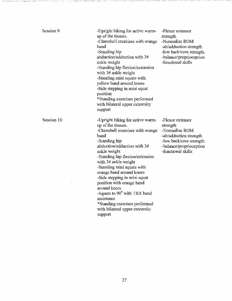
Session 9
Session 10
-Upright biking for active warmup of the tissues. -Clamshell exercises with orange band -Standing hip abduction/adduction with 3# ankle weight -Standing hip flexion/extension with 3# ankle weight -Standing mini squats with yellow band around knees -Side stepping in mini squat position • Standing exercises performed with bilateral upper extremity support
-Upright biking for active warmup of the tissues. -Clamshell exercises with orange band -Standing hip abduction/adduction with 3# ankle weight -Standing hip flexion/extension with 3# ankle weight -Standing mini squats with orange band around knees -Side stepping in mini squat position with orange band around knees -Squats to 900 with TRX band assistance 'Standing exercises performed with bilateral upper extremity support
27
-Flexor extensor strength -Normalize ROM -ab/adduction strength -low back/core strength -balance/proprioception -functional skills
-Flexor extensor strength -Normalize ROM -ab/ adduction strength -low back/core strength -balance/proprioception -functional skills

Session 11 -Upright biking for active warmup of the tissues. -Using the "Keiser" resistance machine closed chain hip abduction, adduction, flexion and extension were performed against resistance. -Semi squat side stepping -Balance on rocker board with eyes open and eyes closed. -Single leg balance.
28
-Flexor extensor strength -Normalize ROM -ab/adduction strength -low back/core strength -balance/proprioception -functional skills

Outcomes
Upon conclusion of these interventions, 2.5 months after this patient was
referred to PT, the patient was again seen orthopedic physician for a follow up
appointment. The patient had progressed in both the afore mentioned long-term
goals (keeping pain at a level of 2/10 or less with ADls and becoming
independent in her home exercise program) as well as one short-term goal
(increasing bilateral hip ROM). Although she had progressed in all three of these
goals, she had yet to meet these goals at the end of her episode of care. The
patient was able to meet one short-term goal (to be able to participate in
isometric strengthening). As was discussed in the interventions section, the
patient did not make any large gains in hip ROM. She was still severely lacking
IR at both hips, and displayed excessive ER bilaterally. Flexion in both hips was
still somewhat limited secondary to pain. The results of therapeutic interventions
are outlined in table 5.
29

Table 5
Clinical Results of Therapeutic Interventions
Category
PAIN
ROM
STRENGTH
Initial Evaluation
Worse with activity, ranges from 5-7/10, "if she is awake pain in present"
IR<100 Bilaterally ER >600Bilaterally Flexion painful above 1000
Bilaterally
3+ in all motions.
Discharge (8 Weeks)
Still exacerbated with activity, ranges from 2-7/10, less frequent, still cannot increase her activity
IR'" 150 Bilaterally ER'" 500 Bilaterally Flexion pain free to '" 1100
Bilaterally
Adduction= 4+/5 Flexion/Extension/Abduction and ER increased to 4/5 bilaterally, IR 3+/5
At the conclusion of the episode of care, the patient was consistently
reporting lower levels of pain when compared to the initial visit and reported that
pain was less frequent and better controlled. Functionally, the patient reported
less pain when climbing stairs at school, carrying items at work and prolonged
standing at work or for leisure activities.
30
The patient was seen by her orthopedic surgeon for a follow-up visit upon
completion of 8 weeks of physical therapy. The surgeon's findings of strength
and ROM were consistent with what both the SPT and PT were able to obtain.
The patient reported to the surgeon, "physical therapy definitely helped." She
also explained to the surgeon that if she tried to increase her activity level at all,
she experienced increased pain in both hips. The surgeon stated that he did not
think that this was acceptable for a 16-year-old girl. His findings were that she
had fairly substantial hip dysplasia, global retroversion, and a deficient lateral and
posterior acetabulum wall. He did not think she was a candidate for arthroscopic
intervention. The surgeon also stated that if the patient was not happy with her
current condition, open surgical intervention would probably be the best course of
action at that point. At that time the surgeon anticipated that patient would require
a rotational osteotomy of the acetabulum and a simultaneous repair of the
labrum.
31

CHAPTER III
DISCUSSION
Throughout the course of this case study, many clinical decisions had to be
made in order to provide the patient with the best care possible. The world health
organization (WHO) provided the intemational classification of function (ICF)
(WHO-ICF) model, which can be utilized when making these clinical decisions. It
was decided by the PT and SPT that it would be best practice to utilize this
model in making decisions regarding this patient. The WHO-ICF model illustrate
the interplay between the "health condition" relative to the body structure, activity,
and participation realms, as well as the influence of intemal and extemal factors
on the outcome of patient care. A representation ofWHO-ICF model applied to
this patient is located in Appendix B.
This potential benefits of PT in the treatment of DOH and FAI are outlined in
this case study. Conservative treatment for femoral acetabular impingement can
be successful5. In order to determine the success of conservative treatment,
goals were established. Successful conservative treatment required achieving all
the PT goals. The decision regarding success or failure of PT was made by the
surgeon in consultation with the patient regarding pain levels and function.
Outcomes. It was an intent of PT, to increase the patient's strength and
normalize her hip ROM to provide proximal stability to the hip joint and
subsequently increase function and decrease pain. These potential outcomes
were supported in the current literature.8,g However, the patient in this case
study was only able to achieve one short-term goal (being able to participate in
32

isometric exercises in order to begin building functional strength) and progress in
a long~term goal (demonstrating pain well managed at a level of 2/1 0 or less with
all ADLs) and another short-term goal (increase bilateral hip ROM to WNL as
pain allows). Throughout the process of conservative treatment, the patient was
able to partially normalize her hip ROM, bilaterally, and increase the majority of
her hip strength.
Although the patient became stronger and obtained a more normalized hip
ROM pattern, she was still unable to increase her activity level without resultant
pain and fatigue. Consequently, her activity level remained unchanged and
classified as sedentary. While the patient's hip pain did decrease in intensity,
duration, and frequency, it remained the limiting factor in this patient's life.
During this episode of care, the patient's overall motivation to participate in
and her confidence in the potential success of the rehabilitation process was in
doubt. Both the patient and her mother, upon initial presentation to physical
therapy, mentioned they believed surgical intervention was the only option for
successful treatment at this point. This belief was demonstrated in poor
compliance in the home exercise program and attendance during PT sessions.
These were likely factors may have been influential regarding the outcomes of
the conservative treatment.
33

Reflective Practice and Conclusions
No QOL measures were implemented, monitored, or documented for this
patient Current published literature6,8,g,10 indicated that when dealing with DDH
or FAI, most often, it is the improvement in QOL that determines successful
treatment outcomes rather than strength and ROM gains. Consequently, it is the
conclusion of this author that this particular case study was limited in its findings
by not gathering this data. It is recommended that in the future, clinicians should
utilize QOL measures when providing intervention for patients with these
disorders to determine if interventions were successful or not.
If the patient had filled out a QOL measure, such as the International Hip
Outcome Tool (iHOT-33)12 during the initial evaluation, at mid-term, and upon
discharge, the clinician could have determined if progress was being made, even
if it was not tangible. Had a QOL measure had been implemented, at the initial
evaluation, then goals could have been created based on the results of this
questionnaire. Consequently, the plan of care created for the patient could have
been designed to improve quality of life, an endpoint that is valued by the patient,
rather than focus on improvements in ROM and strength. For example, if a QOL
measure had been utilized, more time would have been spent in patient
education on how to modify her work, home, and school environments in order to
better serve her, rather than only attempting to strengthen weak muscles and
improve ROM. However, it should be noted, though the literature6,8,g,10 indicates
that, while improvements in QOL should be held superior, strength training and
34
ROM exercises could help increase the patient's QOl. It is recommended that
clinicians should not do away with strength training and ROM exercises, but
rather, attempt to make them applicable to improvements in QOL.
Regarding the patient presenting with an undiagnosed, congenital disorder,
there is evidence supporting the use of serial clinical examination of the hips by a
trained clinician during the periodic health examination of all infants until they are
walking independently.13 Screening for hip dysplasia may prevent the need for
late treatment, which is associated with long term hip deformity, gait disturbance
and arthritis. ,,14 It is anticipated that, had this screening been performed, the
potential for early identification and action may have prevented boney formational
changes. At age 16, there would be little potential to change boney formation
other than through surgical intervention.
While it is unknown if a delay was due to patient compliance or the health
system, it was almost 4 months from the initial onset of hip pain before the
patient reached the physical therapy clinic. By this time, the patient was very
discouraged and in so much pain, that it limited the potential success of PT.
Eventually this patient will most likely have to undergo surgical intervention,
including a rotational osteotomy of the acetabulum and simultaneous repair of
the labrum. This surgical procedure would include at least 6 to 8 weeks of non
weight bearing status and most likely make it difficult for the patient to participate
in school, work, and leisure activities during this period. With the co-morbidity of
depression, a major life event such as this could very well exacerbate her
symptoms as well as cause her to fall behind in school.
35

It recommended that future practice should include a more available and
reasonable way in which to screen all newborns for DOH. Early detection is of
vital importance in the road to recovery. Also, all forms of conservative treatment
should be exhausted prior to seeking surgical intervention to prevent falling
behind in current lifestyle activities (school, work, leisure). Finally, the primary
goal of conservative treatment for patients with DOH, FAlor a combination of
both should have a primary focus on improving the patient's QOl over and above
musculoskeletal improvements such as strength and ROM. However, it is also
recommended that improvements in strength and ROM may be utilized as a
means to reach this end; these parameters should never be used as the primary
indicators of successful treatment.
36

APPENDICES

APPENDIX A
. .,~ "
HEP~GO Hnm" !=v,e>rri"" Program Created by Joseph Taylor, SPT Jun 16th. 2015 View at "v/\vwmy-exercis0-Code com' using code" C3H96K4 HEP Bul{{/er
Repeat 3 Times Hold 10 Seconds complete 3 Sets perform 2 Time(s) a Day
Repeat 10Times Hold 3 Seconds Compiete2 Sets Perform 2 Time(s) a Day
Repeal 10 Times Hold 3 Seconds Complete 2 Sets Perform 2 Time(s) a Day
Repeat 10 Times Hold 3 Seconds Complete2Se.1s Perform 1 nme{s) a Day
HIP INTERNAL ROTATION STRETCH -SEATED
Start by sitting on a chair with your legs spread apart and feet planted on the ground. Next, use your hand to draw your knee inward as shown
CLAM SHELLS
While lying on your side with your knees bent, draw up the top knee while keeping contact of your feet together.
Do not lei your pelvis roll back during the lifting movement.
4 Way Hip #1
Stand on one foot with the resistance attached around the ankle of the other leg. The leg with resistance will move towardS the weight and then away from <
Stand facing four directions (front, righI, back, left) so that the four main hip motions are worked.
STANDING MARCHING - SIGNLE LEG
While standing, draw up your knee, set it down and then repeat on the same side.
Use your arms for support if needed for balance and safety.
Powered by HEP2go.com
38
Repeat 10Times Hold 2 Seconds Complets2Sets Perform 2 Time(s) a Day
Mini squats
Stand at countertop and bend knees for a mini squat
Jun 16th, 2015 - Page 1 of 1

APPENDIXB
World Health Organization ICF Model
HEALTH CONDITION
jJ1 DDH, Femoral Acetabular Impingement, Labral Tear
~ 11 11 U 11 11 BODY STRUCTURE ACTMTY ITASK { PARTICIPATION l
AND FUNCTION ABII1TY LIMITATION
ABILITIES RESIRICTIONS
-Pain with Limited
Decreased strength - Basic mobility increased
- Full time partIcipation in gym (standing,
activity, student dass at school.
bilateral hips, decreased sitting. - Parttime . Feels like she needs
walking) -Pain and lunited employee
extra breaks at
IR, excessive ER, pain ~ Sits in desks stair climbing Ie . Enjoys being work, but doesn't't
rw ability takemem
in both hips with - Walks school with friends and Wanted to play hallway, -Pain \~th
listening to soccer but pain
activity Stands at gening into aod mUSIC
prmntedher fi'om
concerts out of a car. "-
doing so.
-{.;,. "'lr- ..[J,. "'lr- 0 D
t ENVIRONMENTAL
INTERNAL EXTERNAL +Young +Wants to play soccer +Wants to be pain fi'ee +SuPPOItive mother +supportive friends +understanding teachers -Sarcastic - Believes surgel} is the only aoswer - Work Ethic +medication to help with depression -Motivation -Compliance -Accuracy \~th reports - Reliability -Just relocated here (Florida->ND) 4 new ha[ siblings and stepdad--Honesty -hx of depression/aoxiety -<ioesn't't hke to be active mother supportive but lacks involvement
,
39

REFERENCES
1. Loder RT, Skopelja EN. The Epidemiology and Demographics of Hip Dysplasia.lSRN
Orthopedics 20 11;23 :86-87.
2. Loder RT, Shafer C. Seasonal variation in children with developmental dysplasia of
the hip. Journal o/Children's Orthopaedics 2014;8(1):11-22.
3. Lievense. A.M, Bierma-Zeinstra. S.M.A, Verhagen. A.P, Verhaar. J. A. N, Koes. B.W,
Influence of hip dysplasia on the development of osteoarthritis of the hip. Annals 0/
Rheumatic Diseases. 2003; 63(6): 621-626.
4. Dooley Pl. Femoroacetabular impingement syndrome: Nonarthritic hip pain in young
adults. Canadian Family Physician. 2008;54(1): 42-47 .
5. Khaled, E. Wail, S. EL Hausain, M. Khaled, A.E.G. Conservative treatment for mild
femoroacetabular impingement. Journal o/Orthopaedic Surgery 2011;19(1):41-5
6. Amanatullah, D.F. Antkowiak, T. Pillay, K. Et al. Femoroacetabular impingement:
current concepts in diagnosis and treatment. Journal o/Orthopedics. 2015; 38(3): 185-
99. doi: 10.3928/01477447-20150305-07.
7. Shultz, T. Faber's Test. Physio-Pedia.com. mlll:J.LYi"rYLlltrG;;il::
1'."" l"'-'';d'I_''IJ\.'2ci.,""._'E~t 6 -15 -20 15 .
8. Yazbek. P.M. Ovanessian. V, Martin, R.L. Fukuda, T.Y. Nonsurgical treatment of
acetabular labral tears: A Case Series. Journal 0/ Orthopaedic and Sports Physical
Therapy. 2011; 41(5):346-53. DOl: 10.2519/jospt.2011.3225
40

9. Conservative Management for Femoroacetabular Impingement (FAI). Fowler
Kennedy Sports Medicine Clinic website. rlt![l/;li~"iI("J:li.'':!]n.~\LC())n/~yp.::.
",.)C)C),::\.~.\ . ..c."::".I"-"::\~ Updated 9, 2011. Accessed 6, 2015.
10. Zebala LP, Schoenecker PL, Clohisy JC. Anterior Femoroacetabular Impingement: A
Diverse Disease with Evolving Treatment Options. The Iowa Orthopaedic Journal.
2007;27:71-81.
11. Hunt D, Prather H, Harris Hayes M, Clohisy JC. Clinical Outcomes Analysis of
Conservative and Surgical Treatment of Patients With Clinical Indications of
Prearthritic, Intra-articular Hip Disorders. PM & R : the journal of injury, function, and
rehabilitation. 2012;4(7):479-487. doi: 1 0.1016/j.pl11lj.2012.03.012.
12. Mohtadi, N. G. H. Et. AI. The Development and Validation ofa Self-Administered
Quality-of-Life Outcome Measure for Young, Active Patients With Symptomatic Hip
Disease: The International Hip Outcome Tool (iHOT-33). The Journal of Arthroscopic
and Related Surgery. 2012; 28 (5): 595-610.
13. Patel H, the Canadian Task Force on Preventive Health Care. Preventive health care,
2001 update: screening and management of developmental dysplasia of the hip in
newborns. CMAJ: Canadian Medical Association Journal. 2001;164(12):1669-1677.
14. Shorter. D, Hong. T, Osborne D. Screening progranunes for
developmental dysplasia of the hip in newborn infants. Journal of Evidence Based
Child Health. 2013; 8(1): 11-54.
41