diagnostic differentiation plates for hereditaryjb.asm.org/content/86/2/299.full.pdf · diagnostic...
TRANSCRIPT

DIAGNOSTIC COLOR DIFFERENTIATION PLATES FOR HEREDITARYRESPIRATION DEFICIENCY IN YEAST
SUSUMU NAGAI
Biological Laboratories, National Women's University, Nara, Japan.
Received for publication 8 April 1963
ABSTRACT
NAGAI, SUSUMU (National Women's Uni-versity, Nara, Japan). Diagnostic color differ-entiation plates for hereditary respiration de-ficiency in yeast. J. Bacteriol. 86:299-302.1963.-Color differentiation between normalyeasts and their respiration-deficient mutantswas improved by growing yeast colonies onnutrient agar plates containing several selecteddyes and their mixtures. Magdala red (5 to 8mg/liter) was good for single-color plates, givingdeep-red sheen to the mutant colonies in con-trast to the normal ones which tinted light red.A mixture of eosin (8 to 15 mg/liter, either Yor B) with trypan blue (15 to 20 mg/liter) wasexcellent in color and convenient to prepare,giving brilliant purple sheen to the mutantcolonies contrasted to the normal ones whichtinted grayish violet. These color plates weregood over a broad range of Saccharomyces species,although the colony shades and suitable dyeconcentrations varied depending on the speciesand strains.
Various criteria have been employed fordiagnosing hereditary respiration deficiency("petite" or "RD" mutation) in Saccharomycesspecies of yeast since the work by Ephrussi(1953). They include actual oxygen consump-tion, colony size, cytochromes, utilization ofcarbon sources (selective culture media), andcertain color differentiations by suitable indi-cators such as triphenyltetrazolium chloride(TTC) and some other dyes, as reviewed re-cently (Nagai, Yanagishima, and Nagai, 1961).The last-named diagnostic method is convenientparticularly for population scoring of suchrespiratory mutants. This report describes animproved device for better color differentiationwith several selected dyes and their mixtures.
MATERIALS AND METHODS
Single dyes in various concentrations, andmixtures of two dyes (red and blue) in variouscombinations were added to nutrient agarmedium to make color plates. Basal medium anddye solutions were sterilized separately by steam-ing at 100 C for 75 and 50 min, respectively,and were mixed together after cooling to about55 C. In routine practice, 20-ml portions ofappropriately diluted (to 20-fold of desired finalconcentration) single-dye solutions and wateror two-dye solutions in 16-mm tubes were addedto 360 ml of basal agar medium in 500-ml Erlen-meyer flasks to make 400 ml of color medium,which was, after thorough mixing, divided into12 petri dishes (9-cm). Final nutrient compositionwas (w/v): glucose, 2.0%; peptone, 0.15%;dehydrated yeast extract, 0.15%; potassiumdihydrogen phosphate, 0.15%; ammonium sul-fate, 0.15% magnesium sulfate, 0.1%; plus agar,1.2% (to adequate hardness). S. cerevisiaeIFO 0044, S. chevalieri 0210, and S. microel-lipsodes 1016 were used for general tester or-ganisms. Several other yeasts (see Results) werelater used for comparative survey. Mixtures ofnormal and respiration-deficient (RD) mutantcells were spread on the color plates so as toproduce about 100 to 150 colonies per plate, andwere incubated at 30 C for 3 to 7 days.
RESULTS
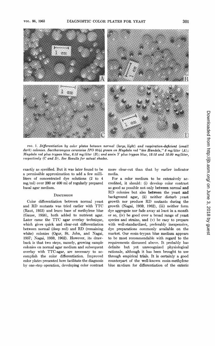
Table 1 summarizes the tests with selecteddyes and their mixtures. Magdala red at 5 to 8mg/liter gave a most desirable color differentia-tion (much better for our eyes than for black-and-white photographs), excelling the other dyeswhen they were applied in single-color plates.The RD mutant colonies appeared in deep-redsheen in contrast to the normal ones which tintedlight red (Fig. 1A). Two preparations labeledas Magdalarot and Magdalarot des Handels(both from Griubler, Leipzig, Germany; manu-
299
on June 3, 2018 by guesthttp://jb.asm
.org/D
ownloaded from
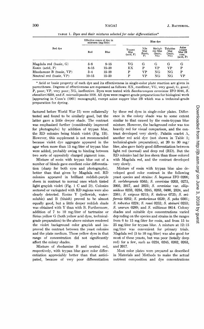
TABLE 1. Dyes and their mixtures selected for color differentiation*Effective concn of dye in Blue dyemixtures (mg/liter)
Red dye Nile Methyl- Toluidne AzineTrypan ble eebu le copper
Red Blue ie bu ne blue blue 2B(acid, P) (basic, (basic, (basic, conc.P) NG) NG) (acid, P)
Magdala red (basic, G) 5-8 8-15 VG G G G GEosin (acid, P) 8-15 15-20 EX P VP VP PRhodamine B (basic, VP) 3-4 15-20 P VP NG NG PNeutral red (basic, VP) 10-15 15-20 P VP NG NG VP
* Acid or basic property of each dye and its effectiveness in single-color plate reaction are given inparentheses. Degrees of effectiveness are expressed as follows: EX, excellent; VG, very good; G, good;P, poor; VP, very poor; NG, ineffective. Dyes were tested with Saccharontyces cerevisiae IFO 0044, S.chevalieri 0210, and S. nticroellipsodes 1016. All dyes were reagent-grade preparations for biological work[appearing in Conn's (1961) monograph], except azine copper blue 2B which was a technical-gradepreparation for dyeing.
factured before World War IJ) were collaterallytested and found to be similarly good, but thelatter gave a little deeper shade. The contrastwas emphasized further (considerably improvedfor photographs) by addition of trypan blue,the RD colonies being bluish violet (Fig. 1B).However, this supplement is not recommendedbecause violet dye aggregate appeared in theagar when more than 15 mg/liter of trypan bluewere added, probably owing to binding betweentwo sorts of oppositely charged pigment ions.
Mixture of eosin with trypan blue out of anumber of blends gave excellent color differentia-tion (sharp for both eyes and photographs),better than that given by Magdala red. RDcolonies appeared in brilliant reddish-purplesheen in contrast to normal ones which tintedlight grayish violet (Fig. 1 C and D). Coloniessectored or variegated with RD regions were alsoclearly detected. Eosins Y (yellowish, water-soluble) and B (bluish) proved to be almostequally good, but a little deeper reddish shadewas obtained with Y than with B. Furthermore,addition of 7 to 10 mg/liter of tartrazine orSirius yellow G (both yellow acid dyes, technical-grade preparation) to the above mixture renderedthe violet background color grayish and im-proved the contrast between the yeast coloniesand the plate medium. Those yellow dyes in thatrange of concentration did not significantlyaffect the colony shades.
Mixture of rhodamine B and neutral red,respectively, with trypan blue gave color differ-entiation appreciably better than that antici-pated, because of very poor differentiation
by these red dyes in single-color plates. Differ-ence in the colony shade was to some extentsimilar to that caused by the eosin-trypan bluemixture. However, the background color was tooheavily red for visual comparison, and the con-trast developed very slowly. Palatin scarlet A,another red acid dye (not shown in Table 1;technical-grade preparation), at 20 to 30 mg/liter, also gave fairly good differentiation betweenlight red (normal) and deep red (RD). But theRD colonies had far less sheen than those coloredwith Magdala red, and the contrast developedvery slowly.
Mixture of eosin with trypan blue also de-veloped good color contrast in the followingyeast species and strains: S. bayanus IFO 0206;S. carlsbergensis 0565; S. cerevisiae 0203, 0275,2005, 2017, and 2023; S. cerevisiae var. ellip-soideus 0233, 0234, 0245, 0263, 0490, 2324, and2361; S. exiguus 0215; S. italicus 0725; S. ovi-formis 0262; S. pastorianus 0539; S. peka 0301;S. robustus 0224; S. rosei 0252; S. steineri 0253;S. uvarum 0289; and S. willianus 0614. Colonyshades and suitable dye concentrations varieddep?nding on the species and strains in the rangesfrom 6 to 15 mg/liter for eosin, and from 15 to25 mg/liter for trypan blue. A mixture at 12:15mg/liter was convenient for primary trials.AMagdala red (5 to 10 mg/liter) was also good formost of these yeasts, but was poor (totally deepred) for a few, such as 0224, 0245, 0262, 0263,and 2017.
M\ost color plates were prepared as describedin AMaterials and Methods to make the actualnutrient composition and dye concentrations
300 NAGAI J. BACTERIOL.
on June 3, 2018 by guesthttp://jb.asm
.org/D
ownloaded from
DIAGNOSTIC COLOR PLATES FOR YEAST
FIG. 1. Differentiation by color plates between normal (large, light) and respiration-deficient (smalldark) colonies. Saccharomyces cerevisiae IFO 0044 grown on Magdala red "des Handels," 5 mg/liter (A);Blagdala red plus trypan blue, 8:15 mg/liter (B); and eosin Y plus trypan blue, 15:15 and 15:20 mg/liter,respectively (C and D). See Results for actual shades.
exactly as specified. But it was later found to bea permissible approximation to add a few milli-liters of concentrated dye solutions (2 to 4mg/ml) over 200 or 400 ml of regularly preparedbasal agar medium.
DiscussIONColor differentiation between normal yeast
and RD mutants was tried earlier with TTC(Raut, 1953) and leuco base of methylene blue(Gause, 1958), both added to nutrient agar.Later came the TTC agar overlay technique,which gives quick and clear-cut differentiationbetween normal (deep red) and RD (remainingwhite) colonies (Ogur, St. John, and Nagai,1957; Nagai, 1959, 1962). However, its draw-back is that two steps, namely, growing samplecolonies on normal agar medium and subsequentoverlay with TTC-agar, are necessary to ac-complish the color differentiation. Improvedcolor plates presented here facilitate the diagnosisby one-step operation, developing color contrast
more clear-cut than that by earlier indicatormedia.
For a color medium to be extensively ac-credited, it should: (i) develop color contrastas good as possible not only between normal andRD colonies but also between the yeast andbackground agar, (ii) neither disturb yeastgrowth nor produce RD mutants during thegrowth (Nagai, 1959, 1962), (iii) neither formdye aggregate nor fade away at least in a monthor so, (iv) be good over a broad range of yeastspecies and strains, and (v) be easy to preparewith well-standardized, preferably inexpensive,dye preparations commonly available on themarket. Our eosin-trypan blue medium appearsto be most recommendable with regard to therequirements discussed above. It probably hasdefinite but yet unrecognized physiologicalrationale, although it has been brought to usethrough empirical trials. It is certainly a goodcounterpart of the well-known eosin-methyleneblue m?dium for differentiation of the enteric
VOL. 86, 1963 301
on June 3, 2018 by guesthttp://jb.asm
.org/D
ownloaded from
J. BACTERIOL.
bacteria and their biochemical mutants. Magdalared is also very good. Bright sheen of the RDcolonies may be somewhat related to the useful-ness of this dye as a vital fluorochrome (Drawert,1955). However, it has been sold only by a fewcompanies from Germany under somewhat con-fusing commercial designations (Drawert, 1955;Fonn, 1961; Nagai, 1962), and is not so readilyavailable to order. Dyes which are found only intechnical-grade preparations are also less con-venient in this respect.The improved color plate method is sufficiently
reliable for scoring RD mutants in mixed popu-lations. It is suggested, however, not to apply thismethod immediately after ultraviolet irradiationand other drastic treatments because the yeastmight suffer complications by heavy exposure tothese dyes, which are otherwise harmless.
ACKNOWLEDGMENTS
Generous supply of the "IFO" yeasts by theInstitute for Fermentation, Osaka, and of dyepreparations by the Osaka City Institute forIndustrial Research is gratefully acknowledged.
This work was supported in part by a grant(No. 4062, to J. Ashida) from the Ministry ofEducation, Japan.
LITERATURE CITED
CONN, H. J. 1961. Biological stains, 7th ed., p.123. The Williams & Wilkins Co., Baltimore.
DRAWERT, H. 1955. Vitalfluorochromierungenmit angeblichem "Magdalarot." Flora (Jena)142:479-488.
EPHRUSSI, B. 1953. Nucleo-cytoplasmic relationsin micro-organisms: their bearing in cellheredity and differentiation. Clarendon Press,Oxford.
GAUSE, G. F. 1958. The search for anticancer anti-biotics. Science 127:506-508.
NAGAI, S. 1959. Induction of the respiration-defi-cient mutation in yeast by various syntheticdyes. Science 130:1188-1189.
NAGAI, S. 1962. Production of respiration-deficientmutants of yeast by some quinone-imine dyes.Exptl. Cell Res. 27: 14-18.
NAGAI, S., N. YANAGISHIMA, AND H. NAGAI. 1961.Advances in the study of respiration-deficient(RD) mutation in yeast and other micro-organisms. Bacteriol. Rev. 25:404-426.
OGUR, M., R. ST. JOHN, AND S. NAGAI. 1957.Tetrazolium overlay technique for populationstudies of respiration deficiency in yeast.Science 125:928-929.
RAUT, C. 1953. A cytochrome deficient mutant ofSaccharomyces cerevisiae. Exptl. Cell Res.4:295-305.
302 NAGAI
on June 3, 2018 by guesthttp://jb.asm
.org/D
ownloaded from