diagnostic endoscopy chapter 4 akash nabh, muhammed...
TRANSCRIPT

Chapter 4
Diagnostic Endoscopy
Akash Nabh, Muhammed Sherid, Charles Spurr andSubbaramia Sridhar
Additional information is available at the end of the chapter
http://dx.doi.org/10.5772/ 52826
1. Introduction
Gastrointestinal (GI) endoscopy is defined as the direct visualization of the digestive tract,with or without therapy. Endoscopic technology has rapidly advanced over the past 40years and has become an integral part of clinical gastroenterology. The utilization of endos‐copy for both diagnostic evaluation and screening has markedly increased over the last twodecades. Many innovations have expandedthe indications for endoscopy. Successful endos‐copy relies upon the ability to recognize abnormalities and diagnose disease. It is imperativefor the endoscopist to detect GI lesions in its early stage to ensure that the patient can re‐ceive less invasive treatment and have better prognosis. To make a correct diagnosis of earlyneoplasm in the GI tract, we first need to detect any lesions with subtle morphologic change.
In this chapter we describe various GI conditions and the role of various endoscopic meth‐ods in their diagnosis.
2. Esophagogastroduodenoscopy (EGD)
EGD is performed by passing a flexible scope through mouth to the esophagus, stomachand duodenum. This procedure is the best method to examine upper gastrointestinal muco‐sa can be performed under conscious sedation in most patients.
The indications for EGD are persistent upper abdominal symtoms despite trial of therapy,upper abdominal symptoms with suspected organic disease (anorexia, weight loss etc.), dys‐phagia, odynophagia, recurrent or persistent gastroesophageal reflux disease (GERD), per‐sistent vomiting, familial adenomatous polyposis (FAP),GI bleeding, portal hypertension totreat and document esophageal varices, management of achalasia/ esophageal strictures/
© 2013 Nabh et al.; licensee InTech. This is an open access article distributed under the terms of the CreativeCommons Attribution License (http://creativecommons.org/licenses/by/3.0), which permits unrestricted use,distribution, and reproduction in any medium, provided the original work is properly cited.

stenotic lesions, removal of foreign bodies, placement of feeding tubes, palliative stenting,surveillance of malignancy in Barrett’s esophagus, and banding of esophageal varices [1].
Contraindications of EGD are medical instability, patient incooperation and suspectedperforation.
Complications of diagnostic EGD are cardiopulmonary events, perforation (0.03%),andbleeding (<0.1%)[2].
2.1. Esophagus
2.1.1. Eosinophilic esophagitis
Eosinophilic esophagitis is a chronic inflammatory disease of the esophagus,usually associ‐ated with allergic syndromes.It is increasingly diagnosed in patients presenting with episo‐dic dysphagia and occurs predominantly in males.Endoscopically it is characterized bylongitudinal furrows (linear furrowing), widespread white spots,diffuse mucosal nodulari‐ty,and multiple rings which fail to disappear with insufflation with air (feline esopha‐gus).Mucosal fragility is frequent. Esophageal mucosa may bleed or become fissured withthe scope passage,particularly in the case of a small-caliber or felinized esophagus[3,4].
Figure 1. Eosinophilic esophagitis with linear furrowing and rings(pointed by arrows)
2.1.2. Pill-induced esophagitis
Prolonged contact with certain medications can irritate the esophageal mucosa causingesophageal ulcer and esophagitis. Medication-induced esophagitis presents with sudden on‐set of odynophagia and retrosternal pain. A clinical diagnosis may be made by history with‐out the requirement for confirmatory endoscopy [5](19). The common culprit medicationsarenon-steroidal anti-inflammatory drugs (NSAIDS), tetracyclines, bisphosphonates, potas‐
Endoscopy of GI Tract38

sium chloride, and iron supplements. Endoscopyallows diagnostic confirmation and is amore sensitive procedurethan barium swallow [5,6].
Endoscopically, pill-induced esophageal injury presents as a discrete ulcer with relativelynormal surrounding mucosa. Exudative inflammation with esophageal thickening and stric‐ture formation are also seen.
The most common sites of injury are the proximal esophagus near the compression from theaortic arch and the distal esophagus in patients with left atrial enlargement.It can also occur inmotility disorders which allow prolonged contact of medications with the esophageal wall [5].
2.1.3. Reflux esophagitis and Gastroesophageal reflux disease (GERD)
GERDis defined as the backward passage of stomach contents through the lower esophagealsphincter. The symptoms of GERD include heart burn, chest pain, water brash and odynophagia.
Endoscopy at initial presentation should be considered in patients who have alarm symp‐toms suggestive of complicated disease or those at risk for Barrett’s esophagus.These alarmsymptoms are failure to respond to appropriate antisecretory medical therapy, dysphagia,upper GI bleeding,anemia, odynophagia, and weight loss [7].
The severity of esophageal erosions is predictive of a patient's response to therapy and ofthe likelihood of relapse after therapy. Therefore it is important to grade the severity of ero‐sive reflux esophagitis.Two grading systems which are commonly usedare Savary-Miller en‐doscopic classification and Los-Angeles grading [7].
The Savary-Miller endoscopic classification system is used widely but usage and interpreta‐tion are very variable. The "MUSE" (Metaplasia, Ulceration, Stricturing, and Erosions) classi‐fication provides clear definitions of the relevant endoscopic features, and it is based on astandardized report form, which allows the endoscopist to make a clear record of esophagi‐tis severity.
Grade I: One or more supravestibular, non-confluent reddish spots, with or without exudates
Grade II: Erosive and exudative lesions in distal esophagus thatmay be confluent, but notcircumferential
Grade III: Circumferential erosions in the distal esophagus, covered by hemorrhagic andpseudomembranous exudates
Grade IV: Chronic complications such as deep ulcers, stenosis, or scarring with Barrett'smetaplasia
The "L.A." (Los Angeles) classification describes four grades of esophagitis severity (A to D),based on the extent of esophageal lesions known as "mucosal breaks," but it does not recordthe presence or severity of other GERD lesions.
Grade A: One or more mucosal breaks each ≤5 mm in length
Grade B: At least one mucosal break >5 mm long, but not continuous between the tops ofadjacent mucosal folds.
Diagnostic Endoscopyhttp://dx.doi.org/10.5772/ 52826
39

Grade C: At least one mucosal break that is continuous between the tops of adjacent mucos‐al folds, but which is not circumferential
Grade D: Mucosal break that involves at leastthree-fourths of the luminal circumference.
Figure 2. LA grade D esophagitis
Chronic GERD can cause esophageal stricture and Barrett’s esophagus(which are describedin later sections)
2.1.4. Barrett’s Esophagus
Barrett’s esophagus is a condition in which metaplastic columnar epithelium that predispos‐es to cancer development replaces the stratified squamous epithelium that normally linesthe distal esophagus. Barrett’s esophagus is well recognized as a complication of GERD. Pa‐tients with GERD who develop Barrett esophagus tend to have a combination of clinical fea‐tures, including hiatal hernia, reduced lower esophageal sphincter (LES) pressures ordelayed esophageal acid clearance time. The annual incidence of esophageal cancer in apopulation of patients with Barrett’s esophagus is approximately 0.5% per year.
Endoscopically,the typical appearance of Barrett’s esophagus is a salmon pink mucosawhich extends down and joins the gastric mucosa.
The American Gastroenterologic Association (AGA) suggests endoscopic screening for Bar‐rett’s esophagus in patients with multiple risk factors associated withesophageal adenocarci‐noma “age 50 years or older,male sex, white race, chronic GERD, hiatal hernia,elevatedbody mass index, and intra-abdominal distributionof body fat”. Once identified, patientswith Barrett esophagus should undergo periodic surveillance endoscopy to identify dyspla‐sia.It is recommended that endoscopic evaluation be performedtaking 4-quadrant biopsiesevery 2 cm with biopsy sampling of any mucosal irregularities. Four-quadrant biopsy speci‐
Endoscopy of GI Tract40

mens be obtainedevery 1 cm in patients with known or suspecteddysplasia.If there is nodysplasia surveillance endoscopy is recommended every 3-5 years.With low grade dyspla‐sia, 6-12 month surveillance intervals are recommended. High grade dysplasia requires en‐doscopic biopsy every 3 months if eradication therapy is not performed.The AGArecommends endoscopic eradication therapywith radiofrequency ablation, photodynamictherapy or endoscopic mucosal resection rather than surveillance for treatment of patientswith high-grade dysplasia with Barrett’s esophagus[8].
Figure 3. Long segment Barrett’s esophagus
2.1.5. Esophagitis related to infections
Candida
Candida species colonize in 20 % of healthy adults.The risk factors are AIDS, cancer, antibiot‐ic or steroid therapy.The causative organism is almost always C. albicans. Candida esophagi‐tis usually present as odynophagia or dysphagia.The diagnosis is based on the endoscopicpicture, microscopic examination and culture of the mucosal brushings, and histologicalexamination of the esophageal mucosa. About two-third of patients have signs of oral thrush(thus its absence does not exclude esophageal involvement).EGD with brushings orbiopsy iscurrently the most sensitive and specific method of diagnosis.Endoscopy demonstrates pat‐chy, whitish plaques covering a friable, erythematous mucosa.When the infection is severe,ulceration may be present as well [9]. Confirmatory biopsy shows the presence of yeasts andpseudohyphae invading mucosal cells, and the culture reveals Candida.
Diagnostic Endoscopyhttp://dx.doi.org/10.5772/ 52826
41

Figure 4. Pale plaques with erythematous mucosa – esophageal candidiasis
Herpes simplex virus
HSV-1 infection of the esophagus is usually seen in immunocompromisedconditions suchasorgan or bone marrow transplantation. Less commonly in HIV patients and occasionallyimmunecompetent patients acquire HSV-1 infection. Endoscopically, there are well circum‐scribed ulcers with raised margins and a punched out” appearance,distinguishing themfrom the ulcers seen in CMV infection.Exudates, plaques, or diffuse erosive esophagitis andvesicles can also be seen. Biopsies should be taken from the edge or margin of the ulcerwhere viral cytopathic effects are most likely to be present [10].
Cytomegalovirus
The most common cause of esophagitis in patients with advanced AIDS is Candida, whereasthe most common viral cause is CMV. CMV esophagitis is seen in post-transplantation,long-term renal dialysis, human immunodeficiency virus (HIV) infection, and AIDS andother debilitating diseases. Endoscopically,extensive ulceration of the esophagus is hallmarkof CMV esophagitis.It may present as asolitary ulcer or multiple ulcers.Most ulcers are not‐ed in the distal esophagus [11]. The multiple biopsy specimens should be taken from thebase of the ulcer.
2.1.6. Esophageal diverticulum
The formation of diverticula occurs due pulsion from increased intraluminal pressure result‐ing in pushing of esophageal mucosa andsubmucosa through the focal weakness of mucosalwall. The risk factors are esophageal dysmotility or stricture which contribute to intralumi‐nal pressure[12].Esophageal diverticula are rare but can occur in any part of the esopha‐gus.If it occurs in the upper esophagus above the upper esophageal sphincter through aweak spot (Killian’s triangle)form above the upper esophageal sphincter in the midline pos‐teriorly at the pharyngoesophageal junction, it is called Zenker’s diverticulum. When it oc‐
Endoscopy of GI Tract42

curs in the distal esophagus just abovethe lower esophageal sphincter, it is called epiphrenicdiverticula. Endoscopically appear round with a wide neck.
Endoscopy does not play important role in the diagnosis but the endoscopistshould beaware and cautious about their presence as perforation can occur with endoscopy especiallywhen side viewing scopes are used. Esophageal diverticula are well seen on barium x-rayexamination, which is the best modalityfor diagnosis.
2.1.7. Esophageal rings and webs
Esophageal webs are thin membrane like structure containing mucosa and submucosawhich can occur anywhere in the esophagus.The patients are asymptomatic or have onlyintermittent dysphagia. It is frequently discovered incidentally during radiographic stud‐ies for other reasons. However, esophageal webs have been described in Plummer-Vin‐son syndrome which present as iron deficiency anemia, glossitis and koilonychia.Endoscopically, the webs are seen with difficulty due to proximal location.They are cov‐ered with squamous mucosa [13,14].
Esophageal rings are thin, fragile structures that partially or completely obstruct the esoph‐ageal lumen.They present with dysphagia if the lumen is <13 mm. They are usually seen inthe distal esophagus. If the ring occurs at squamocolumnar junction covered with squa‐mous mucosa above and columnar epithelium below, it is called Schatzki ring or Type Bring. If the ring occurs about 1.5 cm proximal to the squamocolumnar junction, it is calledType A ring. Endoscopy is less sensitive than the barium esophagram in detecting esopha‐geal rings [13,14].
Endoscopically, an esophageal ring appears as a thin membrane with a concentric smoothcontour that projects into the lumen.
Figure 4. Schatzki ring
Diagnostic Endoscopyhttp://dx.doi.org/10.5772/ 52826
43

Figure 5. Esophageal web
2.1.8. Stricture
The esophageal stricture is narrowing of esophagus which can be benign or malignant.Thesymptoms of esophageal stricture are usually insidious but progressive with dysphagia tosolids followed by dysphagia to liquids. Dysphagia corresponds to the caliber of the stric‐ture; dysphagia to solids is usually present when the esophageal lumen is narrowed to 13mm or less. The causes of esophageal stricture formation are GERD, long-term use of a naso‐gastric (NG) tube, complication of sclerotherapy for varices, infectious esophagitis, post sur‐gical resection for esophageal or laryngeal cancer, caustic ingestion, pill esophagitis, andradiation exposure [15].
Endoscopy is helpful as it allows direct visualization, tissue sampling to rule out malignan‐cy, and dilation of the narrowed part of the esophagus.
Figure 6. Esophageal stricture
Endoscopy of GI Tract44

2.1.9. Hiatus hernia
Hiatus hernia refers to herniation of elements of the abdominal cavity most commonlystomach,into the mediastinum, through the esophageal hiatus of the diaphragm.Endoscopicand radiographic studies have shown a significant relation between GERD and hiatal hernia[16]. The main types of hiatal hernia are sliding type and para-esophageal type.
Sliding hiatal hernia accounts for more than 95 % of cases. It is characterized by widening ofthe muscular hiatal tunnel and circumferential laxity of the phrenoesophageal membrane,allowing a portion of the gastric cardia to herniate upward. Hiatal hernias that are largerthan 2 cm in axial span can be diagnosed easily by barium swallow radiography, endos‐copy, or esophageal manometry. Smaller hernias are more difficult to define. Endoscopical‐ly, the squamocolumnar junction appears 2-3 cm above the diaphragmatic hiatus. Onendoscopic retroflexed view appears as pouch like area just below mucosal junction andabove the diaphragm [17].
Figure 7. Sliding hiatal hernia visible in the esophagus
Para-esophageal herniasaccount for about 5 % of all hiatal hernias.Anatomically,the pouchof stomach herniatesinto the chest adjacent to the esophagus.Most complications of a para-esophageal hernia are related to mechanical problems caused by the hernia. Para-esopha‐geal hernias are best diagnosed with a barium swallow, although their presence is usuallysuggested by endoscopy.Endoscopically, they can cause difficulty in locating the main gas‐tric lumen.
Cameron lesions are erosions or ulcers occurring in the sac of a hiatal hernia. They havebeen described in up to 5.2 % of patients with a hiatal hernia who undergo upper endos‐copy. They are usually an incidental finding but rarely cause acute or chronic upper gastro‐intestinal bleeding and iron deficiency anemia[18].
Diagnostic Endoscopyhttp://dx.doi.org/10.5772/ 52826
45

2.1.10. Motility disorders of the esophagus
Motility disordersin the esophagus present as dysphagia. Evaluation of esophageal motilitydisorders often begins with endoscopy.The diagnosis can be made with endoscopy alone,however some cases require manometry or barium swallowing study for confirmation.
Endoscopy typically reveals a dilated esophagus in achalasia that often contains residualmaterial. A‘‘popping’’effect with difficulty in passing the endoscope through the gastro-esophageal junction may be noted.The esophageal mucosa usually appears normal. Endos‐copy is also essentialin achalasia to exclude malignancy.
Endoscopic findings in spastic disorders of the esophagus such as nutcracker esophagus anddiffuse esophageal spasm are often normal; however, it may reveal sacculations, diverticula,and chaotic contractile activity along the mid or distal esophagus.Endoscopyis usually per‐formed to exclude structuralesophageal obstruction.
In scleroderma, endoscopic findings usually relate to a hypotensive lower esophageal sphinc‐ter [12].
2.1.11. Other benign lesions of the esophagus
The prevalence of benign esophageal tumors is 0.5%. The majority of these benign tumors areasymptomatic which diagnose incidentally. Dysphagia is the most common presenting symp‐tomin patients with symptomatic benign esophageal tumors which occurs with large sizedtumors. Other symptoms include regurgitation, vomiting and retrosternal discomfort [19].
Leiomyomas
Leimyomas are the most common benign tumors of the esophagus. They arise from smoothmuscle cells. Dysphagia can occur only with large sized tumors. Endoscopically, they ap‐pear as submucosal masses with smooth margins and normal overlying mucosa.
Esophageal Cyst
Esophageal cysts are second most common benign tumors. Cysts are usually located in theupper esophagus and are lined by ciliated columnar epithelium. Endoscopically, they ap‐pear as protruding mass in the lumen. Surgical resection is required as they can cause com‐plication line obstruction or hemorrhage.
Fibrovascular polyps
Fibrovascular polyps are thin, solitary polyps which usually occur in upper esophagus.They present with symptomatic dysphagia. Over 75 % are 7cm or larger. They have largemucosal folds or large pedicles containing blood vessel.
Squamous cell papilloma
Squamous cell papilloma is usually solitarysessile, warty lesion less than 1.5cm occurringmost commonly in lower third of the esophagus.Histologically they are finger like projec‐tions of hyperplastic squamous tissue. Etiology of these lesions is felt to be due to humanpapillomavirus (HPV) infection.
Endoscopy of GI Tract46

Inlet patch
Inlet patch is isolated area in the esophagus resembling gastric mucosa usually found in theproximal esophagus.It can be associated with Barrett’s esophagus and esophagitis in about20% cases. Histologically, oxyntic type gastric mucosa is most commonly seen [20].
Figure 8. Inlet patch in the esopghagus
Glycogen acanthosis
Glycogen acanthosis presents as elevated gray-white plaques in the esophagus that range indiameter from 1 to15 mm. They are seen in 20–40% of endoscopic procedures and are moreprominent in the lower third of the esophagus. Histologically, the epithelium is thickenedby the proliferation of large squamous cells filled with glycogen. Glycogen acanthosishasbeen associated with Cowden’s syndrome and celiac disease.
2.1.12. Foreign bodies of the esophagus
Ingestion of foreign bodies occursmost commonly among those with psychiatric disorders,mental retardation, prisoners, and alcoholics.Thepresence of esophageal stricture or ringpredispose to impaction of foreign body or food bolus in the esophagus. Fortunately, mostpass through the gastrointestinal tract harmlessly. However, 10–20% will require non-opera‐tive intervention. Endoscopic extraction is themainstay of non-operative interventionswhich usually attempt after radiographic localization. Foreign bodies at the level of the hy‐popharynx or cricopharyngeus muscle are best treated with rigid laryngoscopy using agrasping clamp.In all other cases esophagoscopy is the method of choice [21].
2.1.13. Esophageal Varices
Varices are dilated veins which develop in the esophagus and stomach due to portal hyper‐tension. Severe upper GI bleeding from varicesas a result of portal hypertension develops in
Diagnostic Endoscopyhttp://dx.doi.org/10.5772/ 52826
47

about 30-40%of cirrhotic patients. Mortality of the first variceal bleed is 25-35%. Endoscopicgrading of size and stigmata is very important in predicting the risk of hemorrhage.Endo‐scopic stigmata which are associated with risk of variceal hemorrhage are red wale mark‐ings, white nipple sign, cherry red and hematocystic spots andvariceallarge size [22]. It isrecommended that all the patients with cirrhosis have a screening test to determine presenceof varices, so that preventive treatments can be recommended to prevent bleeding [23].
Endoscopically, esophageal varices are graded according to their size, as follows [24]:
Small (Grade 1): Small straight varices
Medium (Grade 2): Enlarged tortuous varices occupying less than one third of the lumen
Large (Grade 3): Large coil-shaped varices occupying more than one third of the lumen.
Upper endoscopy plays vital role in diagnosis, management, screening and surveillance ofesophageal varices.
Figure 9. Grade 2 esophageal varices
2.1.14. Mallory Weiss Tear
Mallory Weiss tear is a mucosal lacerationat the level of gastroesophageal junction or gastriccardia usually caused by forceful emesis or retching. Most tears occur with in 2 cm of thecardia side of the gastroesophageal junction on the lesser curvature. The majority of patientspresent with gastrointestinal bleeding. Endoscopy is the diagnostic test of choice which alsohelps in allowing visualization of any active bleeding. Usually,a single tear is noted and themost common location is the right posterior aspect of the cardia. Between 2 and 6 O’clockposition with patient in left lateral decubitus position. If endoscopy is delayed, a healingtear may be seen with grayish or erythematous granulation tissue [25].
Endoscopy of GI Tract48

Figure 10. Mallory Weiss Tear at GE junction (pointed by the arrow)
2.1.15. Esophageal neoplasm
Carcinoma of the esophagus presents with dysphagia to solid food usually which progress‐es gradually to both solids and liquids.There are two main types of esophageal carcinoma:squamous cell type and adenocarcinoma.
Squamous cell carcinoma presents as three typical forms
polypoid mass (most common),
mass with central depressed ulceration,and
diffuse infiltrating form associated with malignant stricture.
Adenocarcinoma usually appears as an infiltrative lesion, with a narrowed lumen with orwithout associatedmass. It often has a nodular appearance with friable and eroded mucosa;stricture may occur.
Endoscopic visualization with multiple biopsies to increase diagnostic yield must be per‐formed to confirm the diagnosis. Chromoendoscopy using Lugol’s iodine (discussed later) ishelpful to direct the biopsies and identify the disease extent. Endoscopic ultrasound alongwith PET /CT further defines the extent of the disease.
2.2. Stomach and duodenum
2.2.1. Peptic ulcer disease
Endoscopy is the most accurate diagnostic test for peptic ulcer disease (PUD) which couldbe benign or malignant ulcers.
Benign Ulcer
Diagnostic Endoscopyhttp://dx.doi.org/10.5772/ 52826
49

Benign ulcers have smooth, regular, rounded edges, with a flat, smooth ulcer base often fil‐led with exudates.The ulcer base is white covered by fibrinous granulation tissue.In theevent of recent bleeding, stigmata of recent bleeding can be seen in the ulcer base.
Figure 11. Large cratered clean based gastric ulcer
Malignant ulcer
An ulcerated mass with nodular looking folds and irregular overhanging, nodularmargin issuggestive of malignant ulcer. The chance of malignancy is greater in large gastric ulcers.Inabout 20 % of cases, endoscopic appearance cannot distinguish benign from the malignantulcer. 4-6 biopsies of the ulcer margin are shown to detect the vast majority of cancers. Mul‐tiple endoscopic biopsies of even benign-appearing gastric ulcerations should be performeddue to the risk they may harbor malignancy [26].
Refractory ulcers
Refractory ulcers have been defined as those that fail to heal despite 8 to 12 weeks of antise‐cretory therapy.I n patients with refractory PUD, surveillance endoscopy should be consid‐ered until healing is documented or until the etiology is defined (eg. NSAID use, highgastrinstates, ischemia).
Bleeding ulcers
Endoscopy is an effective tool in the diagnosis, prognostication, and management of ulcerbleeding. Randomized studies have shown early endoscopic interventions (within 24 hoursof admission) reduce blood transfusionr equirements, shorten intensive care unit and hospi‐tal stays, decrease need for surgery, and lowermortality rate [27]. Patients who are hemody‐namically stable with endoscopy revealing ulcers without high-risk stigmata may be safelydischarged home after endoscopy. Patients with endoscopic stigmata indicating a high riskof rebleeding which includes adherent clots, visible vessels, and active arterial bleedingshould all undergo endoscopic therapy to achieve hemostasis and reduce the risk of rebleed‐ing. Recurrent bleeding may occur in as many as 10% of patients despite endoscopic therapy
Endoscopy of GI Tract50
and the use of high-dose proton pump inhibitors. In patients who rebleed after initial endo‐scopic therapy, repeat endoscopic therapy is suggested before considering surgicalor radio‐logic intervention [28].
2.2.2. Gastric outlet obstruction
Gastric outlet obstruction may occur as a result of PUD with inflammation and scarring of thepylorus or duodenum. Patients typically present with loss of appetite, epigastric pain, bloat‐ing, nausea, vomiting, and weight loss. Endoscopy is important in confirming the diagnosisand differentiating benign from malignant obstruction. Active ulcers may be noted in associ‐ation with gastric outlet obstruction in as many as one third of patients undergoing endos‐copy for this condition [29]. Biopsies to excludem alignancy should be considered.
2.2.3. Gastritis and gastropathy
Gastritis is an inflammatory process while gastropathy demonstrates minimal to no inflam‐mation.
Gastritis
Gastritis is a term that covers entities that induceacute inflammatory changes in the gastricmucosa. The inflammation may involve the entire stomach or a region of the stomach. Acutegastritis is classified as erosive or non-erosive. Erosive gastritis appears as superficial, deepor hemorrhagic erosions. Non erosive gastritis generally caused by Helicobacter pylori.
Vascular gastropathy
Vascular gastropathies are abnormalities in the gastric tissue that involve mucosal vesselswith or without inflammation. The two most important vascular gastropathies are gastricantral vascular ectasias(GAVE) and portal hypertensive gastropathy (PHG).
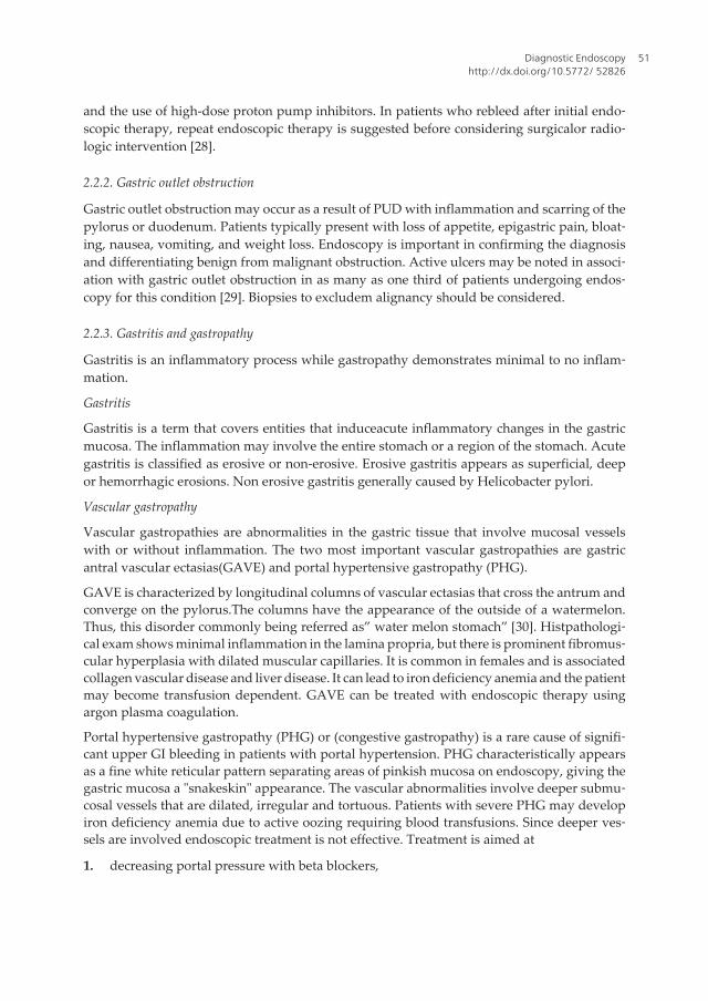
GAVE is characterized by longitudinal columns of vascular ectasias that cross the antrum andconverge on the pylorus.The columns have the appearance of the outside of a watermelon.Thus, this disorder commonly being referred as” water melon stomach” [30]. Histpathologi‐cal exam shows minimal inflammation in the lamina propria, but there is prominent fibromus‐cular hyperplasia with dilated muscular capillaries. It is common in females and is associatedcollagen vascular disease and liver disease. It can lead to iron deficiency anemia and the patientmay become transfusion dependent. GAVE can be treated with endoscopic therapy usingargon plasma coagulation.
Portal hypertensive gastropathy (PHG) or (congestive gastropathy) is a rare cause of signifi‐cant upper GI bleeding in patients with portal hypertension. PHG characteristically appearsas a fine white reticular pattern separating areas of pinkish mucosa on endoscopy, giving thegastric mucosa a "snakeskin" appearance. The vascular abnormalities involve deeper submu‐cosal vessels that are dilated, irregular and tortuous. Patients with severe PHG may developiron deficiency anemia due to active oozing requiring blood transfusions. Since deeper ves‐sels are involved endoscopic treatment is not effective. Treatment is aimed at
1. decreasing portal pressure with beta blockers,
Diagnostic Endoscopyhttp://dx.doi.org/10.5772/ 52826
51

2. portal decompression withtransjugular intrahepatic portosystemic shunt (TIPS), or
3. liver transplantation.
Hypertrophic Gastropathy
Gastric mucosal hypertrophy refers to giant gastric folds. Diffuse mucosal hypertrophy maybe described as hyperplastic or nonhyperplastic.In hyperplastic gastropathy gastric epithe‐lial cells which compose the oxyntic glands may become hyperplastic and give rise to giantmucosal folds. The conditions include: Ménétrier's disease, hyperplastic hypersecretorygastr‐opathy, and Zollinger-Ellison syndrome. In nonhyperplasticgastropathy - gastric mucosa maycontain other cell types which result in enlargement of the gastric folds. These conditionsinclude infiltrative diseases, infections, and malignancy.
Endoscopy with mucosal biopsy is required to distinguish between acute, chronic active andchronic gastritis and gastropathy. All gross abnormalities should be biopsied withmultiplebiopsies of both the corpus and the antrum to establish the diagnosis of Helicobacter pylorior autoimmune gastritis.Biopsies of the duodenum may also be helpful for diagnosing someforms of chronic gastritis such as Crohn’s disease in patients with granulomatous gastritis andceliac disease in patients with lymphocytic gastritis [31].
2.2.4. Dieulafoy’s lesions
Dieulafoy's lesion is a rare but important cause of upper GI bleeding. Arterial bleeding froman aberrant vessel isvisualized without an associated ulcer or mass lesion.The lesion can beeasily missed on endoscopy in the absence of active bleeding. It may look like a raised nippleor visible vessel without an associated ulcer. Endoscopy is the diagnostic modality of choicefor a Dieulafoy's lesion during acute bleeding [32].
Figure 12. Actively bleeding dieulafoy’s lesion
Endoscopy of GI Tract52
2.2.5. Gastric polyps
Gastric polyps are usually found incidentally when upper GI endoscopy performed for anunrelated indication. They are important since some types of polyps have malignant poten‐tial [33]. Adenomatous gastric polyps are at increased risk for malignant transformation andshould be resected completely. Hyperplastic polyps have a rare malignant potential. Endo‐scopic polyp appearance cannot differentiate histologic subtypes, therefore biopsy or poly‐pectomy is recommended when a polyp is encountered. When multiple gastric polyps areencountered, a biopsy of the largest polyps should be performed or they should be excised.Surveillance endoscopy 1 year after removing adenomatous gastric polyps is reasonable toassess recurrence at the prior excision site, new or previously missed polyps, and/or super‐vening early carcinoma. If the results of this examination are negative, repeat surveillanceendoscopy should be repeated no more frequently than at 3- to 5-year intervals. Follow-upafter resection of polyps with high-grade dysplasia and early gastric cancer should be indi‐vidualized. No surveillance endoscopy is necessary after adequate sampling or removal ofnon-dysplastic gastric polyps [34].
The various types of gastric polyps are briefly described below.
Fundic Gland Polyps
Fundic gland polyps are the most common type of polyps detected by endoscopy. These le‐sions are typically less than 5 mm in size, sessile and smooth in appearance. They are locat‐ed in body and fundus. Fundic gland polyps are commonly seen in patients who takeproton pump inhibitors (PPIs) on a long-term basis. They have an extremely low malignantpotential. The PPI-related lesions may regress in 3 months, once use of the PPI is discontin‐ued. A polyp associated with familial polyposis carries a defined 30% to 50% risk of devel‐oping dysplasia. When multiple fundic gland polyps are evident in younger patients,evaluation for familial polyposis should be considered.Biopsy of fundic polyps is done toexclude dysplasia [35].
Hyperplastic polyps
Hyperplastic polyps are caused by an inflamed or atrophic gastric mucosa. They have asmooth, dome-shaped appearance. Hyperplastic polyps can be large in size, and patientsmay present with chronic blood loss or even gastric obstruction. Elimination of the underly‐ing cause, such as H pylori infection, typically results in polyp regression. The risk of malig‐nancy is higher if polyps exceed 2 cm in size. For this reason, large polyps must becompletely excised.
Adenomatous Polyps
Adenomatous polyps occur sporadically or in association with familial polyposis. Thesepo‐lyps are circumscribed, pedunculated, or sessile. They are associated with chronic atrophicgastric metaplasia and have a defined cancer risk. Complete removal should be performed.
Polyposis Syndromes
Diagnostic Endoscopyhttp://dx.doi.org/10.5772/ 52826
53

Polyposis syndromes are characterized by multiple polyps.They include juvenile polyposis,Cronkite-Canada syndrome, Peutz-Jeghers syndrome, and Cowden's disease. Hamartoma‐touspolyps may be present in all of these syndromes. Adenomatous polyps may be found infamilial polyposis.
Gastrointestinal Stromal Tumor
Gastrointestinal stromal tumors (GISTs) make up 1% to 3 % of gastric neoplasms and occurmore frequently in men than in women. GISTs are typically located in the fundus. Biopsy istypically normal. Endoscopic ultrasonography-guided biopsy with fine-needle aspirationprovides the best tissue sample for diagnosis. GISTs are categorized as having malignantpotential ranging from low risk to high risk on the basis of polyp size and level of mitoticactivity. All GISTs should be regarded as having neoplastic potential. Surgical resection isrecommended for lesions larger than 2 cm. Endoscopic resection is an option for smallerGISTs [35].
Pancreatic Heterotopia
Pancreatic heterotopia may present assubmucosal nodular involvement (single or multiple)at the esophagogastric junction or as asubmucosal nodular lesion located in the antrum andprepyloric area.There is a characteristic nodule with a central dimple is seen endoscopically.Histological features resemble normal pancreatic tissue.Pancreatic heterotopia is a benignand asymptomatic condition.
2.2.6. Gastric neoplasm
Esophagogastroduodenoscopy has a diagnostic accuracy of 95% in diagnosing gastric can‐cer. Early gastric cancers may appear as a subtle polypoid protrusion, superficial plaque,mucosal discoloration, depression, or ulcer [33]. Improved detection of abnormal lesionsmay be possible with chromoendoscopy, narrow band imaging and magnification endoscopy(discussed later). Endoscopy is also the primary method for obtaining a tissue diagnosis ofsuspected lesions. Biopsy of any ulcerated lesion should include at least 6 specimens takenfrom around the lesion because of variable malignant transformation.The early use of upperendoscopy in patients presenting with gastrointestinal complaints may be associated with ahigher rate of detection of early gastric cancers. During endoscopy, any suspicious-appear‐ing gastric ulceration should be biopsied. The diagnosis of anaggressive form of diffuse-typecalled "linitisplastica", can be difficult endoscopically. Because these tumors infiltrate thesubmucosa and muscularispropria, superficial mucosal biopsies may be falsely negative.
In selected cases, endoscopic ultrasound may be helpful in assessing depth of penetration ofthe tumor in the layers of the stomach or involvement of adjacent structures.
3. Push enteroscopy
The evaluation of small intestine is difficult due to its length, intraperitoneal location andtortuousity. Recent developments of push enteroscopy, balloon assisted endoscopy, and
Endoscopy of GI Tract54

capsule endoscopy have made endoscopic examination of the entire small bowel examina‐tion practical. Methods used to evaluate the small bowel include push enteroscopy, singleballoon, double balloon enteroscopy and wireless capsule endoscopy [36].
Push enteroscopy using the enteroscope or pediatric or adult colonoscope allows evaluationof small bowel 70-150 cm beyond ligament of Treitz. The disadvantage is looping of scoperesulting in patient discomfort. It helps in diagnosis and therapeutics in small bowel lesionsin the proximal small bowel [36].
4. Deep small bowel enteroscopy
Diagnostic indications for deep small bowel enteroscopy include obscure gastrointestinalbleeding, tattooing of suspected small bowel malignancies or abnormal findings on otherimaging studies and wireless capsule endoscopy, suspected nonsteroidal anti-inflammatorydrug-induced small bowel injury, suspected or established small bowel Crohn's disease, re‐fractory celiac disease,detection of polyps in patients with polyposis syndromes such asfamilial adenomatous polyposis or Peutz-Jeghers syndrome, examination of the gastric rem‐nant in patients who have undergone Roux-en-Y gastric bypass and removal of foreign bod‐ies like retained wireless capsule [37,38].
Deep small bowel enteroscopy can be performed with balloon-assisted or spiral enterosco‐py. Single and double balloon techniques are described. These techniques allow deeper ac‐cess to the small bowel than push enteroscopy.
Single balloon enteroscopy (SBE) uses the scope’s flexible tip to anchor the scope to the bow‐el and intestinal tract is pleated over the overtube and shortened.On the other hand doubleballoon uses a second balloon to anchor the bowel instead of the scope tip.The workinglength of double balloon endoscope (DBE) is about 150-200 cm as a result 150-350 cm ofsmall bowel can be visualized. The success rate of complete inspection of small intestine is40-80%.Balloon-assisted enteroscopy (ie, DBE and SBE) which can be performed orally orper rectum, where as spiral enteroscopy can only be performed orally. The complications ofdouble balloon enteroscopy are ileus, pancreatitis, perforation and prolonged duration ofprocedure [38].
Spiral enteroscopy is a diagnostic and therapeutic intervention of the small bowel. A smallenteroscope is used with overtubethat has helical spirals on the surface. The overtube slidesover the enteroscope.There are no major complications reported.The limitations are in‐creased sedation requirement [36].
5. Wireless capsule endoscopy
Wireless capsule endoscopy is an ambulatory procedure which has become a first line testfor visualizing the mucosa of the small intestine.The PillCam is a capsule comprise of a lens
Diagnostic Endoscopyhttp://dx.doi.org/10.5772/ 52826
55

imager, battery and transmitter. The capsule moves from mouth to the anus with peristalsistaking two images per second at 1:8 magnification. The PillCam SB is FDA approved for vis‐ualization of the small bowel mucosa in adults and children aged >10 years(4).
The most common indications include evaluation of obscure GI bleeding including irondeficiency anemia, suspected Crohn’s Disease, small intestinal tumors and surveillance inpatients with polyposis syndromes,refractory malabsorptive syndromes (eg, Celiac disease)[39].
Contraindications suspected GI obstruction, gastroparesis, swallowing disorders, pregnan‐cy, dementia, strictures or fistulas (based on the clinical picture or preprocedure testing),cardiac pacemakers or other implanted electro-medical devices.
5.1. Small intestine
5.1.1. Celiac disease
Celiac disease is a condition in which the immune system responds abnormally to a proteincalled gluten causing damage to the lining of the small intestine.It affects about 1% of thewestern population.Patients with celiac disease usually have positive IgA endomysial or‐transglutaminase antibody.Patients with positive antibodies should undergo endoscopywith small bowel biopsy.
The duodenal mucosa may appear atrophic with loss of folds, contain visible fissures, havea nodular appearance or the folds may be scalloped. Multiple biopsies should be obtained inthe second and third portion of the duodenum by upper GI endoscopy. Staining techniquesand high resolution magnification endoscopy canalso help identify areas of villous atrophyfor biopsy (discussed later) [40]. Videocapsule endoscopy shows good sensitivity and excel‐lent specificity for the detection of villous atrophy in patients with suspected celiac disease[41]. The advantages of video capsule endoscopy (VCE) are that it is noninvasive, it imagesthe entire length of the small bowel, and it is able to detect minute mucosal details. For thesereasons VCE may be a useful tool for the diagnosis of Celiac disease [42].
5.1.2. Crohn’s disease
Crohn's disease is characterized by transmural inflammation of the gastrointestinal tract.Crohn's disease may involve the entire gastrointestinal tract from mouth to the perianal area.It mostly affects distal small bowel and right colon and 20-30% have disease limited to thesmall bowel only. Thus colonoscopy with ileoscopyand biopsy is a very important test in thediagnosis of Crohn's disease. It can be diagnosed by upper endoscopy, enteroscopy, wire‐less capsule endoscopy or colonoscopy with terminal ileum intubation based on the locationof GI tract involvement. Capsule endoscopy and double balloon enteroscopy have compara‐ble yield. Therefore, capsule endoscopy may be part of an initial evaluation followed by doubleballoon enteroscopy if biopsy or intervention is needed [43]. Capsule retention is the main andonly complication which is indefinite presence of capsule which occurs most commonly inpatients of known Crohn’s disease [39]. This can be avoided by the use of patency capsule(disintegration time-controlled capsule) in patients with high risk of capsule retention [44].
Endoscopy of GI Tract56

Figure 13. Crohn’s disease ulcer in the colon (pointed with an arrow)
On enteroscopy,the findings inCrohn's Disease are linear ulcers,aphthous ulcers,round or ir‐regular ulcers,pseudopolyps,cobble stoning, stricture or stenosis.
5.1.3. Tumors of small bowel
Tumors of the small bowel are relatively uncommon andaccount for approximately 3% ofgastrointestinal neoplasms. As the symptoms are vague and conventional diagnostic testsare unsatisfactory.These tumors often present a clinical, radiological, and endoscopic chal‐lenge. Endoscopy is very accurate in diagnosing and identifying small bowel lesions[45].Capsule endoscopy is helpful in the diagnosis of small bowel tumors[46]. Double balloonendoscopy is shown to have good diagnostic capabilities due to its ability to take biop‐sies[47]. However, isolated mass lesions can be missed on incomplete balloon endoscopy orcapsule endoscopy[48]. The clinical conditions that predispose to small bowel neoplasms areFamilial adematous polyposis, hereditary non-polyposis colorectal cancer (HNPCC), Peutz-Jegherssyndrome, celiac disease, and Crohn’s disease[45].
Adenocarcinomas are the most common type of primary malignant small bowel tumorsmostly occurring in duodenum.They appear as circumscribed,polypoid usually large andcircumferentially involving the bowel wall.
Carcinoids mostly occur in the terminal ileum,less than 1 meter from IC valve. They are usu‐ally small and found incidentally.
GISTs appear as dome shaped submucosal with central ulceration, commonly seen in jejunum.
6. Colonoscopy
Colonoscopy is endoscopic visualization of colonic mucosa.A complete exam is possible in95-99% of patients.The main indications to colonoscopy are screening and surveillance for
Diagnostic Endoscopyhttp://dx.doi.org/10.5772/ 52826
57

colon polyps, pathological bowel wall thickening notedon imaging procedures,diarrhea,malabsorption, rectal bleeding,unexplained iron deficiency anemia, positive fecal occultblood test,suspected short strictures of the colon,rectal foreign bodies,weight loss and ab‐dominal pain [49]. The contraindications of colonoscopy are peritonitis, perforation, fulmi‐nant colitis and recent surgical anastomosis.
Complications of colonoscopy are perforation (0.2% with diagnostic colonoscopy and 0.32%with polypectomy), hemorrhage (0.09% with diagnostic colonoscopy and 1.7% with poly‐pectomy) and postpolypectomy coagulation syndrome( electrocoagulation injury inducingtransmural burn in 0.5-1.2%,occurs 1-5 days of polypectomy, requires no surgical interven‐tion) and sedation related complications [50]. Thus, polypectomy is the single greatest riskfactor for complications of colonoscopy.
6.1. Colon
6.1.1. Screening colonoscopy
Screening for colorectal cancer with colonoscopy is the most common procedure performedby gastroenterologists in the US. Identification of premalignant polyps is primary goal.Somepolyps are only hyperplastic with minimal malignant potential. Polyps of concern are calledadenomas which are premalignant. Guidelines recommend colonoscopy every 10 years be‐ginning at age 50 years. The follow up colonoscopy should be based on number, size andpathologic findings of the adenomatous polyps removed. Patients with 1-2 small (<1cm) tub‐ular adenomas with only low grade dysplasia should get follow up after 5 years, whereaspatients with 3 or more or advanced adenomatous lesions should get repeat colonoscopy in3 years or before if colonoscopy was incomplete, preparation was poor or >10 polyps are re‐moved.If surveillance colonoscopy is normal, follow up colonoscopy is recommend after 5years. Patients with large,sessile adenomatous lesions which are removed in piecemealshould have repeat examination within 2-6 months to exclude and remove remnant poly‐poid tissue [51].
6.1.2. Colonic polyps
Colonic polyps are benign neoplasms that arise from the epithelial cells lining the colon. Colonicpolyps are divided into 3 groups: hyperplastic polyps, adenomas, and polyposis syndromes [52].
Hyperplastic polyps
Hyperplastic polyps comprise about 90% of all polyps. They are rounded and sessile meas‐uring few millimetersin size and cannot be distinguished from adenomas. Hyperplastic pol‐yps most commonly occur in the rectosigmoid region.Theylack malignant potentialespecially if they are located in the rectosimoid area and if the size is few mm. Malignantpotential is present only in the setting of very large polyps.
Polyps with architecture similar to hyperplastic polyps but the cytology is different withsurface mitotic activity, higher nuclear /cytoplasmic ratio and serrated glandular pattern asa result they are termed as “serrated adenomas”[52]..
Endoscopy of GI Tract58
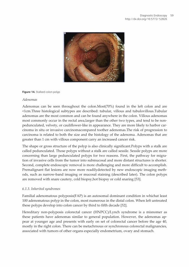
Figure 14. Stalked colon polyp
Adenomas
Adenomas can be seen throughout the colon.Most(70%) found in the left colon and are<1cm.Three histological subtypes are described: tubular, villous and tubulovillous.Tubularadenomas are the most common and can be found anywhere in the colon. Villous adenomasmost commonly occur in the rectal area,larger than the other two types, and tend to be non‐pedunculated, velvety, or cauliflower-like in appearance. They are more likely to harbor car‐cinoma in situ or invasive carcinomacompared toother adenomas.The risk of progression tocarcinoma is related to both the size and the histology of the adenoma. Adenomas that aregreater than 1 cm with villous component carry an increased cancer risk.
The shape or gross structure of the polyp is also clinically significant.Polyps with a stalk arecalled pedunculated. Those polyps without a stalk are called sessile. Sessile polyps are moreconcerning than large pedunculated polyps for two reasons. First, the pathway for migra‐tion of invasive cells from the tumor into submucosal and more distant structures is shorter.Second, complete endoscopic removal is more challenging and more difficult to accomplish.Premalignant flat lesions are now more readilydetected by new endoscopic imaging meth‐ods, such as narrow-band imaging or mucosal staining (described later). The colon polypsare removed with snare cautery, cold biopsy,hot biopsy or cold snaring [53].
6.1.3. Inherited syndromes
Familial adenomatous polyposis(FAP) is an autosomal dominant condition in whichat least100 adenomatous polyp in the colon, most numerous in the distal colon. When left untreatedthese polyps develop into colon cancer by third to fifth decade [52].
Hereditary non-polyposis colorectal cancer (HNPCC)/Lynch syndrome is a misnomer asthese patients have adenomas similar to general population. However, the adenomas ap‐pear at younger age and presents with early on set of colorectal cancer before the age 40,mostly in the right colon. There can be metachrnous or synchronous colorectal malignancies,associated with tumors of other organs especially endometrium, ovary and stomach.
Diagnostic Endoscopyhttp://dx.doi.org/10.5772/ 52826
59

Peutz-Jegherssyndrome is characterized by the presence of hemartomatous polyps occur‐ring more frequently in the small bowel and colon. Melanin spots may be seen on lips andbuccal mucosa.These polyps are not precancerous but patients are prone for tumors ofbreast, lung, ovary and pancreas.
Juvenile polyposis is a conditionpresenting with hamartomatous polyps in the colon, stom‐ach and small bowel. These polyps may be precancerous andrequire close endoscopicsurveillance.
6.1.4. Ischemic Colitis
Ischemic colitis is the most frequent form of mesenteric ischemia, presenting with suddenonset of abdominal pain followed by bloody diarrhea. Colonoscopy or sigmoidoscopy is of‐ten required to establish the diagnosis of ischemic colitis. The examination usually per‐formed without bowel preparation (to avoid reducing blood flow from dehydratingcathartics), and with minimization of air insufflation (to avoid distention and perforation).Colonoscopy is more sensitive in detecting mucosal lesions allows biopsies, and does not in‐terfere with subsequent angiography.
Colonoscopic findings in the acute setting frequently include pale mucosa with petechialbleeding withbluish hemorrhagic nodules may be seen representing submucosal bleeding[54]. Cyanotic mucosa and hemorrhagic ulcerations are seen later in the course [55]. Seg‐mental distribution, abrupt transition between injured and non-injured mucosa and rectalsparing favor the diagnosis of ischemic colitis [54].
Figure 15. Ischemic colitis
Endoscopy of GI Tract60

6.1.5. Pseudomembranous colitis
Pseudomembranes are pathognomonic of pseudomembranous colitis (Clostridium difficileassociated colitis) but are not found in all cases. Clostridium difficiletoxins cause cytoskele‐ton disruption causing shallow ulcerations which exude serum proteins and inflammatorycells forming pseudomembranes. Endoscopic findings include raised yellow or off-whiteplaques up to 2 cm in diameter scattered over the colorectal mucosa which cannot be re‐moved by lavage. These lesions are discrete but may become confluent plaques in more ad‐vanced cases. The other colonic findings are edema, erythema, and inflammation with orwithout pseuodomembranes [56].
Figure 16. Pseudomembranous colitis- pseudomembranes(pointed by arrows)
6.1.6. Diverticular disease of colon
Diverticula are outpouching of mucosa through the muscle wall of the colon. Colonic diver‐ticula are most frequent source of hematochezia followed by angiodysplasia and inflamma‐tory bowel disease (IBD) [55]. Approximately 95% diverticulosis is noted in descending andsigmoid colon. The prevalence increases with age, from less than 5 % at age nearly two-third by age 80.The diverticular bleed presents as painless, acute hematochezia which is ar‐terial in origin occurring at dome or neck of the diverticulum. About 60 % of diverticularbleeds occurs in left colon, however angiography study recognizes diverticular bleedingmore often in the right colon. Bleeding stops spontaneously in 80% cases [55].
Diagnostic Endoscopyhttp://dx.doi.org/10.5772/ 52826
61

Figure 17. Diverticulosis of colon (pointed by the arrows)
6.1.7. Arteriovenous malformations or Angiodysplasia
Angiodysplasia are detected in about 3-12% cases of lower GI bleeding.Most patients areasymptomatic and overt bleeding occurs in presence of coagulopathy or platelet dysfunc‐tion.They are mainly found as multiple lesions in the right colon, appearing as red, circum‐ferential lesions measuring from one millimeter to a few centimeter. The incidence increaseswith age.
Multiple telagiectasias in pale mucosa can also be seen in case of radiation induced proctop‐athy which occurs following radiation therapy for prostatic carcinoma [55].
6.1.8. Inflammatory bowel disease
Endoscopic findings in ulcerative colitis (UC) are mucosal erythema and edema with lossvascular pattern. Granularity of mucosa with friability, spontaneous bleeding and ulcers arealso seen. Some of patients of UC have focal inflammation around the appendiceal orificethat is not contiguous with disease elsewhere in the colon which is known as "cecal patch".Itis important to obtain adequate mucosal biopsies to help distinguish Crohn'sileocolitisfrompan-ulcerative colitis with backwash ileitis (UC with distal ileum involvement).
Endoscopy of GI Tract62

Figure 18. Ulcerative colitis- friable colon mucosa
Endoscopy in Crohns disease reveals aphthous ulcers, cobble stoning or skip lesions .A nor‐mal rectum supports the diagnosis of Crohn's disease, since UC always involves the rectum.The presence of normal vasculature adjacent to affected tissue is seen in Crohn's disease,while loss of vascularity and friability is more typical of UC [57].
6.1.9. Hemorrhoids
Hemorrhoids are clusters of veins, smooth muscle and connective tissue lined by the normalepithelium of the anal canal. They are categorized into internal and external hemorrhoids.These categories are anatomically separated by the dentate (pectinate) line. External hemor‐rhoids are hemorrhoids covered by squamous epithelium below the dentate line, where asinternal hemorrhoids are lined with colonic columnar epithelium proximal to dentate line.Internal hemorrhoids are not supplied by somatic sensory nerves and therefore cannot causepain.Internal hemorrhoids are classified in 4 degrees by the Goligher classification (Table 1).They are best viewed on retroflexed view on flexible endoscopy [58].
First degree Bleeds but donot prolapse
Second degree Prolapse but spontaneously reduce
Third degree Prolapse but require manual reduction
Fourth degree Unable to reduce
Table 1. Stains used in chromoendoscopy
Diagnostic Endoscopyhttp://dx.doi.org/10.5772/ 52826
63

Figure 19. Internal hemorrhoids on retroflexed view
External hemorrhoidal veins are found circumferentially under the anoderm and are inner‐vated by cutaneous nerves that supply the perianal area. Symptoms may occur anywherearound the circumference of the anus [58].
6.1.10. Melanosis coli
Deposition of pigment in the intestinal mucosa is commonly observed on endoscopy, espe‐cially within the colon. Electron microscopy has shown that this pigment is not melanin atall, but lipofuscin deposition in macrophages of colon mucosa. Herbal remedies or anthra‐quinone containing laxatives are often implicated. The pigment intensity is not uniform, be‐ing more intense in the cecum and proximal colon compared to the distal colon. Colorectaladenomas do not contain the melanin-like pigmentation. The association of adenomas withmelanosis coli can be explained by the ease of detection of even tiny polyps as white spotswithin a dark-colored colonic mucosa [59]. The condition is benign requiring no treatment.
Figure 20. Melanosis coli – colon polyp noted (pointed by an arrow)
Endoscopy of GI Tract64

6.1.11. Solitary rectal ulcer syndrome
Solitary rectal ulcer syndrome (SRUS) is a rare disorder of defecation presenting as bleedingper rectum.The term SRUS is a misnomer, as 34% of the endoscopic findings are multiplelesions. Endoscopic findings include mucosal ulcerations, polypoid lesions or simply erythe‐ma. It is a rare and poorly understood disorder that occurs in people with chronic constipa‐tion [60]. Treatments for solitary rectal ulcer syndrome range from changing diet and fluidintake to surgery.
6.1.12. Stercoral ulcer
Stercoral ulceration is the loss ofbowel integrity from the pressure effects of inspissated fe‐ces. It usually occurs in constipated and bedridden patients. Because of associated diseasesin the population at risk, perforation and hemorrhage are the principal complications result‐ing in a mortality exceeding 50%. Endoscopically, it appears as an isolated lesion in the rec‐tosigmoid area [61].
6.1.13. Colorectal cancer
Colorectal cancer presents commonly as abdominal pain, hematochezia, change of bowelhabits, anemia, or weight loss. Early diagnosis depends on routine screening. Colonoscopyis the single best diagnostic test in symptomatic individuals, since it can localize and biopsylesions throughout the large bowel, detect synchronous neoplasms, and remove polyps. Aircontrast barium enema (BE), supplemented with flexible sigmoidoscopy, is also used toevaluate symptomatic patients.
Figure 21. Colon cancer (pointed by an arrow)
Most colon cancers are adenocarcinomas which can be detected on colonoscopy and is un‐doubtedly the single best diagnostic test in symptomatic individuals since it can localize andbiopsy lesions throughout the large bowel, detect synchronous neoplasms, and remove pol‐
Diagnostic Endoscopyhttp://dx.doi.org/10.5772/ 52826
65

yps.Endoscopically,lesions may appear as circular proliferating,exophytic or stenosing le‐sions and uncommonly as plaque like, flat discoid mass with slight depression or ulcer [52].The likelihood of detection of colorectal cancer can be enhanced by novel methods like chro‐moendoscopy, narrow band imaging, confocal laser endomincroscopy and high resolutionand high maginification endoscopy (described later).
7. Novel and adjunct methods with endoscopy
It is shown that certain flat adenomaswith subtle dysplastic and early neoplastic changes aremissed with white light endoscopy as they as they are too small,flat or depressed to be detected.This has led to the development of techniques that compliment conventional endoscopicmethods and help in detection of subtle GI lesions by enhancing the image by high magnifi‐cation or high definition. Image enhanced endoscopy technology can either be dye based(Chromoendoscopy), equipment based (Narrow band imaging), or electronic based [62].
7.1. Chromoendoscopy
Chromoendoscopy involves the topical application of various stains or pigments to subtle GIlesions to improve tissue localization and characterization resulting in targeted biopsies ofthose lesions. The mucosa is pretreated with a mucolytic agent to remove excess mucus fromthe mucosal surface. Most commonly 10 % N-acetylcysteine is used. Targeted spraying via aspray catheter is performed for colon polyps and entire surface is stained inthe evaluationBarrett’s esophagus. Glucagon is administered just before spraying to decrease contractionsand uneven spraying.It is considered to be a safe and nontoxic procedure [63]. The table belowdescribes various stains used in various conditions with their side effects (Table 2).
Stains Conditions Side effects
Methylene Blue Esophagus: Barrett’s mucosa/Post ablation to find Barrett’s
mucosa.
Gastric: Intestinal metaplasia
Colon:Chronic ulcerative colitis
Harmless , transient blue green
discoloration of urine and feces
Toluidine blue Esophagus: Squamous cell cancer None reported
Lugol’s solutionEsophagus: Squamous dysplasia and early squamous cell
carcinoma,
Retrosternal burning and nausea
which can be treated with
application of 5% sodium thiosulfate
which can neutralize residual iodine.
Avoided in patients with Iodine
hypersensitivity and hyperthyroidism
Indigo carmine Colon : Colorectal neoplasia,chronic ulcerative colitis None reported
Table 2. Stains used in chromoendoscopy
Endoscopy of GI Tract66

Chromoendoscopy has been shown to detected higher number of lesions per patient com‐pared with narrow band imaging (NBI), autofluorescence, or white light colonoscopy [64]. Itis an inexpensive, safe and relatively easy to perform but it is not standardized and is sub‐ject to observer interpretation.
7.2. Narrow band imaging
Conventional white light endoscopy uses full visible wavelength (red-green-blue) to pro‐duce an image. On the other hand narrow band imaging uses special filters which increaserelative intensity of the blue band thus enhancing the image quality. NBI used along withmagnifying endoscopy allows the analysis of thesurface architecture of the epithelium (pitpattern) and theanalysis of the vascular network resulting in better characterization of dis‐tinct types of gastrointestinal epithelia (e.g. intestinal metaplasia in Barret’s esophagus),aswell as the disorganization of the vascular pattern ininflammatory disorders and the irregu‐lar pit pattern in earlyneoplastic lesions of the esophagus, stomach and largebowel [65].
The NBI generates a darker field of view than white light and allows adequate inspection ofthe mucosal surface. The tip of the endoscope needs to be closer to the mucosa.The presenceof bile and blood strongly absorb narrow band light thus obscuring the view under NBI. TheNBI images are not yet standardized. There are no reported complications with NBI [66].
7.3. Confocal Laser Endomicroscopy
Canfocal Laser Endomicroscopy (CLE) is new imaging modality of GI endoscopy which al‐lows in vivo imaging of the mucosal layer at cellular and subcellular resolution making invivo histology possible during endoscopy. CLE is based on the principle of illuminating atissue with a low-power laser and then detecting fluorescent light reflected from the tissue.To illuminate the tissue, an exogenousagent ??is applied topically or systemically. Mostcommonly used agent is intravenous fluorescein sodium which highlights the extracellularmatrix enabling confocal imaging. The laser is focused at a specific depth and only light re‐flected back from that plane is refocused and able to pass through the pinhole confocal aper‐ture. As a result, scattered light from above and below the plane of interest is not detected,increasing special resolution. The area being examined is scanned in the horizontal and ver‐tical planes and an image is reconstructed. In this manner, microscopic imaging of biologicaltissue in vivo is possible due to the high lateral resolution of confocal imaging. It helps indifferentiation of neoplastic from non-neoplastic polyps, for example, neoplastic lesion inpatients with Barrett's esophagus [67], or ulcerative colitis, differentiation of benign frommalignant biliary strictures.
Currently, there are two CLE systems used, probe based (confocal probe passes through theaccessory channel of a standard video-endoscope) and integrated endoscopy (CLE integrat‐ed in the distal tip of endoscope).
More data is needed to support these modalities. In future, CLE will develop multicolor analysisof several layers with 3-dimensional reconstruction allowing deeper penetration depth [68].
Diagnostic Endoscopyhttp://dx.doi.org/10.5772/ 52826
67

7.4. High resolution and high magnification endoscopy
High-resolution imaging improves the ability to discriminate detail while magnification en‐larges the image. Magnification endoscopy often utilizes a movable lens controlled by theendoscopist to vary the degree of magnification(100X as compared with 30X in standard en‐doscopy). Both high magnification and high-resolution endoscopes were designed to beused in conjunction with chromoendoscopy. High-resolution and high-magnification endos‐copy may enhance the diagnosis and characterization of some mucosal lesions and may de‐tect changes in vascular architecture of patients with early esophageal cancer. Magnificationchromoendoscopy has been used to characterize Barrett’s esophagus, early gastric cancerand villous atrophy [69]. The magnification endoscopy is simple, inexpensive, requiring nospecial light processors.The disadvantages are lack of standardization and prolongation ofprocedure time [70]. Whether high-resolution or high-magnification endoscopy will de‐crease the need for endoscopic biopsy or increase the diagnostic yield of endoscopic proce‐dures has not yet been determined [71,72].
7.5. Autofluoresence imaging
Autofluorense imaging utilizes changes in concentrations of endogenous fluorophores, forexample flavin adenine dinucleotide, collagenand nicotinamide adenine dinucleotide. Thevideo-endoscopy adds green and red reflectance improving the image quality.The dysplas‐tic tissue there is lack of fluorescence due to lack of collagen resulting in increased red anddecreased green fluorescence.It has been shown useful in detection of dysplasia in Barrett’sesophagus and early esophageal cancer but there is insufficient data to support its routineclinical use [62].
7.6. Endocytoscopy
Endocystoscopy is a new imaging method which provides combination which combineschrmoendoscopy with ultra-high magnification catheter which is passed through the work‐ing channel of the endoscope [73]. Unlike confocal endomicroscopy, it provides images incolor but is limited to superficial layer. It has been shown to give accurate results which arealmost comparable with histological results.The diagnosis based on endocytoscopic imagingis subject to interpretation,and there is no validated criteria regarding tissue diagnosis anddifferentiation for various GI conditions.
Author details
Akash Nabh1*, Muhammed Sherid2, Charles Spurr1 and Subbaramia Sridhar1
*Address all correspondence to: [email protected]
1 Department of Gastroenterology and Hepatology, Georgia Health Sciences University, U.S. A.
Endoscopy of GI Tract68

2 Department of internal medicine, division of gastroenterology, Saint Francis Hosiptal, U.S. A.
References
[1] Cohen, J., Safdi, M. A., Deal, S. E., Baron, T. H., Chak, A., Hoffman, B., Jacobson, B.C., Mergener, K., Petersen, B. T., Petrini, J. L., Rex, D. K., Faigel, D. O., & Pike, I. M.(2006). ASGE/ACG Taskforce on Quality in Endoscopy. Quality indicators for esoph‐agogastroduodenoscopy. Am J Gastroenterol. Apr, 101(4), 886-91.
[2] Eisen, G. M., Baron, T. H., Dominitz, J. A., Faigel, D. O., Goldstein, J. L., Johanson, . J.F., Mallery, J. S., Raddawi, H. M., Vargo, J. J. 2nd, Waring, .J. P., Fanelli, R. D., &Wheeler-Harbough, J. (2002). American Society for Gastrointestinal Endoscopy.Complications of upper GI endoscopy. Gastrointest Endosc. Jun, 55(7), 784-93.
[3] Croese, J., Fairley, S. K., Masson, J. W., Chong, A. K., Whitaker, D. A., Kanowski, P.A., & Walker, N. I. (2003). Clinical and endoscopicfeatures of eosinophilic esophagitisin adults. Gastrointest Endosc. Oct, 58(4), 516-22.
[4] Cantù, P., & Penagini, R. (2010). Eosinophilicoesophagitis: the essentials for daily‐practice. Scand J Gastroenterol. May, 45(5), 528-32.
[5] Zografos, G. N., Georgiadou, D., Thomas, D., Kaltsas, G., & Digalakis, M. (2009).Drug-induced esophagitis. Dis Esophagus Epub Apr 15, 22(8), 633-7.
[6] Kikendall, J. W., Friedman, A. C., Oyewole, Fleischer. D., & Johnson, L. F. (1983). Pill-inducedesophagealinjury. Case reports and review of the medicalliterature. Dig DisSci. Feb, 28(2), 174-82.
[7] Lichtenstein, D. R., Cash, B. D., Davila, R., Baron, T. H., Adler, D. G., Anderson, M.A., Dominitz, J. A., Gan, S. I., Harrison, M. E., 3rd Ikenberry, S. O., Qureshi, W. A.,Rajan, E., Shen, B., Zuckerman, M. J., Fanelli, R. D., & Van Guilder, T. (2007). Stand‐ards of Practice Committee. Role of endoscopy in the management of GERD. Gastro‐intest Endosc. Aug, 66(2), 219-24.
[8] Spechler, S. J., Sharma, P., Souza, R. F., Inadomi, J. M., & Shaheen, N. J. (2011). Amer‐ican Gastroenterological Association. American Gastroenterological Associationmedical position statement on the management of Barrett’s esophagus. Gastroenterol‐ogy, Mar, 140(3), 1084-91.
[9] Underwood, J. A., Williams, J. W., & Keate, R. F. (2003). Clinical findings and riskfactors for Candida esophagitis in outpatients. Dis Esophagus, 16(2), 66-9.
[10] Canalejo, Castrillero. E., García, Durán. F., Cabello, N., & García, Martínez. J. (2010).Herpes esophagitis in healthy adults and adolescents: report of 3 cases and review ofthe literature. Medicine (Baltimore)., Jul, 89(4), 204-10.
Diagnostic Endoscopyhttp://dx.doi.org/10.5772/ 52826
69

[11] Wilcox, C. M., Straub, R. F., & Schwartz, D. A. (1994). Prospectiveendoscopiccharac‐terization of cytomegalovirusesophagitis in AIDS. GastrointestEndosc., Jul-Aug;, 40(4),481-4.
[12] Kopelman, Y., & Triadafilopoulos, G. (2011). Endoscopy in the diagnosis and man‐agement of motilitydisorders. Dig Dis Sci. Mar Epub Feb 1., 56(3), 635-54.
[13] Smith, M. S. (2010). Diagnosis and management of esophageal rings and webs. Gas‐troenterolHepatol (N Y)., Nov, 6(11), 701-4.
[14] Tobin, R. W. (1998). Esophageal rings, webs, and diverticula. J ClinGastroenterol, Dec,27(4), 285-95.
[15] Pregun, I., Hritz, I., Tulassay, Z., & Herszényi, L. (2009). Peptic esophageal stricture:medical treatment. Dig Dis Epub May 8., 27(1), 31-7.
[16] Wright, R. A., & Hurwitz, A. L. (1979). Relationship of hiatal hernia to endoscopical‐lyprovedreflux esophagitis. Dig Dis Sci, Apr, 24(4), 311-3.
[17] Kahrilas, P. J., Kim, H. C., & Pandolfino, J. E. (2008). Approaches to the diagnosis andgrading of hiatal hernia. Best Pract Res ClinGastroenterol, 22(4), 601-16.
[18] Weston, A. P. (1996). Hiatal hernia with cameron ulcers and erosions. GastrointestEn‐doscClin N Am, Oct, 6(4), 671-9.
[19] Choong, . C. K., & Meyers, B. F. (2003). Benign esophageal tumors: introduction, inci‐dence, classification, and clinical features. SeminThoracCardiovasc Surg, Jan, 15(1), 3-8.
[20] Tang, P., Mc Kinley, Sporrer. M., & Kahn, E. (2004). Inlet patch: prevalence, histologictype, and association with esophagitis, Barrett esophagus, and antritis. Arch PatholLab Med, Apr, 128(4), 444-7.
[21] Athanassiadi, K., Gerazounis, M., Metaxas, E., & Kalantzi, N. (2002). Management ofesophageal foreign bodies: a retrospective review of 400 cases. Eur J CardiothoracSurg, Apr, 21(4), 653-6.
[22] Jensen, D. M. (2002). Endoscopicscreening for varices in cirrhosis: findings, implica‐tions, and outcomes. Gastroenterology, May, 122(6), 1620-30.
[23] Merli, M., Nicolini, G., Angeloni, S., Rinaldi, V., De Santis, A., Merkel, C., Attili, A. F.,& Riggio, O. (2003). Incidence and natural history of small esophageal varices in cir‐rhotic patients. J Hepatol, Mar, 38(3), 266-72.
[24] Reliability of endoscopy in the assessment of variceal features. (1987). The ItalianLiver Cirrhosis Project. J Hepatol, Feb, 4(1), 93-8.
[25] Younes, Z., & Johnson, D. A. (1999). The spectrum of spontaneous and iatrogenicesophageal injury: perforations, Mallory-Weiss tears, and hematomas. J ClinGastroen‐terol, Dec, 29(4), 306-17.
Endoscopy of GI Tract70

[26] Graham, D. Y., Schwartz, J. T., Cain, G. D., & Gyorkey, F. (1982). Prospective evalua‐tion of biopsy number in the diagnosis of esophageal and gastric carcinoma. Gastro‐enterology, Feb, 82(2), 228-31.
[27] Barkun, A., Bardou, M., & Marshall, J. K. (2003). NonvaricealUpper GI BleedingCon‐sensus Conference Group. Consensusrecommendations for managingpatients withnonvaricealupper gastrointestinalbleeding. Ann Intern Med, Nov 18;, 139(10), 843-57.
[28] Banerjee, S., Cash, B. D., Dominitz, J. A., Baron, T. H., Anderson, M. A., Ben-Menac‐hem, T., Fisher, L., Fukami, N., Harrison, M. E., Ikenberry, S. O., Khan, K., Krinsky,M. L., Maple, J., Fanelli, R. D., & Strohmeyer, L. (2010). ASGE Standards of PracticeCommittee. The role of endoscopy in the management of patients with peptic ulcerdisease. GastrointestEndosc., Apr, 71(4), 663-8.
[29] Di Sario, J. A., Fennerty, M. B., Tietze, C. C., Hutson, W. R., & Burt, R. W. (1994). En‐doscopic balloon dilation for ulcer-induced gastric outlet obstruction. Am J Gastroen‐tero, Jun, 89(6), 868-71.
[30] Primignani, M., Carpinelli, L., Preatoni, P., Battaglia, G., Carta, A., Prada, A., Cestari,R., Angeli, P., Gatta, A., Rossi, A., Spinzi, G., & De Franchis, R. (2000). Natural histo‐ry of portal hypertensive gastropathy in patients with liver cirrhosis The New ItalianEndoscopic Club for the study and treatment of esophageal varices (NIEC). Gastroen‐terology, Jul, 119(1), 181-7.
[31] Dixon, M. F., Genta, R. M., Yardley, J. H., & Correa, P. (1997). Histological classifica‐tion of gastritis and Helicobacter pylori infection: an agreement at last? The Interna‐tional Workshop on the Histopathology of Gastritis. Helicobacter, Jul, 2(1), S17-24.
[32] Nagri, S., Anand, S., & Arya, Y. (2007). Clinical presentation and endoscopic manage‐ment of Dieulafoy’s lesions in an urban communityhospital. World J Gastroenterol,Aug 28, 13(32), 4333-5.
[33] Kajitani, T. (1981). The generalrules for the gastriccancerstudy in surgery and pathol‐ogy. Part I. Clinical classification. JpnJSurg., Mar, 11(2), 127-39.
[34] Hirota, W. K., Zuckerman, M. J., Adler, D. G., Davila, R. E., Egan, J., Leighton, J. A.,Qureshi, W. A., Rajan, E., Fanelli, R., Wheeler-Harbaugh, J., Baron, T. H., & Faigel,DO. (2006). Standards of PracticeCommittee, AmericanSociety for GastrointestinalEndoscopy. ASGEguideline: the role of endoscopy in the surveillance of prema‐lignantconditions of the upper GI tract. GastrointestEndosc, Apr, 63(4), 570-80.
[35] Goddard, A. F., Badreldin, R., Pritchard, D. M., Walker, M. M., & Warren, B. (2010).British Society of Gastroenterology. The management of gastricpolyps. GutSep EpubJul 30., 59(9), 1270-6.
[36] Voelkel, J. P., & Di Palma, J. A. (2010). Deep enteroscopy. South Med J., Oct, 103(10),1045-8.
Diagnostic Endoscopyhttp://dx.doi.org/10.5772/ 52826
71

[37] Westerhof, J., Weersma, R. K., & Koornstra, J. J. (2009). Investigatingobscuregastroin‐testinal bleeding: capsule endoscopy or double balloon enteroscopy? Neth J Med., Jul-Aug, 67(7), 260-5.
[38] Yano, T., & Yamamoto, H. (2009). Current state of double balloon endoscopy: the lat‐est approach to small intestinal diseases. J GastroenterolHepatol., Feb, 24(2), 185-92.
[39] Eliakim, R. (2010). Videocapsuleendoscopy of the smallbowel. CurrOpinGastroenterol,Mar, 26(2), 129-33.
[40] Lo, A., Guelrud, M., Essenfeld, H., & Bonis, P. (2007). Classification of villousatrophywith enhancedmagnificationendoscopy in patients with celiac disease and tropical‐sprue. GastrointestEndosc, Aug, 66(2), 377-82.
[41] Culliford, A., Daly, J., Diamond, B., Rubin, M., & Green, P. H. (2005). The value ofwireless capsule endoscopy in patients with complicated celiac disease. Gastrointes‐tEndosc, Jul, 62(1), 55-61.
[42] Rondonotti, E., Spada, C., Cave, D., Pennazio, M., Riccioni De, Vitis. I., Schneider, D.,Sprujevnik, T., Villa, F., Langelier, J., Arrigoni, A., Costamagna, G., & de Franchis, R.(2007). Video capsule enteroscopy in the diagnosis of celiac disease: a multicenterstudy. Am J GastroenterolAug Epub Apr 24., 102(8), 1624-31.
[43] Pasha, S. F., Leighton, J. A., Das, A., Harrison, Decker. G. A., Fleischer, D. E., & Shar‐ma, V. K. (2008). Double-balloon enteroscopy and capsule endoscopy have compara‐ble diagnostic yield in small-bowel disease: a meta-analysis.ClinGastroenterolHepatolJun Epub Mar 20., 6(6), 671-6.
[44] Signorelli, C., Rondonotti, E., Villa, F., Abbiati, C., Beccari, G., Avesani, E. C., Vecchi,M., & de Franchis, R. (2006). Use of the Given Patency System for the screening ofpatients at high risk for capsule retention. Dig Liver DisMay Epub Mar 9., 38(5),326-30.
[45] Abu-Hamda, E. M., Hattab, E. M., & Lynch, P. M. (2003). Small bowel tumors. Curr‐Gastroenterol Rep, Oct, 5(5), 386-93.
[46] Sîngeap, A. M., Trifan, A., Cojocariu, C., Sfarti, C., & Stanciu, C. (2010). Capsule en‐doscopy role in diagnosis of small bowel tumors]. [Article in Romanian, Abstract inEnglish]. Rev Med ChirSoc Med Nat Iasi, Oct-Dec, 114(4), 988-92.
[47] Almeida, N., Figueiredo, P., Lopes, S., Gouveia, H., & Leitão, M. C. (2009). Double-balloon enteroscopy and small bowel tumors: a South-European single-center experi‐ence. Dig Dis SciJul Epub 2008 Oct 29., 54(7), 1520-4.
[48] Paski, S. C., & Semrad, C. E. (2009). Small boweltumors. GastrointestEndoscClin N Am,Jul, 19(3), 461-79.
[49] Jechart, G., & Messmann, H. (2008). Indications and techniques for lower intestinalendoscopy. Best Pract Res ClinGastroenterol, 22(5), 777-88.
Endoscopy of GI Tract72

[50] Fisher, D. A., Maple, J. T., Ben-Menachem, T., Cash, B. D., Decker, G. A., Early, D. S.,Evans, J. A., Fanelli, R. D., Fukami, N., Hwang, J. H., Jain, R., Jue, T. L., Khan, K. M.,Malpas, P. M., Sharaf, R. N., Shergill, A. K., & Dominitz, J. A. (2011). ASGE Standardsof Practice Committee. Complications of colonoscopy. GastrointestEndosc, Oct, 74(4),745-52.
[51] Davila, R. E., Rajan, E., Baron, T. H., Adler, D. G., Egan, J. V., Faigel, D. O., Gan, S. I.,Hirota, W. K., Leighton, J. A., Lichtenstein, D., Qureshi, W. A., Shen, B., Zuckerman,M. J., Van Guilder, . T., & Fanelli, R. D. (2006). Standards of Practice Committee,American Society for Gastrointestinal Endoscopy. ASGE guideline: colorectal cancerscreening and surveillance. GastrointestEndosc, Apr, 63(4), 546-57.
[52] Ponz de, Leon. M., & Di Gregorio, C. (2001). Pathology of colorectal cancer. Dig LiverDis, May, 33(4), 372-88.
[53] Tappero, G., Gaia, E., De Giuli, P., Martini, S., Gubetta, L., & Emanuelli, G. (1992).Cold snare excision of small colorectal polyps. GastrointestEndosc, May-Jun, 38(3),310-3.
[54] Barnert, J., & Messmann, H. (2009). Diagnosis and management of lower gastrointes‐tinal bleeding. Nat Rev GastroenterolHepatol, Nov, 6(11), 637-46.
[55] Sherid, M., & Ehrenpreis, E. D. (2011). Types of colitis based on histology. Dis Mon,Sep, 57(9), 457-89.
[56] Hookman, P., & Barkin, J. S. (2009). Clostridium difficile associated infection, diar‐rhea and colitis. World J Gastroenterol, Apr 7;, 15(13), 1554-80.
[57] Waye, JD. (1977). The role of colonoscopy in the differential diagnosis of inflammato‐ry bowel disease. GastrointestEndosc, Feb, 23(3), 150-4.
[58] Appalaneni, V., Fanelli, R. D., Sharaf, R. N., Anderson, M. A., Banerjee, S., Ben-Men‐achem, T., Decker, G. A., Fisher, L., Fukami, N., Harrison, M. E., Strohmeyer, L., Friis,C., Ikenberry, S. O., Jain, R., Jue, T. L., Khan, K. M., Krinsky, M. L., Malpas, P. M.,Maple, J. T., & Dominitz, J. A. (2010). ASGE TECHNOLOGY COMMITTEE.The roleof endoscopy in patients with anorectal disorders. GastrointestEndosc., Dec, 72(6),1117-23.
[59] Freeman, H. J. (2008). Melanosis" in the small and large intestine. World J Gastroenter‐ol, Jul 21;, 14(27), 4296-9.
[60] Chong, V. H., & Jalihal, A. (2006). Solitary rectal ulcer syndrome: characteristics, out‐comes and predictive profiles for persistent bleeding per rectum. Singapore Med J,Dec, 47(12), 1063-8.
[61] Maull, K. I., Kinning, W. K., & Kay, S. (1982). Stercoralulceration. Am Surg, Jan, 48(1),20-4.
Diagnostic Endoscopyhttp://dx.doi.org/10.5772/ 52826
73

[62] Buchner, A. M., & Wallace, M. B. (2008). Future expectations in digestive endoscopy:competition with other novel imaging techniques. Best Pract Res ClinGastroenterol,22(5), 971-87.
[63] Wong, ., Kee, Song. L. M., Adler, D. G., Chand, B., Conway, J. D., Croffie, J. M., Disar‐io, J. A., Mishkin, D. S., Shah, R. J., Somogyi, L., Tierney, W. M., & Petersen, B. T.(2007). ASGE Technology Committee.Chromoendoscopy. GastrointestEndosc Oct;Epub Jul 23., 66(4), 639-49.
[64] Matsumoto, T., Esaki, M., Fujisawa, R., Nakamura, S., Yao, T., & Iida, M. (2009).Chromoendoscopy, narrow-band imaging colonoscopy, and autofluorescence colo‐noscopy for detection of diminutive colorectal neoplasia in familial adenomatouspolyposis. Dis Colon Rectum, Jun, 52(6), 1160-5.
[65] Gheorghe, C. (2006). Narrow-bandimagingendoscopy for diagnosis of malignant andpremalignantgastrointestinallesions. J Gastrointestin Liver Dis, Mar, 15(1), 77-82.
[66] Song, L. M., Adler, D. G., Conway, JD, Diehl, D. L., Farraye, F. A., Kantsevoy, S. V.,Kwon, R., Mamula, P., Rodriguez, B., Shah, R. J., & Tierney, W. M. (2008). ASGETECHNOLOGY COMMITTEE.Narrow band imaging and multiband imaging. Gas‐trointestEndosc, Apr, 67(4), 581-9.
[67] Pohl, H., Rösch, T., Vieth, M., Koch, M., Becker, V., Anders, M., Khalifa, A. C., &Meining, A. (2008). Miniprobeconfocallaser microscopy for the detection of invisible‐neoplasia in patients with Barrett’s oesophagus. Gut Dec; Epub Aug 28., 57(12),1648-53.
[68] Neumann, H., Kiesslich, R., Wallace, M. B., & Neurath, M. F. (2010). Confocal laserendomicroscopy: technical advances and clinical applications. GastroenterologyAuge1-2., 139(2), 388-92.
[69] Boeriu, A. M., Dobru, D. E., & Mocan, S. (2009). Magnifying endoscopy and chro‐moendoscopy of the upper gastrointestinal tract. J Gastrointestin Liver Dis, Mar, 18(1),109-13.
[70] Sharma, P. (2005). Magnification endoscopy. GastrointestEndosc, Mar, 61(3), 435-43.
[71] Stevens, P. D., Lightdale, C. J., Green, P. H., Siegel, L. M., Garcia-Carrasquillo, R. J., &Rotterdam, H. (1994). Combinedmagnificationendoscopy with chromoendoscopy forthe evaluation of Barrett’s esophagus. GastrointestEndosc, Nov-Dec, 40(6), 747-9.
[72] Lee, C. T., Chang, C. Y., Lee, Y. C., Tai, C. M., Wang, W. L., Tseng, P. H., Hwang, J.C., Hwang, T. Z., Wang, C. C., & Lin, J. T. (2010). Narrow-band imaging with magni‐fying endoscopy for the screening of esophageal cancer in patients with primaryhead and neck cancers. Endoscopy, Aug, 42(8), 613-9.
[73] Kwon, R. S., Wong, Kee., Song, L. M., Adler, D. G., Conway, J. D., Diehl, D. L., Far‐raye, F. A., Kantsevoy, S. V., Kaul, V., Kethu, S. R., Mamula, P., Pedrosa, M. C., Ro‐driguez, S. A., & Tierney, W. M. (2009). ASGE Technology Committee.Endocytoscopy.Endosc, Oct, 70(4), 610-3.
Endoscopy of GI Tract74