diagnostic protocol for monilinia fructigena the cause of ... · this version of the national...
TRANSCRIPT
Diagnostic Protocol for Monilinia fructigena the cause of Apple Brown Rot
The methods in this protocol are being updated to incorporate new tests developed to separate M. fructigena
from recently described species.
PROTOCOL NUMBER NDP 1
VERSION NUMBER V2.1
STATUS Endorsed
ISSUE DATE January 2014
REVIEW DATE January 2019
ISSUED BY SPHDS
Prepared for the Subcommittee on Plant Health Diagnostic Standards (SPHDS)
This version of the National Diagnostic Protocol (NDP) for Monilinia fructigena is current as
at the date contained in the version control box on the front of this document.
NDPs are updated every 5 years or before this time if required (i.e. when new techniques become available).
The most current version of this document is available from the SPHDS website: http://plantbiosecuritydiagnostics.net.au/resource-hub/priority-pest-diagnostic-resources/
Disclaimer: The current detection and identification methods in this protocol, while still effective at detecting Monilinia fructigena, cannot distinguish M. fructigena from closely related species recently described, such as M. yunnanensis (Hu et al. 2011) and Monilia polystroma (van. Leeuwen et al. 2002). The method has also not been validated for distinguishing M. fructigena from M. mumeicola and M. mume.
Monilinia yunnanensis and Monilia polystroma were reported on peach and a range of Rosaceae plants, respectively, while Monilinia mume and Monilia mumecola (or M. mumeicola) were known to infect Prunus mume (Batra 1991; van Leeuwen et al., 2002; Harada et al., 2005; Hu et al., 2011).
Batra LR (1991) World species of Monilinia (Fungi): Their ecology, biosystematics and control. Mycological Memoir, No. 16. J. Cramer, Stuttgart, Germany, p. 246.
Harada Y, Nakao S, Sasaki M, Sasaki Y, lchihashi Y, Sano T (2005) Monilia mumecola, anam. nov., a new brown rot fungus on Prunus mume in Japan. Journal of General Plant Pathology 70: 297-307.
Hu M-J, Cox KD, Schnabel G, Luo C-X (2011) Monilinia Species Causing Brown Rot of Peach in China. PLoS ONE 6(9): e24990. doi:10.1371/journal.pone.0024990
van Leeuwen GCM, Baayen RP, Holb IJ, Jeger MJ (2002) Distinction of the Asiatic brown rot fungus Monilia polystroma sp. nov. from M. fructigena. Mycological Research 106: 444-451.
Contents 1. Introduction .................................................................................................................. 1
1.1. Host range .............................................................................................................. 1
1.1.1. Major hosts ...................................................................................................... 1 1.1.2. Minor hosts ...................................................................................................... 2
2. Taxonomic information ................................................................................................ 2
3. Detection ...................................................................................................................... 3
3.1. Symptoms ............................................................................................................... 3
3.1.1. Fruit rot ............................................................................................................ 3 3.1.2. Blossom and twig blight ................................................................................... 6
4. Identification ................................................................................................................ 7
4.1. Morphological and cultural methods ........................................................................ 7
4.1.1. Isolation from plant material ............................................................................. 7 4.1.2. Morphological identification .............................................................................. 8
4.2. Molecular methods ................................................................................................ 11
4.2.1. DNA Extraction from plant tissue or pure culture ............................................ 11 4.2.2. Direct PCR from infected host tissues ............................................................ 12 4.2.3. PCR from pure Monilinia cultures ................................................................... 13
5. Contacts ..................................................................................................................... 16
6. Acknowledgements ................................................................................................... 16
7. References ................................................................................................................. 17
8. Appendix .................................................................................................................... 20
8.1. Monilinia disease cycle ......................................................................................... 20
8.2. DNA Extraction from fungal mycelium ................................................................... 22
8.3. PCR primers for M. fructicola and M. laxa ............................................................. 24
8.4. Direct PCR from infected host tissues ................................................................... 24
8.4.1. Results: Testing Monilinia infected fruit by direct PCR ............................... 25
8.5. Results: Testing Monilinia cultures by PCR ........................................................... 27
1. INTRODUCTION
Monilinia fructigena, commonly referred to as ‘apple brown rot’ is the most common Monilinia species on apple and pear in Europe (Jones and Aldwinckle, 1990). M. fructigena has very similar morphology and biology to M. fructicola and M. laxa and the three species can be easily confused (Byrde and Willetts, 1977; Corazza et al., 1998). Isolates of M. fructigena from Japan were shown to be morphologically and genetically distinct from European strains and have subsequently been reclassified as the new species M. polystroma (Leeuwen et al. 2002). M. fructigena primarily causes fruit rots, both before and after harvest, and less frequently infection of shoots and twigs. The severity of the disease varies from year to year depending on environmental and storage conditions. M. fructigena is highly infectious and is reported to cause considerable losses in orchards during summer when warm temperatures are favourable for disease development (Scopes and Ledieu, 1983; Anon., 1991) and also in stored fruit (Jones and Aldwinckle, 1990; Leeuwen et al., 2000). Latent infections of fruit can also occur, with symptoms only developing after fruit ripening.
Identification of Monilinia species often presents a problem. The necessary observations needed to determine cultural and morphological differences are time consuming and may give inconclusive results as they rely on variable characters. However, identification is possible by combining cultural characteristics such as growth rates, growth pattern and colony colour with morphological data such as conidial dimensions and length of germ tubes. As these various characters are quantitative and may overlap, any identification has to be conducted under standardised conditions and start from pure cultures.
Classical identification methods are not adequate for phytosanitary diagnoses. Consignments of imported soft fruit infected with a Monilinia sp. for example require a method that is both quick and reliable. Molecular diagnostic methods developed by the Central Science Laboratories (CSL), York, United Kingdom for the identification of Monilinia species have enabled the specific and sensitive amplification of fungal DNA directly from the fruit and the differentiation of M. fructigena from the Monilinia species M. laxa and M. fructicola, but not M. polystroma.
1.1. Host range
The disease is most serious on apples and pears but also affects stone fruit, with plum being most susceptible (Anon., 1991; Byrde and Willetts, 1977). Sagasta (1977) stated that M. fructigena had been found on more than 40 species in a number of families.
1.1.1. Major hosts
The main commercial crops hosts are;
Cydonia oblonga (quince), Malus x domestica (apple, includes the synonyms M. pumila, M. sylvestris), Prunus armeniaca (apricot), Prunus avium (sweet cherry), Prunus cerasus (sour cherry), Prunus domestica (plum), Prunus persica (peach), Pyrus communis (European pear) and Pyrus serotina var. culta (Japanese pear) (CABI, 2004).
1

1.1.2. Minor hosts
There are many records on other hosts which include;
Amelanchier canadensis (thicket serviceberry), Berberis sp. (barberries), Capsicum sp. (peppers), Chaenomeles japonica (flowering quince), Cornus mas (cornelian cherry), Corylus avellana (filbert), Cotoneaster, Crataegus laevigata, Diospyros kaki (oriental persimmon), Elaeagnus macrophylla (wild olive), Eriobotrya japonica (loquat), Ficus carica (common fig), Fragaria ananassa (strawberry), Lycopersicon esculentum (tomato), Malus baccata (Siberian crabapple), Mespilus germanica (medlar), Prunus dulcis (almond), Prunus spinosa (blackthorn), Psidium guajava (common guava), Rhododendron (Azalea), Rosa sp. (roses), Rubus occidentalis (black raspberry), Sorbus aucuparia (European Rowan), Sorbus commixta (Scarlet Rowan), Sorbus sp., Vaccinium sp. (blueberries), and Vitis vinifera (grapevine) (CABI, 2004).
2. TAXONOMIC INFORMATION
Category: Fungus
Taxonomic Group: Ascomycetes, Helotiales, Sclerotiniaceae
Scientific Name: Monilinia fructigena (Aderh. & Ruhland) Honey ex Whetzel
Synomyms: Sclerotina fructigena Aderh. & Ruhland
Anamorph Name: Monilia fructigena (Pers. ex Pers.) Eaton
Anamorph Synonyms: Torula fructigena Pers. Oidium fructigenum (Pers.) Kunze & Schmidt Acrosporium fructigenum Pers. ex Pers. Oospora fructigena Wallroth
Common Names: Apple brown rot, Spur canker, Blossom blight of fruit trees, Blossom wilt, Fruit canker, Spur blight, Twig blight, Twig canker and Wither tip.
Monilinia fructigena was previously considered the same species as Monilinia fructicola in some countries (Roberts, 1924; Cline, 2005). These two fungi shared a common earlier homonym [Monilinia fructigena = Sclerotinia fructigena Aderh. & Ruhland 1905; Monilinia fructicola = Sclerotinia fructigena (Pers.: Fr.) Schrot. 1893] and this has attributed to mis-identification of additional records in the American continents (Cline, 2005).
2

3. DETECTION
3.1. Symptoms
M. fructigena causes similar symptoms on all hosts, which are blossom blight, rots on the fruit and cankers on stems. The primary and most frequent symptom being fruit rot (Jones and Aldwinckle, 1990). Stem blights and cankers are observed less commonly (Xu et al., 1998), but can develop from blossom infections. The symptoms caused by the other commonly occurring brown rot fungi, M fructicola and M. laxa, are similar to those described for M. fructigena (Anon., 1991).
3.1.1. Fruit rot
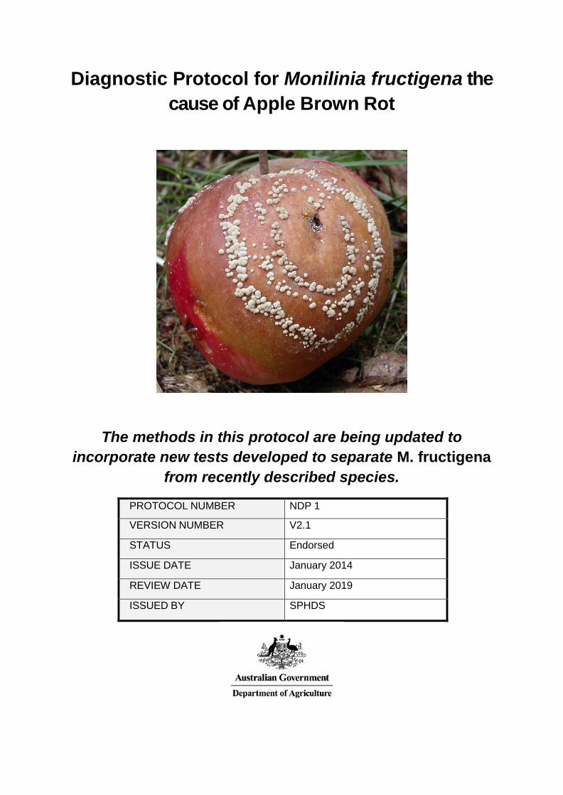
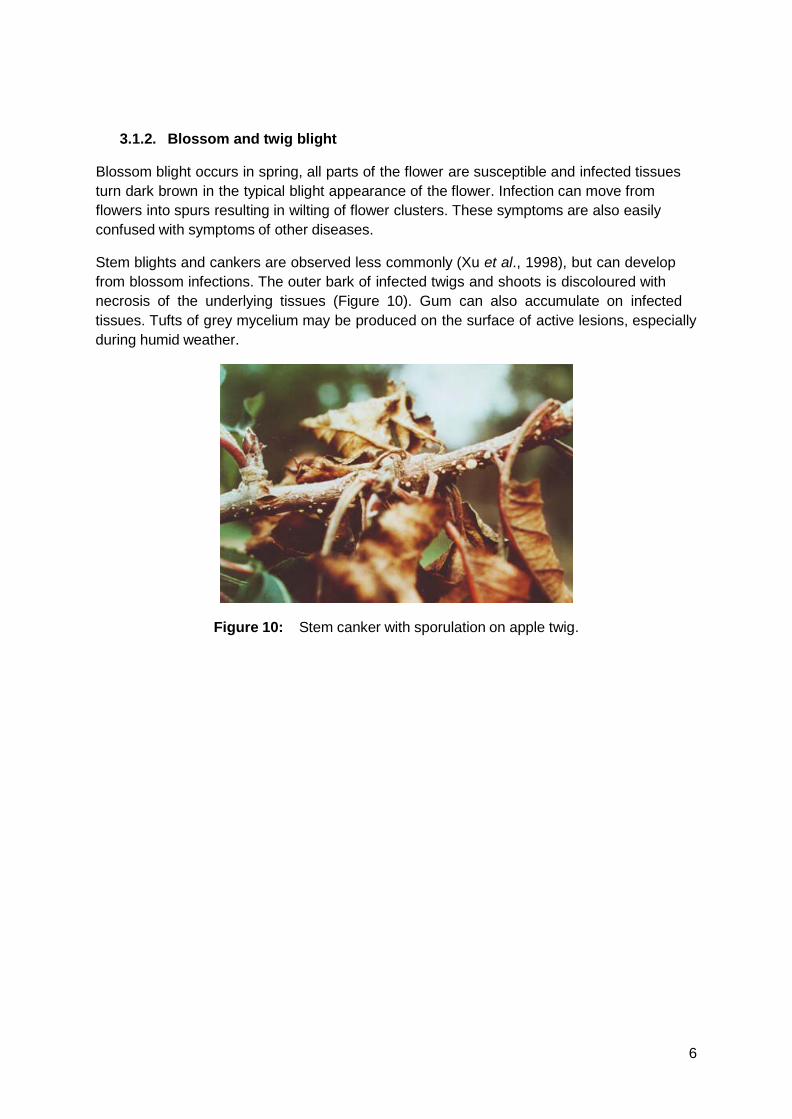
Initial fruit lesions are brown, circular and firm. Tufts of mycelium and conidia sprout from the skin of infected fruit (Figs 1, 2 and cover photo), often arranged in concentric rings. Fruit infection can be easily detected due to visible rots and/or sporulation by the fungus especially if storage or transport conditions were humid for several weeks (Figure 2).When the relative humidity is low and/or when the fruits are not ripe, no mycelium and very few or no conidial tufts develop (CABI, 2004). M. fructigena is most likely to occur on apple, pear fruit, or fruit from other minor hosts (Figs 4-7).
Distinguishing the various species of brown rot fungi requires additional laboratory testing as each species causes similar fruit symptoms (Figs 3 7).
Eventually the entire fruit decays and turns brown. Rotted fruits may either fall to the ground or dry out on the tree, leaving a hard, shrivelled mummy (Anon., 1991). Mummified fruit hang on branches of trees until spring or, alternatively fall to the ground where they remain throughout the winter months, partly or completely buried beneath the soil or leaf litter.
Figure 1: Apple naturally infected with Monilinia fructigena showing large cream or grey/brown tufts of mycelium and conidia.
3
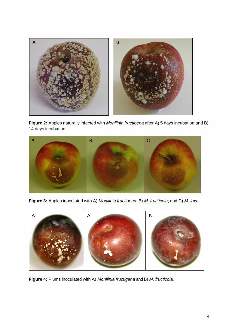
Figure 2: Apples naturally infected with Monilinia fructigena after A) 5 days incubation and B) 14 days incubation.
Figure 3: Apples inoculated with A) Monilinia fructigena; B) M. fructicola; and C) M. laxa.
Figure 4: Plums inoculated with A) Monilinia fructigena and B) M. fructicola.
C B A
A A B
A B
4
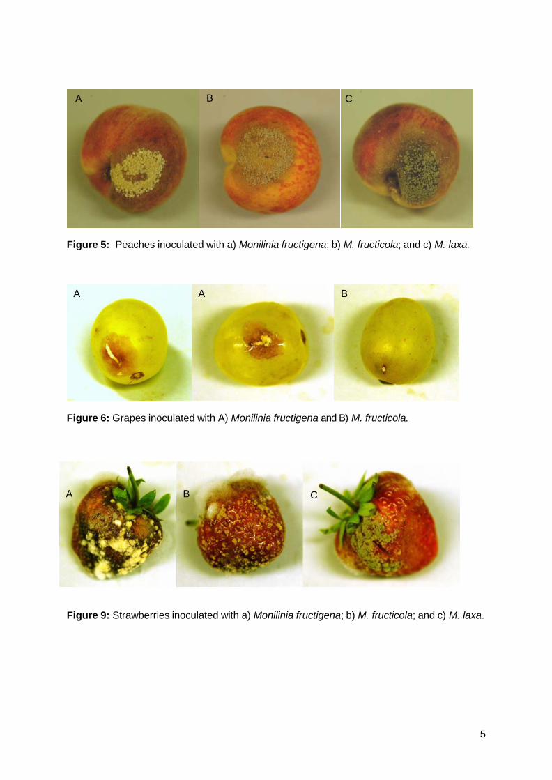
Figure 5: Peaches inoculated with a) Monilinia fructigena; b) M. fructicola; and c) M. laxa.
Figure 6: Grapes inoculated with A) Monilinia fructigena and B) M. fructicola.
Figure 9: Strawberries inoculated with a) Monilinia fructigena; b) M. fructicola; and c) M. laxa.
A A B
B C A
A B C
5
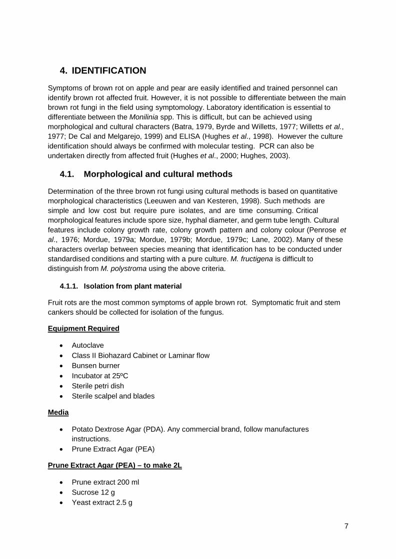
3.1.2. Blossom and twig blight
Blossom blight occurs in spring, all parts of the flower are susceptible and infected tissues turn dark brown in the typical blight appearance of the flower. Infection can move from flowers into spurs resulting in wilting of flower clusters. These symptoms are also easily confused with symptoms of other diseases.
Stem blights and cankers are observed less commonly (Xu et al., 1998), but can develop from blossom infections. The outer bark of infected twigs and shoots is discoloured with necrosis of the underlying tissues (Figure 10). Gum can also accumulate on infected tissues. Tufts of grey mycelium may be produced on the surface of active lesions, especially during humid weather.
Figure 10: Stem canker with sporulation on apple twig.
6
4. IDENTIFICATION
Symptoms of brown rot on apple and pear are easily identified and trained personnel can identify brown rot affected fruit. However, it is not possible to differentiate between the main brown rot fungi in the field using symptomology. Laboratory identification is essential to differentiate between the Monilinia spp. This is difficult, but can be achieved using morphological and cultural characters (Batra, 1979, Byrde and Willetts, 1977; Willetts et al., 1977; De Cal and Melgarejo, 1999) and ELISA (Hughes et al., 1998). However the culture identification should always be confirmed with molecular testing. PCR can also be undertaken directly from affected fruit (Hughes et al., 2000; Hughes, 2003).
4.1. Morphological and cultural methods
Determination of the three brown rot fungi using cultural methods is based on quantitative morphological characteristics (Leeuwen and van Kesteren, 1998). Such methods are simple and low cost but require pure isolates, and are time consuming. Critical morphological features include spore size, hyphal diameter, and germ tube length. Cultural features include colony growth rate, colony growth pattern and colony colour (Penrose et al., 1976; Mordue, 1979a; Mordue, 1979b; Mordue, 1979c; Lane, 2002). Many of these characters overlap between species meaning that identification has to be conducted under standardised conditions and starting with a pure culture. M. fructigena is difficult to distinguish from M. polystroma using the above criteria.
4.1.1. Isolation from plant material
Fruit rots are the most common symptoms of apple brown rot. Symptomatic fruit and stem cankers should be collected for isolation of the fungus.
Equipment Required
• Autoclave • Class II Biohazard Cabinet or Laminar flow • Bunsen burner • Incubator at 25ºC • Sterile petri dish • Sterile scalpel and blades
Media
• Potato Dextrose Agar (PDA). Any commercial brand, follow manufactures instructions.
• Prune Extract Agar (PEA)
Prune Extract Agar (PEA) – to make 2L
• Prune extract 200 ml • Sucrose 12 g • Yeast extract 2.5 g
7
• Agar 32 g • Water 1800 ml
1. Infuse 25 g finely sliced, pitted prunes in 450 ml water for 20 minutes in a beaker, in a hot water bath, stirring frequently.
2. Remove from water bath and leave to cool for at least 5 minutes. Carefully filter warm liquid through two layers of white tissue paper. Pour filtrate into bottles / flasks and sterilise.
3. Once cool, store extract in the refrigerator. 4. Mix together the prune extract, sucrose, and yeast extract and make up to 2 l with
water. Pour in to 500 ml flasks containing 8 g agar. Mix together, seal and autoclave at 120°C for 10 minutes.
NOTE: Do not overcook otherwise the medium becomes cloudy and the agar will hydrolyse and not gel properly.
Method
M. fructigena is easily cultured onto standard media such as prune extract agar (PEA) or potato dextrose agar (PDA) from fruit, shoots/branches. This can be achieved by inducing fruit to sporulate in a humidity chamber and transferring mass spores directly to a suitable culture medium or surface sterilising infected tissue and placing onto culture media.
The following procedures are commonly used for the isolation of Monilinia from infected plant material:
Mass transfer of conidia:
Where sporulation of the fungus is present on infected plant material, directly transfer the spores from the sample to an agar plate with a sterile needle.
Direct isolation from infected tissue:
Samples are cleaned and then sterilised by soaking in 1 % sodium hypochlorite for 3 to 10 minutes (the thicker the tissue the longer the soaking) and rinsing in sterile water three times. Suspected fruit samples can be surface sterilised by wiping the surface with cotton wool dipped in 96 % ethanol.
Small sections (2 to 3 mm3) of the infected tissue are cut out with a sterile scalpel and placed onto an appropriate culture medium. The sections should be cut out from the margin between the healthy and infected area.
Incubation:
Incubate the direct spore transfers or tissue isolations at 22ºC for five days in 12hr light/12hr dark, then examine them for the presence of Monilinia spp.
4.1.2. Morphological identification
The three morphologically similar species of Monilinia can be identified using the synoptic key described by Lane (2002).
Materials and Equipment
8
• Equipment • Incubator (12 h near uv/12 h dark) at 22°C • Dissecting microscope • Autoclave • Balance • Petri dishes • Sterile scalpel blades • Sterile hypodermic syringe needle
Reagents
• Potato dextrose agar
Method
1. Isolate the fungus on PDA; 2. Incubate the cultures at 22°C in the dark for 4 days; 3. Take a 4-mm-diameter plug from the edge of the 4-day-old colony; 4. Place plug centrally on a 9-cm Petri dish containing 12.5 ml PDA; 5. Incubate three replicates per isolate at 22°C with illumination of 12 h near-UV
(wavelength 365.5 nm)/12 h dark; and 6. After 10 days, assess the plates for the seven critical characters described below.
Details of the major cultural characteristics between the two main Monilinia species are presented in Table 1 and visually represented in Figure 11. The critical characters are described as follows:
1. Colony colour: (upper surface of plate) grey (A), yellow (B) or cream/white (C); 2. Growth rate: (mean colony diameter after 10 days growth at 22°C) > 80mm – fast
(D), 70-80mm – medium (E), or <70mm – slow (F); 3. Sporulation: (upper surface of colony, viewed with a dissecting microscope)
abundant (G) or sparse (H); 4. Concentric rings of sporulation: (upper surface of colony viewed with dissecting
microscope) present (I) or absent (J); 5. Colony margin: (underside of the plate) lobed (K) or non-lobed (L); 6. Rosetting: (upper surface of colony) rosetted (showing distinct layers of
mycelium - ‘petals’ - on top of each other, with the appearance of an open rose flower) (M) or not (N); and
7. Black arcs: (lower surface of colony) black arcs or rings associated with ‘petals’ of rosetted isolate (O), black dotted areas or brown arcs or rings (P) or no black arcs or rings present (Q).
Results
The synoptic key to identify the Monilinia species (letters in brackets indicate a character that is not usually produced but can occur in some isolates):
M. fructigena B, (C), (D), E, (F), (G), H, (I), J, L, N, Q M. fructicola A, D, (E), G, I, (J), L, (M), N, (P), Q M. laxa A, (C), (E), F, H, J, K, M, (N), O
9
This method will give a good indication of the species identity but will need to be backed up with a molecular identification.
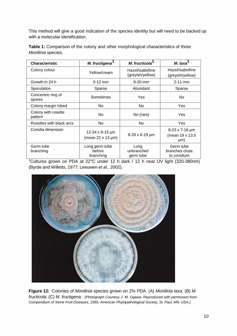
Table 1: Comparison of the colony and other morphological characteristics of three Monilinia species.
Characteristic M. fructigena1 M. fructicola1 M. laxa1 Colony colour
Yellow/cream Hazel/isabelline (greyish/yellow)
Hazel/isabelline (greyish/yellow)
Growth in 24 h 0-12 mm 9-20 mm 2-11 mm Sporulation Sparse Abundant Sparse Concentric ring of spores Sometimes Yes No
Colony margin lobed No No Yes Colony with rosette pattern No No (rare) Yes
Rosettes with black arcs No No Yes Conidia dimension 12-34 x 9-15 μm
(mean 22 x 13 μm) 8-28 x 6-19 μm
8-23 x 7-16 μm (mean 19 x 13.5
μm) Germ tube branching
Long germ tube before
branching
Long unbranched germ tube
Germ tube branches close
to conidium 1Cultures grown on PDA at 22°C under 12 h dark / 12 h near UV light (320-380nm) (Byrde and Willetts, 1977; Leeuwen et al., 2002).
Figure 12: Colonies of Monilinia species grown on 2% PDA. (A) Monilinia laxa; (B) M. fructicola; (C) M. fructigena. (Photograph Courtesy J. M. Ogawa. Reproduced with permission from Compendium of Stone Fruit Diseases, 1995, American Phytopathological Society, St. Paul, MN, USA.)
10
4.2. Molecular methods
4.2.1. DNA Extraction from plant tissue or pure culture
This extraction method uses the Qiagen DNeasy® Plant Mini Kit.
Equipment Required
• Autoclave • Qiagen DNeasy® Plant Mini Kit for DNA isolation from plant tissue • Sterile 1.5 ml microcentrifuge tubes • Appropriate pipettes and sterile filter tips • Sterile Eppendorf Micropestle part: 0030 120.937 • Balance • Microcentrifuge • Freezer/Fridge • Sterile scalpel blades • Vortex • Dry heat block or water bath 65ºC ± 2.5ºC • Sterile glass slide • Safety cabinet • 14. Variable speed electric drill • 15. Dewar liquid nitrogen dispenser and liquid nitrogen (if appropriate)
Reagents
• Ethanol 100% (molecular biology grade)
Method
1. All centrifuge steps are carried out at room temperature. 2. Preheat dry heat block or water bath to 65ºC ± 2.5ºC. 3. Use the Qiagen DNeasy® Plant Mini Kit. 4. Add ethanol 100% to buffers AW and AP3/E as indicated on the bottles if this is the
first use of this kit.
5. Using a sterile scalpel blade excise approximately 0.5 mm3 of fruit flesh or other plant material from the leading edge of the lesion (maximum of 100 mg) for DNA extraction.
6. Grind the plant material using one of the following methods: (a) Micropestle with liquid nitrogen: Add approximately 100 mg of plant tissue to
a 1.5 ml microcentrifuge tube, freeze with liquid nitrogen and grind to a fine powder using a sterile micropestle. Then, add 400 µl of Buffer AP1 and 4 µl of RNase to the ground tissue.
(b) Micropestle without liquid nitrogen: Add approximately 100 mg of plant tissue to a 1.5 ml microcentrifuge tube containing 400 µl of Buffer AP1 and 4 µl of
11
RNase. Using the electric drill and a sterile micropestle, grind the tissue sample vigorously for approximately 3 min.
7. Vortex vigorously. 8. Incubate the mixture for 10 min at 65ºC ± 2.5ºC. Mix 2-3 times during incubation
by inverting the tube. 9. Add 130 µl of Buffer AP2 to the lysate, mix and incubate for 5 min on ice. 10. Centrifuge the lysate for 5 min at full speed. 11. Pipette the lysate to the QIA shredder spin column (lilac) sitting in a 2 ml collection
tube and centrifuge for 2 min at maximum speed. 12. Transfer the flow-through fraction from step 11 to a new sterile 1.5 ml
microcentrifuge tube without disturbing the cell-debris pellet. 13. Add 1.5 volumes of Buffer AP3/E to the cleared lysate and mix by pipetting. 14. Pipette 650 µl of the mixture from step 13, including any precipitate which may have
formed, to the DNeasy mini spin column sitting in a 2 ml collection tube. Centrifuge for 1 min at 8000 rpm and discard the flow- through. Re-use the collection tube in step 15.
15. Repeat step 14. Discard collection tube. 16. Place DNeasy column in a new 2 ml collection tube (supplied in kit), add 500 µl
Buffer AW to the DNeasy column and centrifuge for 1 min at 8000 rpm. Discard flow-through and reuse the collection tube in step 17.
17. Add 500 µl of Buffer AW to the DNeasy column and centrifuge for 2 min at maximum speed to dry the membrane.
18. Transfer the DNeasy column to a sterile 1.5 ml microcentrifuge tube and pipette 50-100 µl of Buffer AE directly onto the DNeasy membrane. Incubate for 5 min at room temperature and then centrifuge for 1 min at 8000 rpm to elute (Elution 1).
19. Optional: Repeat elution (step 18) once (Elution 2).
20. Store DNA elution products at -20ºC ± 5oC or at -80ºC ± 5ºC.
The method used for plant material with Qiagen DNeasy® Plant Mini Kit can also be used to extract DNA from the pure culture.
An additional simplified protocol is available for DNA extraction from fungal mycelium only, which is quick and cost effective. It would be useful where large sample numbers are required to process during a survey. This protocol provides DNA of sufficient quality and quantity for reliable PCR testing (Appendix).
4.2.2. Direct PCR from infected host tissues
This method can be used to obtain a fast but tentative M. fructigena result on potentially infected fruit or other plant material showing limited symptoms. A positive PCR should however be backed up by the isolation of the fungus. The method used is the same as that for PCR from pure culture. Results of PCR from direct testing are presented in the Appendix.
12
4.2.3. PCR from pure Monilinia cultures
A PCR based method can rapidly identify M. fructigena and distinguish between the three Monilinia species (M. fructigena, M. fructicola, and M. laxa). This method is for DNA extracted from pure fungal cultures prior to PCR testing (Ioos and Frey, 2000; Hughes et al., 2001). The protocol by Hughes et al. (2001) is the currently accepted method for separation of the three main Monilinia species. Results and effectiveness are presented in Appendix.
Primer Sequence
Table 2: Primer sets used for the detection of M. fructigena
Primer
Sequence (5'-3') Reference
ITS 1 TCC GTA GGT GAA CCT GCG G White et al. (1990)
Mfg-R2 GGT CAA CCA TAG AAA ATT GGT Hughes et al. (2000)
Specific primers are also available for the detection of M. fructicola and M. laxa (Appendix).
Equipment Required
• Appropriate pipettes and sterile tips • 0.2 ml PCR tubes • 1.5 ml centrifuge tubes • Freezer • Gel tanks • Ice • Microcentrifuge • Microwave • Nitrile gloves • Power pack • Thermocycler • UV transilluminator with camera
Reagents
The reagents listed here for the PCR reaction and gel electrophoresis are those tested but suitable alternatives could also be used.
1. Primers: The recommended primer pair is IST1/ Mfg-R2 (Table 2) can be used to detect M. fructigena in a PCR reaction.
2. Taq polymerase: Invitrogen Platinum Taq DNA Polymerase (Catalogue no. 10966-034) 5U/ul was used. This Taq includes 10 x PCR Rxn Buffer and 50mM MgCl2. If a different Taq polymerase is used, the master mix proportions may need modification.
13

3. DNTPs: 10mM dNTPs Mix (PCR Grade), Invitrogen. Cat #18427-088.
4. DNA ladder: Invitrogen Trackit 100BP ladder. Cat # 10488058
5. PCR Controls: Positive control of total nucleic acid extracted from M. fructigena culture (ATCC 24976). Negative control for PCR master mix is water instead of the DNA template.
6. PCR Master mix: Table 3
7. 10 X TAE Invitrogen Cat. No. 15558-042.
8. Gel 2% (w/v) Agarose gel (2.4 g DNA grade agar in 120 ml of 1 × TAE Buffer, melted) prepared in a medium gel tray.
9. SYBR safe: DNA stain, (Sigma S9430)
10. Loading buffer: per 100 ml 15 g 15% (w/v) Ficoll 400 (Sigma, F1418) 1.4888 g 40 mM EDTA (372.2 g/mol, Sigma, E5134) 0.25 g 0.25% (w/v) Orange-G (Sigma, O3756)
Do not autoclave. Aliquot in 0.5 ml or 1.5 ml Eppendorf tubes and store in the fridge.
Method (PCR Set Up)
1. Label 0.2 ml PCR tubes as appropriate for the number of samples and controls.
2. Prepare "Master Mix" on ice in a 1.5 ml centrifuge tube as per Table 3.
3. Dispense 18 μl of the master mix into each of the 0.2 ml labelled PCR tubes.
4. Add 2 μl of SDD water to the negative control tube, 2 μl of test DNA template to each of the test sample tubes, and 2 μl of the positive control DNA to the positive control tube. Mix and pulse spin to ensure the mix is at the bottom of the tube.
5. Place the tubes in the thermocycler and cycle as per Table 3.
Method (Gel electrophoresis)
6. Mix 10 μl of each PCR product from each of the PCR tubes with 2 μl of
7. Loading Buffer and 1 μl of SYBR green (1:100 dilution in DMSO). 8. Mix 10 μl of DNA of 100 bp ladder with 2 μl of Loading Buffer.
9. Make Gel (2% Agarose) with % SYBR Safe (4-5 μl/ 100 ml) added once cooled but prior to pouring.
10. Load samples and ladder into separate wells of the 2% (w/v) agarose gel.
11. Electrophorese in 1.0 × TAE Buffer at 100V for approximately 1 hour.
12. Visualise and photograph the gel on a UV transilluminator.
14

Table 3: PCR programme and master mix for M. fructigena primers ITS1 and Mfg-R2.
PCR Cycle Checked Reagents1 Volume per reaction (μl)
Master mix (μl) x 20
94ºC 2 min x 1 94ºC 1.0 min 59ºC 1.0 min x 30 72ºC 1.5 min 72ºC 10 min x 1
Sterile deonised water 12.8 256.0
10 x PCR Buffer 2.0 40.0
50 mM MgCl2 0.6 12.0
10 mM dNTPs 0.4 8.0
5 μM ITS1 1.0 20.0
5 μM Mfg-R2 1.0 20.0
5U/μl Taq polymerase 0.2 4.0
PCR product size: 460 bp DNA Template 2.0 -
Total Volume 20.0 400.0
15
5. CONTACTS
International
Dr Renaud Ioos LNPV, Unite de Mycologie Agricole et Forestiere 38 rue Sainte Catherine 54043 Nancy, FRANCE Phone: 33 383304151 Fax: 33 383320045 E-mail: [email protected]
Dr Kelvin Hughes Central Science Laboratory MAFF Sand Hutton, York Y04 1 LZ UNITED KINGDOM Phone: 44 1904 462340 Fax: 44 1904 462147 E-mail: [email protected]
Australia
Ms Alison Mackie Department of Agriculture and Food WA South River Road Carnarvon WA 6701, AUSTRALIA Ph: 61 8 936 83333 Fax: 61 8 9368 3945 E-mail: [email protected]
New Zealand
Plant Health and Environment Laboratory MPI New Zealand
Dr Mark Braithwaite [email protected]
6. ACKNOWLEDGEMENTS
This protocol was drafted from the respective apple brown rot protocols for Australia and New Zealand developed by Drs Alison Mackie (WA) and Mark Braithwaite (NZ).
The protocol has been reviewed by Dr Mark Braithwaite and Dr Wellcome Ho, NZ
Dr Kelvin Hughes for providing DNA samples of Monilinia fructigena extracted from pure cultures and inoculated fruit. Ruth Griffin for testing and evaluating the molecular testing methods in New Zealand.
16
7. REFERENCES
Anon (1991) Brown rot of pome fruit. Plant Quarantine Leaflet No. 37. Australian Quarantine and Inspection Service.
Batra LR (1979) First authenticated North American record of Monilinia fructigena, with notes on related species. Mycotaxon, 8(2):476-484.
Byrde RJW and Willetts HJ (1977) The Brown Rot Fungi of Fruit. Their Biology and Control. Oxford, UK: Pergamon Press, 171pp.
CABI (2004) Monilinia fructigena (brown rot). Crop Protection Compendium. CAB International, Wallingford, UK.
Cline E (2005) Monilinia fructigena and related brown fruit rots. Systematic Mycology and Microbiology Laboratory, ARS, USDA.
Côté MJ, Tardif MC and Meldrum AJ (2004) Identification of Monilinia fuctigena, M. fructicola, Monilinia laxa, and Monilinia polystroma on inoculated and naturally infected fruit using muliplex PCR. Plant Disease 88: 1219-1225.
Corazza L, Cook RTA, Lane CR, Fulton CE, Van Leeuwen GCM. Periera AM, Nazare-Periera A, Melgarejo P and De Cal A (1998) Identification of Monilinia (brown rot) species. Proceedings of the 7th International Congress of Plant Pathology Edinburgh. Paper number 3.372.
De Cal A and Melgarejo P (1999) Effects of long-wave UV light on Monilinia growth and identification of species. Plant Disease, 83:62-65.
Honey EE (1936) North American Species of Monilinia. I. Occurrence, Grouping, and Life-Histories. American Journal of Botany, 23 (2),100-106.
Hughes KJD (2003) DNA extraction from mycological organisms. SOP PLHB/M26. Central Science Laboratory, York.
Hughes KJD, Banksi JN, Rizvii RH, McNaughton J, Lane CR, Stevenson L and Cook RTA (1998) Development of a simple ELISA for identification of Monilinia fructicola. International Congress of Plant Pathology. Edinburgh, UK: BSPP, 1998.
Hughes KJD, Fulton CE, McReynolds D and Lane CR (2000) Development of new PCR primers for identification of Monilinia species. Bulletin OEPP/EPPO Bulletin 30, 507-511.
Hughes KJD, Lane CR and Banks JN (2001) Diagnostic assays for Monilinia brownrot species; members of the family Sclerotinaceae. Proceedings of the International Sclerotinia Workshop, York.
Ioos R and Frey P (2000) Genomic variation within Monilinia laxa, M. fructigena and M. fructicola, and application to species identification by PCR. European Journal of Plant Pathology 106, 373-378.
17
Jones AL and Aldwinckle HS [Eds.] (1990) Brown rot diseases. Compendium of Apple and Pear Diseases. American Phytopathology Society Press. 100 pp.
Lane CR (2002) A synoptic key for differentiation of Monilinia fructicola, M. fructigena and M. laxa, based on examination of cultural characters. Bulletin OEPP/EPPO, Bulletin 32, 489-493.
Leeuwen GC van, Baayen RP, Holb IJ and Jeger M. (2002) Distinction of the Asiatic brown rot fungus Monilinia polystroma sp. Nov. from M. fructigena. Mycological Research 100: 444-451.
Leeuwen GCM van, Stein A, Holb I and Jeger MJ (2000) Yield loss in apple caused by Monilinia fructigena (Aderh. & Ruhl.) Honey, and spatio-temporaldynamics of disease development. European Journal of Plant Pathology 106: 519-528.
Leeuwen GCM van, and Kesteren HA van (1998) Delineation of the three brown rot fungi of fruit crops (Monilinia spp.) on the basis of quantitative characteristics. Canadian Journal of Botany 76: 2042-2050.
Mordue JEM (1979a) Sclerotinia fructicola. In: CMI Descriptions of pathogenic fungi and bacteria, No. 616. Wallingford, UK: CAB International.
Mordue, J.E.M. (1979b) Sclerotinia fructigena. In: CMI Descriptions of pathogenic fungi and bacteria, No. 617. Wallingford, UK: CAB International.
Mordue JEM (1979c) Sclerotinia laxa. In: CMI Descriptions of Pathogenic Fungi and Bacteria, No. 619. Wallingford, UK: CAB International.
Penrose LJ, Tarran J and Wong A-L (1976) First record of Sclerotinia laxa Aderh. & Ruhl. in New South Wales: differentiation from S. fructicola (Wint.) Rehm. by cultural characteristics and electrophoresis. Australian Journal of Agricultural Research, 27(4):547-556.
Roberts JW (1924) The fungus causing the common brown rot of fruits in America. Journal of Agricultural Research 28: 955-960. Available at naldc.nal.usda.gov/download/IND43966911/PDF.
Sagasta EM (1977) Monilinia disease. EPPO Bulletin 7: 105-116.
Scopes N and Ledieu M (1983) Pest disease control handbook. BCPC Publications. Croydon, Great Britain.
Smith I.M, Dunez J, Lelliott RA, Phillips DH and Archer SA(Editors) (1992) Handbook of plant diseases. Madrid, Spain; Ediciones Mundi-Prensa, 671 pp.
White TJ, Burns T, Lee S and Taylor J (1990) Amplification and direct sequencing of fungal ribosomal RNA genes for phylogenetics. In: Innis, M. A., Gelfand, D. H., Sninsky, J. J. and White, T. J. ed. PCR Protocols: a Guide to Methods and Applications, pp. 315- 322. Academic Press, London.
Willetts HJ, Byrde RJW, Fielding AH and Wong A-L (1977) The taxonomy of the brown rot fungi (Monilinia pp.) related to their extracellular cell wall-degrading enzymes. Journal of
18

General Microbiology, 103(1):77-83.
Xu X-M, Berrie AM, and Harris DC (1998) Epidemiology of brown rot (Monilinia fructigena) on apple and pear. Proceedings of the 7th International Congress of Plant Pathology, Edinburgh. Paper number 3.7.52.
7.1. Additional references
Batra LR and Harada Y (1986) A field record of apothecia of Monilinia fructigena in Japan and its significance. Mycologia, 78(6):913-917.
Batra LR (1991) World species of Monilinia (Fungi): their ecology, biosystematics and control. Mycologia Memoir, No 16, 246 pp.
CABI/EPPO (1997) Quarantine Pests for Europe, 2nd edn. CAB International, Wallingford, UK.
CABI/EPPO (1999) Monilinia fructicola. Distribution Maps of Plant Diseases, Map No. 50, Edition 7. CAB International, Wallingford, UK.
CABI/EPPO (2000) Monilinia fructigena. Distribution Maps of Plant Diseases, Map No. 22. CAB International, Wallingford, UK.
Ibragimov GR, Abbasov TF (1976) The occurrence of the ascus state of Monilia fructigena (Schroet.) Honey and M. cydoniae (Schell.) Honey in Azerbaijan. Mikologiya i Fitopatologiya, 10(3):219-222.
19
8. APPENDIX
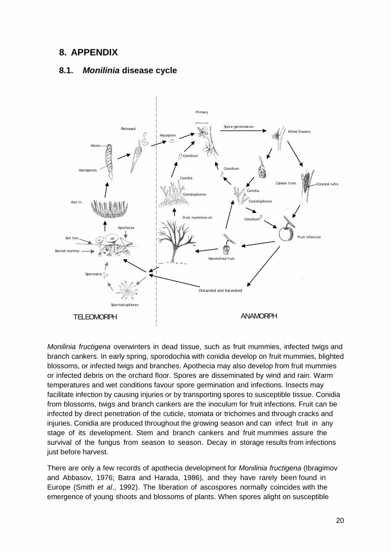
8.1. Monilinia disease cycle
Monilinia fructigena overwinters in dead tissue, such as fruit mummies, infected twigs and branch cankers. In early spring, sporodochia with conidia develop on fruit mummies, blighted blossoms, or infected twigs and branches. Apothecia may also develop from fruit mummies or infected debris on the orchard floor. Spores are disseminated by wind and rain. Warm temperatures and wet conditions favour spore germination and infections. Insects may facilitate infection by causing injuries or by transporting spores to susceptible tissue. Conidia from blossoms, twigs and branch cankers are the inoculum for fruit infections. Fruit can be infected by direct penetration of the cuticle, stomata or trichomes and through cracks and injuries. Conidia are produced throughout the growing season and can infect fruit in any stage of its development. Stem and branch cankers and fruit mummies assure the survival of the fungus from season to season. Decay in storage results from infections just before harvest.
There are only a few records of apothecia development for Monilinia fructigena (Ibragimov and Abbasov, 1976; Batra and Harada, 1986), and they have rarely been found in Europe (Smith et al., 1992). The liberation of ascospores normally coincides with the emergence of young shoots and blossoms of plants. When spores alight on susceptible
Discarded and harvested
Fruit infection
Canker from
Conidial tufts
Killed flowers Spore germination
Primary
blossom
Conidium
Ascospore
Released
Ascus
Ascospores
Asci in
Apothecia
Soil line
Buried mummy
Spermatia
Spermatiophores
Fruit mummies on
Mummified fruit
Conidium
Conidiophores
Conidia
Conidium
Conidiophores
Conidia
TELEOMORPH ANAMORPH
20

tissues under favourable environmental conditions infections are initiated. Microconidia are also produced in abundance within small cavities and on the surfaces of mummified fruit. Monilinia fructigena is a pathogen favoured by rain, fog and other factors that increase relative humidity especially at the beginning of the host growth period. Conidia are generally formed on mummified fruit and blighted twigs at temperatures of >5°C. Germination and germ tube growth are partially inhibited by light, but sporulation is enhanced. Conidia provide the inocula for most primary infections and require free moisture for germination. At 20°C, a period of about 12 h after soaking is required for sporulation to take place; maximum sporulation occurs between 36 and 48 h. The minimum moisture content of mummified fruit in which sporulation can take place at 26°C is 21% (Jenkins and Reinganum, 1968). Inoculum concentration also interacts with temperature and humidity to influence the incubation period, disease incidence, and severity.
Fruit are usually infected through wounds, although healthy fruit can be infected by the growth of mycelium from any diseased fruit with which they are in contact. At harvest, apparently healthy fruit can be contaminated with spores, and decay may then occur during storage and marketing. Latent infections may occur when the fruit is green. In such fruit, the fungus remains quiescent until the fruit starts to ripen (Byrde and Willetts, 1977).
21
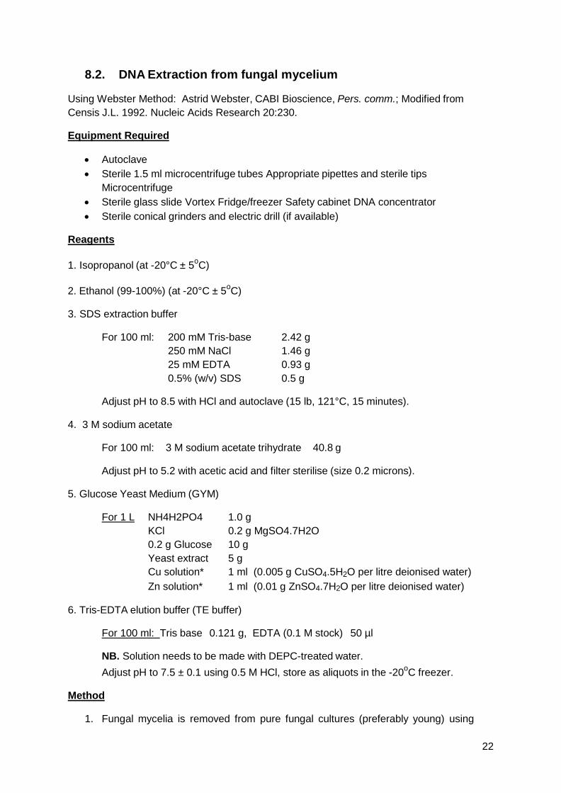
8.2. DNA Extraction from fungal mycelium
Using Webster Method: Astrid Webster, CABI Bioscience, Pers. comm.; Modified from Censis J.L. 1992. Nucleic Acids Research 20:230.
Equipment Required
• Autoclave • Sterile 1.5 ml microcentrifuge tubes Appropriate pipettes and sterile tips
Microcentrifuge • Sterile glass slide Vortex Fridge/freezer Safety cabinet DNA concentrator • Sterile conical grinders and electric drill (if available)
Reagents
1. Isopropanol (at -20°C ± 5oC)
2. Ethanol (99-100%) (at -20°C ± 5oC)
3. SDS extraction buffer
For 100 ml: 200 mM Tris-base 2.42 g 250 mM NaCl 1.46 g 25 mM EDTA 0.93 g 0.5% (w/v) SDS 0.5 g
Adjust pH to 8.5 with HCl and autoclave (15 lb, 121°C, 15 minutes).
4. 3 M sodium acetate
For 100 ml: 3 M sodium acetate trihydrate 40.8 g
Adjust pH to 5.2 with acetic acid and filter sterilise (size 0.2 microns).
5. Glucose Yeast Medium (GYM)
For 1 L NH4H2PO4 1.0 g KCl 0.2 g MgSO4.7H2O 0.2 g Glucose 10 g Yeast extract 5 g Cu solution* 1 ml (0.005 g CuSO4.5H2O per litre deionised water) Zn solution* 1 ml (0.01 g ZnSO4.7H2O per litre deionised water)
6. Tris-EDTA elution buffer (TE buffer)
For 100 ml: Tris base 0.121 g, EDTA (0.1 M stock) 50 µl
NB. Solution needs to be made with DEPC-treated water. Adjust pH to 7.5 ± 0.1 using 0.5 M HCl, store as aliquots in the -20oC freezer.
Method
1. Fungal mycelia is removed from pure fungal cultures (preferably young) using
22
sterile glass slide or from glucose yeast medium and placed in 1.5 ml microcentrifuge tube.
2. Add 300 µl of SDS extraction buffer.
3. Grind the mycelium using a sterile conical grinder (with electric drill if available), for 3 minutes.
4. Add 150 µl of 3 M sodium acetate (pH 5.2) and vortex.
5. Incubate at -20°C ± 5oC for at least 30 minutes.
6. Grind again as per step 3 and incubate at -20 °C ± 5oC for 10 minutes.
7. Centrifuge at 10,000 rpm for 5 minutes.
8. Transfer the supernant using wide-bore pipette tip.
9. Add an equal volume of cold (-20 °C ± 5oC) isopropanol.
10. Incubate at -20°C ± 5oC for at least 30 minutes, or preferably overnight.
11. Centrifuge at 10,000 rpm for 10 minutes.
12. Carefully pipette off the isopropanol without disturbing the DNA pellet.
13. Add 50-80 µl of cold (-20 °C ± 5oC) 70% (v/v) ethanol and vortex.
14. Centrifuge at 10,000 rpm for 5 minutes.
15. Air-dry the pellet for 1-2 hours or use the DNA concentrator.
16. Resuspend the DNA in 100 µl of TE buffer or sterile deionised water
17. Store at -20°C ± 5oC for short term storage or at -80°C ± 5oC for longer term storage.
23
8.3. PCR primers for M. fructicola and M. laxa
Species Primer Sequence
M. fructicola Forward: MFC-F1 5’-TAT GCT CGC CAG AGG ATA ATT A-3’
Reverse: MFC-R1 5’-GAT TTT AGA GCC TGC CAT TS-3’
M. laxa Forward: ML-MFG-F2 5’-GCT CGC CAG AGA ATA ATC-3’
Reverse: ML-MFC-R1 5’-GAT TTT AGA GCC TGC CAT TG-3’
8.4. Direct PCR from infected host tissues
This protocol can be used to obtain a fast but tentative M. fructigena result on potentially infected fruit or other plant material showing limited symptoms. A positive PCR should however be backed up by the isolation of the fungus.
A range of isolates were used to inoculate fruit (Table 4). M. fructigena fruit inoculations and DNA extraction were conducted at the Central Science Laboratories (CSL) (York, UK) using a NucleoSpin® Plant Kit (Clontech). M. fructicola and M. laxa fruit inoculations and DNA extraction using the Qiagen DNeasy® Plant Mini Kit were conducted at MAF Plant Health and Environment Laboratory (PHEL), Auckland.
Table 4: Monilinia species tested and their origins.
Species Isolate numbera Origin (host, country) Monilinia fructigena
CSL 1335* 1336* CSL 1337* CSL 1340* ATCC 24976
Malus pumila, UK CSL Cydonia sp., Portugal Malus pumila, Japan Malus pumila, UK Prunus persica, Holland Monilinia fructicola
ICMP 7639 ICMP 15067 CSL 1326*
Prunus armeniaca, NZ Prunus persica, NZ Malus pumila, Japan
Monilina laxa
ICMP 5475 ICMP8348 CSL 1333*
Prunus glandulosa, NZ Prunus armeniaca, NZ Prunus armeniaca, Australia
a ICMP isolates are maintained by International Collection of Micro-organisms from Plants, Landcare Research, Auckland, New Zealand. ATTC = American Type Culture Collection. * Extracted DNA (1-10 dilution) provided by the Central Science Laboratory, UK either from cultures or infected fruit.
24
8.4.1. Results: Testing Monilinia infected fruit by direct PCR
Results of the PCR from inoculated fruit are presented in Figure 13. The two DNA extraction kits (NucleoSpin® Plant Kit (Clontech) and Qiagen DNeasy® Plant Mini Kit) yielded acceptable quantities of genomic DNA.
The M. fructigena specific primers worked well alone. All isolates of M. fructigena tested were detected and there was no cross reaction with the other two Monilinia species. The addition of general plant primers as an internal control made the reaction inconsistent and unreliable. These primers competed with the M. fructigena primers creating false negative reactions. It was therefore decided not to include an internal control at this stage. Their inclusion may be a potential improvement for the method in subsequent revisions of this protocol.
The specific primer set ITS 1 and Mfg-R2 does not distinguish between M. fructigena and the Japanese fungus M. polystroma. A newly designed set of primers able to separate these two fungi needs evaluation and will be incorporated into this protocol with the next revision (Côté et al., 2004). However, this fungus is also not present in Australia or New Zealand but would be picked up with the M. fructigena primers.
25
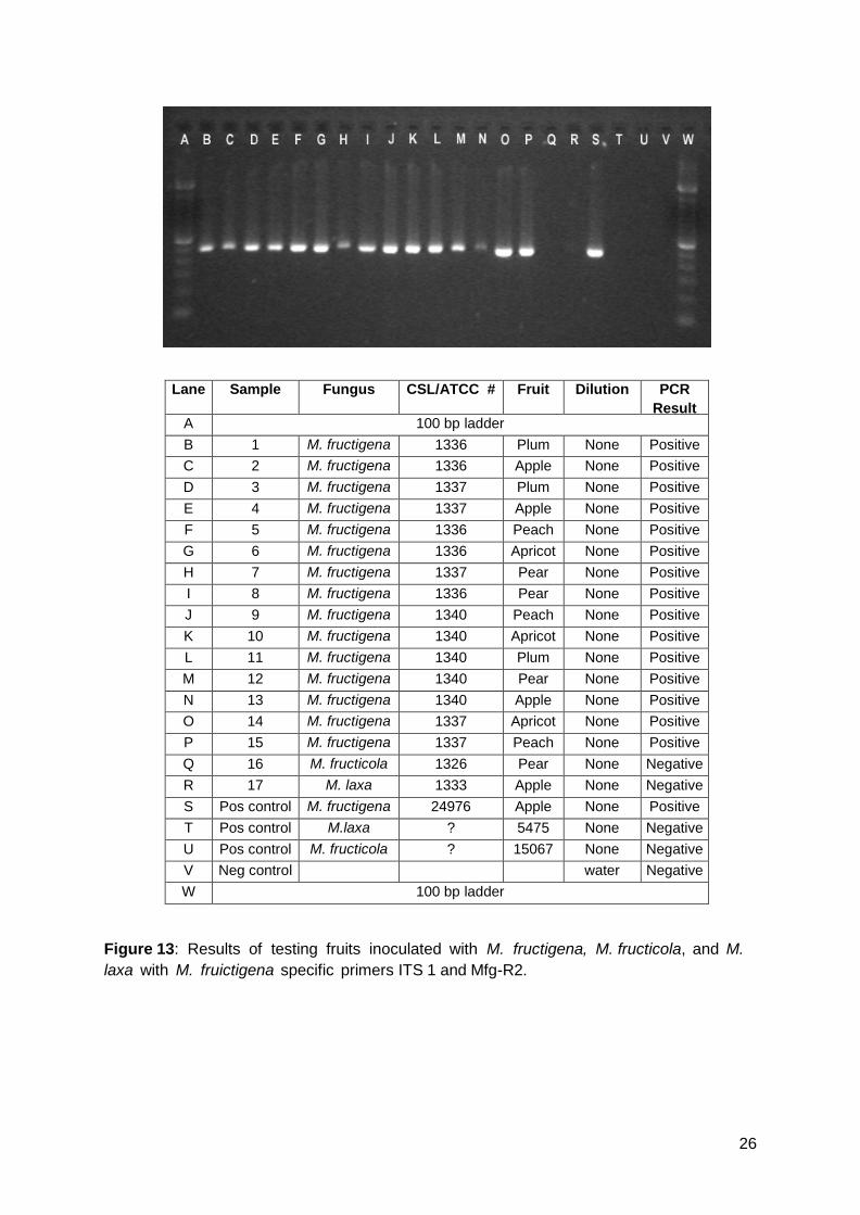
Lane Sample Fungus CSL/ATCC # Fruit Dilution PCR Result
A 100 bp ladder B 1 M. fructigena 1336 Plum None Positive C 2 M. fructigena 1336 Apple None Positive D 3 M. fructigena 1337 Plum None Positive E 4 M. fructigena 1337 Apple None Positive F 5 M. fructigena 1336 Peach None Positive G 6 M. fructigena 1336 Apricot None Positive H 7 M. fructigena 1337 Pear None Positive I 8 M. fructigena 1336 Pear None Positive J 9 M. fructigena 1340 Peach None Positive K 10 M. fructigena 1340 Apricot None Positive L 11 M. fructigena 1340 Plum None Positive M 12 M. fructigena 1340 Pear None Positive N 13 M. fructigena 1340 Apple None Positive O 14 M. fructigena 1337 Apricot None Positive P 15 M. fructigena 1337 Peach None Positive Q 16 M. fructicola 1326 Pear None Negative R 17 M. laxa 1333 Apple None Negative S Pos control M. fructigena 24976 Apple None Positive T Pos control M.laxa ? 5475 None Negative U Pos control M. fructicola ? 15067 None Negative V Neg control water Negative W 100 bp ladder
Figure 13: Results of testing fruits inoculated with M. fructigena, M. fructicola, and M. laxa with M. fruictigena specific primers ITS 1 and Mfg-R2.
26
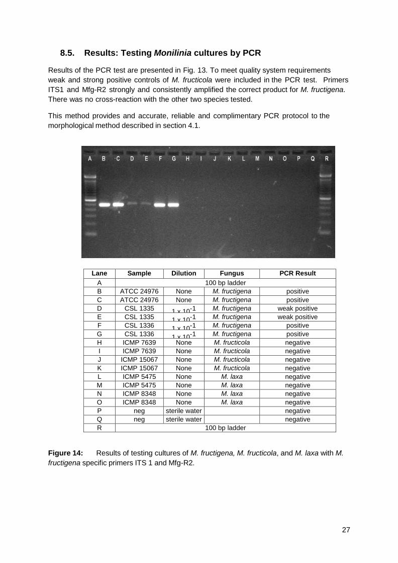
8.5. Results: Testing Monilinia cultures by PCR
Results of the PCR test are presented in Fig. 13. To meet quality system requirements weak and strong positive controls of M. fructicola were included in the PCR test. Primers ITS1 and Mfg-R2 strongly and consistently amplified the correct product for M. fructigena. There was no cross-reaction with the other two species tested.
This method provides and accurate, reliable and complimentary PCR protocol to the morphological method described in section 4.1.
Lane Sample Dilution Fungus PCR Result A 100 bp ladder B ATCC 24976 None M. fructigena positive C ATCC 24976 None M. fructigena positive D CSL 1335 1 x 10⁻¹ M. fructigena weak positive E CSL 1335 1 x 10⁻¹ M. fructigena weak positive F CSL 1336 1 x 10⁻¹ M. fructigena positive G CSL 1336 1 x 10⁻¹ M. fructigena positive H ICMP 7639 None M. fructicola negative I ICMP 7639 None M. fructicola negative J ICMP 15067 None M. fructicola negative K ICMP 15067 None M. fructicola negative L ICMP 5475 None M. laxa negative M ICMP 5475 None M. laxa negative N ICMP 8348 None M. laxa negative O ICMP 8348 None M. laxa negative P neg sterile water negative Q neg sterile water negative R 100 bp ladder
Figure 14: Results of testing cultures of M. fructigena, M. fructicola, and M. laxa with M. fructigena specific primers ITS 1 and Mfg-R2.
27