diana c. g. a. pinto, joão m. p. pereira, artur m. s
TRANSCRIPT

Diana C. G. A. Pinto, João M. P. Pereira, Artur M. S. Silva*11.1 Functional Groups of Biomolecules and their ReactionsAbstract: This chapter starts with a general introduction on some concepts needed to understand the reactivity of organic functional groups. Elementary reaction mecha-nisms are then presented according to their functionality with relevant biological examples. These reactions explain the vast majority of transformations involving bio-molecules. Finally, two examples on the application of the presented concepts are given, namely the metabolism of fatty acids and reactivity of penicillin. Both of these examples call for various types of reactions showing the diversity and simplicity of biological transformations when analysed step by step.
1.1.1 Functional Groups in Biological Systems
The main definition of a functional group in organic chemistry books is as a chemically reactive group of atoms within a molecule that contribute to its characteristic reactivity. Functionality is usually regarded as “implying the presence of heteroatoms and/or unsaturation, but it would not be helpful to attempt to define precisely the limits of application of the term” (IUPAC, Commission on Nomenclature of Organic Chemistry, 1993).
Functional group reactivity may be changed by the presence of other neighbouring functional groups but usually behaves uniformly in every molecule where it can be found. There are several common functional groups that are related to families of organic compounds according to their structural features. However, from those functional groups only a few are found in biological systems (Table 1.1.1). The types of bonding found in these functional groups may be explained by the existence of various hybrid atomic orbitals of the carbon atom created from combination of the one 2s and the three 2p orbitals (Table 1.1.2).
Diana C. G. A. Pinto, João M. P. Pereira, Artur M. S. Silva: Departamento de Química da Universidade de Aveiro, 3810-193 Aveiro, Portugal, *E-mail: [email protected]

Functional Groups in Biological Systems 3
Table 1.1.1: Common functional groups present in biomolecules. In parentheses are the names of the families of compounds, where the group has the highest priority in the compound.
C C
C=C double bond (alkenes)
aromatic ring (arenes)
COH
hydroxyl (alcohols)
CO
C
R-oxyalkyl (ethers)N
C
amino (amines)
CSH
sulfhydryl (thiols/mercaptanes)
CS
C
sulfide (thioethers/sulfides)
SC
CS
disulfide (disulfides)
CH
O
formyl (aldehydes)
C
O
C C
dialkylcarbonyl (ketones)
CC
C
N
imino (imines)
C
O
C OH
carboxyl (carboxylic acids)
C
O
C
C
O
R-oxycarbonyl (ester)
C
O
C N
aminocarbonyl/carboxamide(amides)
C
O
C
C
S
sulfanyl carbonyl (thioesters)
O
C P
O
O
O
alkyl phosphate (phosphates)
OC P
O
OO
C
O
acyl phosphate (mixed anhydrides)
4 Functional Groups of Biomolecules and their Reactions
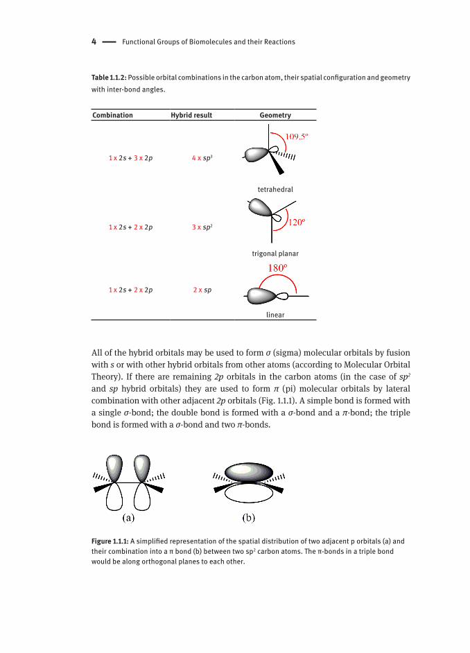
Table 1.1.2: Possible orbital combinations in the carbon atom, their spatial configuration and geometry with inter-bond angles.
Combination Hybrid result Geometry
1 x 2s + 3 x 2p 4 x sp3
tetrahedral
1 x 2s + 2 x 2p 3 x sp2
trigonal planar
1 x 2s + 2 x 2p 2 x sp
linear
All of the hybrid orbitals may be used to form σ (sigma) molecular orbitals by fusion with s or with other hybrid orbitals from other atoms (according to Molecular Orbital Theory). If there are remaining 2p orbitals in the carbon atoms (in the case of sp2 and sp hybrid orbitals) they are used to form π (pi) molecular orbitals by lateral combination with other adjacent 2p orbitals (Fig. 1.1.1). A simple bond is formed with a single σ-bond; the double bond is formed with a σ-bond and a π-bond; the triple bond is formed with a σ-bond and two π-bonds.
Figure 1.1.1: A simplified representation of the spatial distribution of two adjacent p orbitals (a) and their combination into a π bond (b) between two sp2 carbon atoms. The π-bonds in a triple bond would be along orthogonal planes to each other.

Acids and Bases Versus Electrophiles and Nucleophiles 5
These hybridisations have several consequences, such as the electron density of a π-bond lying above and below the plane of the bonding atoms (Fig. 1.1.1), resulting in greater exposure for a reaction. Simultaneously, with the increasing s character of the hybrid orbital:
the formed bond length decreases; –the polarity of a C-H bond increases; –breaking a bond between carbon and a more electronegative atom is more dif- –ficult (e.g. the C-O bond in isopropanol is easier to cleave than the C-O bond in isopropenol).
The electronegativity of an element can also be an important factor to explain some functional groups reactivity. For instance, alcohols, ethers, amines, thiols, sulfides, disulfides and phosphates (Table 1.1.1) all have a carbon forming a single bond with a more electronegative atom, causing the carbon to bear a partial positive charge (δ+). These modifications affect both the σ- and π-bonds, although in the case of some π-bonds the resonance effect should also be considered. The carbonyl group can be classically treated as a resonance hybrid represented by two resonance structures (Scheme 1.1.1), which contributes to the reactivity of the compounds that have this functional group (aldehydes, ketones, carboxylic acids, esters, thioesters, amides, acyl phosphates). Resonance is possible whenever movement of electrons are allowed within the same molecule without movement of atoms.
C
O
C
O
C
O
δ+
δ−
or
Scheme 1.1.1: Carbonyl group resonance structures and equivalent notation, explicitly showing the bond polarity with partial charges.
1.1.2 Acids and Bases Versus Electrophiles and Nucleophiles
Acids and bases are very important in biological transformations, as most require some form of acid or basic catalysis to occur. The simplest acid-base theory is the Brønsted-Lowry theory, which states that acids are molecules that donate protons (hydrogen ions, H+) and bases/alkalis are molecules that accept protons. For example, a carboxylic acid can donate a proton to a base, such as an amine, in a reversible proton-transfer reaction (Scheme 1.1.2).

6 Functional Groups of Biomolecules and their Reactions
R OH
O
R O
O
+ +R1
NH
H
R1N
H
H
acid base conjugate acidconjugate base
H
Scheme 1.1.2: Example of a proton transfer reaction. This specific reaction explains why it is difficult for condensations to happen directly between an amine and a carboxylic acid, as the non-ionic forms of these molecules are more reactive (Chapter 1.1.4.5).
Acids can differ in their ability to donate protons, being classified as strong or weak according to the extent of deprotonation. A strong acid will have a stable conjugate base (or weak conjugate base), resulting in ready donation of a proton. Table 1.1.3 lists the acidity of some typical functional groups (water and ammonium acidity are also given for comparison). The acidity is measured by the acidity constant, Ka or by its pKa (Scheme 1.1.3), where a stronger acid has a smaller pKa and a weaker acid has a larger pKa. The same approach can be applied to bases and their strength.
+ +HA H2O A- H3O+
Scheme 1.1.3: Acidity constant and pKa.
The problem with the Brönsted/Lowry definition is that it only covers the compounds that donate or accept protons. The more general and widely used model is the Lewis definition. A Lewis acid is a molecule that accepts a pair of electrons and a Lewis base is a molecule that donates a pair of electrons. To accept electrons, a Lewis acid must have a vacant low-energy orbital. As a consequence, many species, including H+ itself, metal cations such as Mg2+ and Zn2+, and neutral species such as boron trifluoride (BF3) and carbon dioxide (CO2) are Lewis acids.
Lewis acids and bases are involved in many biological reactions, such as the transformation of carbon dioxide into hydrogen carbonate (Scheme 1.1.4). Lewis bases use unshared electrons to form new bonds with other atoms and are usually referred as nucleophiles (“nucleus-loving”, versus Lewis acids as electrophiles, “electron-loving”). The terms “electrophile” and “nucleophile” are commonly used

Acids and Bases Versus Electrophiles and Nucleophiles 7
in organic and bioorganic transformations. Electrophiles are either positively charged or neutral and have at least one positively polarised, electron-poor atom. Conversely, nucleophiles are either negatively charged or neutral and have a lone pair of electrons that can be donated.
Table 1.1.3: Relative acidity strengths of some functional groups and common reactants.
Functional group Example pKa
carboxylic acid
CH3COH
O 4.76
ammonium NH4+ 9.26
alkylthiol CH3SH 10.3
alkylammonium ion CH3NH3+ 10.66
β-keto ester
CH3CCH2COCH3
O O 10.6
water H2O 15.74
alcohol CH3CH2OH 16.0
ketone
CH3CCH3
O 19.3
thioester
CH3CSCH3
O 21
ester
CH3COCH3
O 25
HOHO
CO
O
+
Lewis acidLewis base
carbonicanhydrase
O
C
O
δ+
δ−
δ−
Scheme 1.1.4: Example of a biochemical reaction involving a Lewis acid and a Lewis base. Reversible conversion of carbon dioxide to hydrogen carbonate can occur rapidly in the active site of a carbonic anhydrase, the equilibrium being regulated by factors such as pH.
8 Functional Groups of Biomolecules and their Reactions
1.1.3 Stereoisomerism and Chirality
Stereoisomers having the spatial orientation of bonds as their unique difference, maintaining the same composition of atoms and bonds.
1.1.3.1 Cis/trans Isomerism
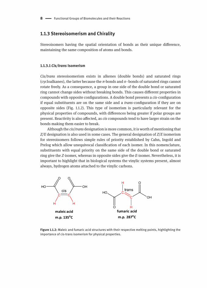
Cis/trans stereoisomerism exists in alkenes (double bonds) and saturated rings (cycloalkanes), the latter because the π-bonds and σ–bonds of saturated rings cannot rotate freely. As a consequence, a group in one side of the double bond or saturated ring cannot change sides without breaking bonds. This causes different properties in compounds with opposite configurations. A double bond presents a cis-configuration if equal substituents are on the same side and a trans-configuration if they are on opposite sides (Fig. 1.1.2). This type of isomerism is particularly relevant for the physical properties of compounds, with differences being greater if polar groups are present. Reactivity is also affected, as cis compounds tend to have larger strain on the bonds making them easier to break.
Although the cis/trans designation is more common, it is worth of mentioning that Z/E designation is also used in some cases. The general designation of Z/E isomerism for stereoisomers follows simple rules of priority established by Cahn, Ingold and Prelog which allow unequivocal classification of each isomer. In this nomenclature, substituents with equal priority on the same side of the double bond or saturated ring give the Z-isomer, whereas in opposite sides give the E-isomer. Nevertheless, it is important to highlight that in biological systems the vinylic systems present, almost always, hydrogen atoms attached to the vinylic carbons.
CC
O
OH
O
HO
CC
O
HO
O
OH
H HH
H
fumaric acidm.p. 287oC
maleic acidm.p. 135oC
cis trans
Figure 1.1.2: Maleic and fumaric acid structures with their respective melting points, highlighting the importance of cis-trans isomerism for physical properties.
Stereoisomerism and Chirality 9
1.1.3.2 Chirality and Enantiomerism
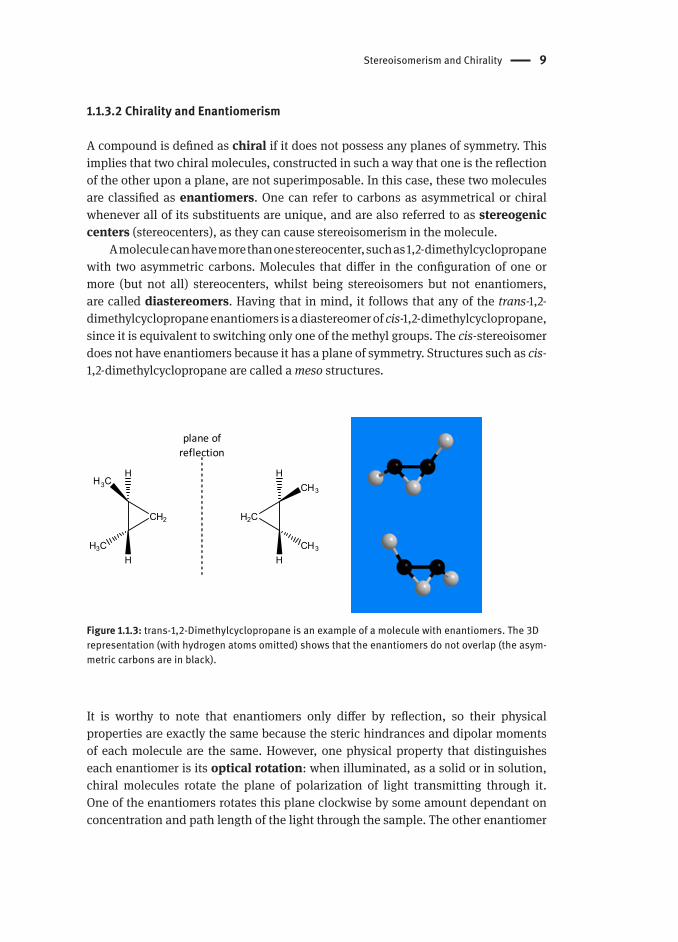
A compound is defined as chiral if it does not possess any planes of symmetry. This implies that two chiral molecules, constructed in such a way that one is the reflection of the other upon a plane, are not superimposable. In this case, these two molecules are classified as enantiomers. One can refer to carbons as asymmetrical or chiral whenever all of its substituents are unique, and are also referred to as stereogenic centers (stereocenters), as they can cause stereoisomerism in the molecule.
A molecule can have more than one stereocenter, such as 1,2-dimethylcyclopropane with two asymmetric carbons. Molecules that differ in the configuration of one or more (but not all) stereocenters, whilst being stereoisomers but not enantiomers, are called diastereomers. Having that in mind, it follows that any of the trans-1,2-dimethylcyclopropane enantiomers is a diastereomer of cis-1,2-dimethylcyclopropane, since it is equivalent to switching only one of the methyl groups. The cis-stereoisomer does not have enantiomers because it has a plane of symmetry. Structures such as cis-1,2-dimethylcyclopropane are called a meso structures.
CH2
H3C
H3C
H2C
CH3
CH3
plane of reflection
H
H
H
H
Figure 1.1.3: trans-1,2-Dimethylcyclopropane is an example of a molecule with enantiomers. The 3D representation (with hydrogen atoms omitted) shows that the enantiomers do not overlap (the asym-metric carbons are in black).
It is worthy to note that enantiomers only differ by reflection, so their physical properties are exactly the same because the steric hindrances and dipolar moments of each molecule are the same. However, one physical property that distinguishes each enantiomer is its optical rotation: when illuminated, as a solid or in solution, chiral molecules rotate the plane of polarization of light transmitting through it. One of the enantiomers rotates this plane clockwise by some amount dependant on concentration and path length of the light through the sample. The other enantiomer

10 Functional Groups of Biomolecules and their Reactions
will rotate light by the same amount but anticlockwise. Racemic mixtures (racemates) are mixtures with same amount of both enantiomers of a compound and have a null optical rotation.
Biological systems are highly stereospecific ‒ in general, only one stereoisomer is reactive towards an enzyme or a certain receptor. As a result, racemate resolution is of extreme importance for drug synthesis, particularly when one enantiomers has a negative effect. This problem is often solved by two different approaches:
Enzymatic resolution: the racemate is transformed in such a way that the 1. resulting product may be then cleaved by an enzyme (e.g. lyase). Because of the stereospecificity of the enzyme, only one of the compounds is cleaved, enabling separation of cleaved and uncleaved product.Diastereomeric resolution: the racemate is made to react with a specific, 2. enantiomerically pure reagent (e.g. L-tartaric acid, to form an ester). The obtained products are diastereomers, which unlike enantiomers possess different properties and are therefore separable. After separation, the reverse reaction is performed to return the original enantiomers.
1.1.4 Common Mechanisms in Biological Chemistry
Reactions that occur in living organisms follow the same rules of those occurring in the laboratory. The solvent, temperature and almost certainly the catalyst can be different, but the fundamental reaction mechanisms are the same. So conveniently, common organic reaction mechanisms can be used to understand the equivalent biological transformations.
1.1.4.1 Nucleophilic Substitution Reactions
Nucleophilic substitution reactions occur when a group attached to an sp3 carbon is substituted for a more nucleophilic one. These reactions may follow two similar mechanisms ‒ bimolecular and unimolecular ‒ but with very different implications for biological systems in terms of stereochemistry.
1.1.4.1.1 SN2 – Bimolecular Nucleophilic Substitution
This type of mechanism (Scheme 1.1.5) is called a bimolecular nucleophilic substitution (SN2) since the determining step involves the reaction of two species, the nucleophile and the substrate (electrophile species).

Common Mechanisms in Biological Chemistry 11
Scheme 1.1.5: General reaction for an SN2 reaction mechanism.
The following happens in one concerted step: a nucleophile (N) attacks the carbon atom as a more electronegative group (X) leaves, with inversion of stereochemistry through an unstable transition state. Note that N can be a neutral protic nucleophile that deprotonates after the substitution. This type of mechanism is typical of primary alkyl halides substitutions. One reaction often performed in a laboratory is an O-methylation using methyl iodide. The oxygen atom (of an alcohol, for example) acts as nucleophile substituting the iodide which is a very good leaving group.
1.1.4.1.2 SN1 ‒ Unimolecular Nucleophilic Substitution ReactionsUnimolecular nucleophilic substitutions (SN1) occur when a carbocation intermediate is stable enough to be transiently formed. In this case, the rate determining step involves reaction of only one species: the substrate where the substitution will take place (Scheme 1.1.6).
Scheme 1.1.6: General reaction for a SN1 reaction mechanism.
A carbocation may be easily formed on tertiary carbons because the carbocation is stabilized through inductive effect by vicinal carbons (R = alkyl or aryl groups). With this in mind, the more electronegative moiety is able to heterolytically cleave its bond to the carbon atom. Since the carbocation is planar, there is no preference for the nucleophile on which side to attack, resulting in a mixture of enantiomers if the product in question in chiral. In enzymes, this does not happen because the active sites are chiral themselves, restricting addition to only one side. This type of mechanism is typical of tertiary alkyl halides substitutions and allylic phosphates. For

12 Functional Groups of Biomolecules and their Reactions
example, geranyl diphosphate cleaves at the C-O bond and the corresponding allylic carbocation, well stabilized by resonance, is attacked by water which deprotonates to produce geraniol (Scheme 1.1.7).
Scheme 1.1.7: SN1 reaction mechanism of geranyl diphosphate forming geraniol.
1.1.4.1.3 Phosphate Group Transfer – the Grey Area of Nucleophilic Substitutions in Biological SystemsNucleophilic substitutions are not restrained to carbon atoms. Phosphate and acyl group transfer reactions, key pieces in metabolic pathways, are also nucleophilic substitution reactions although with some differences. In the phosphorylation of glucose a phosphate group is transferred from ATP to glucose with a phosphorus atom undergoing a nucleophilic substitution (Scheme 1.1.8).
Scheme 1.1.8: Glucose phosphorylation.

Common Mechanisms in Biological Chemistry 13
In this reaction, the phosphorus electrophilicity is reinforced trough chelation with two Mg2+ ions present in the phosphoryl transferase enzyme. In the complex, the most representative resonance structure places a positive formal charge in the P atom and negative charges on the O atoms. This makes the tetrahedral phosphate easy to be attacked by the oxygen atom from the 6-hydroxyl group in glucose, while a base captures the released proton. The mechanism shown in Scheme 1.1.8 is a simplified version of what has been observed. The preferred reaction path is not well defined in biological systems, since it is highly dependent on the nature of the nucleophile and the enzyme scaffold. Intermediates may or may not be involved. Either a pentavalent trigonal bipyramidal (associative intermediate) or a metaphosphate (dissociative intermediate) intermediate may be formed (Scheme 1.1.9). A concerted reaction mechanism has also been observed, where the substitution is done smoothly in one step (i.e. SN2 like).
R P
-O O-
O-
R 1P
O O-
O
a b
Scheme 1.1.9: a) Phosphorane intermediate ‒ analogue of the activated complex ‒ in red are the axial positions (collinear) and in blue are the equatorial positions (coplanar); b) metaphosphate (planar, stabilised by resonance) ‒ analogue of a carbocation.
1.1.4.2 Electrophilic Addition Reactions
Electrophilic reagents can react with compounds that are electron rich in certain exposed regions, from which alkenes are a typical example. In these systems, the π bond results from overlapping of p orbitals and provides regions of increased electron density above and below the plane of the molecule. π electrons are more loosely bound than those of a σ-bond so they can interact more easily with a positively charged electrophilic species, forming a new σ-bond and a carbocation (Scheme 1.1.10). This in turn rapidly reacts with a nucleophile to form another σ-bond. In this case, the nucleophile is either an anion or a neutral moiety with free pairs of electron that will become neutral again eliminating a group or an atom.

14 Functional Groups of Biomolecules and their Reactions
C CE
C CENu
C CE Nu
Scheme 1.1.10: Electrophilic addition reaction mechanism.
1.1.4.2.1 Synthesis of α-Terpineol ‒ Intramolecular AdditionIn the following example we can see the simplified (without enzyme interactions) biosynthetic mechanism of α-terpineol from linalyl diphosphate, which occurs through an electrophilic addition (Scheme 1.1.11).
Scheme 1.1.11: Synthesis of α-terpineol from linalyl diphosphate.
It should be noted that in this intramolecular addition, the electrophile is generated via diphosphate ion (PPO-) elimination. The diphosphate ion leaves easily because it is itself a stable anion and the generated carbocation is also stable, as it is an allylic carbocation stabilized by resonance. The delocalized positive charge turns the terminal carbon into a strong electrophile capable of adding to the double bond; the deficiency of electrons in the electrophile does not always coincide with the atom that will be attacked. Biological examples of electrophilic reactions occur frequently in metabolic processes, such as in the β-oxidation pathway of the fatty acid metabolism (Chapter 1.1.5).

Common Mechanisms in Biological Chemistry 15
1.1.4.3 Aromatic Substitutions
In simple terms, aromaticity may be described as a chemical property that arises from delocalization of electrons in a ring. These electrons may be provided by conjugated unsaturation, lone pair electrons or even orbitals; the system becomes aromatic when agreeing with the Huckel rule, which are state that the number of the total conjugated electrons must be 4n+2 electrons. It is a particularly strong form of resonance stabilization making aromatic compounds more stable than expected otherwise. The orbital alignment required for aromatic stabilization turns the aromatic (aryl) moieties planar (Figs. 1.1.4 and 1.1.5). Because of the intermediate character of its bonds (Fig. 1.1.4) the reactivity of aromatic compounds is not identical to the reactivity of other unsaturated compounds.
Figure 1.1.4: Clarification of the aromatic stabilization of benzene. Hypothetically, cyclohexatriene would have two different types of bonds: simple and double bonds, but spectroscopic data show that all bonds are equivalent. The bonds in benzene have an intermediate character between a simple and a double bond with the electrons delocalized (density evenly distributed).

16 Functional Groups of Biomolecules and their Reactions
Figure 1.1.5: Cytosine is an example of a biomolecule exhibiting aromaticity in its imidic acid form. Furan is an example of a heteroaromatic compound where a lone pair of electrons of the heteroatom is delocalized into the ring to allow aromaticity. The Fig. explicitly shows all the resonance forms in the rings of the compounds.
1.1.4.3.1 Electrophilic Aromatic SubstitutionA strong electrophile may capture electrons from the ring, forming a very unstable intermediate. Since the loss of aromaticity is energetically unfavourable, a nucleophile does not add to the cation. Instead, the ring eliminates a proton (Scheme 1.1.12).
Scheme 1.1.12: Mechanism of an electrophilic aromatic substitution reaction.
The reactivity of the aromatic ring is enhanced if electron donating (hydroxyl, amino and alkoxyl) groups are attached to the ring, increasing the electron density and making the attack to the electrophile easier.

Common Mechanisms in Biological Chemistry 17
1.1.4.3.2 Nucleophilic Aromatic SubstitutionsNucleophilic aromatic substitutions are also possible when electron withdrawing groups are attached to the ring (e.g. nitro group and carbonyl moieties) allowing it to accommodate a carbanion, even though aromatic rings are already dense in electrons. The leaving group also needs to be quite electronegative to leave easily and drive the reaction forward (Scheme 1.1.13). The exact mechanism varies dependently on the leaving group having some parallels to normal nucleophilic substitution.
Scheme 1.1.13: General reaction of a nucleophilic aromatic substitution.
1.4.3.3 Hallucinogen Synthesis ‒ Aromatic Substitution on FungiErgot fungi produce a variety of alkaloids often with strong hallucinogenic effects upon consumption. The first pathway-specific step in their synthesis is the alkylation of tryptophan by dimethylallyl diphosphate obtaining the dimethylallyl tryptophan (DMAT) (Scheme 1.1.14). This step consists of an electrophilic aromatic substitution, catalysed by DMAT synthase.
Scheme 1.1.14: Reaction mechanism for the synthesis of DMAT from tryptophan and dimethylallyl phosphate.

18 Functional Groups of Biomolecules and their Reactions
1.4.4 Eliminations Reactions
An elimination reaction is a type of reaction upon which there is a net elimination of a molecule from another. To clarify this point, look at the example shown in Scheme 1.1.15.
Scheme 1.1.15: Dehydrogenation of ethane to form ethene through two different mechanisms: proton-hydride transfer and a radical mechanism, both leading to the elimination of what is equi-valent to a hydrogen molecule (the arrows without origin represent electron transfers to/from other molecules).
A dehydrogenation reaction involves the net elimination of a hydrogen molecule, but does not necessarily release a hydrogen molecule. It may proceed through abstraction of a proton connected to one of the carbons followed by transfer of a hydride from the other. It may also follow a radical mechanism by hydrogen atom abstraction from both carbons (Scheme 1.1.15). In a dehydrogenation, the elimination is oxidative because the oxidation state of each carbon atoms goes from -3 to -2, a net molecular change of +2 (Chapter 1.1.4.8).
In mechanistic terms, non-oxidative eliminations may occur in various steps having its variations similarly to the nucleophilic substitutions. In all of the following cases, the reactions shown are [1,2]-eliminations, meaning that the reaction implicates electron movement between two vicinal carbons. Other types of eliminations may occur involving more distant carbon atoms and heteroatoms ‒ [1,3] in decarboxylations or [1,4] in aldol condensations (Chapter 1.1.4.6).
1.1.4.4.1 E1 ‒ Unimolecular EliminationAn E1 reaction mechanism occurs when a relatively stable carbocation may be formed by elimination of a stable anion (X-), such as a chloride ion (Cl-) for a good leaving group. The only rate determining (slow) step is the dissociation of the leaving group to form a carbocation (hence a unimolecular reaction). A base (B) captures the proton released from one carbon away of the carbocation formed in the E1 reaction mechanism. It is worthy to note that the E1 mechanism competes with the SN1 mechanism since the nucleophile (B) may react directly at the halogenated carbon (substitution) or at the

Common Mechanisms in Biological Chemistry 19
neighbouring hydrogen atom (elimination). Steric hindrance both at the base and at the carbocation, as well as stronger bases promote elimination.
C C
H
X
C C
H
C C
B
+ BH+
+ X-
Scheme 1.1.16: General E1 reaction mechanism.
1.1.4.4.2 E2 ‒ Bimolecular Elimination
C C
H
X
C CB + BH+
+ X-
Scheme 1.1.17: General E2 reaction mechanism.
E2 mechanisms occur through a concerted transfer of a set of electron pairs from the base to the more electronegative group (X), the latter leaving as its anion and a vicinal proton is transferred to the base (Scheme 1.1.17). This mechanism is preferred if no stable ionic intermediate can be formed. The double bond is formed in a step involving the two reagents, thus the elimination mechanism is bimolecular. It is noteworthy that the E2 mechanism competes with the SN2 mechanism, as both reactions involve a base and an electronegative leaving group. More steric hindrance and stronger bases favour an E2 mechanism over SN2.
1.1.4.4.3 E1cB ‒ Unimolecular Elimination through Conjugate Base
C C
H
X
C C
X
C CB
BH+ X-
Scheme 1.1.18: General E1cB reaction mechanism.

20 Functional Groups of Biomolecules and their Reactions
E1cB mechanism is the symmetric version of the E1 mechanism. First, the proton is removed from the main molecule to form its conjugate base, a carbanion, which promotes the elimination of the electrophilic group. This mechanism is preferred whenever a carbanion intermediate is stabilized, in most of the cases by resonance (Scheme 1.1.18). This mechanism is particularly relevant in biological transformation, as it is by far the most frequent elimination mechanism because of the high occurrence of carbonyl compounds which form relatively stable carbanions.
1.1.4.5 Nucleophilic Carbonyl Addition Reactions
This type of reaction happens between a nucleophile and a carbonyl group where a pair of electrons from the nucleophile is transferred to the carbonyl carbon (Scheme 1.1.19).
R1
CR
2
O
δ+
δ−
Nu R1
CR
2
ONu
Scheme 1.1.19: General simplified scheme for a nucleophilic addition reaction
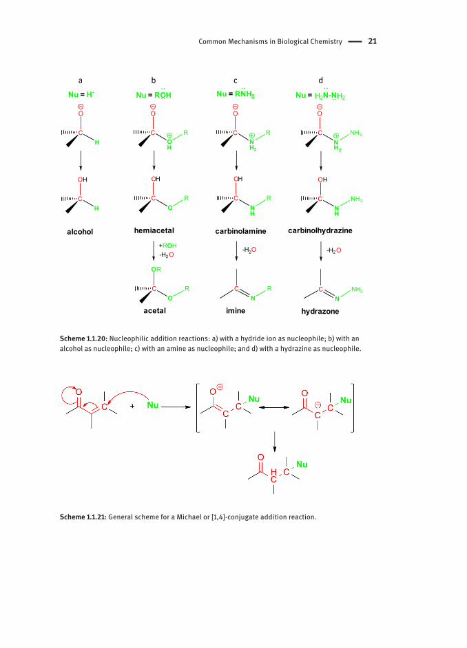
If the nucleophile is neutral and gives a pair of electrons then it will acquire a positive charge, which in turn is compensated by a deprotonation of the introduced group (as in the formation of imines; Scheme 1.1.20c). If the nucleophile is anionic, no positive charge is generated. In both cases, a negative charge is generated at the most electronegative atom, the carbonyl oxygen, which is usually neutralized through protonation (Scheme 1.1.20). Scheme 1.1.20 illustrates some of typical nucleophilic addition reactions of different nucleophiles to aldehydes and ketones.
All the reactions depicted in Scheme 1.1.20 are called “direct” or [1,2]-additions. A special case of nucleophilic addition reactions is the Michael or [1,4]-conjugate addition. This reaction is quite frequent in biochemical pathways and consists of a nucleophile addition to the β–position of an α,β-unsaturated carbonyl system (Scheme 1.1.21).
Common Mechanisms in Biological Chemistry 21
C
O
H
C
OH
H
C
O
NH2
R
C
OH
NH
R
-H2O
CN
R
alcohol carbinolamine
imine
C
O
OH
R
C
OH
OR
CO
R
hemiacetal
OR
acetal
-H2O+ROH
a b cNu = H- Nu = RNH2Nu = ROH
C
O
NH2
NH2
C
OH
NH
NH2
-H2O
CN
NH2
carbinolhydrazine
hydrazone
d
Nu = H2N-NH2
Scheme 1.1.20: Nucleophilic addition reactions: a) with a hydride ion as nucleophile; b) with an alcohol as nucleophile; c) with an amine as nucleophile; and d) with a hydrazine as nucleophile.
Scheme 1.1.21: General scheme for a Michael or [1,4]-conjugate addition reaction.

22 Functional Groups of Biomolecules and their Reactions
As with electrophilic addition reactions, the nucleophile may be added to any of the p orbitals, leading to the formation of enantiomer mixtures (in the case of α,β-unsaturated ketones, monosubstituted at β-position or disubstituted with two different groups). However, enzymes can be enantioselective and produce only one enantiomer.
1.1.4.5.1 Nitrofurantoin ‒ a SemicarbazoneSemicarbazones (Scheme 1.1.22) are a family of compounds classified as imine derivatives, originating from the action of semicarbazines on aldehydes or ketones (instead of amines). The –NH2 group of a semicarbazide is akin to a primary amino group. Thus, the reaction mechanism is the same as with other nucleophilic carbonyl addition reactions.
Scheme 1.1.22: General scheme for a semicarbazone synthesis.
There are some semicarbazones with pharmacological interest, such as nitrofurantoin (non-systematic name). Nitrofurantoin is a nitrofuran-based antibiotic considered an essential medicine by the World Health Organization. Scheme 1.1.23 presents the mechanism of the nitrofurantoin synthesis from 5-nitrofuran-2-carbaldehyde and 1-aminoimidazolidine-2,4-dione.
Scheme 1.1.23: Synthesis of Nitrofurantoin from 5-nitrofuran-2-carbaldehyde and 1-aminoimidazolidine-2,4-dione.

Common Mechanisms in Biological Chemistry 23
The reaction steps are the same as in any other nucleophilic carbonyl addition reaction followed by dehydration:
nucleophilic attack of the nucleophilic amino moiety to the electrophilic carbo-1. nyl carbon;deprotonation of the positive nitrogen atom and protonation of the negative 2. oxygen atom;elimination of water by protonation of the hydroxyl group and deprotonation at 3. the nitrogen atom.
This synthetic route as a whole is a condensation reaction: two molecules produce a larger molecule with the loss of a small molecule, in this case water. In fact, imine and hydrazone synthesis are condensations too, but were introduced as additions (see above) since the condensation product is often readily generated from the unstable addition product unless very strict reaction conditions are used.
1.1.1.4.6 Acyl Substitution ReactionsAcyl substitutions are another class of reactions involving carbonyl groups (Scheme 1.1.24). An acyl substitution reaction is favoured over a simple addition whenever an electronegative group is attached to the carbonyl group. This property is a main feature presented by carboxylic acids and their derivatives.
Scheme 1.1.24: General mechanism for an acyl substitution reaction.
The first step of this mechanism is shared with nucleophilic addition reactions and consists in the nucleophilic addition to the carbonyl group. The second step is a regeneration of the carbonyl group with elimination of the anion. It is important to note that the whole reaction is reversible unless some product stabilization is provided, such as if the leaving group is a stable anion, which is unlikely to be able to attack the carbonyl again. Acyl phosphates are considered activated analogues of carboxylic acids, as the leaving phosphate is a very stable anion and it is unlikely

24 Functional Groups of Biomolecules and their Reactions
that a better leaving group is attached to a carbonyl group. This shifts the equilibrium towards product formation. On the other hand, a simple carboxylic acid would have the hydroxyl as leaving group which is a strong nucleophile capable of adding onto the carbonyl again. This is the reason why esters, thioesters and acyl phosphates play a major role in promoting substitution reactions within biological systems. In the laboratory environment it is more common the use of carboxylic acid anhydrides and acyl chlorides or bromides. The resulting anions (carboxylates and halogens, respectively) are very weak bases promoting the completeness of the reaction. Because of that, these compounds are very sensitive to hydrolysis thus rendered useless in biological systems.
1.1.4.6.1 Aspirin ‒ Esterifications and TransesterificationsAspirin, acetylsalicylic acid or, by its systematic name, 2-acetoxybenzoic acid, is a nonsteroidal anti-inflammatory drug that inhibits the formation of prostaglandins and thromboxanes by inactivating cyclooxygenases (COXs). It is synthesized by esterification of salicylic acid with acetic anhydride (Scheme 1.1.25). One of its action pathways involves the acetylation of a serine residue in the enzyme through a transesterification reaction (Scheme 1.1.26).
Esterification is an acyl substitution reaction where an acyl group is transferred to the oxygen atom of the alcohol. In the synthesis of aspirin, acetic anhydride is used for efficiency reasons as it is a much better acetylating agent than acetic acid (Scheme 1.1.25).
Scheme 1.1.25: Simplified mechanism for the synthesis of aspirin from salicylic acid and acetic anhy-dride. This reaction is normally performed under acid catalysis with sulfuric or phosphoric acids.
Transesterifications are acyl substitution reactions where the nucleophile is an alcohol and the electrophile is an ester. Essentially, the alkoxyl moiety in the ester is substituted by another. In the case of aspirin, the alcohol/nucleophile is the side chain of the serine residue in the COX enzyme which substitutes the alcohol moiety of the salicylic acid (Scheme 1.1.26).

Common Mechanisms in Biological Chemistry 25
Scheme 1.1.26: Simplified mechanism of the transesterification reaction of aspirin with the serine residue.
1.1.1.4.7 Carbonyl Condensation ReactionsAs we have seen before, a condensation reaction joins two molecules together to form a bigger one and liberates a small one (e.g. water, methanol, acetic acid). Carbonyl condensations occur with two carbonyl compounds. This type of reaction is a very important one, adding to the chemical versatility that carbonyl groups grant to a system. Carbonyl condensations are essentially nucleophilic substitutions that allow easy carbon-carbon bond formation in relatively mild conditions (certainly within the reach of an enzyme), adding chain formation and polymerization to the list of enzyme-catalysed reactions.
1.1.4.7.1 Aldol ReactionAn aldol reaction involves carbonyl compounds such as aldehydes or ketones in which at least one species has an α-proton (Scheme 1.1.27). Although it is not formally a condensation reaction unless dehydration happens, it is often named as aldol condensation in biochemical fields. The “proper” aldol condensation product though is the result of a dehydration of the formed aldol.

26 Functional Groups of Biomolecules and their Reactions
Scheme 1.1.27: General mechanism for an aldol condensation reaction. The aldol adduct suffers a dehydration for which the mechanism is not specified since the precise step sequence may vary according to catalyst used.
First of all, the enolate is generated from a ketone or an aldehyde by an α-deprotonation with a base. The enolate is a strong nucleophile and a nucleophilic addition occurs unto an aldehyde (or ketone) acting as electrophile. The resultant anion, the aldolate, is protonated to produce the aldol, a β-hydroxy-aldehyde or ketone. The aldol may then eliminate a water molecule yielding an α,β-unsaturated aldehyde or ketone (Scheme 1.1.27).
1.1.4.7.2 Claisen CondensationClaisen condensation is the base-catalysed condensation reaction of an ester with another carbonyl compound taking place through the mechanism depicted in Scheme 1.1.28.
Scheme 1.1.28: General mechanism for a Claisen condensation reaction.

Common Mechanisms in Biological Chemistry 27
Firstly, the enolate is generated from an ester by α-deprotonation with a base. The enolate performs an acyl substitution followed by elimination of the alcoxyl group, giving a β-keto ester (Scheme 1.1.28).
1.1.4.7.3 Aldolases ‒ Stabilization StrategiesIn order to better perform aldol additions, enzymes create stabilized intermediate forms that provide a lower energy reaction path (Scheme 1.1.29). In the case of class II fructose-biphosphate aldolases, a zinc(II) ion is used to further polarize the carbonyl C=O bond of dihydroxyacetone phosphate (DHAP). The latter deprotonates easily, as the enolate intermediate is stabilized by the zinc ion. The enolate then attacks the carbonyl carbon of the glyceraldehyde 3-phosphate (GA3P or GAP), producing the addition product fructose biphosphate. This reaction is completely stereospecific as with all enzyme catalysed reactions this statement in parenthesis should be in the description of Scheme 1.1.29. The reverse reaction (hydrolysis) is also catalysed by another enzyme.
Scheme 1.1.29: Mechanism of an aldol addition in a class I fructose-biphosphate aldolase.
Class I fructose-biphosphate aldolases have another stabilization mechanism that does not resemble a regular aldol reaction, though the final product is the same (Scheme 1.1.30). The mechanism occurs in the following abbreviated steps:
A carbinolamine is formed by nucleophilic addition of a lysine residue to DHAP1. The carbinolamine eliminates water through acid/base catalysis, forming an 2. enamineThe enamine acts as an enol (through analogous resonance structures) and adds 3. to GAPWater is added to the resulting iminium ion (hydrolysis is the reverse of the imine 4. synthesis)The carbonyl and lysine residue are regenerated from the new carbinolamine.5.
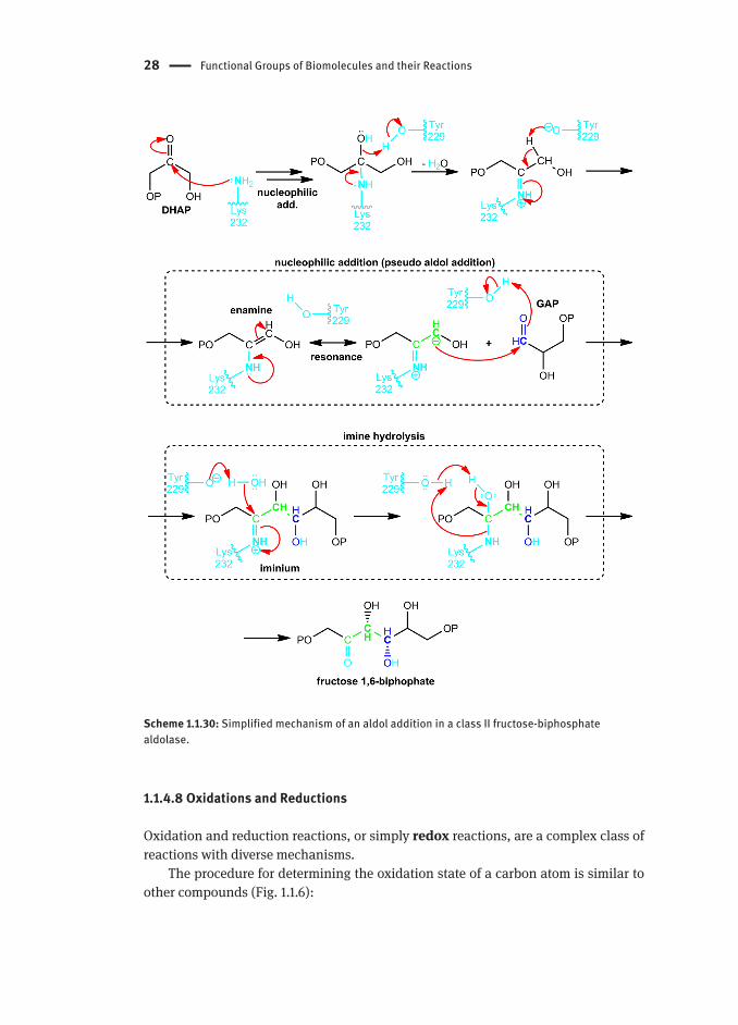
28 Functional Groups of Biomolecules and their Reactions
Scheme 1.1.30: Simplified mechanism of an aldol addition in a class II fructose-biphosphate aldolase.
1.1.4.8 Oxidations and Reductions
Oxidation and reduction reactions, or simply redox reactions, are a complex class of reactions with diverse mechanisms.
The procedure for determining the oxidation state of a carbon atom is similar to other compounds (Fig. 1.1.6):

Common Mechanisms in Biological Chemistry 29
For each bond to less electronegative atoms, such as a hydrogen atoms, count 1. as -1For each bond to more electronegative atoms, such as oxygen atoms, count as +2. Bonds between carbon atoms do not affect the oxidation state, unlike other 3. elements.
HC
H
H OH
-2H
CH
O
0
H3CC
OH
O
+3H3C
CNH2
H SH
+1OH
CH2N
O
+4
HC
H
H CH3
-3
H3CC
OH
Cl CH3
+2
HC
H
H H
-4H3C
CH
H OPO32-
-1
Figure 1.1.6: Compounds with the oxidation state of the highlighted carbon indicated below each structure. In order: methane, ethane, methanol, ethyl phosphate, formaldehyde, (R)-1-aminoethane-1-thiol, 1,1-dichloroethan-1-ol, acetic acid and carbamic acid.
The simplest redox reaction in a biological system is the oxidation of an alcohol to a carbonyl compound. In a laboratory, a metal in a high oxidation state is usually used as oxidant, where it attaches to the oxygen of the alcohol then acts as a leaving group with an E2-like mechanism (Scheme 1.1.31).
Scheme 1.1.31: General mechanism for the oxidation of an alcohol to a carbonyl compound with a metal or its complex (M). Note that the M leaves in a lower oxidation state.
In the case of an aldehyde, the carbonyl group may be oxidized to carboxyl by nucleophilic attack of water generating a hydrated aldehyde, in which one of the hydroxyl groups is then oxidized to carbonyl yielding the carboxyl group.
30 Functional Groups of Biomolecules and their Reactions
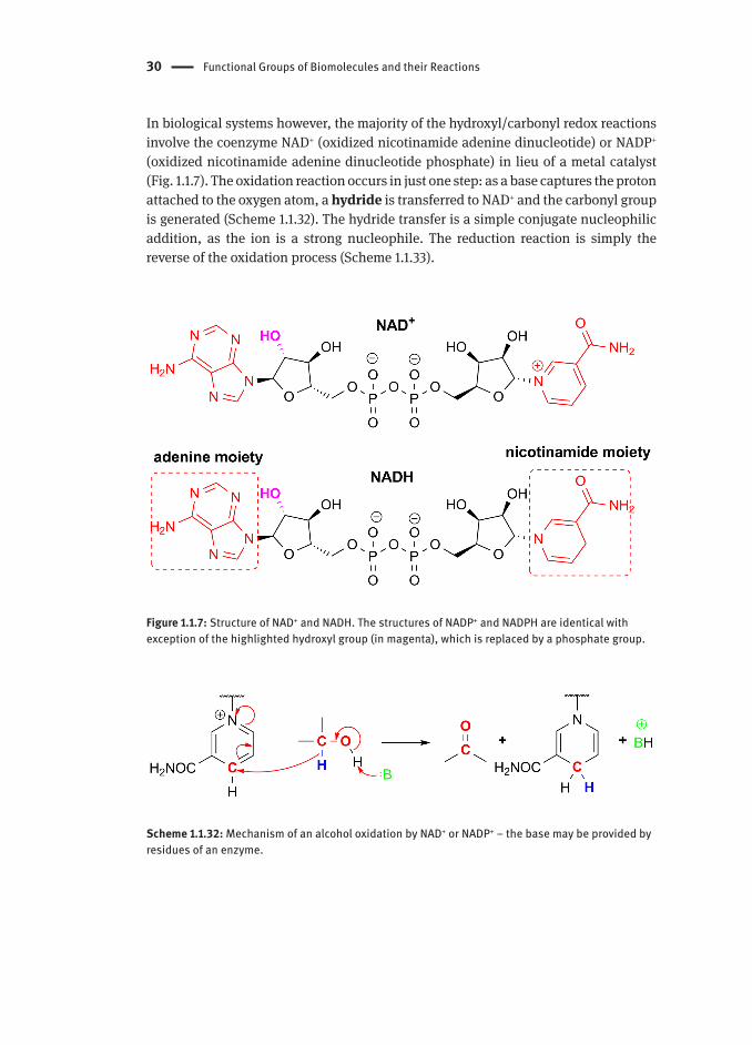
In biological systems however, the majority of the hydroxyl/carbonyl redox reactions involve the coenzyme NAD+ (oxidized nicotinamide adenine dinucleotide) or NADP+ (oxidized nicotinamide adenine dinucleotide phosphate) in lieu of a metal catalyst (Fig. 1.1.7). The oxidation reaction occurs in just one step: as a base captures the proton attached to the oxygen atom, a hydride is transferred to NAD+ and the carbonyl group is generated (Scheme 1.1.32). The hydride transfer is a simple conjugate nucleophilic addition, as the ion is a strong nucleophile. The reduction reaction is simply the reverse of the oxidation process (Scheme 1.1.33).
Figure 1.1.7: Structure of NAD+ and NADH. The structures of NADP+ and NADPH are identical with exception of the highlighted hydroxyl group (in magenta), which is replaced by a phosphate group.
Scheme 1.1.32: Mechanism of an alcohol oxidation by NAD+ or NADP+ ‒ the base may be provided by residues of an enzyme.

Common Mechanisms in Biological Chemistry 31
Scheme 1.1.33: Mechanism of an alcohol reduction of a carbonyl group by NAD or NADP, in this case of an acetylated acyl carrier protein (ACP), an important part in the fatty acid synthesis.
1.1.4.8.1 Disulfide Bridges ‒ Oxidized ThiolsDisulfide bridges are essential in a protein to allow stable structural scaffolds. They are S‒S bonds between cysteine residues (Scheme 1.1.34). Note that the sulfur loses a bond to a hydrogen and gains one to another sulfur atom, so the formation of this linkage is a redox reaction with thiols being oxidized. The oxidant (to be reduced) is a glutathione dimer (GSSG), which consists in two glutathione molecules connected by a disulfide bridge (Scheme 1.1.35).
Scheme 1.1.34: Disulfide bridge formation.
Scheme 1.1.35: Mechanism of disulphide linkage formation by a glutathione dimer, releasing two reduced glutathione molecules.

32 Functional Groups of Biomolecules and their Reactions
1.1.5 The Organic Mechanisms of Biological Transformations
Previously, we highlighted that common organic reaction mechanisms can be used to understand the biosynthetic pathways. Herein some illustrative examples are presented.
1.1.5.1 Cis/trans-Isomers Interconversion in the Vision Pathway
It is common knowledge that vitamin A, retinol (Scheme 1.1.36), plays an important role in our vision. Although the sequence of reactions and detailed transformations are not in the scope of this chapter, it is remarkable that a simple oxidation and change in configuration is ultimately responsible for a complex process such as vision. Cis/trans isomerase enzymes, through a cysteine residue, are responsible for this isomerization. The next step is Schiff base (imine) formation through the reaction of retinal with a lysine residue of the protein opsin to produce rhodopsin, which isomerizes upon absorption of visible light. This change in geometry causes an electrical signal that is sent to the brain. Finally, the hydrolysis of the imine linkage regenerates the opsin protein and (11E)-retinal (Scheme 1.1.36).
Scheme 1.1.36: The chemistry of vision.

The Organic Mechanisms of Biological Transformations 33
1.1.5.2 Metabolism of Fatty Acids ‒ β-Oxidation Pathway
The catabolism of fatty acids (saturated or unsaturated) starts with chemical activation by esterification with coenzyme A (Scheme 1.1.37), with the following steps occurring in an acyl-CoA synthase:
The carboxylate acts as a nucleophile to attack the double P‒O bond in ATP;1. The diphosphate group leaves, resulting in an activated acid in the form of a 2. mixed anhydride (acyl AMP). (PPi is a good leaving group as it is stable and not nucleophilic)Another acyl substitution is performed on the mixed anhydride by the thiol 3. moiety in CoA (a moderate nucleophile). Again, this is facilitated because AMP is a good leaving group.
Scheme 1.1.37: Activation of a fatty acid via esterification.
With the thioester (activated fatty acid) formed, β-oxidation may now occur through a sequence of dehydrogenation, hydration and dehydrogenation reactions:
The Cα-Cβ single bond is oxidised to a double bond, with flavin adenine nucleotide 1. (FAD) as the oxidant. This step occurs in a family of acyl-CoA dehydrogenases where the products are FADH2 (reduced FAD) and α,β-unsaturated acyl-CoA. The mechanistic details of this step are not yet fully resolved, though it is known that one of the α protons may be first attacked by 376-Glu and a β-hydride is abstracted from the fatty acid by FAD.
CH2
O
SCoA
H2C
CH
O
SCoA
HC
FAD FADH2
acyl CoA(thioester)
a ,b-unsaturated acyl CoA
Enoyl-CoA hydratase performs a nucleophilic addition of water to the unsaturated 2. double bond. The hydroxyl (nucleophile) is added exclusively in the β-position to the carbonyl since the enzyme is stereospecific.

34 Functional Groups of Biomolecules and their Reactions
The formed hydroxymethylene group is oxidised to a carbonyl group with 3. a β-hydroxyacyl-CoA dehydrogenase. This reaction yields an NADH ion, formed from the coenzyme NAD+ acting as the oxidant, and the β-ketoester.
A retro-Claisen reaction cleaves the β-ketothioester yielding two thioesters, 4. producing the initial acyl-CoA shortened by two carbons and acetyl-CoA (this proceeds to the citric acid cycle, ultimately being oxidized to CO2).
The pathway is repeated until all of the fatty acid is oxidised. If, during the cleavage process, a cis-oriented unsaturation is encountered, the stereochemistry of the double bond is switched to trans by an isomerase and the reaction sequence continues as for saturated fatty acids, since the enoyl-CoA hydratase is stereospecific for a trans-configuration.
The Organic Mechanisms of Biological Transformations 35
1.1.5.3 Penicillin ‒ a Strong Acylating Agent
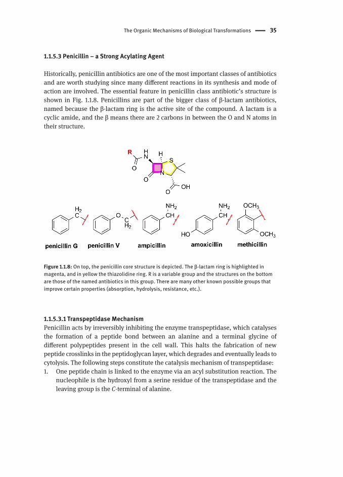
Historically, penicillin antibiotics are one of the most important classes of antibiotics and are worth studying since many different reactions in its synthesis and mode of action are involved. The essential feature in penicillin class antibiotic’s structure is shown in Fig. 1.1.8. Penicillins are part of the bigger class of β-lactam antibiotics, named because the β-lactam ring is the active site of the compound. A lactam is a cyclic amide, and the β means there are 2 carbons in between the O and N atoms in their structure.
Figure 1.1.8: On top, the penicillin core structure is depicted. The β-lactam ring is highlighted in magenta, and in yellow the thiazolidine ring. R is a variable group and the structures on the bottom are those of the named antibiotics in this group. There are many other known possible groups that improve certain properties (absorption, hydrolysis, resistance, etc.).
1.1.5.3.1 Transpeptidase MechanismPenicillin acts by irreversibly inhibiting the enzyme transpeptidase, which catalyses the formation of a peptide bond between an alanine and a terminal glycine of different polypeptides present in the cell wall. This halts the fabrication of new peptide crosslinks in the peptidoglycan layer, which degrades and eventually leads to cytolysis. The following steps constitute the catalysis mechanism of transpeptidase:
One peptide chain is linked to the enzyme via an acyl substitution reaction. The 1. nucleophile is the hydroxyl from a serine residue of the transpeptidase and the leaving group is the C-terminal of alanine.

36 Functional Groups of Biomolecules and their Reactions
Another acyl substitution reaction involves a second peptide chain that displaces 2. the link to the hydroxyl of a serine residue resulting in the two peptide chains being linked.
1.1.5.3.2 Transpeptidase InhibitionAs seen above, the core transformation in the transpeptidase reaction is an acyl substitution reaction. Penicillin (Scheme 1.1.38) is similar to the normal transpeptidase substrates, so it mimics the substrate and binds irreversibly to the enzyme active site. What makes penicillin so effective is that the β-lactam ring is under considerable strain, making the reaction irreversible. The thiazolidine ring further increases the strain by distorting the bonds and removing resonance stabilization. The β-lactam in anionic form is also protected from hydrolysis so the absorption is more efficient.
Scheme 1.1.38: Mechanism of the acyl substitution reaction occurring with penicillin and the serine residue from transpeptidase.

The Organic Mechanisms of Biological Transformations 37
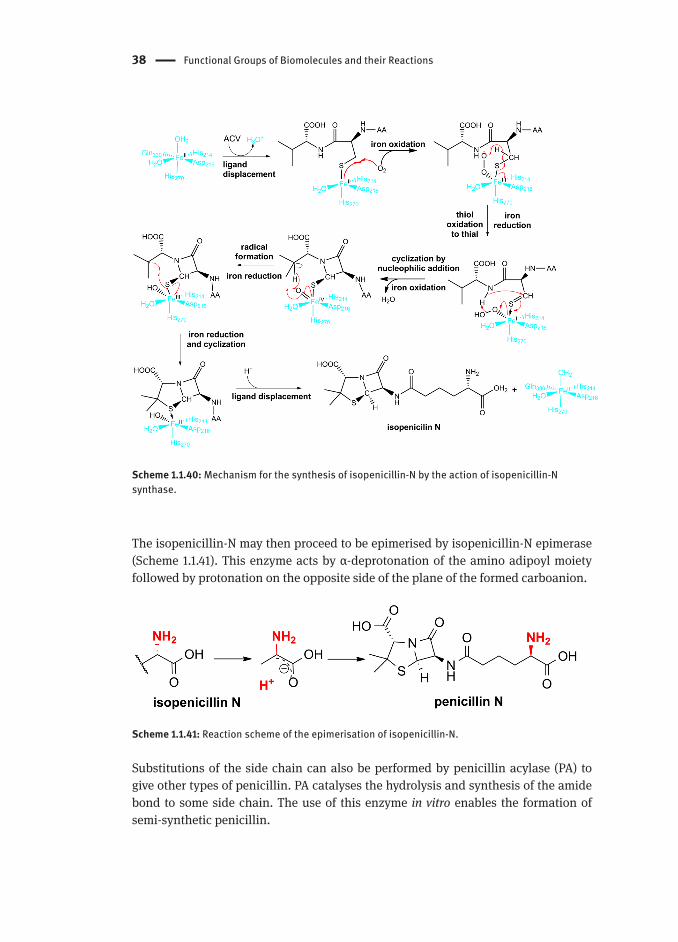
1.1.5.3.3. Penicillin BiosynthesisPenicillin biosynthesis begins with the formation of the tripeptide L-δ-(α-aminoadipoyl)-L-cysteinyl-D-valine (ACV) (Scheme 1.1.39) by condensation of three amino acids catalysed by ACV synthase. ACV is then processed by isopenicillin-N synthase, an enzyme of the oxyreductase family, with the following mechanistic steps (Scheme 1.1.40):
Attachment of the cysteine thiol moiety by displacing a water ligand1. Oxidation of Fe(II) to Fe(III) by molecular oxygen, creating a radical species2. Intramolecular hydrogen transfer, oxidation of the thiol to thioaldehyde 3. (extremely reactive due higher dipole moment than that of an aldehyde) and reduction of Fe(III) to Fe(II)Amide deprotonation by the hydroperoxide ligand and nucleophilic addition of 4. the nitrogen to the thiol with oxidation of Fe(II) to Fe(IV). This transformation results in the lactam ring formationRadical formation by hydrogen abstraction by the oxide (turns into an hydroxide 5. ligand) with reduction of Fe(IV) to Fe(III)Radical attack of the sulfur atom, closing the thiazolidine ring and reducing 6. Fe(III) to Fe(II)Displacement of the sulfur by water to restore the enzyme active site and release 7. isopenicillin-N.
Scheme 1.1.39: Equation for the synthesis of ACV.
38 Functional Groups of Biomolecules and their Reactions
Scheme 1.1.40: Mechanism for the synthesis of isopenicillin-N by the action of isopenicillin-N synthase.
The isopenicillin-N may then proceed to be epimerised by isopenicillin-N epimerase (Scheme 1.1.41). This enzyme acts by α-deprotonation of the amino adipoyl moiety followed by protonation on the opposite side of the plane of the formed carboanion.
Scheme 1.1.41: Reaction scheme of the epimerisation of isopenicillin-N.
Substitutions of the side chain can also be performed by penicillin acylase (PA) to give other types of penicillin. PA catalyses the hydrolysis and synthesis of the amide bond to some side chain. The use of this enzyme in vitro enables the formation of semi-synthetic penicillin.

The Organic Mechanisms of Biological Transformations 39
1.1.5.4 NAD+ − a Classical Coenzyme
An enzyme cannot catalyse oxidation reactions unless a coenzyme is present. In some sense, the enzyme’s role is to hold the substrate and coenzyme together to facilitate the oxidation reaction. One of the most commonly used coenzymes is nicotinamide adenine dinucleotide (NAD+) (Fig. 1.1.9). There is evidence that sirtuins are proteins related to several diseases and are NAD+ dependent.
Figure 1.1.9: Structure of nicotinamide adenine dinucleotide (NAD+).
The most known intervention of coenzyme NAD+ is its role in the ethanol metabolism, where it acts as a common hydride acceptor (Scheme 1.1.42).
Scheme 1.1.42: Mechanism of ethanol oxidation in cells.
40 Functional Groups of Biomolecules and their Reactions
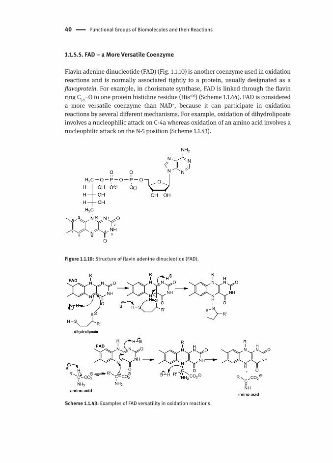
1.1.5.5. FAD − a More Versatile Coenzyme
Flavin adenine dinucleotide (FAD) (Fig. 1.1.10) is another coenzyme used in oxidation reactions and is normally associated tightly to a protein, usually designated as a flavoprotein. For example, in chorismate synthase, FAD is linked through the flavin ring C(2)=O to one protein histidine residue (His106) (Scheme 1.1.44). FAD is considered a more versatile coenzyme than NAD+, because it can participate in oxidation reactions by several different mechanisms. For example, oxidation of dihydrolipoate involves a nucleophilic attack on C-4a whereas oxidation of an amino acid involves a nucleophilic attack on the N-5 position (Scheme 1.1.43).
Figure 1.1.10: Structure of flavin adenine dinucleotide (FAD).
Scheme 1.1.43: Examples of FAD versatility in oxidation reactions.

The Organic Mechanisms of Biological Transformations 41
Furthermore, FAD oxidation mechanisms are controversial because in some proposals, such as the chorismate synthase reaction (Scheme 1.1.44), the authors suggest the involvement of ionic and radical structures.
Scheme 1.1.44: Chorismate synthase reaction.
1.1.5.6 Biotin and Carboxylation Reactions
Biotin-dependent enzymes are common in living organisms and are involved in carboxylation reactions. Biotinylation occurs by the addition of a biotin molecule to a specific lysine residue (Fig. 1.1.11).
Figure 1.1.11: Biotin and enzyme-bound biotin.

42 Functional Groups of Biomolecules and their Reactions
The carboxylation catalysed by biotin-dependent enzymes use carbonate (HCO3⁻) as source of the carboxyl group, ATP to activate it and Mg2+ to decrease the overall negative charge. The mechanism involves a nucleophilic attack of the biotin moiety on the activated carbonate, resulting in the formation of carboxybiotin (Scheme 1.1.45). Nucleophilic attack by the substrate on carboxybiotin results in the transfer of the carboxyl group from biotin to the substrate, as shown in Scheme 1.1.45 with acetyl-CoA.
Scheme 1.1.45: Acetyl-CoA carboxylation mechanism.
ReferencesAllen, K. N., & Dunaway-Mariano, D. (2004). Phosphoryl group transfer: evolution of a catalytic
scaffold. Trends in Biochemical Sciences, 29(9), 495–503.Cleland, W., & Hengge, A. (1995). Mechanisms of phosphoryl and acyl transfer. FASEB J, 9(15),
1585–1594.Fushinobu, S., Nishimasu, H., Hattori, D., Song, H.-J., & Wakagi, T. (2011). Structural basis for the
bifunctionality of fructose-1,6-bisphosphate aldolase/phosphatase. Nature, 478(7370), 538–541.
Gebler, J. C., Woodside, A. B., & Poulter, C. D. (1992). Dimethylallyltryptophan synthase. An enzyme-catalyzed electrophilic aromatic substitution. Journal of the American Chemical Society, 114(19), 7354–7360.
Imai, S.-i. & Guarente, L. (2014). NAD+ and sirtuins in aging and disease. Trends in Cell Biology, 24(8), 464-470.
IUPAC, Commission on Nomenclature of Organic Chemistry. (1993). A Guide to IUPAC Nomenclature of Organic Compounds (Recommendations 1993). Blackwell Scientific publications.
Bibliography 43
Kirchberg, K., Kim, T.-Y., Haase, S. & Alexiev, U. (2010). Functional interaction structures of the phochromic retinal protein rhodopsin. Photochemical & Photobiological Sciences, 9, 226-233.
Kitzing, K., Auweter, S., Amrhein, N. & Macheroux, P. (2004). Mechanism of Chorismate Synthase. The Journal of Biological Chemistry, 279(5), 9451-9461.
Lietzan, A. D., Lin, Y. & St. Maurice, M. (2014). The role of biotin and oxamate in the carboxyltransferase reaction of pyruvate carboxylase. Archives of Biochemistry and Biophysics, 562, 70-79.
McMurry, J. E. & Bagley, T. P. (2005). The Organic Chemistry of Biological Pathways. Robeerts and Company Publishers.
Yuan, H. & Marmorstein, R. (2012). Structural basis for sirtuin activity and inhibition. Journal of Biological Chemistry, 287(14), 42428-42435.