differential g protein subunit expression by prostate cancer
TRANSCRIPT

El-Haibi et al. Molecular Cancer 2013, 12:64http://www.molecular-cancer.com/content/12/1/64
RESEARCH Open Access
Differential G protein subunit expression byprostate cancer cells and their interactionwith CXCR5Christelle P El-Haibi1, Praveen Sharma2, Rajesh Singh3, Pranav Gupta3, Dennis D Taub4, Shailesh Singh3
and James W Lillard, Jr3*
Abstract
Background: Prostate cancer (PCa) cell lines and tissues differentially express CXCR5, which positively correlatewith PCa progression, and mediate PCa cell migration and invasion following interaction with CXCL13. However,the differential expression of G protein α, β, and γ subunits by PCa cell lines and the precise combination of theseproteins with CXCR5 has not been elucidated.
Methods: We examined differences in G protein expression of normal prostate (RWPE-1) and PCa cell lines (LNCaP,C4-2B, and PC3) by western blot analysis. Further, we immunoprecipitated CXCR5 with different G protein subunits,and CXCR4, following CXCL13 stimulation. To investigate constitutive coupling of CXCR5 with CXCR4 and PAR-1 weperformed invasion assay in PCa cells transfected with Gαq/i2 or Gα13 siRNA, following CXCL13 treatment. We alsoinvestigated Rac and RhoA activity by G-LISA activation assay in PCa cells following CXCL13/thrombin stimulation.
Result: Of the 22 G proteins studied, Gαi1-3, Gβ1-4, Gγ5, Gγ7, and Gγ10 were expressed by both normal and PCa celllines. Gαs was moderately expressed in C4-2B and PC3 cell lines, Gαq/11 was only present in RWPE-1 and LNCaP celllines, while Gα12 and Gα13 were expressed in C4-2B and PC3 cell lines. Gγ9 was expressed only in PCa cell lines.Gα16, Gβ5, Gγ1-4, and Gγ13 were not detected in any of the cell lines studied. Surprisingly, CXCR4 co-immunoprecipitatedwith CXCR5 in PCa cell lines irrespective of CXCL13 treatment. We also identified specific G protein isoforms coupled toCXCR5 in its resting and active states. Gαq/11/Gβ3/Gγ9 in LNCaP and Gαi2/Gβ3/Gγ9 in C4-2B and PC3 cell lines, werecoupled to CXCR5 and disassociated following CXCL13 stimulation. Interestingly, Gα13 co-immunoprecipitated withCXCR5 in CXCL13-treated, but not in untreated PCa cell lines. Inhibition of Gαq/i2 significantly decreased the ability ofcells to invade, whereas silencing Gα13 did not affect CXCL13-dependent cell invasion. Finally, CXCL13 treatmentsignificantly increased Rac activity in Gαq/i2 dependent manner, but not RhoA activity, in PCa cell lines.
Conclusions: These findings offer insight into molecular mechanisms of PCa progression and can help to designsome therapeutic strategies involving CXCR5 and/or CXCL13 blockade and specific G protein inhibition to abrogatePCa metastasis.
Keywords: Prostate cancer, Chemokine, G proteins, G protein-coupled receptor
* Correspondence: [email protected] of Microbiology, Biochemistry, & Immunology, MorehouseSchool of Medicine, Atlanta, GA, USAFull list of author information is available at the end of the article
© 2013 El-Haibi et al.; licensee BioMed Central Ltd. This is an Open Access article distributed under the terms of the CreativeCommons Attribution License (http://creativecommons.org/licenses/by/2.0), which permits unrestricted use, distribution, andreproduction in any medium, provided the original work is properly cited.
El-Haibi et al. Molecular Cancer 2013, 12:64 Page 2 of 11http://www.molecular-cancer.com/content/12/1/64
BackgroundG protein-coupled receptors (GPCRs) are divided intothree broad classes based on the similarity of the trans-membrane sequences and the nature of their ligand [1].Chemokine receptors are categorized under the super-family of Class A Rhodopsin-like GPCRs [2]. GPCRsinteract with heterotrimeric guanine nucleotide-bindingproteins (G proteins) composed of α, β, and γ subunitspresent on the inner surface of the plasma membrane.After ligand binding, the receptor elicits a conform-ational alteration resulting in the exchange of guanosinediphosphate (GDP) for guanosine triphosphate (GTP) bythe Gα subunit. This leads to heterotrimer dissociationand stimulation of downstream effector molecules to ini-tiate intracellular signaling cascades [1,3,4]. Gα subunitsare divided into four families Gαs, Gαi, Gαq/11, and Gα12/13
based on sequence homology and functional similarities.Gαs proteins are known to stimulate adenylyl cyclases(AC), while Gαi proteins inhibit AC and activate phospho-diesterases. Alternatively, Gαq/11 proteins regulate the ac-tivity of phosphatidylinositol-specific phospholipases togenerate lipid second messengers, and Gα12/13 proteinsregulate the small guanine triphosphate (GTPases). Onthe other hand, G protein β and γ subunits function as atightly associated complex to modulate the activity of se-veral effectors including AC, protein tyrosine kinases (e.g.,Src family tyrosine kinases), phosphoinositide-3 kinase(PI3K) γ, GPCR kinases (GRKs), and Ca+2 as well as K+ ionchannels [4,5].Gα subunits are encoded by 17 genes (Gnas, Gnasxl,
Gnal, Gnai1, Gnai2, Gnai3, Gnao, Gnaz, Gnag, Gnat1,Gnat2, Gnaq, Gna11, Gna14, Gna15, Gna12, andGna13). There are five known genes encoding Gβ sub-units (Gnb1, Gnb2, Gnb3, Gnb4, and Gnb5) and 12genes encoding Gγ subunits (Gngt1, Gngt2, Gng2, Gng3,Gng4, Gng5, Gng7, Gng8, Gng10, Gng11, Gng12, andGng13) [3,6,7]. A large number of potential combina-tions of Gα/β/γ heterotrimers can form; however, not allassociations are functional and they vary in their affinityfor distinct GPCRs [8,9]. G proteins also exhibit tissue-specific expression. Most G proteins are ubiquitouslypresent in several tissues, but a smaller subset is con-fined to specialized cell types [7,10].Several studies have reported the role of G proteins in
different human diseases [11]. Comparatively, less isknown regarding the expression of these signaling pro-teins by PCa cells. PCa cells express a repertoire of che-mokine receptors that contribute to disease progressionand metastasis [12,13]. In this regard, we have shownthat PCa cell lines differentially express CXCR5, and thisexpression positively correlates with the ability of celllines to migrate and invade extracellular matrix com-ponents following interaction with CXCL13 [14,15].To our knowledge, neither the differential expression
of G protein α, β, and γ subunits by PCa cell lines norspecific G protein interactions with CXCR5 have been de-scribed. Here, we elucidate the differences in G proteinisoforms expressed by normal and tumorigenic prostatecell lines. We also identified the specific G proteinisoforms coupled to CXCR5 in the presence or absence ofCXCL13 stimulation.
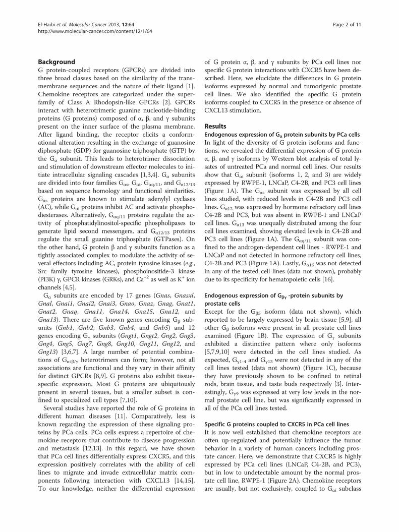
ResultsEndogenous expression of Gα protein subunits by PCa cellsIn light of the diversity of G protein isoforms and func-tions, we revealed the differential expression of G proteinα, β, and γ isoforms by Western blot analysis of total ly-sates of untreated PCa and normal cell lines. Our resultsshow that Gαi subunit (isoforms 1, 2, and 3) are widelyexpressed by RWPE-1, LNCaP, C4-2B, and PC3 cell lines(Figure 1A). The Gαs subunit was expressed by all celllines studied, with reduced levels in C4-2B and PC3 celllines. Gα12 was expressed by hormone refractory cell linesC4-2B and PC3, but was absent in RWPE-1 and LNCaPcell lines. Gα13 was unequally distributed among the fourcell lines examined, showing elevated levels in C4-2B andPC3 cell lines (Figure 1A). The Gαq/11 subunit was con-fined to the androgen-dependent cell lines - RWPE-1 andLNCaP and not detected in hormone refractory cell lines,C4-2B and PC3 (Figure 1A). Lastly, Gα16 was not detectedin any of the tested cell lines (data not shown), probablydue to its specificity for hematopoietic cells [16].
Endogenous expression of Gβγ -protein subunits byprostate cellsExcept for the Gβ5 isoform (data not shown), whichreported to be largely expressed by brain tissue [5,9], allother Gβ isoforms were present in all prostate cell linesexamined (Figure 1B). The expression of Gγ subunitsexhibited a distinctive pattern where only isoforms[5,7,9,10] were detected in the cell lines studied. Asexpected, Gγ1-4 and Gγ13 were not detected in any of thecell lines tested (data not shown) (Figure 1C), becausethey have previously shown to be confined to retinalrods, brain tissue, and taste buds respectively [3]. Inter-estingly, Gγ9 was expressed at very low levels in the nor-mal prostate cell line, but was significantly expressed inall of the PCa cell lines tested.
Specific G proteins coupled to CXCR5 in PCa cell linesIt is now well established that chemokine receptors areoften up-regulated and potentially influence the tumorbehavior in a variety of human cancers including pros-tate cancer. Here, we demonstrate that CXCR5 is highlyexpressed by PCa cell lines (LNCaP, C4-2B, and PC3),but in low to undetectable amount by the normal pros-tate cell line, RWPE-1 (Figure 2A). Chemokine receptorsare usually, but not exclusively, coupled to Gαi subclass
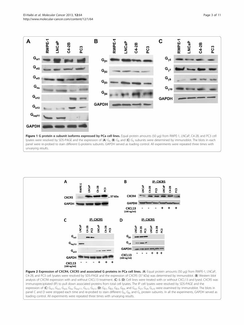
Figure 1 G protein α subunit isoforms expressed by PCa cell lines. Equal protein amounts (50 μg) from RWPE-1, LNCaP, C4-2B, and PC3 celllysates were resolved by SDS-PAGE and the expression of (A) Gα (B) Gβ and (C) Gγ subunits were determined by immunoblot. The blots in eachpanel were re-probed to stain different G-proteins subunits. GAPDH served as loading control. All experiments were repeated three times withunvarying results.
Figure 2 Expression of CXCR4, CXCR5 and associated G proteins in PCa cell lines. (A) Equal protein amounts (50 μg) from RWPE-1, LNCaP,C4-2B, and PC3 cell lysates were resolved by SDS-PAGE and the expression of CXCR5 (37 kDa) was determined by immunoblot. (B) Western blotanalysis of CXCR4 expression with and without CXCL13 treatment. (C) & (D) Cell lines were treated with or without CXCL13 and lysed. CXCR5 wasimmunoprecipitated (IP) to pull down associated proteins from total cell lysates. The IP cell lysates were resolved by SDS-PAGE and theexpression of (C) Gαi1, Gαi2, Gαi3, Gαs, Gαq/11, Gα12, Gα13 (D) Gβ1, Gβ2, Gβ3, Gβ4, and Gγ5, Gγ7, Gγ9, Gγ10 were examined by immunoblot. The blots inpanel C and D were stripped each time and re-probed to stain different Gα, Gβ, and Gγ protein subunits. In all the experiments, GAPDH served asloading control. All experiments were repeated three times with unvarying results.
El-Haibi et al. Molecular Cancer 2013, 12:64 Page 3 of 11http://www.molecular-cancer.com/content/12/1/64

El-Haibi et al. Molecular Cancer 2013, 12:64 Page 4 of 11http://www.molecular-cancer.com/content/12/1/64
of G proteins [17]. In this study, we demonstrate thatonly Gαi2 co-immunoprecipitated with CXCR5 in un-treated C4-2B and PC3 cell lines in the absence of agon-ist, while Gαq/11 associates with CXCR5 in untreatedLNCaP cells. Gα13 co-immunoprecipitated with CXCR5in all three PCa cell lines treated with CXCL13, but wasnot detected in untreated cells (Figure 2C). Gβ3 and Gγ9
co-immunoprecipitated with CXCR5 in the absence ofCXCL13 in all PCa cell lines used (Figure 2D). This Gβ3/γ9
complex was not detected following CXCL13 stimulationindicating its ligand-induced dissociation from the recep-tor. The other Gα (i1, i3), Gs, Gα12, Gβ (1, 2, 4) and Gγ (5, 7, 10)
subunits which were detected in PCa cell lines (Figure 1Band 1C) were not co-immunoprecipitated with CXCR5 inpresence or absence of agonist (data not shown).
Validation and significance of Gαq/11/Gβ3/Gγ9 and Gαi2/Gβ3/
Gγ9 binding to CXCR5 in LNCaP, and C4-2B, and PC3 celllines respectivelyTo further validate differences observed in Gα subunit(s)coupling and uncoupling to CXCR5 in CXCL13-treatedversus untreated cells, we separately immunoprecipitatedGαq/11 and Gαi2 subunits in untreated and CXCL13-treatedPCa cells and immunoblotted for CXCR5. Our results pro-vide the first evidence of multifunctional coupling ofCXCR5 to different types of G proteins favoring a pertussis
Figure 3 Validation of Gαq/11, Gαi2, and Gα13 protein association with(A) Gαq/11 and (B) Gαi2 were immunoprecipitated (IP) from total cell lysateswas examined by immunoblot. (C) Identification of CXCR4 and CXCR5 couor without CXCL13 and lysed. Antibody against Gα13 was used to immunopby SDS PAGE and immunoblotted for CXCR5 followed by CXCR4, after stripexperiments were repeated three times with unvarying results.
toxin-insensitive signaling pathway mediated by Gαq/11 inLNCaP cells and a pertussis toxin-sensitive signaling path-way mediated by Gαi2 in C4-2B and PC3 cells (Figure 3).
Association of Gα13 protein, CXCR4, and PAR-1 withCXCR5 in CXCL13-treated PCa cell linesOne surprising result was the association of the Gα13
subunit with CXCR5 in PCa cell lines treated withCXCL13, but not in untreated cells. Thus, it was criticalto confirm this finding by immunoprecipitating Gα13
protein from CXCL13-treated and untreated PCa cells,and immunoblotting for CXCR5. Results confirm thatcoupling of Gα13 to CXCR5 is specific to CXCL13-treated cells (Figure 3C). It has been reported that pro-teinase activated receptor-1 (PAR-1) is capable ofbypassing signaling through Gαi-pathway to supportGα12/13-dependent mechanisms, enhancing cellular pro-liferation, invasion, and metastasis [18]. We thereforeexamined the association of PAR-1 with Gα13 andshowed that CXCR5 and PAR-1 are linked to Gα13 fol-lowing treatment with CXCL13 (Figure 4A).The presence of CXCR4 in CXCR5 immunoprecipitants
(with or without CXCL13 treatment) offers the first evi-dence of CXCR5 association with CXCR4 (Figure 2B).These interactions could potentially support CXCR4-CXCR5 signaling crosstalk. Moreover, the ability of
CXCR5. Cell lines were treated with or without CXCL13 and lysed.. The IP cell lysates were resolved by SDS-PAGE and CXCR5 expressionpled to Gα13 following CXCL13 stimulation. Cell lines were treated withrecipitate (IP) it from total cell lysates. The IP cell lysates were resolvedping. In all the experiments, GAPDH served as loading control. All

Figure 4 (See legend on next page.)
El-Haibi et al. Molecular Cancer 2013, 12:64 Page 5 of 11http://www.molecular-cancer.com/content/12/1/64

(See figure on previous page.)Figure 4 Gα13 association with PAR-1 and CXCR5, and Gα13 and Gαi2 contribution to PCa cell lines invasion and Rac/Rho activation.(A) Cell lines were treated with or without CXCL13 and lysed. Antibody against Gα13 was used to immunoprecipitate (IP) it from total cell lysates.The IP cell lysates were resolved by SDS PAGE and immunoblotted for PAR-1 followed by CXCR5, after stripping. GAPDH served as a loadingcontrol. (B) Invasion of LNCaP, C4-2B and PC3 cells was assessed using BD Matrigel™ invasion chamber. The assay was performed using LNCaP(open bars), C4-2B (solid bars) and PC3 (hashed bars) cells transfected and/or treated with control siRNA, Gαq/αi2 siRNA, or Gα13 siRNA duplex, andCXCL13 and/or Thrombin for 8 h and the cells that migrated to the lower surface of the membrane were counted by microscopy at 40Xmagnification. CXCL13-treated cells exhibited an enhanced ability to invade Matrigel. Abrogation of Gαq/i2 decreased the ability of cells to invadewhereas silencing of Gα13 did not affect cell invasion. (C) Rac and RhoA protein expression were determined in CXCL13 and/or thrombin treatedLNCaP, C4-2B, and PC3 cells. (i) shows differential expression of Rac protein, involved in lamellipodia formation, in response to CXCL13, thrombin,CXCL13 followed by thrombin and from cells transfected with Gαq/i2/13 siRNA in different experiments. (ii) shows differential expression of RhoAprotein, involved in stress fiber formation and cell adhesion, in response to CXCL13, thrombin, CXCL13 followed by thrombin and from cellstransfected with Gα13 siRNA in different experiments. All experiments were repeated at least three times and results were in accordance witheach other.
El-Haibi et al. Molecular Cancer 2013, 12:64 Page 6 of 11http://www.molecular-cancer.com/content/12/1/64
CXCR4 to engage in Gα13-mediated cell signaling eventsthat activate Rho pathways leading to cell adhesion hasbeen previously demonstrated [19]. Gα13 association withCXCR5, CXCR4 and PAR-1 after CXCL13 treatment(Figures 3C & 4A) alludes to chemokine receptor oligo-mer formation or the recruitment of other GPCR-Gα13
associated signaling complexes after stimulation, whichcould presumably potentiate synergistic or additionalbiological events, respectively [20,21].It is plausible that the CXCL13:CXCR5 axis regulates
cell migration by desensitizing CXCR4 and conditionalcoupling of CXCR5 with PAR-1. Therefore, constitutivecoupling of CXCR5 with CXCR4 and PAR-1 afterCXCL13 ligation in PCa cells could be another mechan-ism through which CXCL13 sequesters factors hamper-ing cell migration. To investigate whether thishypothesis holds true, we allowed LNCaP, C4-2B, andPC3 cells previously transfected with Gαq/i2 or Gα13
siRNA duplexes to invade across a Matrigel membranefollowing treatment with CXCL13 or thrombin, whichare activating ligands of CXCR5 and PAR-1, respectively.Control siRNA duplex-treated PCa cells exhibited in-creased invasive potential to CXCL13 (Figure 4B). Whileabrogation of Gαq/i2 significantly decreased the ability ofcells to invade, silencing Gα13 did not affect CXCL13-dependent cell invasion. In contrast, PCa cell lines did notinvade in response to thrombin alone, but were moderatelyinvasive in the presence of CXCL13 and thrombin. Thisinvasive potential was also Gαq/i2 -dependent, but Gα13 -independent. Taken together, these observations suggestCXCL13 is signaling independently of the PAR-1/Gα13
complex and mainly through CXCR5/Gαq/i2 to promotePCa cell invasion.
CXCL13, Thrombin, Gαq/i2 protein, and Gα13 proteinmediated Rac and RhoA activation in PCa cell linesG proteins have been shown to differentially activatethree members of the Rho family of GTPases (Rac,Cdc42, and RhoA). Our data show that Gαq/11/β3/γ9 andGαi2/β3/γ9 proteins dissociated from CXCR5 after
CXCL13 stimulation. This uncoupling is thought to bethe result of G protein subunit activation, which stimu-lates downstream effector molecules, including RhoAand Rac. We therefore performed Rac and RhoA activityassays on CXCL13 and thrombin-treated PCa cells.CXCL13 treatment resulted in a 395% increase in Racactivity, but no change in RhoA activity (Figure 4C).Correspondingly, thrombin-treated PCa cells displayedno significant increase in Rac activity. CXCL13-mediatedRac activation was Gαq/i2 -dependent, while thrombin-induced RhoA activation was Gα13 -dependent and Gαq/i2 -independent. Interestingly, treatment of cells withCXCL13, 5 min before thrombin stimulation did not sig-nificantly effect Rac activation, but abrogated thrombin-dependent RhoA activation. Together, our results showCXCL13 stimulation biases PCa cells to invade or migrate,instead of adhere, even in the presence of a potent adhe-rence signal, i.e., thrombin-PAR-1 interactions.
DiscussionGPCR mediated heterotrimeric G protein signaling isknown to regulate cellular motility, growth and differen-tiation, and gene transcription, three factors central tothe biology of cancer. Depending on the physiologicfunction, expression of G protein(s) subunit isoformsmay vary from one cell type to other. Gαi subunit in-hibits the production of cAMP from ATP. In our study,we found constitutive expression of Gαi subunit isoformsin all the cell lines tested. This is in tune with the earlierreports stating that Gαi subunit isoforms are the mostubiquitously expressed G protein α isoforms [7,10].Moreover, studies of tissue samples obtained from pa-tients with T2 stage PCa revealed low levels of Gαs sub-unit compared to high levels in normal controls [22].Gα12 and Gα13 levels were significantly elevated by PC3and DU-145 cell lines, than compared to PrEC andLNCaP cell lines [23,24]. We found similar results,where Gα12 was detected only in hormone refractoryC4-2B and PC3 cell lines, whereas Gα13 was significantlyelevated in these cell lines. Gβ1-4 and Gγ5,7,9,10 were

El-Haibi et al. Molecular Cancer 2013, 12:64 Page 7 of 11http://www.molecular-cancer.com/content/12/1/64
expressed in all the cell lines tested. If all of these Gβ1-4
and Gγ5,7,9,10 proteins could combine to form a dimer,there would be 16 potential arrangements in PCa cells.Emerging evidences suggest that most pairs can indeedform, with some noted exceptions in specific expressionsystems [4,9,25]. For instance, Gβ1 can combine with Gγ2
and Gγ5 but not Gγ3; and Gβ2 can form a pair with Gγ5
but not with Gγ1 [26]. Also, Gβ3 pairing with Gγ1 andGγ2 is structurally impossible [9]. Gγ13 can form stabledimers with Gβ1, Gβ3, and Gβ4, while Gγ10 is capable ofinteracting with Gβ1, Gβ2, but not Gβ3 [9,27,28]. FutureX-ray crystallography studies will be necessary to unravelthe precise structural and functional relationship(s)among G protein subunit isoforms.Malignant cells, which express a wide repertoire of
chemokine receptors, respond to chemokines with in-creased directional migration, proliferation, and/or sur-vival [29]. We have recently demonstrated CXCR5expression in tissues obtained from PCa patients, andshowed that elevated levels of CXCR5 correlate with ad-vanced disease [15]. Furthermore, we established a rolefor CXCL13 and CXCR5 interaction in prostate tumorprogression and elucidated some of the molecular andcellular processes mediated by activation of this chemo-kine receptor [14]. In confirmation we investigated theexpression of CXCR5 and its association with G proteinsubunits in both androgen sensitive and hormone refrac-tory PCa cells. However, five minutes after CXCL13stimulation, the G protein subunits (Gαi2 and Gαq/11)that bind to CXCR5 were not detected in cell lysates.The plausible explanation for this finding is that bindingof CXCL13 to CXCR5 causes conformational changesthat elicit the classical dissociation of these G proteins,allowing them to stimulate downstream signaling cas-cades. Indeed, static and dynamic light scattering mea-surements of protein complexes will be used to quantifythe strength of these interactions, including potentialhomo- and hetero-associations. In addition to the stoi-chiometry of these protein-protein associations, futurestudies will also include isothermal titration calorimetrycharacterization of these interactions to provide infor-mation on the enthalpy, entropy and binding kinetics be-tween these proteins.Oncogenic mutations of Gαi2 protein have been identi-
fied in ovarian and adrenocortical tumors suggesting apotential role in cellular transformation [30]. Gαi2 hasalso been reported to promote B lymphocyte traffickingand motility within lymph nodes in response to CXCL13[31]. The characteristic Gαi2 coupling to CXCR5, a che-mokine receptor aberrantly expressed by C4-2B and PC3cell lines, offers a new perspective on the role of G pro-teins in CXCL13:CXCR5-mediated PCa cell migration.While the LNCaP cell line is androgen-responsive, C4-
2B and PC3 cell lines have hormone-refractory properties
[20,32]. This might explain the differential expression ofG proteins we observed in LNCaP and C4-2B cell lines,even though the C4-2B cell line was derived fromLNCaP cells. Androgen is known to regulate the cellularcomposition of the normal prostate and acts on a set ofspecific genes, which impact the protein repertoire of acell [33]. This dissimilarity in PCa cell line sensitivity toandrogen might account for the variation in G proteinexpression, and could ultimately mandate CXCR5-medi-ated G protein coupling in these cell types. Our resultsalso suggest that androgen receptor (AR) activation and/or inhibition may contribute to G protein expression inPCa tumors. However, defining the contributions of ARin CXCR5 signaling will be the subject of a differentstudy.It has been demonstrated that G protein α subunits
undergo post-translational lipidation, which increasetheir affinities for G protein β and γ subunits. These co-valent modifications largely determine which G protein αisoforms associate with specific G protein βγ-complexes[34]. Inhibition of the Gβγ subunits in general preventsPCa formation and growth in vivo [35]. It is worthnoting that a polymorphism in the gene encoding Gβ3
subunit is associated with oncogenesis and risk ofbone metastasis in patients with breast cancer, whilethe homozygous Gβ3 genotype conferred protectionagainst disease progression [36]. Hence, the identifica-tion of Gβ3/γ9 coupling to CXCR5 is of considerableinterest and the functional relevance of this finding isa matter for future studies. It has also been noted thatfree Gβγ complexes can effect other second messen-gers, e.g., phospholipase A2 and phospholipase C, orgating ion channels, e.g., G protein coupled inwardrectifying potassium channels and L-type calciumchannels. While this has not been observed followingCXCR5 signaling, future studies will be needed to de-termine the potential signaling events induced by theGβ3-γ9 complex following CXCR5 stimulation.We also found that Gα13 protein associates with
CXCR5 following CXCL13 stimulation. While multiplescenarios could exist to explain this result, Gα13 associ-ation with active CXCR5 could be the product of ligand-mediated G protein switching. It has been reported thatG protein isoforms switch their coupling to receptors inresponse to ligand binding in a cAMP-dependent pro-tein kinase (PKA) fashion to presumably initiate a newset of signaling cascades [37]. This phenomenon hasbeen described in CHO cells, where the β2-adrenergicreceptor switches its coupling specificity from Gαs to Gαi
in response to agonist binding [38].Previously it has been shown that CXCR4 is widely
expressed by PCa cell lines and migration and invasivepotential of these cells were significantly impaired byanti-CXCR4 antibodies [39]. In our study, we found a

El-Haibi et al. Molecular Cancer 2013, 12:64 Page 8 of 11http://www.molecular-cancer.com/content/12/1/64
constitutive coupling of CXCR4 to CXCR5 and a likelyoligomerization with other GPCRs upon CXCR5 activa-tion (Figure 5). This interaction can sequester Gα13 and/or associated receptors to apparently diminish theirfunctions, e.g. adhesion. While co-immunoprecipitationis considered the gold standard for determining protein-protein interactions of endogenous untagged proteins,futures studies will be needed to ascertain the affinityand confirmation of these interactions. Indeed, it will beimportant for potential molecular drug development ef-forts to determine the binding constants and the preciseregions where CXCR5 (or CXCR4) and Gαq/11, Gαi2,Gα13, Gβ3 and Gγ9 proteins interact.The ability of GPCRs to differentially couple to
multiple classes of G proteins (Gαi, Gαq/11, Gα12/13)has also been described for sphingosine-1-phosphatereceptors, and the liver pancreastatin receptor [40,41].While the possibility of CXCR5 switching from Gαi toGα13 signaling pathways requires further investigation,the possibility of its occurrence presents a means fortumor cells to acquire new signaling machinery thatcould promote disease progression. Hence, it is morelikely that CXCR5 binds Gα13 protein as a mechanismto sequester and prevent it from signaling, whichwould favor Rac > > RhoA activation and cell migra-tion. To explain, Gα12/13 family of G proteins have
Figure 5 Hypothetical model of CXCR5 interactions in PCa cells. CXCRin androgen-dependent LNCaP cell line or Gαi2/Gβ3/Gγ9 heterotrimers in holigand, CXCL13. Upon CXCL13 stimulation, G proteins dissociate from CXCRassociates or sequesters Gα13protein favoring signals that would promote P
been shown to stimulate RhoA activation and subse-quent actin cytoskeletal rearrangements characterizedby the formation of stress fibers for focal adhesion[42].RhoA activation causes the formation of stress fi-
bers and focal adhesions. Rac activation leads to la-mellipodia formation and membrane ruffling, whilecdc42 activation results in filopodia formation. Thesecellular processes are particularly important for cellmigration and adhesion [43]. Compelling evidencesuggest that Rac are primarily activated by Gαi andGαq subunits [44]. RhoA has shown to be activateddownstream of Gα12/13 subunits and to a lesser extent byGαq, while Gβγ complexes are thought to contribute to ac-tivation of both RhoA and Rac pathways through directstimulation of PI3K [45].
ConclusionsWe show differential G protein expression by PCa celllines and establish specific heterotrimeric coupling toCXCR5 in an androgen-sensitive (LNCaP) and hormonerefractory (C4-2B and PC3) manner. We also provideevidence for Gα13 protein association with CXCR5 fol-lowing CXCL13 stimulation, which could inhibit or po-tentiate various cellular processes. Moreover, we identifyfor the first time the constitutive coupling of CXCR4 to
5 associates with CXCR4 and couples with Gαq/11/Gβ3/Gγ9 heterotrimersrmone refractory C4-2B and PC3 cell lines in the absence of its specific5 to activate effector molecules. In addition, CXCL13-activated CXCR5Ca cell motility.

El-Haibi et al. Molecular Cancer 2013, 12:64 Page 9 of 11http://www.molecular-cancer.com/content/12/1/64
CXCR5. Clearly, there is much to learn about how spe-cific heterotrimeric G protein compositions are regu-lated, and how these associations dictate uniquesignaling pathways. It will also be important to deter-mine the clinical relevance of the Gαq/11/Gβ3/Gγ9
heterotrimer in early and Gαi2/Gβ3/Gγ9 in advanced orhormone refractory PCa.Several observations have described chemokine recep-
tor oligomer formation resulting in unusual G proteinsignaling [46]. The hetero-dimerization between CCR2and CCR5 has been extensively explored and suggests amechanism of differential receptor coupling to pertussistoxin-sensitive to -insensitive G proteins [47,48]. Evi-dence also supports the ability of CCR5 to interact withnon-chemokine receptors including opioid receptors[49]. While CXCR4 is present in almost all invasive can-cers, CXCR5 has been implicated in advanced stages ofchronic myelogenous leukemia, head and neck cancers,colon, and prostate cancer [1,12,29,50]. There is growingevidence to suggest transactivation of chemokine recep-tors will result in signal amplification at the receptorlevel, providing a means for tumor cells to metastasizeand grow [21,46].The signaling cascade following CXCL13-CXCR5 in-
teractions is indeed complex. These signals support Racactivation and invasion in a Gαq/i2 protein dependentfashion. Further, CXCR5 associates with CXCR4 and fol-lowing activation can sequester Gα13 and/or associatedreceptors to seemingly diminish their functions.No doubt, CXCR5 and/or CXCL13 blockade and spe-
cific G protein inhibition might prove to be effectivetherapeutic strategies to disrupt CXCR5 (and possiblyCXCR4) signaling to abrogate PCa cell metastasis.
MethodsCell lines and cultureHuman prostate cancer cell lines (LNCaP, C4-2B, andPC3) and the epithelial cell line RWPE-1 derived fromnormal prostate were used in this study. All the cell lineswere obtained from ATCC. To authenticate the celllines, we carried out short tandem repeats genotyping.RWPE-1 cell line (ATCC # CRL-11609) is an establishednormal prostate epithelial cell line that was cultured inkeratinocyte serum free media (K-SFM) supplementedwith bovine pituitary extract (0.05 mg/ml) and epidermalgrowth factor (5 ng/ml) at 37°C in a humidified atmos-phere with 5% CO2. LNCaP cell line (ATCC # CRL-1740)is derived from the left supraclavicular lymph node of ametastatic prostate adenocarcinoma patient and is re-sponsive to 5-alpha-dihydrotestosterone. C4-2B cell lineis derived from the LNCaP cell line; however, it is hor-mone refractory. The PC3 cell line (ATCC # CRL-1435)was derived from a bone metastasis of a grade IV pros-tatic adenocarcinoma patient. All three PCa cell lines
were cultured in complete RPMI 1640 media sup-plemented with 10% fetal bovine serum (FBS) andmaintained in a cell culture incubator at 37°C in a hu-midified atmosphere with 5% CO2. Cell lines were serumstarved overnight prior to treatment with 100 ng/ml ofCXCL13 (Pepro Tech, NJ, USA) or 1U/ml of thrombin(Sigma, MO, USA).
ImmunoprecipitationRWPE-1, LNCaP, C4-2B and PC3 cells were lysed in acell lysis buffer containing 1% NP40, 1% Triton X-100,0.25% deoxycholate, 100 mM NaCl, 50 mM Tris–HCl,pH7.4, and protease and phosphatase inhibitors (Roche,IN, USA). The protein concentrations of whole cell ly-sates were determined by bicinchoninic acid (BCA) pro-tein determination assay (Pierce, IL, USA). To determineselective G protein isoforms coupled to CXCR5, equalamounts (100 μg) of LNCaP, C4-2B, and PC3 cell lysateswere incubated with 1 μg of mouse anti-CXCR5 (R&Dsystems, MN, USA), mouse anti-Gαi2, rabbit anti-Gαq/11,or goat anti-Gα13 antibodies (Santa Cruz, CA, USA) for2 h at 4°C. Immune complexes were collected by adding20 μl of Agarose A/G PLUS beads (Santa Cruz, CA,USA) overnight at 4°C. Following incubation proteincomplexes were washed twice with lysis buffer by centri-fugation at 10,000 × g for 10 min at 4°C and releasedfrom the beads by boiling in sample buffer for 5 min.The resultant immunoprecipitates were further analyzedby immunoblot analysis.
Immunoblotting and antibodiesWestern blot analysis was conducted on immuno −precipitants generated as described above or directly oncell lysates containing 50 μg of protein. Samples were de-natured by boiling in Laemmli buffer for 5 min, resolvedby electrophoresis on 4-15% gradient SDS-polyacrylamidegel as needed, and transferred to nitrocellulose membranesusing a semi-dry transfer cell system (Bio-Rad, CA, USA).Membranes were blocked for 1 h at room temperature(RT) in 5% non-fat milk in 1X TTBS (30 mM Tris-Base,150 mM NaCl, and 0.1% Tween 20), followed by washingwith 1X TTBS. Primary antibodies against G proteins αi1,αi2, αi3, αs, αq/11, α12, α13, α16, β1, β2, β3, β4, β5, γ1, γ2, γ3, γ4,γ5, γ7, γ9, γ10, γ13, CXCR5 (Santa Cruz, CA, USA), andCXCR4 (R&D systems, MN, USA) were added to themembranes and incubated overnight at 4°C in 5% non-fatmilk. Membranes were then washed and correspondinghorseradish peroxidase (HRP)-conjugated secondary anti-bodies (Santa Cruz, CA, USA) were added for 1 hfollowed by additional washes. Immunoreactive proteinswere visualized by a chemiluminescent detection reagent(Amersham, PA, USA) on autoradiographic films. Theblots were re-probed each time to stain different G proteinsubunit isoforms. Following development for G proteins,

El-Haibi et al. Molecular Cancer 2013, 12:64 Page 10 of 11http://www.molecular-cancer.com/content/12/1/64
all membranes were stripped and re-probed with antibodyagainst GAPDH (Ambion, NY, USA) to ensure equalloading.
Invasion assayPCa cell invasion was assessed using BD Matrigel™ in-vasion chamber (BD Biosciences). Briefly, Matrigel in-serts were hydrated for 2 h with 500 μl of DMEM at37°C with 5% CO2. CXCL13 (100 ng/ml) or thrombin(1 U/ml) was added to the bottom chamber containingserum-free RPMI medium. LNCaP, C4-2B, and PC3cells were transfected with 1 μg control siRNA, Gαq/i2
siRNA, or Gα13 siRNA duplex (Santa Cruz, CA, USA)prior to harvest, and added to the top chambers inserum-free RPMI medium at 10,000 cells per well. Thecells were allowed to invade for 8 h at 37°C with 5%CO2. Non-invading cells on the upper surface of themembrane were removed with a cotton swab. The cellsthat migrated to the lower surface of the membranewere fixed with methanol at RT for 5 min, stained withcrystal violet for 2 min, and washed with distilled water.The membranes were peeled and mounted on glassslides. Cells were then counted by microscopy at 40Xmagnification. Experiments were performed in triplicateand repeated three times.
Rac and RhoA G-LISA activation assaysRac and RhoA activity were determined from cell lysatescollected from LNCaP, C4-2B, and PC3 cells treated withor without CXCL13, thrombin, control siRNA, Gαq/i2
siRNA and/or Gα13 siRNA. PCa cells were transfectedwith 1 μg of control, Gαq/i2 siRNA, or Gα13 siRNA du-plexes (Santa Cruz, CA, USA) as before. Optimal knock-down of RNA and resulting protein knockdown occurred72 h after transfection, which was confirmed by RT-PCRand Western blot analysis. Transfected PC3 cell cultureswere pre-treated with media alone, 100 ng/ml of CXCL13or 1 U/ml of thrombin for 30 min. Subsequently, cul-tures were treated with these CXCR5 or PAR-1 ligandsto determine Rac and RhoA activities. After 10 min. ofstimulation, protein lysates were isolated and assayedusing the colorimetric-based G-LISA™ Rac activity andluminescence-based G-LISA™ RhoA activation assay kits(Cytoskeleton, CO, USA), according to the manufac-turer’s instructions. Briefly, proteins were isolated usingthe provided cell lysis buffer and lysates were collectedby centrifugation at 10,000 rpm at 4°C for 2 min. Pro-tein concentrations from each sample were quantifiedand then adjusted to contain protein concentrations of2 mg/ml for the assay. Absorbance and luminescencewere detected as suggested by the manufacturer.Changes in Rac and RhoA activity among conditions arereported as fold difference normalized to the samplewith no additions.
AbbreviationsATCC: American Type Culture Collection; BCA: Bicinchoninic acid; GPCRs:G protein coupled receptors; FBS: Fetal bovine serum; HRP: Horseradishperoxidase; PCa: Prostate cancer; PI3K: Phosphoinositide-3 kinase;RPMI: Roswell Park Memorial Institute; SDS-PAGE: Sodium dodecyl sulfate-polyacrylamide gel electrophoresis; TTBS: Tris-Tween Buffered Saline.
Competing interestsThe authors declare that they have no competing interests.
Authors’ contributionsCH carried-out all experiments, quantified protein levels, and analyzed datawith the assistance of PS, RS, PG, DT, and SS. JL conceived the study,participated in its design with all authors, coordinated and helped to draftthe manuscript with the assistance of all authors. All authors read andapproved the final manuscript.
AcknowledgmentsThe content of this manuscript benefited from many fruitful conversationswith members of the Morehouse School of Medicine. This work benefitedfrom the cooperation between investigators from the Morehouse School ofMedicine and the Wallace Tumor Institute at the University of Alabama atBirmingham via the National Cancer Institute sponsored “ComprehensiveMinority Institution / Cancer Center Partnership”. This study was supportedby funds from National Institute of Health Grants S21MD000101,G12RR03034, G12MD007602, U54CA118638, and Department of Defense(DoD) Prostate Cancer Research Program Award W81XWH-06-1-0562, andW81XWH-11-1-0109.
Author details1Department of Pathology, Beth Israel Deaconess Medical Center, HarvardMedical School, Boston, MA, USA. 2School of Natural Sciences, Center of LifeSciences, Central University of Jharkhand, Brombe, Ranchi, India.3Department of Microbiology, Biochemistry, & Immunology, MorehouseSchool of Medicine, Atlanta, GA, USA. 4Laboratory of Immunology, NationalInstitute on Aging, National Institute of Health, Bethesda, Maryland, USA.
Received: 12 February 2013 Accepted: 5 June 2013Published: 18 June 2013
References1. Boege FF, Neumann EE, Helmreich EJE: Structural heterogeneity of
membrane receptors and GTP-binding proteins and its functionalconsequences for signal transduction. FEBS J 1991, 199:1–15.
2. Pierce KL, Premont RT, Lefkowitz RJ: Seven-transmembrane receptors.Nat Rev Mol Cell Biol 2002, 3:639–650.
3. Offermanns S, Simon MI: Organization of transmembrane signalling byheterotrimeric G proteins. Cancer Surv 1996, 27:177–198.
4. Oldham WM, Hamm HE: Heterotrimeric G protein activation by G-protein-coupled receptors. Nat Rev Mol Cell Biol 2008, 9:60–71.
5. Beecroft MD, Taylor CW: Incremental Ca2+ mobilization by inositoltrisphosphate receptors is unlikely to be mediated by theirdesensitization or regulation by luminal or cytosolic Ca2+. Biochem J1997, 326:215–220.
6. Simon MIM, Strathmann MPM, Gautam NN: Diversity of G proteins insignal transduction. Science 1991, 252:802–808.
7. Downes GB, Gautam N: The G protein subunit gene families. Genomics1999, 62:544–552.
8. Clapham DE, Neer EJ: New roles for G-protein beta gamma-dimers intransmembrane signalling. Nature 1993, 365:403–406.
9. Clapham DE, Neer EJ: G protein beta gamma subunits. Annu RevPharmacol Toxicol 1997, 37:167–203.
10. Offermanns S: G-proteins as transducers in transmembrane signalling.Prog Biophys Mol Biol 2003, 83:101–130.
11. Spiegel AM: Defects in G protein-coupled signal transduction in humandisease. Annu Rev Physiol 1996, 58:143–170.
12. Waugh DJJ, Wilson C, Seaton A, Maxwell PJ: Multi-faceted roles forCXC-chemokines in prostate cancer progression. Front Biosci 2008,13:4595–4604.

El-Haibi et al. Molecular Cancer 2013, 12:64 Page 11 of 11http://www.molecular-cancer.com/content/12/1/64
13. Bonfil RD, Chinni S, Fridman R, Kim H-R, Cher ML: Proteases, growthfactors, chemokines, and the microenvironment in prostate cancer bonemetastasis. Urol Oncol 2007, 25:407–411.
14. Singh S, Singh R, Sharma PK, Singh UP, Rai SN, Chung LWK, Cooper CR,Novakovic KR, Grizzle WE, Lillard JW: Serum CXCL13 positively correlateswith prostatic disease, prostate-specific antigen and mediates prostatecancer cell invasion, integrin clustering and cell adhesion. Cancer Lett2009, 283:29–35.
15. Singh S, Singh R, Singh UP, Rai SN, Novakovic KR, Chung LWK, Didier PJ,Grizzle WE, Lillard JW: Clinical and biological significance of CXCR5expressed by prostate cancer specimens and cell lines. Int J Cancer 2009,125:2288–2295.
16. Amatruda TT, Steele DA, Slepak VZ, Simon MI: G alpha 16, a G proteinalpha subunit specifically expressed in hematopoietic cells. Proc NatlAcad Sci USA 1991, 88:5587–5591.
17. Rodríguez-Frade JM, Martínez-A C, Mellado M: Chemokine signalingdefines novel targets for therapeutic intervention. Mini Rev Med Chem2005, 5:781–789.
18. Nguyen Q-D, Faivre S, Bruyneel E, Rivat C, Seto M, Endo T, Mareel M, EmamiS, Gespach C: RhoA- and RhoD-dependent regulatory switch of Galphasubunit signaling by PAR-1 receptors in cellular invasion. FASEB J 2002,16:565–576.
19. Tan W, Martin D, Gutkind JS: The Galpha13-Rho signaling axis is requiredfor SDF-1-induced migration through CXCR4. J Biol Chem 2006,281:39542–39549.
20. George SR, O’Dowd BF, Lee SP: G-protein-coupled receptoroligomerization and its potential for drug discovery. Nat Rev Drug Discov2002, 1:808–820.
21. Rodríguez-Frade JM, Mellado M, Martínez-A C: Chemokine receptordimerization: two are better than one. Trends Immunol 2001, 22:612–617.
22. García-Fernández MO, Solano RM, Sánchez-Chapado M, Ruiz-Villaespesa A,Prieto JC, Carmena MJ: Low expression of Galpha protein subunits inhuman prostate cancer. J Urol 2001, 166:2512–2517.
23. Kelly P, Casey PJ, Meigs TE: Biologic functions of the G12 subfamily ofheterotrimeric g proteins: growth, migration, and metastasis. Biochemistry2007, 46:6677–6687.
24. Kelly P, Stemmle LN, Madden JF, Fields TA, Daaka Y, Casey PJ: A role for theG12 family of heterotrimeric G proteins in prostate cancer invasion. J BiolChem 2006, 281:26483–26490.
25. Yan K, Kalyanaraman V, Gautam N: Differential ability to form the Gprotein betagamma complex among members of the beta and gammasubunit families. J Biol Chem 1996, 271:7141–7146.
26. Wu D, Katz A, Simon MI: Activation of phospholipase C beta 2 by thealpha and beta gamma subunits of trimeric GTP-binding protein. ProcNatl Acad Sci USA 1993, 90:5297–5301.
27. Blake BL, Wing MR, Zhou JY, Lei Q, Hillmann JR, Behe CI, Morris RA, HardenTK, Bayliss DA, Miller RJ, Siderovski DP: G beta association and effectorinteraction selectivities of the divergent G gamma subunit G gamma(13). J Biol Chem 2001, 276:49267–49274.
28. Ray K, Kunsch C, Bonner LM, Robishaw JD: Isolation of cDNA clonesencoding eight different human G protein gamma subunits, includingthree novel forms designated the gamma 4, gamma 10, and gamma 11subunits. J Biol Chem 1995, 270:21765–21771.
29. Zlotnik A: Chemokines and cancer. Int J Cancer 2006, 119:2026–2029.30. Farfel Z, Bourne HR, Iiri T: The expanding spectrum of G protein diseases.
N Engl J Med 1999, 340:1012–1020.31. Han S-B, Moratz C, Huang N-N, Kelsall B, Cho H, Shi C-S, Schwartz O, Kehrl
JH: Rgs1 and Gnai2 regulate the entrance of B lymphocytes into lymphnodes and B cell motility within lymph node follicles. Immunity 2005,22:343–354.
32. Liu AY, Brubaker KD, Goo YA, Quinn JE, Kral S, Sorensen CM, Vessella RL,Belldegrun AS, Hood LE: Lineage relationship between LNCaP and LNCaP-derived prostate cancer cell lines. Prostate 2004, 60:98–108.
33. Debes JD, Tindall DJ: The role of androgens and the androgen receptorin prostate cancer. Cancer Lett 2002, 187:1–7.
34. Casey PJ: Protein lipidation in cell signaling. Science 1995, 268:221–225.35. Bookout AL, Finney AE, Guo R, Peppel K, Koch WJ, Daaka Y: Targeting
Gbetagamma signaling to inhibit prostate tumor formation and growth.J Biol Chem 2003, 278:37569–37573.
36. Clar H, Langsenlehner U, Krippl P, Renner W, Leithner A, Gruber G, HofmannG, Yazdani-Biuki B, Langsenlehner T, Windhager R: A polymorphism in the
G protein beta3-subunit gene is associated with bone metastasis risk inbreast cancer patients. Breast Cancer Res Treat 2008, 111:449–452.
37. Martin NP, Whalen EJ, Zamah MA, Pierce KL, Lefkowitz RJ: PKA-mediatedphosphorylation of the beta1-adrenergic receptor promotes Gs/Giswitching. Cell Signal 2004, 16:1397–1403.
38. Zamah AM, Delahunty M, Luttrell LM, Lefkowitz RJ: Protein kinaseA-mediated phosphorylation of the beta 2-adrenergic receptor regulatesits coupling to Gs and Gi. Demonstration in a reconstituted system. J BiolChem 2002, 277:31249–31256.
39. Taichman RS, Cooper C, Keller ET, Pienta KJ, Taichman NS, McCauley LK: Useof the stromal cell-derived factor-1/CXCR4 pathway in prostate cancermetastasis to bone. Cancer Res 2002, 62:1832–1837.
40. Kim E-S, Kim J-S, Kim SG, Hwang S, Lee CH, Moon A: Sphingosine1-phosphate regulates matrix metalloproteinase-9 expression and breastcell invasion through S1P3-Gαq coupling. J Cell Sci 2011, 124:2220–2230.
41. Santos-Alvarez J, Sánchez-Margalet V: G protein G alpha q/11 and G alphai1,2 are activated by pancreastatin receptors in rat liver: studies withGTP-gamma 35S and azido-GTP-alpha-32P. J Cell Biochem 1999,73:469–477.
42. Buhl AM, Johnson NL, Dhanasekaran N, Johnson GL: G alpha 12 andG alpha 13 stimulate Rho-dependent stress fiber formation and focaladhesion assembly. J Biol Chem 1995, 270:24631–24634.
43. Gratacap MP, Payrastre B, Nieswandt B, Offermanns S: Differentialregulation of Rho and Rac through heterotrimeric G-proteins and cyclicnucleotides. J Biol Chem 2001, 276:47906–47913.
44. Booden MA, Siderovski DP, Der CJ: Leukemia-associated Rho guaninenucleotide exchange factor promotes G alpha q-coupled activation ofRhoA. Mol Cell Biol 2002, 22:4053–4061.
45. Whitehead IP, Zohn IE, Der CJ: Rho GTPase-dependent transformation byG protein-coupled receptors. Oncogene 2001, 20:1547–1555.
46. Mellado M, Rodríguez-Frade JM, Vila-Coro AJ, Fernández S, Martín de Ana A,Jones DR, Torán JL, Martínez-A C: Chemokine receptor homo- orheterodimerization activates distinct signaling pathways. EMBO J 2001,20:2497–2507.
47. Rodríguez-Frade JM, del Real G, Serrano A, Hernanz-Falcón P, Soriano SF,Vila-Coro AJ, de Ana AM, Lucas P, Prieto I, Martínez-A C, Mellado M:Blocking HIV-1 infection via CCR5 and CXCR4 receptors by acting intrans on the CCR2 chemokine receptor. EMBO J 2004, 23:66–76.
48. Vàzquez-Salat N, Yuhki N, Beck T, O’Brien SJ, Murphy WJ: Gene conversionbetween mammalian CCR2 and CCR5 chemokine receptor genes: apotential mechanism for receptor dimerization. Genomics 2007,90:213–224.
49. Chen C, Li J, Bot G, Szabo I, Rogers TJ, Liu-Chen LY: Heterodimerizationand cross-desensitization between the mu-opioid receptor and thechemokine CCR5 receptor. Eur J Pharmacol 2004, 483:175–186.
50. Meijer J, Zeelenberg IS, Sipos B, Roos E: The CXCR5 chemokine receptor isexpressed by carcinoma cells and promotes growth of colon carcinomain the liver. Cancer Res 2006, 66:9576–9582.
doi:10.1186/1476-4598-12-64Cite this article as: El-Haibi et al.: Differential G protein subunitexpression by prostate cancer cells and their interaction with CXCR5.Molecular Cancer 2013 12:64.
Submit your next manuscript to BioMed Centraland take full advantage of:
• Convenient online submission
• Thorough peer review
• No space constraints or color figure charges
• Immediate publication on acceptance
• Inclusion in PubMed, CAS, Scopus and Google Scholar
• Research which is freely available for redistribution
Submit your manuscript at www.biomedcentral.com/submit