differential susceptibility of rae-1 isoforms to mouse
TRANSCRIPT

Differential susceptibility of RAE-1 isoforms to mouse cytomegalovirus 1
2
Jurica Arapović1, Tihana Lenac
1, Ronald Antulov
1, Bojan Polić
1, Zsolt Ruzsics
2, 3
Leonidas N. Carayannopoulos3, Ulrich H. Koszinowski
2, Astrid Krmpotić
1 and Stipan 4
Jonjić1*
5
1Department of Histology and Embryology, Medical Faculty University of Rijeka, 51000 6
Rijeka, Croatia 7
2Max von Pettenkofer Institute, LMU, 80336 Munich, Germany 8
3Division of Pulmonary and Critical Care Medicine, Washington University School of 9
Medicine, St. Louis, MO 63110, USA. 10
11
Running title: RAE-1δ resistance to MCMV 12
13
*Corresponding author: 14
Stipan Jonjić, MD, Ph.D. 15
Department of Histology and Embryology, 16
Medical Faculty University of Rijeka, 17
51000 Rijeka, Croatia 18
E-mail: [email protected] 19
Phone: +385 51 651 206 20
Fax: +385 51 651 176 21
22
Abstract contains 169 words 23
Text contains 4,663 words 24
25
26
Copyright © 2009, American Society for Microbiology and/or the Listed Authors/Institutions. All Rights Reserved.J. Virol. doi:10.1128/JVI.02549-08 JVI Accepts, published online ahead of print on 3 June 2009
on January 30, 2018 by guesthttp://jvi.asm
.org/D
ownloaded from

2
Abstract 1
The NKG2D receptor is one of the most potent activating NK cell receptors involved in 2
antiviral responses. Mouse NKG2D ligands, MULT-1, RAE-1 and H60 are regulated by 3
mouse cytomegalovirus (MCMV) proteins m145, m152 and m155, respectively. In 4
addition, the m138 protein interferes with the expression of both MULT-1 and H60. Here 5
we show that one of five RAE-1 isoforms, RAE-1δ, is resistant to down-regulation by 6
MCMV and that this escape has functional importance in vivo. Although m152 retained 7
newly synthesized RAE-1δ and RAE-1γ in the endoplasmic reticulum, no viral regulator 8
was able to affect the mature RAE-1δ form which remains expressed on the surface of 9
infected cells. This differential susceptibility to down-regulation by MCMV is not a 10
consequence of faster maturation of RAE-1δ as compared to RAE-1γ but rather an 11
intrinsic property of the mature surface-resident protein. This difference can be attributed 12
to the absence of a PLWY motif from RAE-1δ. Altogether, these findings provide 13
evidence for a novel mechanism of host escape from viral immunoevasion of NKG2D-14
dependent control. 15
16
17
18
19
20
21
22
23
on January 30, 2018 by guesthttp://jvi.asm
.org/D
ownloaded from

3
Introduction 1
Cytomegaloviruses (CMVs) are ubiquitous pathogens causing morbidity in 2
immune suppressed and immunodeficient hosts (34). Since CMVs are strictly species 3
specific viruses, the infection of mice with mouse CMV (MCMV) represents a widely 4
used model to study CMV infection and disease (22, 40). 5
Natural killer (NK) cells play a crucial role in the control of many viruses and are 6
among the first cells to sense proinflammatory cytokines, as well as the perturbations in 7
the expression of MHC class I molecules and other surface molecules induced by viral 8
infection (13). Both human CMV (HCMV) and MCMV have evolved strategies to 9
compromise innate immunity mediated by NK cells (20, 49). 10
Although pro-inflammatory cytokines released during the early stage of MCMV 11
infection induce NK cell activation this is usually not sufficient for virus control (11). 12
Namely, most mouse strains fail to mount an effector phase of NK cell response against 13
infected cells (42) in spite of the fact that MCMV infection causes down-modulation of 14
MHC I molecules (17), which should activate NK cells via a “missing-self” mechanism 15
(31). The lack of NK cell activation by MCMV is even more puzzling considering that 16
NK cells possess activating receptors which recognize cellular ligands induced by 17
infection. Among these is the activating receptor NKG2D, a C-type lectin-like receptor 18
encoded by a single gene in humans and rodents (39). Engagement of NKG2D transduces 19
a strong activating signal to promote NK cell stimulation. NKG2D also serves as a 20
costimulatory receptor on CD8+ T cells (2). Several NKG2D ligands have been described 21
in mice: MULT-1, H60a, b and c and RAE-1α, β, γ, δ and ε isoforms (4-6, 10, 14, 32, 22
35, 44). What prevents the activation of NK cells via the NKG2D receptor during 23
on January 30, 2018 by guesthttp://jvi.asm
.org/D
ownloaded from

4
MCMV infection? We and others have characterized four MCMV proteins involved in 1
the down-modulation of NKG2D ligands (15, 23, 24, 26, 28, 29). Furthermore, the 2
deletion of any of the four MCMV inhibitors of NKG2D ligands rendered virus mutants 3
susceptible to NK cell control in vivo. The MCMV immunoevasin of NKG2D described 4
first was the glycoprotein gp40, encoded by the gene m152 (23). Note that m152 also 5
compromises the CD8+ T cell response by down-regulation of MHC class I molecules 6
(25, 54). Later it was noticed that m152 also affects the expression of RAE-1 proteins 7
(28). It is important to point out that mouse strains express different RAE-1 isoforms. 8
Strains, like BALB/c, express RAE-1α,β,γ, while others, like C57BL/6, express RAE-1δ 9
and ε (28). All five RAE-1 isoforms are GPI-linked proteins and contain MHC class I-10
like α1 and α2 domains (6, 10, 14, 35). 11
Based on our initial observation that there is NKG2D-dependent control of wild 12
type (WT) MCMV in certain mouse strains, we postulated NKG2D ligands that resist 13
virus mediated down-modulation. Here we show that the RAE-1 proteins differ in their 14
susceptibility to down-regulation by MCMV. In contrast to RAE-1γ, representing the 15
sensitive isoform, surface resident RAE-1δ remains present on MCMV-infected cells. 16
The differential down-modulation of RAE-1 isoforms during MCMV infection is caused 17
by differences in the stability of the mature RAE-1 molecules associated with a sequence 18
motif absent in RAE-1δ. 19
20
Materials and methods 21
Mice and blockade of NKG2D. BALB/c (H-2d), C3H/J (H-2
k), CBA/J (H-2
k), 22
C57BL/6 (H-2b) and DBA/2 (H-2
d) mice used in this study were housed under SPF 23
on January 30, 2018 by guesthttp://jvi.asm
.org/D
ownloaded from

5
conditions at the Central Animal Facility, Faculty of Medicine, University of Rijeka. 1
Mice were injected i.v. with 3×10⁵ PFU of tissue-culture-grown (TC) WT or ∆m157 2
MCMV (3). Salivary-glands-derived MCMV (SGV) was injected i.p. in a dose of 5×10⁴ 3
PFU. Virus titers in tissues were analyzed at indicated days p.i. as described previously 4
(21). All the protocols used for breeding of mice and different kinds of treatment were 5
approved by the Ethics Committee of the University of Rijeka and were performed in 6
accordance with Croatian Law for Protection of Laboratory Animals which has been 7
harmonized with the existing EU legislation (EC Directive 86/609/EEC). Handling of 8
animals, identification, surgical procedures and anaesthesia were performed using the 9
recommendation described in Guide for the Care and Use of Laboratory Animals 10
(National Academies Press) and the Act on the Welfare of Animals (NN 19/1999). 11
The blocking of NKG2D in vivo was performed by i.p. injection of anti-mouse NKG2D 12
mAb (clone C7, kindly provided by W.M. Yokoyama) (18) one day before infection. 13
CD4 and CD8 T cells were depleted by i.p. injection of rat mAbs YTS191.1 and 14
YTS169.4, respectively (8). The statistical significance of the difference between 15
experimental groups was determined by the Mann-Whitney exact rank test. 16
Cells and transfection conditions. SVEC4-10 (ATCC CRL-2181) and NIH 3T3 17
(ATCC CRL-1658) cells were cultivated in DMEM supplemented with 10% FCS. MEFs 18
prepared from C3H/J mice were cultivated in DMEM supplemented with 3% FCS (21). 19
FLAG-tagged RAE-1γ was cloned into the SalI restriction site of pB45-Neo plasmid 20
(kindly provided by E.R. Podack, University of Miami School of Medicine, Miami, FL) 21
(37) to generate pB45-Neo-RAE-1γ (described in (1)). The FLAG-tag was introduced 22
into the RAE-1δ gene (GenBank accession no. AAF97631) and the RAE-1δ gene lacking 23
on January 30, 2018 by guesthttp://jvi.asm
.org/D
ownloaded from

6
the PLWY by gene synthesis (Ezbiolab). Both synthetic ORFs were re-cloned into the 1
SalI site of pB45-Neo plasmid generating pB45-Neo-RAE-1δ and pB45-Neo-RAE-1δ-2
PLWY. These plasmids carrying FLAG-tagged RAE-1γ, RAE-1δ and RAE-1δ-PLWY 3
constructs were then transfected into NIH 3T3 fibroblasts using SuperFect Transfection 4
Reagent (QIAGEN) according to the manufacturer’s instructions and cell lines NIH-5
RAE-1γ, NIH-RAE-1δ and NIH-RAE-1δ-PLWY expressing the respective constructs 6
were selected in DMEM supplemented with 10% FCS and 750 µg/ml of G418 (Sigma). 7
Viruses. A bacterial artificial chromosome (BAC)-derived MCMV, MW97.01, 8
has previously been shown to be biologically equivalent to the MCMV Smith strain 9
(ATCC VR-1399) and here is referred to as wild-type (WT) MCMV (47). MCMV-gfp 10
recombinant virus, a derivative of the MCMV strain MW97.01 is here referred to as WT-11
gfp MCMV (33). The recombinant strains lacking the m152 or m157 gene (MCMV-12
∆m152 and MCMV-∆m157, respectively) were propagated as described previously (3, 13
46). Tissue culture-grown virus preparations were used for in vivo and in vitro 14
experiments (21). SGV-MCMV was used as a third passage and prepared as described 15
elsewhere (21). 16
Flow cytometry. SVEC4-10, NIH 3T3, RAE-1-transfected cells or C3H/J MEF 17
were infected with WT MCMV or mutant viruses. Cells were harvested after 2 to 12 h 18
and washed in FACS medium, then stained with PE-labeled NKG2D-tetramer (23), 19
biotynylated-mouse anti-FLAG M2 (Sigma) mAb, rat anti-MULT-1 mAb (24), rat anti-20
RAE-1αβγ (R&D Systems), rat anti-RAE-1αβδε (R&D Systems), rat anti-RAE-1ε mAb 21
(R&D Systems) or mouse anti-RAE-1δ mAb (IgG1 - clone RD33). The latter was 22
generated by immunizing mice with baculovirus-expressed RAE-1δ (4) followed by 23
on January 30, 2018 by guesthttp://jvi.asm
.org/D
ownloaded from

7
standard hybridoma generation using the Ag8x63 fusion partner. The binding of primary 1
mAbs was visualized by streptavidin-PE, PE-labeled goat anti-rat IgG (Caltag), FITC-2
labeled goat anti-rat IgG F(ab)2 (Santa Cruz Biotechnology), or biotinylated goat anti-3
mouse IgG following FITC- or PE-labeled streptavidin (BD Pharmingen). 4
Confocal microscopic analysis. RAE-1-transfected NIH 3T3 cells were infected 5
for 12 h with 4 PFU per cell of WT MCMV, ∆m152 MCMV or left uninfected. The cells 6
were washed in PBS, fixed and permeabilized using ice cold methanol or fixed in 2.5% 7
PFA and permeabilized using 0.1% Triton X-100. Unspecific binding was blocked in 8
0.2% fish skin gelatin. For mAbs used refer to the flow cytometry section. Samples were 9
analyzed with Olympus FV300 confocal laser scanning microscope. 10
Immunoprecipitation and Western blot. The cells were mock treated or 11
infected, as indicated in the specific experiments. The proteins were extracted using NP-12
40 lysis buffer (150 mM NaCl, 10 mM Na3PO4 pH7.2, 1 mM PMSF, 2mM EDTA 13
pH8,1% NP-40) for 20 minutes on ice. To analyse the RAE1 maturation by Western blot 14
(WB) the cell lysates (100-150 µg of proteins) were incubated with 5 mU of EndoH 15
(Roche) at 37°C overnight. If it was required to inhibit glycosilation, before the lysis, the 16
cells were treated with tunicamycin (2 µg/ml) for indicated period of time. For 17
immunoprecipitation (IP) the lysates were precleared with protein G-sepharose 18
(Amersham) followed by the incubation with anti-FLAG M2 sepharose (Sigma) for 60 19
min. Then the sepharose beads were washed and eluted (5 min, 95°C) with non reducing 20
loading buffer (200 mM Tris pH8.8, 1 M Sucrose, 1% SDS, 5 mM EDTA pH8.0, 0.02% 21
Bromophenol blue) and separated by 11.5% SDS polyacrylamide gel electrophoresis 22
(SDS-PAGE). The separated proteins were transferred to Hybond-C membrane 23
on January 30, 2018 by guesthttp://jvi.asm
.org/D
ownloaded from

8
(Amersham) and the specific signals were obtained by using rat anti-RAE-1αβδε mAbs 1
(R&D Systems) and polyclonal goat anti-RAE-1γ (R&D Systems) followed by 2
peroxidase-labeled goat anti-rat F(ab)2 Abs (Amersham) and rabbit anti-goat IgG (Wako), 3
respectively. Detection was performed with BM Chemiluminiscence Western Blot Kit 4
(Roche). 5
For cell surface biotinylation, RAE-1 transfectants were grown in 12 well plates, washed 6
twice with PBS before biotinylation buffer (10 mM Na2B4O7·10H2O, 5 mM NaCl, 2 mM 7
CaCl2, and 1 mM MgCl2; pH 8.8) containing 0.05 mg/ml of Sulfo-NHS-SS-Biotin 8
(Pierce) was added. The biotinylation reaction was performed on ice and stopped after 20 9
min with the cold stop solution (10 mM Glicin in PBS) for 10 min on ice. After three 10
washings with PBS, medium supplemented with 10% FBS was added and cells were 11
incubated for different periods of time. Cells were lysed using NP-40 lysis buffer for 20 12
min on ice. The immunoprecipitation was performed using anti-FLAG M2 Sepharose 13
(Sigma) followed by immunobloting by SA-POD (Roche). Detection was performed with 14
BM Chemiluminiscence Western Blot Kit (Roche). 15
For metabolic labeling, cells were labeled either with 300 µCi/ml of [35
S]methionine 16
(Amersham) for 2 h or with 500 µCi/ml of [35
S]methionine for 1 h at 37° C, washed and 17
chased at 37°C for indicated periods of time in DMEM supplemented with 10% FCS. 18
Then, the cells were washed in PBS and lysed in NP-40 lysis buffer. The cell lysates were 19
precleared with protein G-sepharose (Amersham), incubated for 1.5 h at 4°C either with 20
3 µg of anti-RAE-1αβδε mAbs (R&D Systems), goat anti-RAE-1γ Abs (R&D Systems) 21
or anti-RAE-1γ mAbs (eBioscience) followed by 1.5 h of incubation with 60 µL of 22
protein G-sepharose (Amersham). Samples were eluted (5 min, 95° C) with the reducing 23
on January 30, 2018 by guesthttp://jvi.asm
.org/D
ownloaded from

9
loading buffer (200 mM Tris pH8.8, 1 M Sucrose, 1% SDS, 5 mM EDTA pH8.0, 0.02% 1
Bromophenol blue, 0.05M DTT). If EndoH treatment was performed the beads were 2
eluted in 100 mM citrate buffer (pH 5.6) containing 10mM β-Mercaptoethanol (Sigma) 3
and 2 mM PMSF (5 min, 95° C), then treated with 10 mU of EndoH (Roche) for 12 h or 4
left untreated. Samples were boiled on 95° C and separated by 11.5% SDS-PAGE. Dried 5
gels were analyzed by autoradiography by exposition of BioMaxMR films (Kodak) at -6
80° C for 2 to 3 days. 7
8
Results 9
NKG2D-dependent control of MCMV in vivo. We and others reported that 10
down-modulation of NKG2D ligands prevents the NK cell response in BALB/c mice to 11
WT MCMV infection (23, 28). This is in accordance with in vitro studies showing the 12
complete absence of NKG2D ligands on BALB/c derived fibroblasts infected with WT 13
MCMV using NKG2D tetramers. In an attempt to explain the resistance of different 14
mouse strains to early MCMV infection, we noticed that in some mouse strains the 15
blocking of NKG2D activity by in vivo administration of anti-NKG2D monoclonal 16
antibodies (mAb) resulted in a modest but consistent increase of virus titers in spleen as 17
compared to BALB/c mice, where no effect could be detected (Fig. 1A). This effect of 18
NKG2D blocking was less pronounced in lungs as well as in liver (data not shown). Note 19
that CBA/J, C3H/J and C57BL/6 mice express RAE-1δ and ε in contrast to BALB/c 20
mice, which express RAE-1α, β and γ (Supplementary Fig. 1 posted at 21
http://www.medri.hr/~jstipan/jonjic/). To test the putative role of NKG2D at later stages 22
of infection, C57BL/6 mice were treated with blocking anti-NKG2D mAbs either alone 23
on January 30, 2018 by guesthttp://jvi.asm
.org/D
ownloaded from

10
or in combination with anti-CD4 and anti-CD8 mAbs to deplete T cells. As shown on 1
Fig. 1B, even 7 days post infection anti-NKG2D treatment had only modest effects on 2
virus titers in lungs. However, when anti-NKG2D was combined with T cell depletion, an 3
increase of virus titer was observed exceeding the titers observed by depletion of T cells 4
alone. Thus, the results indicated that the role of NKG2D in NK cell dependent virus 5
control is detectable in the absence of T cells. The effect of NKG2D blocking was not 6
restricted to control of low virulent TC MCMV since similar results were obtained in 7
DBA/2 mice 11 days post infection with more virulent SGV MCMV (Fig. 1C). These 8
findings prompted us to investigate whether the NKG2D-dependent antiviral activity in 9
WT MCMV infected mice is a consequence of differential susceptibility of NKG2D 10
ligands to viral regulation. 11
RAE-1δ is exempt from the down-regulation by MCMV. We analyzed the 12
expression of NKG2D ligands on the cell lines derived from individual mouse strains. In 13
contrast to WT MCMV-infected NIH 3T3 cells, in which infection abolishes the 14
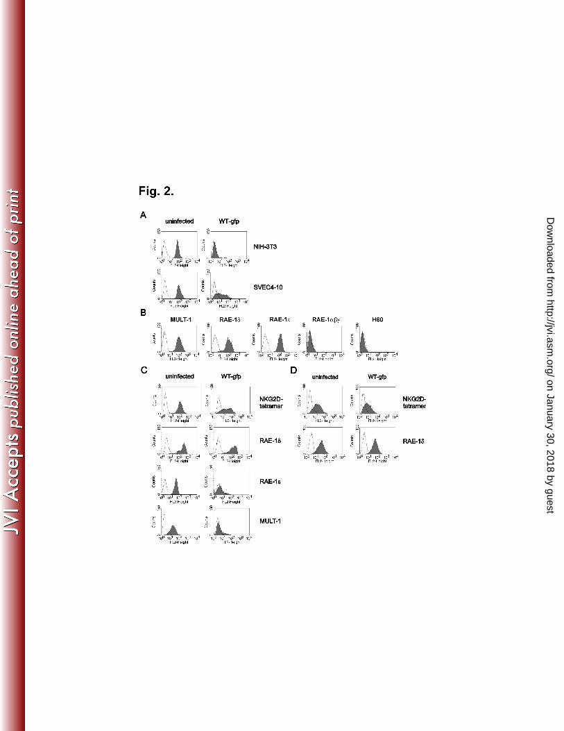
expression of NKG2D ligands, SVEC4-10 cells (C3H/J derived) showed only partial 15
reduction of NKG2D binding (Fig. 2A). Specific mAbs demonstrated MULT-1, RAE-1δ 16
and RAE-1ε on the plasma membrane of SVEC4-10 cells, whereas H60, RAE-1α, β and 17
γ were absent (Fig. 2B). In addition, WT MCMV infection resulted in complete down-18
modulation of surface MULT-1 and RAE-1ε, whereas the expression of RAE-1δ was 19
preserved (Fig. 2C). Similar results were obtained with primary mouse embryonic 20
fibroblasts (MEFs) derived from C3H/J (Fig. 2D), CBA/J, and C57BL/6 mice as well as 21
with C57BL/6 derived cell lines TpnT and IC-21 (data not shown). Altogether, the results 22
showed that RAE-1δ escapes MCMV mediated down-regulation. 23
on January 30, 2018 by guesthttp://jvi.asm
.org/D
ownloaded from

11
m152 abolishes the surface expression of RAE-1γγγγ but does not prevent the 1
expression of RAE-1δδδδ. Down-modulation of RAE-1 molecules is mediated by MCMV 2
m152 (23, 28). Conventional mouse strains express either RAE-1α, β and γ or RAE-3
1δ and ε (28). To avoid the possible impact of MHC class I haplotypes on the 4
susceptibility of RAE-1 isoforms to regulation by MCMV, we generated NIH 3T3 cells 5
stably transfected with RAE-1γ and RAE-1δ. The cell lines were infected with either WT 6
MCMV, the mutant lacking the m152 (∆m152) or left uninfected, and surface expression 7
of RAE-1 was analyzed at different time points post infection (p.i.). In accordance with 8
published data (28), MCMV infection led to down-modulation of RAE-1γ, which was 9
visible already 4 h p.i. (Fig. 3A, left). The effect was m152-dependent, since the level of 10
RAE-1γ expression in ∆m152-infected cells was comparable to that of not infected cells. 11
In contrast, the expression of RAE-1δ on infected cells was only marginally affected 12
(Fig. 3A, right). Similar to WT MCMV, the m152-revertant virus strain (19) showed 13
differential effect on RAE-1γ and RAE-1δ (Supplementary Fig. 2A posted at 14
http://www.medri.hr/~jstipan/jonjic/). 15
m152 differentially affects the maturation pattern of RAE-1δδδδ and RAE-16
1γγγγ. . . . To gain insight into the mechanism of RAE-1 isoform control by MCMV we 17
performed immunoprecipitation (IP) and confocal analysis on the RAE-1γ and RAE-1δ 18
transfectants. Uninfected, WT MCMV-infected, or ∆m152-infected cells were analyzed 19
12 h p.i. Different to RAE-1δ precipitated from control cells or cells infected with 20
∆m152, additional RAE-1δ forms of lower molecular size were immunoprecipitated from 21
the lysate of WT MCMV-infected cells (Fig. 3B). The same results were obtained on 22
transfectans expressing RAE-1γ and RAE-1δ after infection with BAC-derived m152-23
on January 30, 2018 by guesthttp://jvi.asm
.org/D
ownloaded from

12
revertant virus (Supplementary Fig. 2B posted at http://www.medri.hr/~jstipan/jonjic/). 1
The effect of m152 on RAE-1γ was also revealed by accumulation of bands of lower 2
molecular weight representing the immature protein. However, in contrast to RAE-1δ, the 3
loss of mature RAE-1γ protein was almost complete in lysates of WT MCMV-infected 4
cells and RAE-1γ was also not detectable on the surface of WT MCMV-infected cells by 5
confocal microscopy (Fig. 3C). The infection with ∆m152 MCMV resulted in the 6
preservation of RAE-1γ display. 7
This accumulation of lower molecular weight forms of RAE-1δ and RAE-1γ 8
resembles the m152 effect on MHC class I molecules, which are retained in the ER-Golgi 9
intermediate compartment (ERGIC) (54). However, the RAE-1δ expression on the 10
surface of infected cells is in clear contrast to RAE-1γ and MHC class I down-regulation. 11
One explanation could be that newly synthesized RAE-1δ can bypass the m152 block, 12
alternatively the mature cell membrane form of RAE-1δ might be more stable than RAE-13
1γ, or RAE-1δ might mature and transverse to the cell surface at a faster rate thus being 14
less sensitive to control by an MCMV early gene such as m152. 15
The PLWY motif determines resistance of RAE-1δδδδ to MCMV. In an attempt 16
to explain the differential susceptibility of RAE-1 to MCMV, we aligned the sequence of 17
RAE-1 isoforms (Vector NTI AlignX;NCBI Pub-Med database search) (Fig. 4A). As 18
described previously (6), the sequences are highly conserved. However, among other 19
differences, the amino acid sequence PLWY (aa 49-52) present in RAE-1α,β,γ was 20
absent in RAE-1δ, whereas a LPWC sequence in RAE-1ε was found at the same position. 21
Therefore, transfectants of RAE-1δ with an insertion of the PLWY were generated (RAE-22
on January 30, 2018 by guesthttp://jvi.asm
.org/D
ownloaded from

13
1δ-PLWY) and tested for the effect of WT MCMV and ∆m152 MCMV (Fig. 4B). The 1
insertion of the PLWY motif into the RAE-1δ sequence indicated RAE-1δ susceptibility 2
to MCMV regulation. These findings were corroborated by IP (Fig. 4C) and confocal 3
studies (Fig. 4D). Clearly, and in contrast to wild type RAE-1δ showing both immature 4
and mature forms in lysates of cells infected with WT MCMV, only the immature form 5
of the RAE-1δ-PLWY could be precipitated from WT MCMV-infected cells. Two 6
models of PLWY regulation of RAE-1 isoforms are possible. First, PLWY itself could 7
serves as a site for the interaction of RAE-1γ with m152, and second, the PLWY 8
sequence might affects the maturation of the RAE-1 molecules. The lower molecular 9
weight of the mature RAE-1δ-PLWY suggests an effect of the motif on RAE-1 10
glycosylation (Fig. 4C). 11
m152 does not affect the mature form of RAE-1. Due to the presence of m152 12
in MCMV-infected cells the MHC class I proteins mature only to the stage
of EndoH 13
sensitive glycosylation, indicating that the protein does not exit the ER (54). We tested 14
whether the RAE-1δ forms of lower molecular weight represent proteins retained by the 15
m152 in a compartment
with ERGIC/cis-Golgi properties (Fig. 5A). The
EndoH 16
treatment of lysates from WT MCMV-infected cells showed that the lower molecular 17
band of RAE-1δ is EndoH sensitive. However, we noticed a shift in mobility after EndoH 18
treatment even in case of mature forms of RAE-1δ and RAE-1γ suggesting that some of 19
the N-linked carbohydrate chains are still EndoH sensitive. Similar findings were 20
observed for some other cellular (38, 52) and viral glycoproteins (30). 21
The above results demonstrated that m152 can retain immature RAE-1δ in the ER 22
despite simultaneous surface expression of this ligand observed on the WT MCMV-23
on January 30, 2018 by guesthttp://jvi.asm
.org/D
ownloaded from

14
infected cells. We therefore tested whether this discrepancy could be explained by the 1
incomplete block of newly synthesized RAE-1δ. Τo that aim, the cells were labeled with 2
[35
S]methionine between 6 to 8 h p.i. and chased for different periods of time (Fig. 5B). 3
From uninfected cells at zero time of chase both RAE-1δ forms, the lower and higher 4
molecular weight bands, corresponding to immature and mature forms, were 5
immunoprecipitated. As early as after 2 h of chase, a complete maturation of RAE-1δ 6
was observed. However, in WT MCMV-infected cells the protein remained immature 7
throughout the chase period (Fig. 5B), similar to the effect of m152 on MHC class I 8
molecules (54). 9
Next we tested RAE-1δ maturation in comparison to RAE-1γ maturation. To that 10
aim RAE-1δ and RAE-1γ transfectants were pulsed with [35
S]methionine for 1 h and 11
chased. As shown in the Figure 5C, the rate of maturation of RAE-1δ was comparable to 12
that of RAE-1γ. As early as after one hour of chase, RAE-1γ reached the fully mature 13
form, whereas a small proportion of RAE-1δ still remained EndoH sensitive. These 14
results strongly argue against different maturation rates as a reason for resistance to 15
MCMV infection. 16
Next we tested whether surface expression of RAE-1δ in WT MCMV-infected 17
cells is a consequence of the inability of the virus to affect the RAE-1δ which had 18
matured already prior to infection and m152 expression. To that aim, RAE-1δ or RAE-19
1γ transfectants were pulsed with [35
S]methionine for 2 h prior to infection and chased for 20
up to 12 h. As shown in Fig. 5E, RAE-1δ reached the mature form and remained in this 21
form stably expressed also in WT MCMV-infected cells. The results document a 22
differential susceptibility of immature and mature forms of RAE-1δ to MCMV. In 23
on January 30, 2018 by guesthttp://jvi.asm
.org/D
ownloaded from

15
addition there were differences between the half-lives of mature RAE-1δ and RAE-1
1γ proteins (Fig. 5E). Prior to chase, both immature and mature RAE-1γ forms were 2
present, similar to RAE-1δ, whereas during chase also mature RAE-1γ protein were lost 3
from lysates of uninfected and MCMV-infected cells. However, the disappearance of the 4
mature RAE-1γ protein was faster during infection conditions. 5
The PLWY motif determines the stability of RAE-1 proteins. The lack of a 6
PLWY motif determined the resistance of surface RAE-1δ to MCMV (Fig. 4) and its role 7
in the RAE-1 glycosylation pattern has been indicated. Since RAE-1δ surface expression 8
in WT MCMV-infected cells cannot be explained by escape of newly synthesized RAE-9
1δ from the m152-mediated export block (Fig. 5B), we tested whether the PLWY motif 10
defines the stability of RAE-1 isoforms. Therefore, RAE-1γ, RAE-1δ and RAE-1δ-11
PLWY transfectants were treated with the glycosylation inhibitor, tunicamycin, and 12
surface biotinylation was carried out. RAE-1 proteins were immunobloted from the 13
lysates of the untreated cells or cells incubated with tunicamycin (Fig. 5D). The mature 14
form of RAE-1γ was visualized in untreated cells, whereas after 12 h of tunicamycin 15
treatment mainly the immature, unglycosylated form of protein was detected. A similar 16
effect on RAE-1γ was observed upon the cycloheximide treatment (data not shown). In 17
contrast, a significant fraction of mature RAE-1δ was still present after 12 h of 18
tunicamycin treatment. The differential stability of the mature RAE-1δ and RAE-19
1γ forms was affected by the presence of PLWY, since the mature form of RAE-1δ-20
PLWY showed the pattern of RAE-1γ. The same differences between RAE-1δ, RAE-1γ, 21
and RAE-1δ-PLWY were seen when the analysis addressed mainly the stability of 22
on January 30, 2018 by guesthttp://jvi.asm
.org/D
ownloaded from

16
surface resident molecules by biotinylation (Fig. 5F). The reduction of the RAE-1δ signal 1
during a 12 h period after surface labeling is small, compared to the short half life of 2
RAE-1γ and RAE-1δ-PLWY. Altogether, the differential susceptibility of surface RAE-3
1γ and RAE-1δ to down-regulation during MCMV infection is determined by the PLWY 4
motif which affects the intrinsic stability of the mature protein forms. 5
Discussion 6
NKG2D is a dominant NK cell activating receptor present on almost all human 7
and mouse NK cells. HCMV and MCMV encode a set of immunevasins which avoid 8
activation of this receptor through down-modulation of NKG2D ligands (20). Here we 9
report the inability of MCMV to down-modulate RAE-1δ from the surface of infected 10
cells. Our results also showed the functional significance of RAE-1δ escape from MCMV 11
regulation. Although m152 is able to affect all RAE-1 isoforms, it affects only their 12
immature forms. We show that RAE-1γ and RAE-1δ isoforms possess intrinsic 13
differences with respect to the stability of their mature forms. The differential stability of 14
RAE-1 proteins and their susceptibility to MCMV is associated with a PLWY motif 15
which affects RAE-1 glycosylation and leads to the shorter half-life of the mature RAE-1 16
that possesses this motif. 17
After co-transfection of 293T cells with m152 and RAE-1 cDNAs (α, β, γ, δ or ε), 18
all RAE-1 proteins were found to be susceptible to the impact of m152 (28). We have 19
shown that the molecular mechanism of the m152 mediated effect is the retention of 20
RAE-1 molecules in the ER/cis-Golgi compartment. However, in contrast to the model 21
used by Lodoen et al. in which the m152 effect was tested in isolation, here we used cell 22
lines which constitutively or stably express RAE-1δ. Here, MCMV is unable to down-23
on January 30, 2018 by guesthttp://jvi.asm
.org/D
ownloaded from

17
regulate surface-resident RAE-1δ. It is of note that RAE-1 proteins are rapidly induced 1
(6, 16) also upon MCMV infection (28, 44). We demonstrate that newly synthesized 2
RAE-1 molecules mature in less than 2 hours. It is to be expected that after MCMV 3
infection significant amounts of RAE-1 proteins are displayed on the plasma membrane 4
well before the function of m152 can take place. 5
The m152 was first described by its ability to cause the retention of MHC class I 6
in the ER/cis-Golgi compartment (54). The cytoplasmic tail and the transmembrane 7
region of m152 are not relevant for the MHC class I retention by m152 (53). Since RAE-8
1 proteins are GPI anchored (6, 10), the logical presumption was that the luminal domain 9
of m152 is also involved in its effect on RAE-1. The m152 and MHC class I molecules 10
do not co-precipitate (53) and we were also unable to find complexes of the m152 with 11
RAE-1. Although m152 is able to affect all mouse MHC class I haplotypes (54), there are 12
allele-specific differences in MHC class I down-regulation (46). Although all RAE-1 13
proteins are target for m152, the RAE-1β isoform shows the highest degree of 14
susceptibility (28). When we first noticed RAE-1δ resistance to MCMV, we predicted 15
that m152 might have lower affinity for this isoform, and in that way, a substantial 16
portion of RAE-1δ could escape m152-mediated block. However, IP/WB analysis 17
showed nascent RAE-1δ susceptibility. The m152-retained RAE-1δ molecules were not 18
degraded, which resembles the m152 effect on MHC class I molecules (53). 19
The differential stability of the mature RAE-1 γ and δ was lost after the insertion 20
of PLWY motif in the RAE-1δ sequence. The PLWY motif did not affect RAE-1δ 21
susceptibility to m152, but affected the stability of the ligand. Insertion of PLWY in 22
RAE-1δ resulted not only in the altered sensitivity to MCMV, but also in a significantly 23
on January 30, 2018 by guesthttp://jvi.asm
.org/D
ownloaded from

18
lower molecular weight of precipitated proteins resembling the pattern of RAE-1γ. One 1
could speculate that RAE-1δ is differently glycosylated due to structural effect of the 2
missing PLWY. The crystal structure of murine RAE-1β (27) can serve as a template to 3
determine the PLWY-containing loop and its relationship to the N-linked carbohydrate 4
addition sites. As shown in Supplementary figure S3 5
(http://www.medri.hr/~jstipan/jonjic/) the PLWY loop lies close to the N70 glycosylation 6
site, while surface representation shows that both are in a prominent solvent exposed 7
region. The lack of PLWY, as observed in RAE-1δ, would eliminate the bulky PLWY 8
exposed loop and possibly make N70 more accessible as well. In other words, PLWY 9
might limit the accessibility of N70 to the glycosylation machinery needed for the full 10
maturation of RAE-1 molecules. 11
Certain human ligands of NKG2D receptor also show differential resistance to 12
viral inhibitors. HCMV UL16 arrests ULBP-1, ULBP-2 and MICB proteins in the ER, 13
but not the ULBP-3, ULBP-4 and MICA family members (9, 12, 41, 48, 51). Sequence 14
differences in the MICA and MICB α2 domains precluded UL16 from binding to MICA 15
and in addition, the replacement of the MICA α2 with the MICB α2 domain resulted in 16
the UL16 sensitive recombinant protein (43). However, another HCMV encoded viral 17
inhibitor, UL142, was later characterized as a specific down-regulator of MICA (7, 50). 18
Interestingly, UL142 is not able to affect surface expression of all MICA alleles; the 19
truncated form of MICA, which is the most common, is completely resistant to the 20
HCMV regulation (55). The K5 protein of the Kaposi sarcoma-associated herpesvirus 21
(KSHV) downregulates the NKG2D ligands MICA and MICB as well as the AICL 22
on January 30, 2018 by guesthttp://jvi.asm
.org/D
ownloaded from

19
ligand for the NKp80 receptor (45). Like in the case of UL142, the truncated MICA form 1
lacking the cytoplasmic tail is resistant to K5. 2
Variations in RAE-1 isoforms are probably consequences of selective pressure of 3
the virus during the evolution. The unique absence of PLWY motif in RAE-1δ may 4
represent a host escape from MCMV. Another counterpart of RAE-1, RAE-1ε, possesses 5
a LPWC motif instead of PLWY. Recently we could show that deletion of m152 only is 6
not sufficient to prevent down-modulation of RAE-1ε. MCMV has other means to 7
complete RAE-1ε down-modulation, namely the viral protein m138/fcr1 (1). RAE-1ε 8
possesses the highest affinity for the NKG2D receptor of all RAE-1 isoforms. Notably, 9
RAE-1δ has the lowest affinity (5, 36). This might explain a more intensive viral effort to 10
regulate RAE-1ε than RAE-1δ surface expression. Still, the phenomenon of RAE-1δ 11
resistance in vivo points to a threat to the virus since the increased resistance of some 12
mouse strains to acute MCMV infection can be attributed to NK/NKG2D-mediated 13
control. Thus, the results presented in this work emphasize a continuous evolutional 14
struggle between viruses and their hosts. NKG2D as a dominant activation receptor 15
important not only for the innate but also for the adaptive immune response is an obvious 16
target. 17
18
Acknowledgments 19
We thank David H. Margulies and Li Zhi for help and critical reading of the manuscript, 20
Mark Lötzerich and Frederic Lemmnitzer for valuable information, Hermine Muehlbach 21
and Christian Mohr for help with transfectants and Edvard Razic for technical help. Our 22
work is supported by Croatian Ministry of Science - grants: 0621261-1263 (SJ) and 23
on January 30, 2018 by guesthttp://jvi.asm
.org/D
ownloaded from

20
0621261-1268 (AK). AK is supported by the Howard Hughes Medical Institute 1
International Research Scholars grant. UHK and ZR are supported by the Deutsche 2
Forschungsgemeinschaft through SFB 455. LC was supported by NIAID K08 AI057361. 3
4
5
References 6
1. Arapovic, J., T. Lenac Rovis, A. B. Reddy, A. Krmpotic, and S. Jonjic. 2009. 7
Promiscuity of MCMV immunoevasin of NKG2D: m138/fcr-1 down-modulates 8
RAE-1varepsilon in addition to MULT-1 and H60. Mol Immunol. 9
2. Bauer, S., V. Groh, J. Wu, A. Steinle, J. H. Phillips, L. L. Lanier, and T. 10
Spies. 1999. Activation of NK cells and T cells by NKG2D, a receptor for stress-11
inducible MICA. Science 285:727-9. 12
3. Bubic, I., M. Wagner, A. Krmpotic, T. Saulig, S. Kim, W. M. Yokoyama, S. 13
Jonjic, and U. H. Koszinowski. 2004. Gain of virulence caused by loss of a gene 14
in murine cytomegalovirus. J Virol 78:7536-44. 15
4. Carayannopoulos, L. N., O. V. Naidenko, D. H. Fremont, and W. M. 16
Yokoyama. 2002. Cutting edge: murine UL16-binding protein-like transcript 1: a 17
newly described transcript encoding a high-affinity ligand for murine NKG2D. J 18
Immunol 169:4079-83. 19
5. Carayannopoulos, L. N., O. V. Naidenko, J. Kinder, E. L. Ho, D. H. Fremont, 20
and W. M. Yokoyama. 2002. Ligands for murine NKG2D display heterogeneous 21
binding behavior. Eur J Immunol 32:597-605. 22
6. Cerwenka, A., A. B. Bakker, T. McClanahan, J. Wagner, J. Wu, J. H. 23
Phillips, and L. L. Lanier. 2000. Retinoic acid early inducible genes define a 24
ligand family for the activating NKG2D receptor in mice. Immunity 12:721-7. 25
7. Chalupny, N. J., A. Rein-Weston, S. Dosch, and D. Cosman. 2006. Down-26
regulation of the NKG2D ligand MICA by the human cytomegalovirus 27
glycoprotein UL142. Biochem Biophys Res Commun 346:175-81. 28
8. Cobbold, S. P., A. Jayasuriya, A. Nash, T. D. Prospero, and H. Waldmann. 29
1984. Therapy with monoclonal antibodies by elimination of T-cell subsets in 30
vivo. Nature 312:548-51. 31
9. Cosman, D., J. Mullberg, C. L. Sutherland, W. Chin, R. Armitage, W. 32
Fanslow, M. Kubin, and N. J. Chalupny. 2001. ULBPs, novel MHC class I-33
related molecules, bind to CMV glycoprotein UL16 and stimulate NK 34
cytotoxicity through the NKG2D receptor. Immunity 14:123-33. 35
10. Diefenbach, A., A. M. Jamieson, S. D. Liu, N. Shastri, and D. H. Raulet. 2000. 36
Ligands for the murine NKG2D receptor: expression by tumor cells and activation 37
of NK cells and macrophages. Nat Immunol 1:119-26. 38
on January 30, 2018 by guesthttp://jvi.asm
.org/D
ownloaded from

21
11. Dokun, A. O., S. Kim, H. R. Smith, H. S. Kang, D. T. Chu, and W. M. 1
Yokoyama. 2001. Specific and nonspecific NK cell activation during virus 2
infection. Nat Immunol 2:951-6. 3
12. Dunn, C., N. J. Chalupny, C. L. Sutherland, S. Dosch, P. V. Sivakumar, D. C. 4
Johnson, and D. Cosman. 2003. Human cytomegalovirus glycoprotein UL16 5
causes intracellular sequestration of NKG2D ligands, protecting against natural 6
killer cell cytotoxicity. J Exp Med 197:1427-39. 7
13. French, A. R., and W. M. Yokoyama. 2003. Natural killer cells and viral 8
infections. Curr Opin Immunol 15:45-51. 9
14. Girardi, M., D. E. Oppenheim, C. R. Steele, J. M. Lewis, E. Glusac, R. Filler, 10
P. Hobby, B. Sutton, R. E. Tigelaar, and A. C. Hayday. 2001. Regulation of 11
cutaneous malignancy by gammadelta T cells. Science 294:605-9. 12
15. Hasan, M., A. Krmpotic, Z. Ruzsics, I. Bubic, T. Lenac, A. Halenius, A. 13
Loewendorf, M. Messerle, H. Hengel, S. Jonjic, and U. H. Koszinowski. 2005. 14
Selective down-regulation of the NKG2D ligand H60 by mouse cytomegalovirus 15
m155 glycoprotein. J Virol 79:2920-30. 16
16. Hayakawa, Y., J. M. Kelly, J. A. Westwood, P. K. Darcy, A. Diefenbach, D. 17
Raulet, and M. J. Smyth. 2002. Cutting edge: tumor rejection mediated by 18
NKG2D receptor-ligand interaction is dependent upon perforin. J Immunol 19
169:5377-81. 20
17. Hengel, H., U. Reusch, A. Gutermann, H. Ziegler, S. Jonjic, P. Lucin, and U. 21
H. Koszinowski. 1999. Cytomegaloviral control of MHC class I function in the 22
mouse. Immunol Rev 168:167-76. 23
18. Ho, E. L., L. N. Carayannopoulos, J. Poursine-Laurent, J. Kinder, B. 24
Plougastel, H. R. Smith, and W. M. Yokoyama. 2002. Costimulation of 25
multiple NK cell activation receptors by NKG2D. J Immunol 169:3667-75. 26
19. Holtappels, R., J. Podlech, M. F. Pahl-Seibert, M. Julch, D. Thomas, C. O. 27
Simon, M. Wagner, and M. J. Reddehase. 2004. Cytomegalovirus misleads its 28
host by priming of CD8 T cells specific for an epitope not presented in infected 29
tissues. J Exp Med 199:131-6. 30
20. Jonjic, S., M. Babic, B. Polic, and A. Krmpotic. 2008. Immune evasion of 31
natural killer cells by viruses. Curr Opin Immunol 20:30-8. 32
21. Jonjic, S., A. Krmpotic, J. Arapovic, and U. H. Koszinowski. 2008. Dissection 33
of the antiviral NK cell response by MCMV mutants. Methods Mol Biol 415:127-34
49. 35
22. Krmpotic, A., I. Bubic, B. Polic, P. Lucin, and S. Jonjic. 2003. Pathogenesis of 36
murine cytomegalovirus infection. Microbes Infect 5:1263-77. 37
23. Krmpotic, A., D. H. Busch, I. Bubic, F. Gebhardt, H. Hengel, M. Hasan, A. 38
A. Scalzo, U. H. Koszinowski, and S. Jonjic. 2002. MCMV glycoprotein gp40 39
confers virus resistance to CD8+ T cells and NK cells in vivo. Nat Immunol 40
3:529-35. 41
24. Krmpotic, A., M. Hasan, A. Loewendorf, T. Saulig, A. Halenius, T. Lenac, B. 42
Polic, I. Bubic, A. Kriegeskorte, E. Pernjak-Pugel, M. Messerle, H. Hengel, 43 D. H. Busch, U. H. Koszinowski, and S. Jonjic. 2005. NK cell activation 44
through the NKG2D ligand MULT-1 is selectively prevented by the glycoprotein 45
encoded by mouse cytomegalovirus gene m145. J Exp Med 201:211-20. 46
on January 30, 2018 by guesthttp://jvi.asm
.org/D
ownloaded from

22
25. Krmpotic, A., M. Messerle, I. Crnkovic-Mertens, B. Polic, S. Jonjic, and U. 1
H. Koszinowski. 1999. The immunoevasive function encoded by the mouse 2
cytomegalovirus gene m152 protects the virus against T cell control in vivo. J Exp 3
Med 190:1285-96. 4
26. Lenac, T., M. Budt, J. Arapovic, M. Hasan, A. Zimmermann, H. Simic, A. 5
Krmpotic, M. Messerle, Z. Ruzsics, U. H. Koszinowski, H. Hengel, and S. 6 Jonjic. 2006. The herpesviral Fc receptor fcr-1 down-regulates the NKG2D 7
ligands MULT-1 and H60. J Exp Med 203:1843-50. 8
27. Li, P., G. McDermott, and R. K. Strong. 2002. Crystal structures of RAE-1beta 9
and its complex with the activating immunoreceptor NKG2D. Immunity 16:77-10
86. 11
28. Lodoen, M., K. Ogasawara, J. A. Hamerman, H. Arase, J. P. Houchins, E. S. 12
Mocarski, and L. L. Lanier. 2003. NKG2D-mediated natural killer cell 13
protection against cytomegalovirus is impaired by viral gp40 modulation of 14
retinoic acid early inducible 1 gene molecules. J Exp Med 197:1245-53. 15
29. Lodoen, M. B., G. Abenes, S. Umamoto, J. P. Houchins, F. Liu, and L. L. 16
Lanier. 2004. The cytomegalovirus m155 gene product subverts natural killer cell 17
antiviral protection by disruption of H60-NKG2D interactions. J Exp Med 18
200:1075-81. 19
30. Lu, X., D. G. Kavanagh, and A. B. Hill. 2006. Cellular and molecular 20
requirements for association of the murine cytomegalovirus protein m4/gp34 with 21
major histocompatibility complex class I molecules. J Virol 80:6048-55. 22
31. Ljunggren, H. G., and K. Karre. 1990. In search of the 'missing self': MHC 23
molecules and NK cell recognition. Immunol Today 11:237-44. 24
32. Malarkannan, S., P. P. Shih, P. A. Eden, T. Horng, A. R. Zuberi, G. 25
Christianson, D. Roopenian, and N. Shastri. 1998. The molecular and 26
functional characterization of a dominant minor H antigen, H60. J Immunol 27
161:3501-9. 28
33. Mathys, S., T. Schroeder, J. Ellwart, U. H. Koszinowski, M. Messerle, and U. 29
Just. 2003. Dendritic cells under influence of mouse cytomegalovirus have a 30
physiologic dual role: to initiate and to restrict T cell activation. J Infect Dis 31
187:988-99. 32
34. Mocarski, E. S., and C. T. Courcelle. 2001. Cytomegaloviruses and Their 33
Replication, p. 2629-2673. In D. M. Knipe and P. M. Howley (ed.), Virology, vol. 34
2. Lippincott Williams and Wilkins, Philadelphia. 35
35. Nomura, M., Z. Zou, T. Joh, Y. Takihara, Y. Matsuda, and K. Shimada. 36
1996. Genomic structures and characterization of Rae1 family members encoding 37
GPI-anchored cell surface proteins and expressed predominantly in embryonic 38
mouse brain. J Biochem (Tokyo) 120:987-95. 39
36. O'Callaghan, C. A., A. Cerwenka, B. E. Willcox, L. L. Lanier, and P. J. 40
Bjorkman. 2001. Molecular competition for NKG2D: H60 and RAE1 compete 41
unequally for NKG2D with dominance of H60. Immunity 15:201-11. 42
37. Ohe, Y., D. Zhao, N. Saijo, and E. R. Podack. 1995. Construction of a novel 43
bovine papillomavirus vector without detectable transforming activity suitable for 44
gene transfer. Hum Gene Ther 6:325-33. 45
on January 30, 2018 by guesthttp://jvi.asm
.org/D
ownloaded from

23
38. Omary, M. B., and I. S. Trowbridge. 1981. Biosynthesis of the human 1
transferrin receptor in cultured cells. J Biol Chem 256:12888-92. 2
39. Raulet, D. H. 2003. Roles of the NKG2D immunoreceptor and its ligands. Nat 3
Rev Immunol 3:781-90. 4
40. Reddehase, M. J., J. Podlech, and N. K. Grzimek. 2002. Mouse models of 5
cytomegalovirus latency: overview. J Clin Virol 25 Suppl 2:S23-36. 6
41. Rolle, A., M. Mousavi-Jazi, M. Eriksson, J. Odeberg, C. Soderberg-Naucler, 7
D. Cosman, K. Karre, and C. Cerboni. 2003. Effects of human cytomegalovirus 8
infection on ligands for the activating NKG2D receptor of NK cells: up-regulation 9
of UL16-binding protein (ULBP)1 and ULBP2 is counteracted by the viral UL16 10
protein. J Immunol 171:902-8. 11
42. Scalzo, A. A., A. J. Corbett, W. D. Rawlinson, G. M. Scott, and M. A. Degli-12
Esposti. 2007. The interplay between host and viral factors in shaping the 13
outcome of cytomegalovirus infection. Immunol Cell Biol 85:46-54. 14
43. Spreu, J., T. Stehle, and A. Steinle. 2006. Human cytomegalovirus-encoded 15
UL16 discriminates MIC molecules by their alpha2 domains. J Immunol 16
177:3143-9. 17
44. Takada, A., S. Yoshida, M. Kajikawa, Y. Miyatake, U. Tomaru, M. Sakai, H. 18
Chiba, K. Maenaka, D. Kohda, K. Fugo, and M. Kasahara. 2008. Two novel 19
NKG2D ligands of the mouse H60 family with differential expression patterns 20
and binding affinities to NKG2D. J Immunol 180:1678-85. 21
45. Thomas, M., J. M. Boname, S. Field, S. Nejentsev, M. Salio, V. Cerundolo, 22
M. Wills, and P. J. Lehner. 2008. Down-regulation of NKG2D and NKp80 23
ligands by Kaposi's sarcoma-associated herpesvirus K5 protects against NK cell 24
cytotoxicity. Proc Natl Acad Sci U S A 105:1656-61. 25
46. Wagner, M., A. Gutermann, J. Podlech, M. J. Reddehase, and U. H. 26
Koszinowski. 2002. Major histocompatibility complex class I allele-specific 27
cooperative and competitive interactions between immune evasion proteins of 28
cytomegalovirus. J Exp Med 196:805-16. 29
47. Wagner, M., S. Jonjic, U. H. Koszinowski, and M. Messerle. 1999. Systematic 30
excision of vector sequences from the BAC-cloned herpesvirus genome during 31
virus reconstitution. J Virol 73:7056-60. 32
48. Welte, S. A., C. Sinzger, S. Z. Lutz, H. Singh-Jasuja, K. L. Sampaio, U. 33
Eknigk, H. G. Rammensee, and A. Steinle. 2003. Selective intracellular 34
retention of virally induced NKG2D ligands by the human cytomegalovirus UL16 35
glycoprotein. Eur J Immunol 33:194-203. 36
49. Wilkinson, G. W., P. Tomasec, R. J. Stanton, M. Armstrong, V. 37
Prod'homme, R. Aicheler, B. P. McSharry, C. R. Rickards, D. Cochrane, S. 38 Llewellyn-Lacey, E. C. Wang, C. A. Griffin, and A. J. Davison. 2008. 39
Modulation of natural killer cells by human cytomegalovirus. J Clin Virol 41:206-40
12. 41
50. Wills, M. R., O. Ashiru, M. B. Reeves, G. Okecha, J. Trowsdale, P. Tomasec, 42
G. W. Wilkinson, J. Sinclair, and J. G. Sissons. 2005. Human cytomegalovirus 43
encodes an MHC class I-like molecule (UL142) that functions to inhibit NK cell 44
lysis. J Immunol 175:7457-65. 45
on January 30, 2018 by guesthttp://jvi.asm
.org/D
ownloaded from

24
51. Wu, J., N. J. Chalupny, T. J. Manley, S. R. Riddell, D. Cosman, and T. Spies. 1
2003. Intracellular retention of the MHC class I-related chain B ligand of NKG2D 2
by the human cytomegalovirus UL16 glycoprotein. J Immunol 170:4196-200. 3
52. Yuan, W., A. Dasgupta, and P. Cresswell. 2006. Herpes simplex virus evades 4
natural killer T cell recognition by suppressing CD1d recycling. Nat Immunol 5
7:835-42. 6
53. Ziegler, H., W. Muranyi, H. G. Burgert, E. Kremmer, and U. H. 7
Koszinowski. 2000. The luminal part of the murine cytomegalovirus glycoprotein 8
gp40 catalyzes the retention of MHC class I molecules. Embo J 19:870-81. 9
54. Ziegler, H., R. Thäle, P. Lucin, W. Muranyi, T. Flohr, H. Hengel, H. Farrell, 10
W. Rawlinson, and U. H. Koszinowski. 1997. A mouse cytomegalovirus 11
glycoprotein retains MHC class I complexes in the ERGIC/cis-Golgi 12
compartments. Immunity 6:57-66. 13
55. Zou, Y., W. Bresnahan, R. T. Taylor, and P. Stastny. 2005. Effect of human 14
cytomegalovirus on expression of MHC class I-related chains A. J Immunol 15
174:3098-104. 16
17
18
Figure legends 19
FIG. 1. NKG2D-dependent virus control in vivo. (A) CBA/J, C3H/J and 20
BALB/c mice were injected i.v. with 3×10⁵ PFU of WT MCMV (MW 97.01). C57BL/6 21
mice received the same dose of ∆m157 MCMV. Mice were injected i.p. with PBS or with 22
blocking anti-NKG2D mAbs. Virus titers were determined 3 days post infection. There 23
were significant differences in virus titers in spleen between the groups of untreated mice 24
and groups treated with anti-NKG2D mAbs: for CBA/J mice (P=0.008), and for 25
C57BL/6 mice (P=0.038). Because of small number of animals per group (n=3) for 26
C3H/J mice, although virus titer differences are indicative, there were no statistically 27
significant differences. (B) C57BL/6 mice, injected i.p. with either PBS or blocking 28
NKG2D mAbs either alone or in combination with cytolytic anti CD4 and anti CD8 29
mAbs, were injected i.v. with 2×10⁵ PFU of ∆m157 virus. Virus titers were determined 7 30
days post infection. There were significant differences between untreated group and T 31
cell depleted group (P=0.029) as well as between the untreated group and group treated 32
on January 30, 2018 by guesthttp://jvi.asm
.org/D
ownloaded from

25
with anti-NKG2D, anti-CD4 and anti-CD8 mAbs (P=0.049). (C) DBA/2 mice, were 1
injected i.p. with PBS or blocking NKG2D mAbs or in addition also with cytolytic anti 2
CD4 and anti CD8 mAbs. They were also injected i.p. with 5×10⁴ PFU of salivary gland-3
derived (SGV)-WT MCMV. Virus titers were determined 11 days post infection. There 4
were significant differences between untreated group and T cell depleted group 5
(P=0.049) as well as untreated group and group treated with anti-NKG2D, anti-CD4 and 6
anti-CD8 mAbs (P=0.029). Titers for individual mice (circles) and median values 7
(horizontal bars) are shown. 8
9
FIG. 2. Different susceptibility of NKG2D ligands to MCMV. (A) NIH 3T3 10
and SVEC4-10 cells were infected for 12 h with 1 PFU/cell of WT-gfp MCMV or left 11
uninfected. Cells were stained with PE-labeled NKG2D tetramer (filled histograms). AV-12
PE was used as a control (open histograms). (B) Expression of NKG2D ligands on 13
SVEC4-10 cells was tested by specific mAbs: rat anti-MULT-1, mouse anti-RAE-1δ, rat 14
anti-RAE-1ε, rat anti-RAE-1αβγ and rat anti-H60 followed by biotinylated-goat anti-rat 15
IgG or biotinylated-goat anti-mouse IgG and PE-labeled streptavidin (filled histograms). 16
Isotype-matched rat IgG2a and mouse IgG1 mAbs were used as a negative control (open 17
histograms). (C) SVEC4-10 and (D) C3H/J derived MEFs were infected for 12 h with 1 18
PFU/cell of WT-gfp MCMV or left uninfected. Cells were stained with PE-labeled 19
NKG2D tetramer or with mAbs to RAE-1δ, RAE-1ε and MULT-1 (filled histograms). 20
Isotype-matched irrelevant mAbs and AV-PE were used as controls for staining with 21
specific antibodies and tetramer, respectively (open histograms). 22
23
on January 30, 2018 by guesthttp://jvi.asm
.org/D
ownloaded from

26
FIG. 3. m152 affects the maturation of RAE-1 proteins. RAE-1γ- and RAE-1δ-1
transfected NIH 3T3 cells were infected with 4 PFU/cell of WT MCMV, ∆m152 MCMV 2
or left uninfected. Cells were analyzed with specific anti-RAE-1 mAbs: (A) by flow 3
cytometry, (B) by immunoblot (IB) after the immunoprecipitation (IP) with anti FLAG 4
M2 sepharose and (C) by confocal microscopy. 5
6
FIG. 4. The PLWY motif of RAE-1 protein modulates sensitivity to MCMV. 7
(A) Comparison of the amino acid sequences of RAE-1 isoforms using the Vector NT 8
AlignX program (NCBI Pub-Med database search). PLWY motif (aa 49 to 52) is marked. 9
(B) RAE-1γ-, RAE-1δ- and RAE-1δ-PLWY-transfected NIH 3T3 cells were infected for 10
12 h with indicated viruses and analyzed for the expression of RAE-1 isoforms using the 11
anti FLAG M2 mAb. (C) RAE-1δ- and RAE-1δ-PLWY-transfected NIH 3T3 cells were 12
infected for 12 h with 4 PFU/cell of indicated viruses and analyzed either by 13
immunobloting (IB) using the anti-RAE-1 mAbs after the immunoprecipitation (IP) with 14
anti FLAG-M2 sepharose or (D) by confocal microscopy. 15
16
FIG. 5. Differential stability of RAE-1δ and RAE-1γ γ γ γ mature forms. (A) RAE-1δ-17
transfected NIH 3T3 cells were infected with 4 PFU/cell of WT MCMV, ∆m152 MCMV 18
or left uninfected. RAE-1δ was immunobloted from EndoH treated or untreated lysates 19
with anti-RAE-1δ mAbs. (B) RAE-1δ- or empty vector-transfected NIH 3T3 cells were 20
infected with 4 PFU of WT MCMV or left uninfected. Cells were metabolically labeled 21
with 300 µCi/ml of [35
S]methionine 6h to 8h after infection, chased for indicated periods 22
of time and immunoprecipitation was performed using anti-RAE-1δ mAbs followed by 23
on January 30, 2018 by guesthttp://jvi.asm
.org/D
ownloaded from
27
protein G-Sepharose. (C) RAE-1δ- or RAE-1γ-transfected NIH 3T3 cells were labeled 1
with 500 µCi/ml of [35
S]methionine for 1h and chased for indicated periods of time. After 2
immunoprecipitation with either anti-RAE-1δ or anti-RAE-1γ mAbs followed by G-3
sepharose, eluted proteins were treated with EndoH or left untreated. (D) RAE-1γ-, RAE-4
1δ- and RAE-1δ-PLWY-transfected NIH 3T3 cells were untreated or treated for 12 h 5
with tunicamycin (2 µg/ml). Cell lysates were immunobloted using specific anti-RAE-1 6
antibodies. (E) RAE-1δ-, RAE-1γ- or empty vector-transfected NIH 3T3 cells were 7
metabolically labeled with 300 µCi/ml of [35
S]methionine for 2h before infection with 4 8
PFU of WT MCMV. Cells were chased for indicated periods of time and 9
immunoprecipitation was performed using either anti-RAE-1δ mAbs or anti-RAE-1γ 10
antibodies followed by protein G-Sepharose. (F) RAE-1δ-, RAE-1γ- and RAE-1δ-11
PLWY-transfected NIH 3T3 cells were surface biotinylated. After indicated periods of 12
time RAE-1 molecules were immunoprecipitated using anti-FLAG M2-sepharose 13
followed by immunobloting with SA-POD. Arrows indicate different maturation forms of 14
RAE-1 proteins: R, resistant to EndoH; S, sensitive to EndoH; D, digested with EndoH. 15
RAE-1 Tun.R indicates matured form of RAE-1 which is resistant to Tunicamycin 16
whereas RAE-1 Tun.S indicates deglycosylated form of RAE-1 proteins by Tunicamycin. 17
18
19
20
on January 30, 2018 by guesthttp://jvi.asm
.org/D
ownloaded from