direct recording and pressures within the
TRANSCRIPT
Brit. Heart J., 1963, 25, 549.
DIRECT RECORDING OF SOUNDS AND PRESSURESWITHIN THE HEART
BY
D. W. BARRITT AND D. H. DAVIESFrom the Cardiac Department, Bristol Royal Hospital, Bristol
Received October 22, 1962
Pressure waves and vibrations in the heart and great vessels may be conducted to a manometeroutside the body by a system of tubing filled with fluid. More than a metre of tubing is usuallyneeded as the circulation is entered at an arm vein. Any such system has the following defects.
1. Delay in the transmission of the low frequency pressure waves to the manometer: the delaywill vary slightly with different tubes and with the temperature and density of the fluid within.
2. Unwanted vibrations set up along the length of the tubing as its sides are set in motion by theheart chambers- that it traverses.
3. Smoothing-out of the pressure waves or overshoot is almost unavoidable as a result of theelasticity of the tubing and the inertia of the fluid.
4. The high frequency, low amplitude vibrations that constitute audible sounds and murmursare so poorly conducted that no satisfactory sound record can be made.
Elimination of the long fluid column by the. use of a miniature manometer introduced into theheart may overcome all these difficulties. Such a manometer is now available and Soulie and histeam have reported their extensive use of the Allard-Laurens micromanometer (Soulie et al.,1961). The present paper reports our preliminary experience with this instrument.
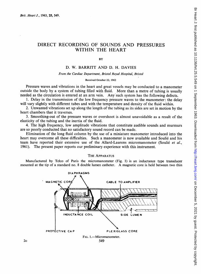
THE APPARATUSManufactured by Telco of Paris the micromanometer (Fig. 1) is an inductance type transducer
mounted at the tip of a standard no. 8 double lumen catheter. A magnetic core is held between two thin
DIA PHRAGMS
MAGNETIC C CABLE TO AMPLIFIER
CA P
COI L SIDE LUME
PLEXIGLASS CORE
FIG. 1.-Micromanometer.5492o
on Decem
ber 5, 2021 by guest. Protected by copyright.
http://heart.bmj.com
/B
r Heart J: first published as 10.1136/hrt.25.5.549 on 1 S
eptember 1963. D
ownloaded from

550 BARRITT AND DAVIES
I SIGNAL
INiTMA NOM.
EXTMA NOM
I* O*. ~ . ~4
14'
FIG. 2.-Transmission delay curve.
diaphragms and moves axially with pressure change. Its movement modifies the inductance of a coilaround it. The signals are amplified and low frequency vibrations give the pressure waves and high fre-quencies record sounds. The sound channel accepts frequencies of the order of 30 to 2000 cycles persecond. Thus, sounds and pressures are recorded separately and simultaneously from the catheter tip withno transmission delay. The second lumen of the catheter allows blood sampling and a standard pressure
INTRACARDIAC PHONO.
EXT PHONO. PA,,, A2
A
/~~.-1..,"IfW
MICRO. MANOM., A
STANDARD MANOM.
EGIPLOAYATRECG. X PULMONARY ARTE:RY
FIG. 3.-Pulmonary valvular stenosis with ventricular septal defect.
fs
on Decem
ber 5, 2021 by guest. Protected by copyright.
http://heart.bmj.com
/B
r Heart J: first published as 10.1136/hrt.25.5.549 on 1 S
eptember 1963. D
ownloaded from
DIRECT RECORDING OF SOUNDS AND PRESSURES WITHIN THE HEART 551
recording through the side hole. Sounds have been recorded at a paper speed of 80 mm. per second usingN.E.P. galvanometers and camera. The internal manometer is calibrated by superimposing the pressurecurve upon that from a standard external manometer (N.E.P.) on an oscilloscope. The pressure range hasalways been taken from the standard pressure record and calibrated against a mercury manometer aftereach recording.A comparison of the transmission delay of the two manometers using a method as described by Norman
(1958) is shown in Fig. 2. After inserting the catheter into a glass cylinder with a thin rubber covering apressure of 100 mm. Hg has been applied. Rupture of the covering allows the pressure to fall rapidly tozero. There is no measurable delay in the onset of pressure change recorded by the internal manometerand the whole range is registered in less than 0-01 sec. The initial fall in pressure registered by the standardmanometer at the end of the saline column is slightly delayed and the full range of pressure fall occupies0 04 sec. No artificial damping has been applied and motion artefacts are very evident.
Passage of Catheter. Over 60 patients have been studied, all but 4 having congenital heart disease.Although the catheter is somewhat rigid and has no curve at the tip, little difficulty has been experienced ingaining the pulmonary trunk even in children as young as 3 years. Only once has the catheter been passedfrom the saphenous vein and on this occasion the right ventricle could not be entered.
The time taken to obtain good records has been an important factor. In consequence our only additionaltechniques have been measurement of pulmonary blood flow, careful sampling of blood for oxygen saturation,and angiocardiography, when called for.
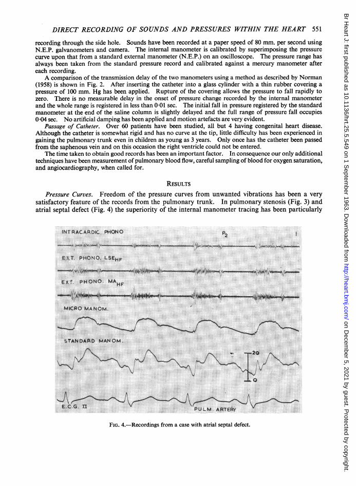
RESULTSPressure Curves. Freedom of the pressure curves from unwanted vibrations has been a very
satisfactory feature of the records from the pulmonary trunk. In pulmonary stenosis (Fig. 3) andatrial septal defect (Fig. 4) the superiority of the internal manometer tracing has been particularly
INT RACARDIC PHONO P2
EXT. PHONO. LSEHF
EXT. PHONO. MAHF
MICRO MANOM.
STANDAFkD MANOM.
-20
0
E-C GII PULM ARTE-RyFIG. 4.-Recordings from a case with atrial septal defect.
on Decem
ber 5, 2021 by guest. Protected by copyright.
http://heart.bmj.com
/B
r Heart J: first published as 10.1136/hrt.25.5.549 on 1 S
eptember 1963. D
ownloaded from

BARRITT AND DAVIES
evident. In these conditions a low pulmonary arterial pressure and vigorous right ventricularmovement often spoils the standard pressure curve. A clear record of the pulmonary dicroticnotch allows pulmonary valve closure to be timed with confidence and the steadiness of the fallingpressure wave during diastole makes possible a worth-while analysis of pulmonary vascular compli-ance (Shaw, 1961).
Pressure tracings from the ventricles are free from the overshoot on the upstroke and down-stroke that is so commonly seen in catheter records (Fig. 5 and 6). The onset of rise in pressure inthe ventricle can be timed accurately and the relation of pressure changes to valve movements morereadily studied.
The rapid low pressure changes in the atria are also damped by a long fluid column. Atrialtracings from the internal transducer are characterized by a very sharp x descent usually synchronouswith the atrioventricular valve closure (Fig. 7).
Heart Sounds and Murmurs. Heart sounds and murmurs are localized more exactly with aninternal transducer than with microphones on the chest wall. In the right side of the heart, forinstance, sounds and murmurs can be localized by passing the transducer back and forth in thearea of maximal vibrations until the site of origin of the sound is clear. Some difficulty results from
INTRACAROIC PHONOS, sm A2 I i
E t T. PHONO.LSEF x, A2P2EAT. PHON M
EX T. PHONO kiW4AMF
_
_|~~~~~~~~~~~~~~~~~~~~~~~~~~~~~~~~~~~~~~~~~~~
FIG. 5.-Atrial septal defect. The arrows at the ventricular pressurecurve indicate the moment of valve movement.
the fact that more intense sounds are transmitted across the cardiac septa and recorded on the otherside of the heart. For instance, the aortic valve closure sound is usually well recorded in the rightventricle (Fig. 8) and a loud pulmonary ejection murmur is also well recorded in the superior venacava and in the left atrium (Fig. 9).
Almost all sounds and murmurs have been recorded best when the transducer was placed distalto the site of origin of the sound. The sounds have been carried forwards in the blood stream with
552
on Decem
ber 5, 2021 by guest. Protected by copyright.
http://heart.bmj.com
/B
r Heart J: first published as 10.1136/hrt.25.5.549 on 1 S
eptember 1963. D
ownloaded from

DIRECT RECORDING OF SOUNDS AND PRESSURES WITHIN THE HEART 553
INTRACARDIAC PHONO ?Mcs A2
EXT. PHONO. LSEHF g A2 P2
-..E.n%z ii S!0.> ai.4
MICRO MANOM.
STAND. MANOM ;
E.C.G.E LEFT VENTRICLE
FIG. 6.-Atrial septal defect.
INTRACARDIAC PHONOo...........INSR
-~~~~~~~~~O 4 MI "NOW U- vu'Wiof r-fis"OM I Vomw
TCS DM TCs
EXT PHONO. LSEHF % A2 P2-t*n*_ _ _ E__~~1-
MICRO MANOM W
RIGHT ATRIUM
CTANDUD MANOM.
E. C.G.n
FIG. 7.-Atrial septal defect.
on Decem
ber 5, 2021 by guest. Protected by copyright.
http://heart.bmj.com
/B
r Heart J: first published as 10.1136/hrt.25.5.549 on 1 S
eptember 1963. D
ownloaded from

BARRITT AND DAVIES
IIY'Wn hIUWWIL*r n wq I N1 S P 1 K A TA 0 N |~~~~~~~~~~EXT. PMONOP4F
EXt. PHOhOMAn
FIG. 8.-Atrial septal defect. The aortic closure sound is clearly recorded in the right ventricle.
INTRACARDIC PHONOSM
EXT. PHONO-MAHF
E.C.G U
MICRO MANOM.
LEFT ATRIUM
FIG. 9.-The ejection murmur of pulmonary valvular stenosis is clearly recordedin the left atrium.
554
I
I
.o v~~~~O&r_.A
on Decem
ber 5, 2021 by guest. Protected by copyright.
http://heart.bmj.com
/B
r Heart J: first published as 10.1136/hrt.25.5.549 on 1 S
eptember 1963. D
ownloaded from

DIRECT RECORDING OF SOUNDS AND PRESSURES WITHIN THE HEART 555
..ME
EXT PHONO I>SF'H
MICRO MANOM
S TADNDA MANOM .
S,
6v1,;^^ ,4 f/t
FIG. 10.-Withdrawal tracing P.A. to R.V. in atrial septal defect. The ejection murmur is lost as the manometerenters the right ventricle.
little attenuation. The ejection murmur of pulmonary stenosis or of atrial septal defect is wellrecorded in the branches of the pulmonary trunk and is usually intensified as the transducer iswithdrawn towards the pulmonary valve. As the catheter tip passes backwards below the valve toenter the right ventricle the murmur is at once attenuated or lost (Fig. 10).
INTRACARDAC PHONO
EXT PMONO LSEHF-I._ E...n ne ~ . 4 F _
A
^t
ll/ ''.i'jJi,''\Si'A '
FIG. 11.-Tricuspid stenosis. The atrial systolic murmur is recorded clearly in the right ventricle and lost as themanometer is withdrawn into the right atrium.
q II II III
I -INTRACARDIAC PHONOII
-
II
4ftA, 0 4kow,*kw O 'Iftoomill ON foo, .4 I WpAkov A kV ow-, oa .%A! ovt-im .
on Decem
ber 5, 2021 by guest. Protected by copyright.
http://heart.bmj.com
/B
r Heart J: first published as 10.1136/hrt.25.5.549 on 1 S
eptember 1963. D
ownloaded from

BARRIlT AND DAVIES
INTRACARAIAC PHO ! i;_,,.,9.: FIT p..,.j4| ,
- r"-4M u
f
EX T. PHONO. PA
I - .AL. i-l i_z qp 4~W ~* 4qz1e*- tF-:a_--_-1.k. _X.,.. I dm i mn
MICRO MANOMN i
- ECG t
I~~~~~~~~~~~~~~A
STANDARD MANOM.
(B)FIG. 12.-(A) Pulmonary valvular stenosis withdrawal tracing. (B) Record with the tip of the
manometer above the stenosis and the side hole below the valve.
556
MRS!rTIMP -,-n
on Decem
ber 5, 2021 by guest. Protected by copyright.
http://heart.bmj.com
/B
r Heart J: first published as 10.1136/hrt.25.5.549 on 1 S
eptember 1963. D
ownloaded from

DIRECT RECORDING OF SOUNDS AND PRESSURES WITHIN THE HEART 557
Similarly, the murmur of patent ductus arteriosus is recorded well throughout the pulmonaryarteries but is not seen in the aortic record until the transducer is entering the ductus.
Right atrial gallop sounds and atrial systolic murmurs (Fig. 11) are well seen in the lower partof the right ventricle.
Pulmonary valvular, or infundibular, stenosis may be distinguished with more confidence whena sound record is added to the withdrawal tracing. When the stenosis is valvular the standardpressure record from the side hole first shows the pressure gradient, and the site of the valve may beaccurately shown with the diaphragm of the intracardiac manometer above the valve and the sidehole below (Fig. 12). Here, the ejection murmur is intense. As the diaphragm passes below thevalve an abrupt rise in pressure occurs and the systolic murmur ceases with the first beat. In thecase of an infundibular stenosis (Fig. 13) there is no pressure gradient across the valve and no changein the murmur as the tip enters the infundibular chamber. Below the stenosis the harsh ejectionmurmur is replaced at once by the less intense murmur of ventricular septal defect, which begins alittle earlier and has no accentuation in mid systole. Recordings from the chest wall resemble thepulmonary ejection murmur which is the louder of the two.
Murmurs produced by ruptured sinus of Valsalva allow the exact site of the rupture to be identi-fied. The record overleaf (Fig. 14) illustrates the localization of such a fistula opening into theoutflow tract of the right ventricle.
_..
j SC>GeN /f1li#; L '11 i'X iW 'a M
FIG. 13.-Ventricular septal defect and infundibular pulmonary stenosis. Withdrawal tracing from the infund-ibular chamber to the main right ventricle. The harsh murmur changes in character as the manometerpasses below the stenosis.
O al. wilmomol ---w-
Wr
1:./I
on Decem
ber 5, 2021 by guest. Protected by copyright.
http://heart.bmj.com
/B
r Heart J: first published as 10.1136/hrt.25.5.549 on 1 S
eptember 1963. D
ownloaded from

BARRITT AND DAVIES.....A0M Ab AA A
MPA. A V.HIGHA}LOW LV.
FIG. 14.-Ruptured sinus of Valsalva into the outflow tract of the right ventricle. As the catheter is with-drawn from the right ventricular outflow tract to the lower part of the ventricle the very harsh murmur islost. In the left ventricle the murmur of aortic regurgitation is seen.
DISCUSSIONIntracardiac sound records have been made by at least three methods. Yamakawa and his
colleagues (1954) in Tokyo used a capacitance method. Lewis et al. (1957, 1959 a and b) employed acatheter with activated barium titanate at its tip as their microphone. Both of these methods havethe great disadvantage that the pressure tracing to which the sounds are referred is recorded outsidethe body. The Allard-Laurens intracardiac manometer on which this paper is based allows thesimultaneous recording of sounds and pressures within the heart. Its great value, therefore, is thatexact timing of the relation of sounds and pressures can be studied. Furthermore, the pressurerecords are relatively free from the motion artefacts and damping that mar previous catheter tracings.
Two reservations need to be made. The localization of sounds and murmurs by the transduceris incomplete in that intense sounds are transmitted fairly widely through the heart and may be foundon the record in an adjacent chamber. On the other hand, a sound may be lost from the record if thetransducer is moved a short distance within the same chamber. There is no certainty, therefore,that a sound present in any particular heart chamber will appear on the tracing. Critical study isneeded or the interpretation of the record may be incorrect. At times, doubt will still remain.
SUMMARYThe Allard-Laurens intracardiac manometer has been found to give clear records of pressures
and sounds within the heart. Elimination of transmission delay and the damping effect of a longsaline column on the pressure tracing allow a more confident assessment of the relation betweenpressure change and heart sounds and murmurs.
558
on Decem
ber 5, 2021 by guest. Protected by copyright.
http://heart.bmj.com
/B
r Heart J: first published as 10.1136/hrt.25.5.549 on 1 S
eptember 1963. D
ownloaded from

DIRECT RECORDING OF SOUNDS AND PRESSURES WITHIN THE HEART 559
REFERENCESLewis, D. H., Deitz, G. W., Wallace, J. D., and Brown, J. R. (1957). Intracardiac phonocardiography in man.
Circulation, 16, 764., ---9, , and (1959a). Intracardiac phonocardiography. Progr. cardiovasc. Dis., 2, 85.,Ertugrul, A., Deitz, G. W., Wallace, J. D., Brown, J. R., and Moghadam, A. (1959b). Intracardiac phonocardio-graphy in the diagnosis of congenital heart disease. Pediatrics, 23, 837.
Norman, J. (1958). Transmission delay times in catheter/manometer systems. J. Soc. Cardiolog. Technicians, 3,197.
Shaw, D. (1961). Pulmonary artery pressure wave analysis: a method of assessing pulmonary vascular elasticresistance. Brit. Heart J., 23, 461.
Soulie, P., Baculard, P., Bouchard, F., Comu, C., Laurens, P., and Wolff, F. (1961). Intracardiac sound. Arch. Mal.Caeur, Suppl. 1.
Yamakawa, K., Shionoya, Y., Kitamura, K., Nagai, T., Yamamoto, T., and Ohta, S. (1954). Intracardiac phono-cardiography. Amer. Heart J., 47, 424.
on Decem
ber 5, 2021 by guest. Protected by copyright.
http://heart.bmj.com
/B
r Heart J: first published as 10.1136/hrt.25.5.549 on 1 S
eptember 1963. D
ownloaded from