directed at attachment damage - kanaal.nvve.com · pulpal obliteration ... pathologic root...
TRANSCRIPT
3/24/13
1
Current Treatment Options Of Dental Trauma
Guidelines of the International Association of Dental Traumatology
Asgeir Sigurdsson, cand odont, MS Chairman of the Department of Endodontics
NYU College of Dentistry Past President of IADT
Part 2
Luxation Injuries
Emergency Treatment
Directed at Attachment
Damage
Emergency Treatment “Prevention”
Minimize additional PDL damage
Limit initial inflammatory response
Stimulate cemental healing
Luxation Injuries
Extrusion
Concussion
Subluxation
Lateral Luxation
Intrusion
Always causes damage to the PDL. May cause a damage to the pulp.
Luxation injury
Forces on Teeth During Traumatic Injury
(Pileggi and Holland, 2009)
Luxation Injuries
The activation of these proteins is an essential cellular mechanism designed to protect against a variety of environmental stresses.
- Ferrets - 4 groups. - Right canine was traumatized with the contra lateral canines acting as undamaged controls. - Concussion injury with a standardized force. - Observation time 24, 48, 72 and 168 h.
The expression of heat shock protein 70 in the dental pulp following trauma.
(Pileggi and Holland, 2009)
Luxation Injuries
Ferrets There was a statistically significant difference in Hsp70 staining between traumatized and non-traumatized teeth only in the group observed 24 h after the trauma
The expression of heat shock protein 70 in the dental pulp following trauma.

3/24/13
2
Luxation Injuries
Concussion - No abnormal loosening - Reaction to percussion
Luxation Injuries
Subluxation - Abnormal loosening but no displacement - Reaction to percussion - Often negative Sensibility
Luxation Injuries
Concussion / Subluxation Treatment inside the office: - Rule out root fracture (Radiographs) - Adjust occlusion – splint only for patient comfort. - Baseline Sensibility tests
Luxation Injuries
Follow-up Concussion 4 weeks 6-8 weeks 1 year All appointments incl: Sensibility test and Radiographic evaluation
Luxation Injuries
Follow-up Subluxation 2 weeks 4 weeks 6-8 weeks 6 months 1 year All appointments incl: Sensibility test and Radiographic evaluation
Luxation Injuries
Lateral, Extrusive Luxation - Displacement - Reaction to percussion - Most often negative Sensibility
? Fracture of root or alveolar process

3/24/13
3
Luxation Injuries
Luxation Treatment outside the office:
Reposition tooth if easy - otherwise refer to dental office ASAP
Luxation Injuries
Luxation Treatment inside the office : 1. Radiographs at different angles. 2. Reposition. 3. Functional splint.
Luxation Injuries
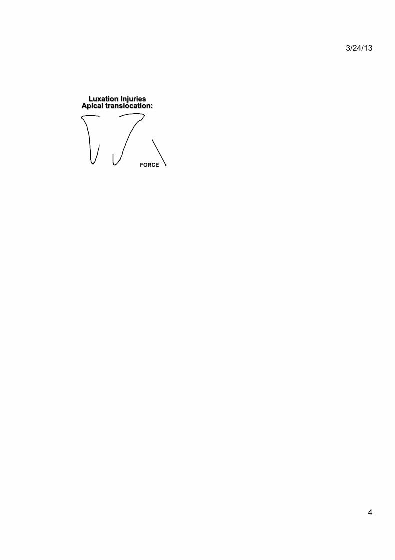
Lateral Luxation Apical translocation? Two possibilities:
- Apex in its original location - Apex moved facially
Luxation Injuries Apical translocation:
FORCE
Luxation Injuries Apical translocation:
FORCE
Luxation Injuries Apical translocation:
3/24/13
4
FORCE
Luxation Injuries Apical translocation:
Luxation Injuries Apical translocation:
FORCE
Extrusion and Lateral Luxation Treatment OPEN apex:
- Anesthesia (? vasoconstrictor). - Reposition the tooth into normal position. - Confirm the position with radiograph. - Splint for 2 weeks if needed. - Follow-up 2 weeks, 4 weeks, 6-8 weeks, 6 & 12 months and then yearly for at least 5 years. - Initiate root canal therapy as soon as
symptoms indicate.
Traumatic Injuries
Luxation Injuries
Possible complications:
ü Pulpal obliteration
"Of 122 teeth showing partial or total pulpal obliteration, 16 (13%) teeth showed periapical signs of pulpal necrosis"
(Jacobsen & Kerekes 1977)
Traumatic Injuries
Luxation Injuries
Follow-up Extrusive luxation 2 weeks; splint removal 4 weeks 6-8 weeks 6 months 1 year and yearly for 5 years All appointments incl: Sensibility test and Radiographic evaluation
Second Visit
7-10 days Treatment Objective
Prevent or treat
pulpal infection
Closed Apex:

3/24/13
5
Intrusion
Does always cause massive injury to the periodontal ligament. Does almost always (>95%) cause pulpal necrosis in case of closed apex.
Intrusion
Pulpal Prognosis:
0
20
40
60
80
100
1 yr. 10 yr. 5 yr. Andreasen and Vestgaard Pedersen 1985
Open Apex
% S
urvi
val
Closed Apex
Traumatic Injuries
Intrusion
Incisors intruded > 6 mm had significantly decreased survival compared with incisors intruded < 3 mm.
(Humphrey et al. 2003)
Intrusion
Treatment options Permanent teeth: - Spontaneous re-eruption. - Orthodontic forced eruption. - Surgical reposition.
Intrusion Intrusive luxation of permanent incisors in Norwegians aged 6-17 years: a retrospective study of treatment and outcome. Teeth in children (6 – 12 years old, 51 teeth): Did in many cases erupt spontaneously
37 spontaneous, 7 othro, 7 surgical Pulpal survival 43%. Inflammatory RR 26% (treatable with Ca(OH)2) Replacement RR 12% (not treatable).
(Wigen TI. et al. 2008)
Intrusion
Treatment options Permanent teeth:
When the injury to the tooth was severe, orthodontic extrusion initiated 5-7 days after trauma had little effect on repositioning of the injured tooth. Resulted in undesirable movement of the anchorage teeth. When the injury was less severe, orthodontic forces facilitated repositioning of the affected tooth.
(Turley et al. 1984)
A dog model/observation:

3/24/13
6
Intrusion Clinical and histological alterations in the surrounding periodontium of dog's teeth submitted for an intrusive luxation. 12 teeth, Ca(OH)2 placed after 14 d. Ortho extrusion 40 d. observation: The teeth that were moved immediately after the trauma had lesser degree of replacement resorption compared with those that were extruded 7 days after the trauma.
(Gomes JC. et al. 2008)
Intrusion
Treatment options Permanent teeth: - Surgical reposition.
Is quick and cost effective way. Can cause additional damage to the tooth and alveolar bone.
Closed Apex - Allow eruption without intervention if
tooth intruded less than 3 mm. - If no movement after 2–4 weeks,
reposition surgically or orthodontically before ankylosis can develop.
- If tooth is intruded beyond 7 mm, reposition surgically.
Intrusion
Follow-up 2 weeks; splint removal 4 weeks 6-8 weeks 6 months 1 year and then yearly for min 5 years. All appointments incl: Sensibility test and Radiographic evaluation
Intrusion
Avulsion
One of the few real emergency situation in dentistry.
At the site of the injury give the following advise:
ü Keep the patient calm. ü Find the tooth and pick it up by the crown. ü If the tooth is dirty, wash briefly under cold running
water and replant. ü If replant not possible place the tooth in a glass of
milk or other suitable storage medium. ü Seek emergency dental treatment immediately.
Avulsion
Known Factors Affecting Prognosis: ü Time out of the socket ü Storage condition ü Splinting technique and time

3/24/13
7
Avulsion
Known Factors Affecting Prognosis: ü Time out of the socket ü Storage condition ü Splinting technique and time ü Condition of the alveolus ü Stage of root development
Avulsion
Known Factors Affecting Prognosis:
ü Time out of the Socket – 90% of teeth replanted within 30 minutes
were without root resorption – 43% of teeth replanted 31 - 90 minutes
were without root resorption – 7% of teeth replanted after 90 minutes
were without root resorption
(Andreasen and Hjörting-Hansen, 1966)
Avulsion
Consequences:
ü Attachment damage ü Pulpal necrosis ü Bacteria contamination
Treatment of Avulsion Place the tooth in physiologic storage medium immediately once in the office, while examining the pt.! Less than 60 min dry time: ü Clean the root with a stream of saline. ü Administer local anesthesia (vasoconstriction??) ü Irrigate the socket with saline. ü Examine the alveolar socket, if fractured gently
reposition the fragments. ü Replant the tooth, slowly with slight digital pressure.
NO force!
Treatment of Avulsion Place the tooth in physiologic storage medium immediately once in the office! ü Suture gingival lacerations ü Verify normal (correct) position of the tooth, clinically
AND radiographically. ü Apply a flexible splint for up to two weeks, away from
the gingiva. ü Administer systemic antibiotics. ü Check tetanus protection ü Give patient instruction (incl. hygiene, diet, pain med) ü Initiate root canal therapy in 7 to 10 days, before
removing the splint if apex is closed.
Treatment of Avulsion If the tooth was replanted at the site of the injury: ü Leave the tooth in place. ü Clean the area with water spray, saline or
chlorhexidine. ü Suture gingival lacerations. ü Verify normal (correct) position of the tooth, clinically
AND radiographically. ü Apply a flexible splint for up to two weeks, away from
the gingiva. ü Etc.

3/24/13
8
Consequences of Tooth Avulsion
Pathologic root resorption due to dental injuries is always (at least initially) inflammatory in origin. It is either:
Self-limiting if the only stimulus for the resorption is the injury itself. Progressive if after the initial injury an additional stimulus is present or there is a severe damage to the protective layer.
Root Resorption
Diagnosis of root resorption: - Multiple radiographs with different angulations.
Difficult, if not impossible, to assess true extent of the lesion, AND more importantly confirm location, facial or lingual!
Root Resorption
Diagnosis of root resorption: - Multiple radiographs with different angulations, - Cone-Beam Computed Tomography:
CBCT is it as good as we think? Comparison of two cone beam computed tomographic systems versus
panoramic imaging for localization of impacted maxillary canines and detection of root resorption. In vivo study, n=60 “The results of this study suggest that CBCT is more sensitive than conventional radiography for both canine localization and identification of root resorption of adjacent teeth.”
(Alqerban A. et al . 2011a)
Root Resorption
External Root Resorption Surface Resorption
Ø Localized injury to PDL and/or cementum Ø No significant inflammatory changes in PDL Ø Self limiting Ø Spontaneous repair with cementum Ø Not related to contents of root canal Ø Hard to detect on radiograph
Surface Root Resorption
It is important to not mis-interpret these cases as progressive in nature.
If the pulp is vital but some surface changes on the root are seen on a radiograph:
- no treatment should be performed - a wait and see attitude taken - allow spontaneous healing to take place!

3/24/13
9
Root Resorption
External Root Resorption Replacement Resorption
Ø Fusion of alveolar bone with root surface Ø Absence of vital PDL Ø Continuous replacement of tooth substance Ø No cementum repair Ø No direct relationship with content of root canal Ø Tooth structure fuse with bone on radiograph
Replacement Root Resorption
It is important to remember that only the initial inflammatory resorption is pathologic and the subsequent osseous replacement should be considered physiologic.
Replacement Root Resorption
Intervention for Treating Traumatized Ankylosed Permanent Front Teeth Cochrane Database Syst Rev 2010
SELECTION CRITERIA:
- Randomized controlled trials (RCTs) - Comparing any intervention for treating displaced ankylosed permanent front teeth in individuals - Any age. - No language restrictions.
(de Souza et al. 2010)
Replacement Root Resorption
Intervention for Treating Traumatized Ankylosed Permanent Front Teeth Cochrane Database Syst Rev 2010
MAIN RESULTS:
ü The search retrieved 77 references to studies. ü None matched the inclusion criteria and therefore all were excluded.
(de Souza et al 2010)
Replacement Root Resorption
Intervention for Treating Traumatized Ankylosed Permanent Front Teeth Cochrane Database Syst Rev 2010
AUTHORS' CONCLUSIONS: “There is no evidence from RCTs about the comparative effectiveness of the different treatment options for ankylosed permanent front teeth. “
(de Souza et al 2010)
Infra-Positioned Tooth (Ankylosed)
What can be done? Nothing
Not a good esthetic option. Orthodontics: Not possible – the tooth will not move. Composite buildup on incisal edge
Possible with low smile line and minor infra position.

3/24/13
10
Infra-Positioned Tooth (Ankylosed)
What can be done? Extraction followed by auto transplantation of a tooth:
A good option for a patient with foreseeable crowding in the arch. Issues with cost and timing of treatment.
Infra-Positioned Tooth (Ankylosed)
What can be done? Decoronation and transplant or osseous implant later.
Benefits are preservation of the alveolar bone and possibly vertical growth of the bone!
Infra-Positioned Tooth (Ankylosed)
What can be done for a young individual? Root submersion and transplant or osseous
implant later.
Benefits are preservation of the alveolar bone and possibly vertical growth of the bone!
Decoronation
- The bone that forms in the area is of good quality and placement of implants. - No apparent complications in cases where there are some remnants of the root still visible on radiographs (Malmgren et al. 2002). - Therefore there does not seem to be any need to remove those remnants prior to placement of an implant into the area.
Decoronation
ü It perseveres the alveolar process’ width and
height. ü It is likely to negate the need for expensive
and invasive surgical alveolar ridge augmentation procedure.
ü Studies indicate that vertical bone apposition is possible after the crown is removed.
The Advantages of Decoronation Procedures are:
Decoronation
The main disadvantages of the Decoronation procedure are its surgical nature;
- may be challenging in young children, - the necessity for a long-term esthetic space maintainer.

3/24/13
11
Root Resorption
External Root Resorption Inflammatory Resorption
Ø Injury to PDL and cementum Ø Significant inflammation of PDL Ø Continuous replacement of tooth substance
Ø No cementum repair Ø Direct relationship with content of root canal Ø Tooth structure and bone loss on radiograph
Internal Inflammatory Root Resorption
Usually asymptomatic and is first recognized clinically
through routine radiographs. For internal resorption to be active, at least part of the
pulp must be vital.
Internal Inflammatory Root Resorption
Internal root resorption is treated with the endodontic
methods Pulpectomy removes the blood supply to the
granulomatous tissue and the rest of the treatment is concentrated on removing tissue from the irregular resorptive defect and obturating the space.
Consequences of Tooth Avulsion
In summary: In order for root resorption to occur two requirements must be met:
1. A change has to occur in the protective attachment layer (pre-dentin internally or pre-cementum externally) of the root.
2. An inflammatory process must be present and be maintained adjacent to this damaged root surface*.
(* except in replacement root resorption)
Management of the
Emergency Patient
Management of the Emergency Patient
Treatment focus: Minimize inflammation due to attachment damage

3/24/13
12
Emergency Management Outside the dental office
Best TX - Replant if possible
Emergency Management Outside the dental office
Place in appropriate storage medium
- specialized media - milk - saline - vestibule of mouth - (((water)))
Milk Is Good!
Has physiological osmolarity (230-270 mOsm/kg). pH is in physiological range (6.5-6.9). Can provide some nutrients to cells. Pasteurized milk has very low bacterial count.
(Blomlöf et al. 1981)
Milk is Good!
At 8 h, PDL cell viability in regular pasteurized milk and long shelf-life milk were significantly greater than in Save-A-Tooth (p < or = 0.001) (no refrigeration) There was no significant difference between regular pasteurized milk and long shelf-life milk at any time period. Access to long shelf-life milk is usually easier than regular milk.
(Marino TG et al. 2000)
Milk is Good!
Skim milk and 0.5% milk maintained cell viability at significantly greater levels than milks of greater fat content over the first 15 to 30 min. Cell viability was also significantly greater in these maintenance media after 210 min.
(Harkacz OM et al. 1979)
Propolis: Mean 10E4 cells ml-1
0
50
100
150
200
250
Saline HBSS Milk P50% P100%
Martin and Pileggi 2004
* * P<0.0001
New Transport Medium

3/24/13
13
Emergency Visit Root Preparation
Mature Tooth
Immature Tooth
Dry time < 60 min > 60 min
Emergency Management Inside the dental office
Root preparation- Mature Tooth
Dry time < 60 minutes
Replant
Emergency Visit
Inside the dental office
Splint
16 and 35 lb monofilament fishing line
Guidelines of IADT What is new? Any physiological and hygienic splint acceptable; monofilament fishing line 16 to 25 lbs!
Splinting Time: Type of Injury Splinting Time
Subluxation 2 weeks
Extrusive luxation 2 weeks
Avulsion 2 weeks
Lateral luxation 2 weeks
Root fracture (middle 1/3) 4 weeks
Alveolar fracture 4 weeks
Root fracture (cervical 1/3) 4 months
(Andersson et al. 2012)
Emergency Treatment Adjunctive Therapy
Systemic Antibiotics? The International Association for Dental Trauma has suggested to use Doxycycline (tetracycline) for anyone above 8 to 12 years of age.

3/24/13
14
Emergency Treatment
Adjunctive Therapy * Systemic Antibiotics
(Doxycycline > 8 to 10 y. old)
* Systemic NSAIDS
* Chlorhexidine rinses
* Tetanus booster ?
Prevention of Resorption
Calcium Hydroxide-Ca(OH)2
Current recommendations include instrumentation and placement of Ca(OH)2 into the root canal space within 7-14 days following replantation.
Prevention of Resorption
Calcium Hydroxide-Ca(OH)2 Ca(OH)2 placed in the root canal of teeth with damaged root surfaces may induce ankylosis due to the necrotizing effect on cells repopulating the root surface.
(Hammarström et al, 1986)
When to place the Ca(OH)2?
"Effect of delayed calcium hydroxide treatment on periodontal healing in contaminated replanted teeth". Intracanal calcium hydroxide treatment of teeth with compromised periodontal ligament may cause replacement resorption if left in the canal for a long time or changed repeatedly.
(Lengheden et al. 1990)
Effect of Ca(OH)2 on Strength of the Tooth
It has been proposed that immature teeth are weakened by filling of the root canals with calcium hydroxide dressing and gutta-percha.
(Cvek 1992)
Effect of Ca(OH)2 on Strength of the Tooth
(Andreasen et al. 2002)
0!
2!
4!
6!
8!
10!
12!
14!
16!
18!
od! 2w! 1m! 2m! 3m! 6m! 9m! 12m!
Question about comparison control??

3/24/13
15
Effect of Ca(OH)2 on Strength of the Tooth
Comparison of Fracture Resistance in Root Canals of Immature Sheep Teeth After filling with Calcium Hydroxide. 30 immature teeth (80% of growth), Instron
Saline Ca(OH)2 paste MTA Ca(OH)2 paste + MTA after 30 days
(Andreasen et al. 2006)
Effect of Ca(OH)2 on Strength of the Tooth
“The long-term effect of calcium hydroxide application on dentin fracture strength of endodontically treated teeth”. Extracted teeth – stored for 30, 90, 180, 270, 360, and 540 days Instron Machine 15 teeth each group PROBLEM: NO CONTROL TEETH AT ANY TIME.
- Control teeth tested only after one week of storage!!!!!!!!!
(Batur YF et al. 2013)
Prevention of Resorption
Calcium Hydroxide-Ca(OH)2
No direct anti-inflammatory action provided.
Prevention of Resorption
Corticosteroids Block production of inflammatory stimulators including prostaglandins and leukotrienes, produced by cyclo-oxygenase and lipooxygenase pathways.
Prevention of Resorption
Ledermix® Paste
• 1% Triamcinolone - Corticosteroid
• 3% Demeclocycline - Broad spectrum antibiotic
Prevention of Resorption
Ledermix® Paste Ledermix
® has been shown to diffuse
through dentin.
Ledermix has a rapid initial release followed by a slow steady release.
(Abbott, et al 1988, 1989)

3/24/13
16
Purpose
To evaluate the usefulness of Ca(OH)2 and Ledermix
® in attenuating or
arresting external root resorption secondary to avulsion in a dog dental trauma model (Note – no infection)
(Dr. Bryson et al. 2002)
58.7
13.8
0
10
20
30
40
50
60
Ledermix Ca(OH)2
Results 60 min Dry time
% Favorable
Healing
p=0.004
(Dr. Bryson et al. 2002)
5.39
2.19
0
1
2
3
4
5
6
Ledermix Ca(OH)2
Results 60 min Dry-time
Avg Remaining
Root Structure
p=0.002
(Dr. Bryson et al. 2002)
The effect of intracanal anti-inflammatory medicaments on external root resorption of replanted
dog teeth after extended extra-oral dry time
Dog Study: The canals were filled with:
- Ledermix, - Triamcinolone, - Demeclocycline,
Replanted after 60 min dry time.
(Chen et al. 2006)
The effect of intracanal anti-inflammatory medicaments on external root resorption of replanted
dog teeth after extended extra-oral dry time
Dog Study: The groups treated with: - Ledermix 75.8% - Triamcinolone 69.8% - Demeclocycline 52.4% Statistically significantly more favorable healing and more remaining root structure than those without treatment.
(Chen et al. 2006)
Effect of Intracanal Corticosteroids on Healing of Replanted Dog Teeth after Extended Dry Times
Dog Study: The effect of potent intracanal corticosteroids on periodontal healing of replanted avulsed teeth. Systemic absorption of these corticosteroid.
(Kirakozova, et al. 2009)

3/24/13
17
Effect of Intracanal Corticosteroids on Healing of Replanted Dog Teeth after Extended Dry Times
Dog Study: No statistically significant difference in plasma cortisol levels was found between different time points of the study period (days 0, 1, 30) p = .1005
(Kirakozova, et al. 2009)
Other Ways in Prevention of Resorption
Intracanal bisphosphonate: Does it inhibit replacement resorption?
Monkey study, incisors, 60 min extra-oral dry. 8 week observation time. 3 groups
Neg control (contamination with dental plaque) bisphosphonate (etidronate disodium) Placement of calcium hydroxide 8 days post avulsion.
(Thong, YL et al. 2009)
Other Ways in Prevention of Resorption
Intracanal bisphosphonate: Does it inhibit replacement resorption?
46% root loss in untreated teeth (inflammatory) < 30% root loss with calcium hydroxide 39% root loss with bisphosphonate
(Thong, YL et al. 2009)
Flurbiprofen is one of the most potent cyclooxygenase inhibitor, and it’s effect diminishing the rate of alveolar bone loss (associated with periodontal disease) in beagles and humans has already been reported.
(Williams RC et al. 1974, Williams RC et al. 1989)
The Effect of NSAID’s
Results
Statistical analysis performed using Pair Student T-Test showed no significant difference among groups and treatment rendered.
Dr. Franciso Banch
32 incisors and premolar roots, 2 dogs, dry time 30, 40 and 60 min
Prevention of Resorption Minocycline
The roots with and without minocycline treatment showed no significant differences between the remaining root mass or the percentage of favorably healed root surfaces. The use of minocycline is not currently recommended for prevention or attenuation of external root resorption following avulsion in a dog trauma model.
(Bryson et al 2003)
48 premolars, 6 dogs, uniform cemental defect created

3/24/13
18
Pulpal Necrosis
Pulp revascularization is favored when the apical foramen is not completely formed. (Öhman, 1965; Skoglund and Tronstad, 1981; Kristerson and Andreasen, 1984; Kling et al., 1986; Cvek et al., 1990a).
Immature Non-Vital Tooth TX Approaches
• Long term calcium hydroxide apexification • MTA “quick” apexification
Pulpal Necrosis
The occurrence of pulp revascularization is enhanced if the apical foramen is more than 0.5 mm wide in monkeys
(Cvek et al., 1990) and 1.1 mm wide in humans
(Kiling et al. 1986).
Pulpal Necrosis
If the tooth is replanted within 45 minutes of the injury, the prognosis for pulp revascularization is favorable.
(Kiling et al. 1986).
Pulpal Necrosis
AND pulp revascularization is highly dependent on the presence or absence of bacteria in the pulpal lumen.
(Cvek et al. 1990 and 1990)
Pulpal Necrosis
Revascularization of the pulp in a tooth with an open apex could be expected to be some where between 18 and 25% - more likely the larger the apical opening is.
(Cvek et al. 1990 and 1990)

3/24/13
19
Bacterial contamination !!!!
The principle factor for failure of revascularization is
(Cvek et al, 1990)
Revascularization Topical Treatment with
Doxycycline before replantation (1mg/20ml saline)
Increases the incidence of complete revascularization from 18% to 41%
in a monkey model (Cvek et al,1990)
Statistical Analysis: Fisher’s exact test
There was a significant difference between groups with and without doxycycline soak (p=0.024)
Complete vitality with doxycycline 60% No doxycyline 36%
(Yampiset et al. 2000)
Research Question
Is Minocycline (Arestin or Dentomycin) more effective than Doxycycline in preventing bacterial penetration along the PDL after replantation?
(Ritter et al. 2004)
Minocycline G 1 (n=11): specimens were kept dry for 5 min, covered with minocycline hydrochloride microspheres and replanted. G 2 (n=11): specimens were kept dry for 5 min, then soaked in 1mg/20ml of Doxicycline solution for 5 minute and replanted. G 3 (n=11): specimens were kept dry for 5 min, then soaked in saline for 5 minute and replanted.
(Ritter et al. 2004)
Results - Histology
Saline: - 33.33% Vital pulp
Doxycycline topical treatment: 72.73% Vital pulp + osteoid tissue
(Ritter et al. 2004)

3/24/13
20
Results - Histology
Minocycline topical treatment: 90.96% Vital pulp + osteoid tissue
(Ritter et al. 2004)
Revascularization of Non-vital Teeth With Apical Periodontitis
Necrotic Pulp When the pulp in an immature tooth becomes necrotic and the pulpal space infected the success of any endodontic treatment is severely reduced.
- Difficult to treat - Inadequate strength
Necrotic Pulp
Until now it has not been an option to wait for possible revascularization because once bacteria is in the pulpal space there is no possibility of re-growth of tissue. And it has been believed the pulp progenitor cells necessary for the proliferation of pulpal tissue can not survive the infection.
(Cvek et al 1990, Iwaya et al 2001)
Three Mix Antibiotic
Dental pulp regeneration is aided by blood and blood substitutes after experimental removal of the pulpal tissue in immature teeth.
(Nygaard-Ostby 1961)
Where it all started
Dental pulp regeneration aided by blood and blood substitutes after experimentally induced periapical infection
Myers and Fountain Oral Surg Oral Med Oral Pathol. 1974;37:441-50

3/24/13
21
Disinfection of a Root Canal
Study on immature teeth with necrotic and infected canals showed that the revascularization failed primarily because of inadequate disinfection before inducing bleeding into the canal space.
(Myers and Fountain 1974)
12 anterior teeth in 4 Ciebus apellae monkeys:
Necrotic and Infected Pulp
Is it possible completely disinfect the pulpal space such that revascularization will occur? Do the pulp progenitor cells necessary for the proliferation of pulpal tissue survive the infection?
Where it all started Sterilization of infected root-canal dentine by topical application of a mixture of ciprofloxacin, metronidazole and minocycline in situ
Sato, Ando-Kurihara, Kota, Iwaku, Hoshino International Endodontic Journal 1996;29:118-24
In - vitro antibacterial susceptibility of bacteria taken from infected root dentine to a of 0.5 mg mixture of ciprofloxacin, metronidazole and minocycline
Hoshino, Kurihara-Ando, Sato, Uematsu, Sato, Kota, Iwaku International Endodontic Journal 1996;29:125-30.
Sato et al. 1996
Studied the potential of a mixture of: ciprofloxacin, metronidazole minocycline Is the mixture able to penetrate through root dentin? And can the mixture kill cultured bacteria in deep layers of extracted tooth root dentine?
Sato et al. 1996
Confirmed that in extracted teeth there was penetration of the antibiotic paste through dentine. And the mix had the antibacterial efficacy expected against bacteria infecting the dentine.
Hoshino et al. 1996 Studied the antibacterial effect of a mixture of:
ciprofloxacin, metronidazole minocycline,
on bacteria taken from infected dentine of
extracted root canal walls under strict anaerobic conditions.

3/24/13
22
Hoshino et al. 1996
The efficacy was estimated in vitro by measuring bacterial recovery on BHI-blood agar plates in the presence or absence of the drug combination.
The respective drug alone (10, 25, 50 and 75 micrograms ml-1) substantially decreased the bacterial recovery but could not kill all the bacteria.
Disinfection abilities in vivo?
First study in vivo: Dog model with immature teeth (36 teeth and 6 controls) All samples cultured positive for bacteria with a mean CFU count of 1.7X10⁸ prior to irrigation of NaOCl. After two weeks of the paste 70% of the samples cultured bacteria-free with a mean CFU count of only 26. Reductions in mean CFU counts between S1, S2 and S3 were significant (p<0.0001).
(Windley et al 2005)
Does it work?
Revascularization of an immature permanent tooth with apical periodontitis and sinus tract
Iwaya, Ikawa, Kubota Dental Traumatology 2001;17:185-7
Does a simpler protocol work?
Protocol The root canal system is thoroughly irrigated with 5.25% NaOCl and 0.1% Chlorhexidine solution BUT minimally instrumented. Canals dried with paper points. The three antibiotics, ciprofloxacin, metronidazole and minocycline are mixed together in equal portions and then a paste is created with adding saline
(Banchs and Trope 2004)
Treatment Protocol
The paste is pipetted into the canal and 2/3 of the canal filled with the paste. Care is to be taken to not leave ANY of the paste in the coronal portion of the tooth because the mix will seriously stain the crown.
Treatment Protocol
After three weeks the patient is recalled and the paste irrigated out with saline
DO NOT use epinephrine containing local anesthetics.
Bleeding is created in the canal space with a sterile endo explorer and files.*

3/24/13
23
Three Mix Antibiotic
Some kind of a scaffold for the in-growing tissue seems to be essential. Previous case reports used a blood cloth to serve as a scaffolding. However it is rather difficult control and to maintain during restoration.
Treatment Protocol
Effort is made to have the blood fill the whole canal space. This blood should be allowed to begin its initial clotting prior to closing the access.
After three weeks the patient is recalled and the paste irrigated out with saline
DO NOT use epinephrine containing local anesthetics. Bleeding is created in the canal space with a sterile endo explorer and files.*
Treatment Protocol
* Some clinicians do draw blood from the patients and transfer it to the tooth rather that rely upon getting adequate bleeding into the canal. Others have advocated additionally spinning the collected blood and only use platelet rich plasma. To date no research published that confirms the advantages of that.
Discoloration
Recently it has been suggested to apply bonding agent (even 2 layers) on the inside of the access preparation and down in to the cervical area prior to applying the three mix.
(Reynolds et al. 2009)
Discoloration Few authors have in case reports and alike suggested to only use ciprofloxacin and metronidazole but not minocycline to prevent discoloration. NO study so far published on the bacteriological effectiveness of those two on endodontic infections!
(Kim JH et al. 2010)
Discoloration Other authors have in case reports and alike suggested ciprofloxacin, metronidazole and amoxicillin or clindamycin. Again NO study so far published on the bacteriological effectiveness of that mixture on endodontic infections!
(Thompson and Kahler 2010)

3/24/13
24
Does Tri-Mix work? Response to Intracanal Medication in Immature Teeth with Pulp Necrosis: An Experimental Model in Rat Molars: (rats n=36, split mouth, molars left open for 3 weeks) - Vital teeth showed increase of root length and hard tissue thickness throughout the experimental periods. - Induction of necrosis arrested root formation. ed with the control (P < .05).
(Scarparo et al. 2011)
Does Tri-Mix work? Response to Intracanal Medication in Immature Teeth with Pulp Necrosis: An Experimental Model in Rat Molars: (rats n=36, split mouth, molars left open for 3 weeks) - Teeth disinfected with NaOCl and triple antibiotic paste had:
- significant reduction of periapical lesions, - gain in root length, - increased wall thickness compared with the control (P < .05).
(Scarparo et al. 2011)
A Retrospective Evaluation of Radiographic Outcomes in Immature Teeth With Necrotic Root Canal Systems Treated With Regenerative Endodontic Procedures
R Bose, P Nummikoski, and K Hargreaves, J Endodon, 2009
- NOTE: The position of Ca(OH)2 was critical. When Ca(OH)2 was radiographically restricted to the coronal half of the root canal system, it produced better results than when it was placed beyond the coronal half.
A case series study (20 human teeth) using only Ca(OH)2 for disinfection showed that the outcome of continued root development was not as predictable as increased thickening of the canal walls. However all pericapical lesions healed and 8/20 did respond to EPT eventually.
So the Future? Or What is Next?
Is the antibiotic mixture needed at all?
(Chen et al. 2011)
So the Future
A Journey from Dental Pulp Stem Cells to a Bio-tooth
Ming Yan & Yan Yu & Guangdong Zhang &
Chunbo Tang & Jinhua Yu Stem Cell Rev and Rep (2011) 7:161–171