directed natural forces of affinity between a bacterium and mineral
TRANSCRIPT

DIRECTED NATURAL FORCES OF AFFINITY BETWEEN A BACTERIUMAND MINERAL
STEVEN K. LOWEROhio State University, 125 South Oval Mall, 275 Mendenhall Laboratory, Columbus,
Ohio 43210 USA; [email protected]
ABSTRACT. The formation of a bond between a bacterium and a mineral isultimately controlled by forces that operate over length scales of a few nanometers.This manuscript presents evidence that bacteria may actively modulate forces at thecell-mineral interface to promote contact with specific mineral phases. Nano- topico-Newton forces were measured between goethite (FeOOH) and each of twospecies of Gram negative bacteria (Escherichia coli and Shewanella oneidensis) inaqueous solution of varying oxygen concentration. The interactions were dominatedby electrostatic and steric forces as either bacterium approached to within 10 to 12 nmof the surface of goethite. The van der Waals force exhibited an influence on eachbacterium-mineral pair at separations of �2 nm. These types of nonspecific forceswere all that were observed for the E. coli-goethite pair. However, S. oneidensisexhibited a selective disposition to form a specific bond with goethite, particularlyunder anaerobic conditions. These data suggest that S. oneidensis is able to perceive andrecognize the surface of metal oxyhydroxides and regulate attractive forces at thecell-oxide interface. This may be a vestige of the close evolutionary linkage betweeniron oxyhydroxides and metal-reducing bacteria like Shewanella, which can use Fe(III)in the crystal structure of a mineral as a terminal electron acceptor.
introduction
Few things in nature are as specific as the interactions between a protein and itscomplementary ligand. Intra and intermolecular forces between reactive functionalgroups direct the specificity of such reactions. For quite some time, such specificity hasbeen known to focus cell-cell binding reactions within biofilms inhabiting the humanoral cavity (Kolenbrander, 1989; Whittaker and others, 1996). In such environments,steriospecific “fit” has been identified as the primary factor controlling the binding of aparticular bacterium to another cell or organic conditioning film (Ellen and others,1997). Tantalizing hints that such affinity may exist between bacteria and inorganicsurfaces can be found in the literature. For example, some researchers have shown thatAcidithiobacillus ferrooxidans expresses an affinity for particular sulfide minerals thatserve as a source of electrons for energy generation (Devasia and others, 1993;Ohmura and others, 1993; Arredondo and others, 1994; Ohmura and others, 1996;Dziurla and others, 1998). However, these types of studies rely on indirect evidence forbacterium-mineral “recognition”, such as, microscopy images that show a highernumber of cells attached to one mineral relative to another.
Lower and colleagues have recently measured intermolecular forces betweenShewanella oneidensis and iron versus aluminum hydroxides (Lower and others, 2001a;Lower and others, 2002). These data have provided the first direct evidence thatbacteria may indeed recognize inorganic crystalline surfaces by directing natural forcesof affinity from cell-surface biopolymers towards a mineral face. The work containedherein will explore further the specific nature of biological recognition of inorganicsurfaces by presenting force microscopy measurements between goethite (FeOOH)and S. oneidensis versus Escherichia coli. Both of these microorganisms are Gram negativebacteria meaning that the general composition and architecture of their outer surfacesare very similar. Figure 1 presents the external face of the outer membrane (OM) of aGram negative bacterium.
[American Journal of Science, Vol. 305, June, September, October, 2005, P. 752–765]
752

Each cell is surrounded by a lipid-protein membrane called the outer membrane(OM). The outer face of the OM is composed of a lipopolysaccharide (LPS), which ismade of three distinct parts: lipid A, which anchors the LPS into the OM; a corepolysaccharide, which contains ketodeoxyoctonate and several hexose and heptosesugars; and an O-polysaccharide, which consists of strain-specific sugar residues (seefig. 1). The polysaccharide chains of the LPS extend 2 to 40 nm outwards from the cellwall, depending on the bacteria strain (Nikaido, 1996; Pink and others, 2003; Stoicaand others, 2003). The OM also contains a number of proteins composed of relativelylong chains of amino acids. Some of these proteins are inducible and are expressedunder specific physiological conditions, while others are constitutive or expressedunder all growth conditions.
The fact that the outer surfaces of both E. coli and S. oneidensis are very similar (seefig. 1) suggests that either bacterium should interact with goethite in much the samefashion. Nonspecific forces such as van der Waals and electrostatic interactions wouldbe expected to governor how or if an interface is formed between goethite and E. colior S. oneidensis. However, one must also consider the fact that S. oneidensis, unlike E. coli,has outer membrane proteins that are capable of shuttling electrons to Fe(III) inminerals under anaerobic conditions. (Myers and Myers, 1992, 1993, 1997; Myers andMyers, 1998, 2001; Myers and Myers, 2003). This has lead some researchers toconjecture that dissimilatory metal reducing bacteria like Shewanella, were among thefirst biological cells to inhabit and propagate the early Earth, which did not haveoxygen as a terminal electron acceptor (Lovley, 1991; Vargas and others, 1998).Presumably, these early metal reducing bacteria evolved the proteins that their presentday counterparts possess for energy generating reactions in which electrons are
Fig. 1. Schematic diagram showing the general architecture and biomolecular structure of the exteriorcell surface of Gram negative bacteria (based on the work of Beveridge, 1981; Nikaido, 1996; Raetz, 1996;Rick and Silver, 1996; Beveridge, 1999; Madigan and others, 2003). The force microscope used in this studyallows one to probe forces between a mineral surface and biological molecules on a living cell. The forcemicroscope is also capable of measuring the biomechanical topology of macromolecules (such as, outermembrane proteins) that form a bond with a mineral surface. Abbreviations are as follows: N-acetyl-glucosamine (GlcN), phosphate (P), ketodeoxyoctonate (KDO), heptose sugar (Hep), glucose (Glc),galactose (Gal), amino acid (AA), repeating amino acid residues (brackets with an “n” subscript). TheGram-negative strains used in this study (E. coli K12 and S. oneidensis MR-1) possess only the coreoligosaccharide and lack the O-side chain (T. Beveridge, personal communication).
753Steven K. Lower 753

shuttled from a bacterium to Fe(III) in the crystal structure of a mineral. It is possiblethat this intimacy between metal reducing bacteria and ferric containing minerals ispreserved in the genetic makeup of a species such that S. oneidensis expresses a naturaland selective affinity for metal hydroxides, like goethite, that may serve as terminalelectron acceptors under deoxygenated conditions.
This study presents evidence that S. oneidensis forms specific bonds with metaloxyhydroxides. This was accomplished by using a force microscope to probe inter- andintra-molecular forces between goethite and S. oneidensis versus E. coli. The observedforce measurements were compared to theoretical models describing van der Waals,electrostatic, and steric forces between goethite and each of the two Gram-negativebacteria. These experiments indicate that E. coli and S. oneidensis perceive a mineralsurface through intermolecular forces that operate over length scales of a few nanome-ters. S. oneidensis actively regulates forces at the goethite interface by targeting proteinsto the cell wall, which in turn form specific bonds with goethite.
methods
Growth of Bacteria and Preparation of Biologically-Active-Force-ProbesE. coli K12 was purchased from BioRad Laboratories (Hercules, CA) and cultured
in 25 g/L Luria-Bertani (LB) medium (Fisher Scientific, Pittsburg, PA) at 22 to 25oCand pH �7. S. oneidensis MR-1 (ATCC 700550) was purchased from the American TypeCulture Collection (Manassas, VA) and cultured in LB medium or in defined M1medium (Myers and Nealson, 1990) at 22 to 25°C. For anaerobic cultures of S.oneidensis, LB or M1 medium was supplemented with 15 mM lactate as the carbon andenergy source and 2 mM ferric citrate as the electron acceptor.
For aerobic growth of each bacteria species, a glass bottle containing LB or M1medium was inoculated with an overnight, aerobic culture and grown with vigorousaeration using a shaker table. Anaerobic growth of S. oneidensis was performed insidean anaerobe chamber (Coy Laboratory Products, Ann Arbor, MI) with anaerobicmedia and solutions. E. coli or S. oneidensis cells were harvested at mid to lateexponential growth phase by centrifugation. These cells were rinsed in sterile NaClsolutions and used immediately to fabricate biologically-active-force-probes.
Biologically-active-force-probes were created with E. coli or S. oneidensis cells asdescribed previously (Lower and others, 2000; Lower and others, 2001b; Kendall andLower, 2004). Briefly, glass beads (�10 �m diameter) were coated with amino-silane,washed to remove excess silane, and then placed in a suspension of bacterial cells. Asingle bacteria-coated bead was then attached to the end of a silicon nitride cantileverwith epoxy. As shown previously (Lower and others, 2001a), the silane molecule doesnot have any impact on the observed forces as it is confined to the interface betweenthe bacteria and glass bead, rather than the surface of the bacteria that will interactwith a mineral during force measurements. Further, the silane linker did not appear toharm the cells as the bacteria were still viable after their use. This was confirmed byplacing a biologically-active-force-probe on an agar plate subsequent to force measure-ments. For the data presented herein, a distinct, single colony was observed under theend of each of the cantilevers.
Scanning laser confocal microscopy was used to image each force-probe prior toits use in the force microscope (Lower and others, 2000). Prior to fabricatingbiologically-active-force-probes, bacteria were transformed with a plasmid for thegreen fluorescence protein, pSMC2, kindly provided by G. O’Toole (Lower andothers, 2000; Lower and others, 2001a). The position of E. coli or S. oneidensis cells on acantilever could be imaged by detecting the fluorescent signal emitted by living cells. Itwas impossible to control the orientation of bacterial cells on a bead attached to thecantilever. Therefore, the true contact geometry of a bacterial cell is not known.
754 Steven K. Lower—Directed natural forces of affinity

Biological Force Microscopy Measurements and Data AnalysesCommercial force microscopes (NanoScope IIIa Multimode SPM and NanoScope
IV Bioscope, Veeco-Digital Instruments) were used to measure attractive or repulsiveforces between each of the two bacterial species and the (010) face of goethite(FeOOH) in aerobic or anaerobic solutions at circumneutral pH and 10 to 100 mMNaCl. Forces were measured as a bacterium approached and was subsequently re-tracted from the surface of goethite. Approach force data were compared to theoreti-cal models describing nonspecific forces such as van der Waals, electrostatic, and stericforces between a bacterium and the surface of goethite. Retraction force data weresearched for evidence of specific forces of interaction between a bacterium andgoethite. The operation of a force microscope has been described previously (Lowerand others, 2000; Lower and others, 2001b). The specifics of these particular forcemeasurements are described in detail by Lower and others (see this issue of theAmerican Journal of Science). It should be noted that atomic force microscopy, biologicalforce microscopy, and force microscopy are all names describing the same forcemeasuring technique.
resultsIn a typical experiment, approach and retraction forces were measured after
varying the contact time between goethite and either S. oneidensis or E. coli in aerobic oranaerobic solutions. Figure 2 illustrates intermolecular forces that were detected at a
S. oneidensis
E. coli
Fig. 2. Forces recorded as the (010) surface of goethite (FeOOH) approaches either E. coli (crosssymbols) or S. oneidensis (open circles) in a 0.1M NaCl solution at circumneutral pH. The two solid blackcurves show the average values for E. coli and S. oneidensis. Error bars, corresponding to the 95% confidenceinterval, are shown for the S. oneidensis data. Error bars are omitted from the E. coli data for clarity. Repulsiveforces have a positive sign, whereas attractive forces are negative. Dotted, black lines correspond to thetheoretically predicted intermolecular forces calculated using the DLVO theory (eqs. 1 and 2) and“extended” DLVO theory (eqs. 1, 4, and 5). Parameters for these models were taken from table 1 and arelisted on the figure. Abbreviations are as follows: Hamaker constant (Ha), IS (ionic strength), LPS(lipopolysaccharide).
755between a bacterium and mineral

bacterium-goethite interface upon approach of the cells towards the mineral. As S.oneidensis approached to within 8 nm of the goethite surface, attractive forces causedthe bacterium to make contact with the mineral to a maximum force of 0.2 nN (fig. 2).E. coli experienced similar forces with the exception that a repulsive force of �0.05 nNpreceded the attractive “jump to contact” feature, which was observed to occur at aseparation of 4 nm (fig. 2). Another notable difference was that the maximumattractive force between E. coli and goethite (-0.05 nN) was less than that for goethiteand S. oneidensis (-0.20 nN). In general, the forces measured upon approach weresimilar for either of the two species of Gram negative bacterium. Neither bacterialspecies experienced any type of interaction until they were within 10 nm of the mineralsurface. Further, both E. coli and S. oneidensis experienced an attractive force oncecontact was established with the (010) surface of goethite.
Figure 3 shows the retraction measurements collected between goethite and eachof the two bacteria species. E. coli exhibited an average adhesive force towards goethiteof �0.35 nN. While a few retraction traces exhibited longer range force interactions, E.coli typically broke free of any force fields at a distance of �10 nm (see fig. 3). Thisseparation length is similar to the distance at which E. coli first felt the goethite surfaceupon approach (see fig. 2).
S. oneidensis, on the other hand, exhibited a very different relationship withgoethite upon being pulled away from the mineral surface (fig. 3). The S. oneidensisretraction curves display a much stronger affinity at contact particularly under anaero-bic conditions. Another notable difference is the jagged, “sawtooth” like profile, whichappear to be a hallmark signature for S. oneidensis interactions with goethite underanaerobic conditions. These sawtooth features are regions of the retraction curvewhere the force increases nonlinearly and then recoils back towards zero force. Somesawteeth extend outwards for more than 500 nm.
Fig. 3. Forces recorded as the (010) surface of goethite is retracted from E. coli in aerobic solution(upper, solid black lines) or S. oneidensis in anaerobic solution (lower, dotted black lines). All forces shownhere are negative which indicates attraction.
756 Steven K. Lower—Directed natural forces of affinity

It should be noted that the amount of contact time played a significant role indetermining whether sawtooth patterns were detected in the retraction profiles. Therewas a very strong, positive correlation between the amount of time S. oneidensis spent incontact with goethite and the number of sawtooth features detected in the retractionprofiles. In general, contact times greater than 20 to 30 minutes resulted in far moresawtooth features in the force curves. No such relationship was noted for E. coli -goethite retraction curves. As mentioned above, most retraction traces between E. coliand goethite did not even contain sawtooth signatures.
discussion
The results discussed above present quantitative measurements of pico- to nano-Newton forces between the (010) surface of goethite and each of two living bacteria (E.coli and S. oneidensis) in aqueous solution. These measurements reveal two distinctforce-distance relationships between a bacterium and mineral, that is, those forcesobserved upon approach of a cell towards a mineral versus the force-distance relation-ship observed when a cell is pulled from contact with a mineral. These two data sets willbe interpreted with several well-established theoretical constructs that describe force-distance relationships between surfaces. The discussion below will demonstrate thatthe approach data describe nonspecific intermolecular forces that govern the ap-proach of a bacterium towards the mineral; whereas the retraction data probe specificinteractions between goethite and macromolecules on the outer surface of a bacte-rium.
Over fifty years ago, four scientists presented a theory that is now widely known asthe DLVO model (Derjaguin and Landau, 1941; Verwey and Overbeek, 1948).Originally developed to understand dispersion and aggregation phenomena of col-loids in solution, it has since been used to study the intermolecular forces at thecell-mineral interface. The DLVO theory sums van der Waals and electrostatic interac-tions to determine attractive or repulsive energy (E in Joules) or force (F in Newtons)as a function of the distance (D) between a bacterium and mineral in aqueous solution.Force and energy are related via F(D) � �dE(D)/dD, which defines the force at aspecified distance as the negative of the energy gradient at that distance. In general,the van der Waals force is attractive, while the electrostatic force may be attractive forparticles of unlike charge, or repulsive for particles with the same sign of charge.
The van der Waals expression describing the interactions between a bacterium,treated as a sphere, and goethite, treated as a flat plane, is given by (Israelachvili, 1992;Butt and others, 1995; Leckband and Israelachvili, 2001)
F�D� ��Har6D 2 (1)
where, Ha is the Hamaker constant (in J), D is the separation distance (in m) betweenthe sphere and flat plane, and r (in m) equals the radius of the sphere. The van derWaals force arises because of spontaneous electrical and/or magnetic polarizationsbetween particles at close separation (Israelachvili, 1992; Elimelech and others, 1995).
The electrostatic force, which arises between charged particles, is a more compli-cated interaction to model. Commonly, this force type is described by approximating asolution to the Poisson-Boltzmann equation, which provides a mathematical relation-ship between a particle’s charge density and electrical potential in an electrolytesolution (Elimelech and others, 1995). Among the most common approximationscited in the literature are the linear superposition approximation of Gregory (1975)and the constant potential approximation of Hogg and others (1966). The reader isreferred to Elimelech and others (1995) for a thorough description of these and otherapproximations such as the constant charge approximation.
757between a bacterium and mineral

Gregory’s linear superposition model (1975) for electrostatic forces between asphere and flat surface is:
F�D� � 64�r εε0��kBTzec
�2
12e��D (2)
where D and r are defined as above, ε is the dielectric constant of water (78.54 at 298K), ε0 is the permittivity of free space (8.854 10�12 C2 J�1 m�1), kB is Boltzmann’sconstant (1.381 10�23 J K�1), T is temperature (in K), z is the valence of electrolyteions (1 for NaCl), and ec is the charge of an electron (1.602 10�19 C). The inverseDebye length (�, in m�1) describes the thickness of the diffuse double layer ofcounterions that surrounds charged particles in solution. For monovalent electrolytesat 298 K, the Debye length (��1, in nm) is given by 0.304/(c)1/2, where c is theconcentration of the electrolyte (mol L�1). The final parameter in Gregory’s equationis the dimensionless surface potential () described as
tanh�zec�x
4kBT� (3)
where �x is the surface potential (in V) of particle “x”, and the other parameters (z, ec,kB, and T) are as defined above.
Hoggs’ constant potential model (1966) for electrostatic forces between a sphereand flat surface is:
F�D� � �2�r εε0��kBTzec
�2��12 � �2
2 � 2�1�2e �D
�e�D � 1��e�D � 1� � (4)
where parameters are as defined above. The reduced potential (�x) of particle “x”(that is, a bacterium or mineral) is equal to (z ec �x/kB T). In most cases, a particle’s zetapotential ( , in V) is used as a proxy for the surface potential (�) in equationsdescribing the electrostatic force.
Figure 4 shows the theoretical van der Waals and electrostatic forces betweengoethite and a Gram negative bacterium under different conditions. The surfacepotential (or surface charge) and Hamaker constant for these particular bacteria-mineral-solution systems were taken from values published in the literature (table 1).Both the van der Waals and electrostatic forces are expected to be attractive for eitherbacterial species interacting with goethite (see fig. 4). Neither force is expected toextend beyond a length scale of �10 nm. In general, the electrostatic force is expectedto be larger range than the van der Waals force. Although, for a Hamaker constant of10�20 J in 0.1M NaCl, the range of the van der Waals force is the same as that of theelectrostatic force even with a bacterium surface potential as high as �80 mV (and amineral surface potential of 80 mV). For the most part, the electrostatic force ispredicted to be larger than the van der Waals force until the separation is �1 nm (datanot shown on fig. 4).
Summing the electrostatic and van der Waals forces leads to the theoretical DLVOdescription of the interactions between goethite and either S. oneidensis or E. coli.Figure 2 provides a comparison of the measured and theoretical forces between eachof the two bacteria species and goethite. Intermolecular forces between S. oneidensisand goethite are well described by the linear superposition approximation (Gregory,1975) for a bacterium with a radius of 0.5 �m and a surface potential of �80 mV (�84mC m�2) interacting with a mineral that has a surface potential of 80 mV (84 mC m�2).The Hamaker constant for the S. oneidensis-goethite pair was selected as 10�21 J. Theseparameters (surface potential values and Hamaker constant) were taken from theliterature (table 1).
758 Steven K. Lower—Directed natural forces of affinity

There is a notable discrepancy between the observed forces and theoreticaldescription at separations �6 nm for the S. oneidensis-goethite system (see fig. 2). Themeasured forces are significantly less than predicted at this length scale. A potentialreason for this has to do with the spring constant of the cantilever used in these forcemeasurements. When the actual force gradient between two surfaces exceeds thespring constant of the cantilever (0.07-0.11 nN nm�1 for the cantilevers used in thisresearch), the cantilever jumps to contact. So called “jump to contact” features arecommon with force microscopy investigations, which detect attractive forces as twosurfaces come together. At a distance of �6 nm the theoretical force gradient (thegradient between 4 and 6 nm is �0.125 nN nm�1) begins to exceed the spring constantof the cantilever. Therefore, the disparity between the measurements and DLVOtheory may be due to the instrument itself. An alternative explanation is that theinherent surface roughness of a bacterium and/or mineral may lead to a decrease inthe observed forces. Recent work on the theoretical aspects of DLVO theory suggeststhat a particles’ surface roughness is expected to decrease the magnitude of intermo-lecular forces between two surfaces (Cooper and others, 2000; Hoek and others, 2003).The van der Waals and electrostatic equations (eqs 1, 2, and 4) shown in this paperwere derived for smooth surfaces.
Contrary to the case with S. oneidensis, the E. coli-goethite system cannot beadequately described by the DLVO theory for any reasonable parameters selected from
Fig. 4. Theoretically predicted force-distance relationships between goethite and E. coli or S. oneidensis.Equation (1) was used to determine the van der Waals force, where the Hamaker constant (Ha) was 10�21 or10�20 J (two curves with open triangles). Equations (2) or (4) were used to determine the electrostatic force,where the surface potential of the mineral was 80 mV and that of a bacterium (with a radius of 0.5 �m) wasselected as �20 mV or �80 mV (two curves with open circles). The ionic strength (IS) was selected as 0.1 MNaCl to calculate the electrostatic force. Equation (5) was used to determine the steric force, where thedensity and thickness of the lipopolysaccharide (LPS) layer was chosen as 2 1017 to 5 1017 m�2 and 2.7nm to 4.8 nm, respectively (two curves with open squares). The parameters for these three force types (vander Waals, electrostatic, and steric) were taken from table 1. Positive forces represent repulsive forces,whereas attractive forces take on a negative sign. Also shown are the observed approach forces (bold blackcurves) between goethite and E. coli or S. oneidensis taken from figure 2.
759between a bacterium and mineral
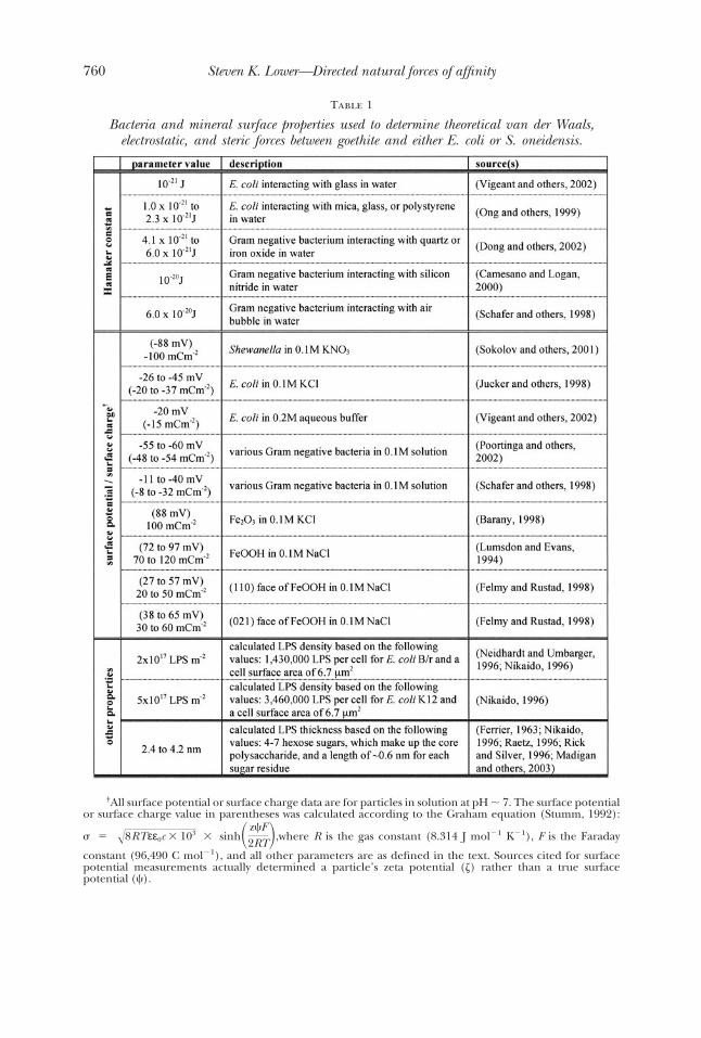
Table 1
Bacteria and mineral surface properties used to determine theoretical van der Waals,electrostatic, and steric forces between goethite and either E. coli or S. oneidensis.
†All surface potential or surface charge data are for particles in solution at pH � 7. The surface potentialor surface charge value in parentheses was calculated according to the Graham equation (Stumm, 1992):
� � �8RTεε0c � 103 � sinh� z�F2RT�,where R is the gas constant (8.314 J mol�1 K�1), F is the Faraday
constant (96,490 C mol�1), and all other parameters are as defined in the text. Sources cited for surfacepotential measurements actually determined a particle’s zeta potential ( ) rather than a true surfacepotential (�).
760 Steven K. Lower—Directed natural forces of affinity

table 1. This is because the van der Waals and electrostatic forces are expected to beattractive at all separations (see fig. 4). However, the measurements reveal a repulsiveforce between E. coli and goethite at a range of 4 to 10 nm (see fig. 2). The observedrepulsive force may be due to non-DLVO type of forces such as steric interactions(Israelachvili and McGuiggan, 1988; Israelachvili, 1992). Steric forces may arisebetween two particles if at least one of the particles contains a polymer layer on itssurface. As the two surfaces approach one another, the polymers become confined tothe intervening space and they are no longer free to move at random. This loss ofdynamic movement for chain molecules creates an entropic repulsive force at closeseparation.
Steric interactions, like DLVO forces, have been described in terms of energy-distance relationships (Alexander, 1977; de Gennes, 1987). Based on these priordescriptions, Butt and others (1999) and Camesano and Logan (2000) derived atheoretical force-distance relationship between a sphere and a flat surface, one ofwhich is coated with a polymer. This steric force is given as (Butt and others, 1999):
F�D� � 50rkBTL 0 �3/2e�2�D/L0 (5)
where L0 is the equilibrium thickness of the polymer (in m), � is the density of thepolymer on the surface (in m�2), and all other parameters are as defined above.
Figure 4 shows the steric force for a Gram-negative bacterium interacting withgoethite using values for biopolymer thickness and surface density taken from theliterature (table 1). The observed forces between E. coli and goethite are modeledreasonably well when the repulsive steric force is added to the van der Waals and theconstant potential approximation of the electrostatic force (see fig. 2). Both themeasured forces and the “extended” DLVO theory reveal an initial repulsive forcebetween E. coli and goethite that is overcome by an attractive force at very closeseparation.
In the context of bacterial recognition of a mineral surface, which is the objectiveof this paper, the approach force data provide some very important information. Themagnitude and range of the measured forces can be described in terms of nonspecificforces of interaction such as, van der Waals, electrostatic, and steric forces. All of theseforce types are very short range in these solution conditions. The bacteria must get towithin 5 to 10 nm of goethite before the cell senses the presence of the mineral. Inother words, a bacterium must come into direct physical contact with a mineral beforeit has a chance to recognize that surface. However, these approach force data do notprovide evidence of specific interactions between goethite and either bacteria. Theretraction force data, on the other hand, contain information about the energeticaffinity between a bacterium and goethite and details about specific biopolymers thatmay form a bond with goethite.
Figure 3 illustrates the forces recorded as E. coli or S. oneidensis were pulled awayfrom the mineral surface. Force profiles between E. coli and goethite are relativelysimple showing, in most cases, that E. coli can be separated from the mineral within afew nanometers. S. oneidensis, conversely, remained linked to the surface of goethite forhundreds of nanometers resulting in more complicated retraction profile (see fig. 3).
The overall characteristics of retraction curves can be described in terms of themaximum force recorded at a distance of zero, the so-called “adhesion force”. For theShewanella-goethite bacteria-mineral pair, this would seem to be an oversimplificationof the retraction data because the adhesion force is only one point on the entireretraction profile. The energy or work required to separate a bacterium from a mineralcan be quantified by numerically integrating the retraction force profiles with respectto distance (Lower and others, 2001a). This provides a physically relevant parameterthat takes into account the entire retraction data.
761between a bacterium and mineral

Even though the surfaces of S. oneidensis and E. coli are very similar (fig. 1), there isa striking difference in the energetic affinity for goethite exhibited by these two Gramnegative bacteria. Integrating force with respect to distance for retraction curves likethose shown in figure 3 yields an average affinity between E. coli and goethite of 7 � 3attoJoules (10�18 J). This affinity was indifferent to oxygen concentration. Ionicstrength, rather than oxygen concentrations, appears to have a much stronger impacton retraction forces for E. coli (see Lower and others, 2000). Also, the work to separatethe bacterium from the mineral did not change with the amount of time E. coliremained in contact with goethite (up to 30 min).
The situation was very different for S. oneidensis. The affinity between S. oneidensisand goethite was determined as 144 � 44 aJ (10�18 J) under anaerobic conditionswhen S. oneidensis remained in direct physical contact with goethite for 20 to 25minutes (see also Lower and others, this issue of the American Journal of Science). WhenS. oneidensis was placed in contact with goethite for only 5 min under anaerobicconditions, the energetic attraction was only 60 aJ (Lower and others, 2001a). Thesolution concentration of oxygen also impacted the energetic affinity of S. oneidensistowards goethite. Under aerobic conditions, only 25 aJ of energetic affinity wasobserved between S. oneidensis and goethite, regardless of the amount of “contact time”between the cells and mineral (Lower and others, 2001a). Finally, it should be notedthat forces have been measured between S. oneidensis and diaspore (AlOOH) (Lowerand others, 2001a; Lower and others, see this issue of the American Journal of Science).Diaspore has very similar surface properties to goethite, but the Al in the mineralstructure of diaspore cannot serve as a terminal electron acceptor for microorganisms.S. oneidensis expressed a significantly smaller affinity for AlOOH (�40 aJ) relative toFeOOH (�140 aJ, see above) under anaerobic conditions.
These energy or work determinations can be summarized into a few key points.First, S. oneidensis exhibits a much higher affinity for goethite relative to anotherGram-negative bacterium. Second, S. oneidensis has a selective affinity for iron oxidesrelative to other minerals like aluminum oxides, which have very similar surfaceproperties. Third, S. oneidensis alters its affinity for iron oxyhydroxides as a result of theoxygen concentration in the intervening solution. A higher affinity is noted for S.oneidensis towards goethite under anaerobic conditions when Fe(III) in the mineral isthe only available terminal electron acceptor. Fourth, a period of time is necessary forS. oneidensis to recognize the surface of goethite. Presumably, this is due to the amountof time it takes a cell to express and/or localize macromolecules to the bacterium-mineral interface. Taken together, the energy determinations suggest that S. oneidensispossesses a rare trait of being able to recognize certain minerals such that it expresses aselective affinity for these minerals under environmental conditions that wouldmaximize its chance for survival in nature.
While the energy or work calculations provide a suitable means of characterizingthe overall attributes of the retraction data, it would be useful if one could provideevidence that specific macromolecules, like proteins, are targeted by S. oneidensis to thecell-mineral interface. Such information can, in fact, be established by looking forforce-signatures that are characteristic of proteins that form a bond between abacterium and mineral. Lower and others (2005) have shown that sawtooth-like forcesignatures denote protein bonds between a cell and solid surface.
The retraction profiles between E. coli and goethite are void of all but anoccasional sawtooth (see fig. 3). However, retraction profiles for S. oneidensis andgoethite contain discrete sawtooth features (fig. 3). This indicates that outer mem-brane proteins are used by S. oneidensis, but not E. coli, to form a specific bond withgoethite. Protein force-signatures are primarily detected between S. oneidensis andgoethite under anaerobic conditions when Fe(III) may have served as terminal
762 Steven K. Lower—Directed natural forces of affinity

electron acceptor. This phenomena is described in more detail by Lower and others(see this issue of the American Journal of Science). Taken together, these observationssuggest that S. oneidensis recognizes the surface of goethite as a terminal electronacceptor such that it increases its affinity for FeOOH and targets putative mineral-specific proteins to promote contact with the mineral and perhaps transfer electrons tothe mineral.
conclusion
The ability to probe forces between a living bacterium and another surface isproviding an entirely new awareness of the way bacteria perceive solid phases such asminerals. This manuscript has presented quantitative measurements of the forces ofattraction (or repulsion) between goethite and two Gram negative bacteria species: E.coli and S. oneidensis. One of these bacteria, S. oneidensis, expresses a selective affinitytowards goethite under conditions that would promote its survival in the environment.Intermolecular forces measured as S. oneidensis approached the (010) face of goethitewere very short range (�8 nm). This means, in essence, that S. oneidensis must comeinto physical contact before it senses the presence of the mineral surface. Once incontact with the mineral, a period of time of more than �15 to 20 minutes is necessaryfor S. oneidensis to recognize goethite as a potential terminal electron acceptor. Suchrecognition is highly selective as S. oneidensis appears to differentiate between Fe-hydroxides and closely related minerals such as Al-hydroxides, which have similarsurface properties but cannot serve as terminal electron acceptors. Recognitiontriggers the expression and/or localization of putative mineral-specific proteins thatdirect natural forces of affinity towards goethite, presumably to enhance electrontransfer between the bacterium and Fe(III) in the crystal structure of the mineral.
acknowledgmentsThis work was supported by the Department of Energy (DE-FG02-04ER15590)
and the National Science Foundation (EAR-0417712; EAR-0411935). B. Lower, T.Beveridge, T. Camesano, and an anonymous reviewer provided valuable input thatgreatly improved this manuscript. I would also like to acknowledge the support of J.Tak, without whom this would not be possible.
References
Alexander, S. J., 1977, Adsorption of chain molecules with a polar head: A scaling description: Physique,v. 38, p. 983–987.
Arredondo, R., Garcia, A., and Jerez, C. A., 1994, Partial removal of lipopolysaccharide from Thiobacillusferrooxidans affects its adhesion to solids: Applied and Environmental Microbiology, v. 60, p. 2846–2851.
Barany, S., 1998, Complex electrosurface investigations of dispersed microphases: Advances in Colloid andInterface Science, v. 75, p. 45–78.
Beveridge, T. J., 1981, Ultrastructure, chemistry, and function of the bacterial wall: International Review ofCytology, v. 72, p. 229–317.
–––––– 1999, Structures of Gram-negative cell walls and their derived membrane vesicles: Journal ofBacteriology, v. 181, p. 4725–4733.
Butt, H. J., Jaschke, M., and Ducker, W., 1995, Measuring surface forces in aqueous electrolyte solution withthe atomic force microscope: Bioelectrochemistry and Bioenergetics, v. 38, p. 191–201.
Butt, H. J., Kappl, M., Mueller, H., Raiteri, R., Meyer, W., and Ruhe, J., 1999, Steric forces measured with theatomic force microscope at various temperatures: Langmuir, v. 15, p. 2559–2565.
Camesano, T. A., and Logan, B. E., 2000, Probing bacterial electrosteric interactions using atomic forcemicroscopy: Environmental Science and Technology, v. 34, p. 3354–3362.
Cooper, K., Ohler, N., Gupta, A., and Beaudoin, S., 2000, Analysis of contact interactions between a roughdeformable colloid and a smooth substrate: Journal of Colloid and Interface Science, v. 222, p. 63–74.
de Gennes, P. G., 1987, Polymers at an interface: A simplified view: Advances in Colloid and InterfaceScience, v. 27, p. 189–209.
Derjaguin, B. V., and Landau, L. D., 1941, Theory of the stability of strongly charged lyophobic sols and theadhesion of strongly charged particles in solutions of electrolytes: Acta Physicochim URSS, v. 14,p. 733–762.
763between a bacterium and mineral

Devasia, P., Natarajan, K. A., Sathyanarayana, D. N., and Rao, G. R., 1993, Surface-Chemistry of Thiobacillus-Ferrooxidans Relevant to Adhesion on Mineral Surfaces: Applied and Environmental Microbiology,v. 59, p. 4051–4055.
Dong, H. L., Onstott, T. C., Ko, C. H., Hollingsworth, A. D., Brown, D. G., and Mailloux, B. J., 2002,Theoretical prediction of collision efficiency between adhesion-deficient bacteria and sediment grainsurface: Colloids and Surfaces B-Biointerfaces, v. 24, p. 229–245.
Dziurla, M. A., Achouak, W., Lam, B. T., Heulin, T., and Berthelin, J., 1998, Enzyme-linked immunofiltrationassay to estimate attachment of Thiobacilli to pyrite: Applied and Environmental Microbiology, v. 64,p. 2937–2942.
Elimelech, M., Gregory, J., Jia, X., and Williams, R., 1995, Particle Deposition and Aggregation: Measure-ment, Modeling, and Simulation: Oxford, Butterworth-Heinemann, 441 p.
Ellen, R. P., Lepine, G., and Nghiem, P. M., 1997, In vitro models that support adhesion specificity inbiofilms of oral bacteria: Advances in Dental Research, v. 11, p. 33–42.
Felmy, A. R., and Rustad, J. R., 1998, Molecular statics calculations of proton binding to goethite surfaces:Thermodynamic modeling of the surface charging and protonation of goethite in aqueous solution:Geochimica et Cosmochimica Acta, v. 62, p. 25–31.
Ferrier, W., 1963, The crystal and molecular structure of [beta]-d-glucose: Acta Crystallographica, v. 16,p. 1023–1031.
Gregory, J., 1975, Interaction of unequal double layers at constant charge: Journal of Colloid and InterfaceScience, v. 51, p. 44–51.
Hoek, E. M. V., Bhattacharjee, S., and Elimelech, M., 2003, Effect of membrane surface roughness oncolloid-membrane DLVO interactions: Langmuir, v. 19, p. 4836–4847.
Hogg, R. I., Healey, T. W., and Fuerstenau, D. W., 1966, Mutual coagulation of colloidal dispersions:Transactions of the Faraday Society, v. 62, p. 1638–1651.
Israelachvili, J., 1992, Intermolecular and Surface Forces: London, Academic Press, 450 p.Israelachvili, J. N., and McGuiggan, P. M., 1988, Forces between surfaces in liquids.: Science, v. 241,
p. 795–800.Jucker, B. A., Zehnder, A. J. B., and Harms, H., 1998, Quantification of polymer interactions in bacterial
adhesion: Environmental Science and Technology, v. 32, p. 2909–2915.Kendall, T. A., and Lower, S. K., 2004, Forces between minerals and biological surfaces in aqueous solution:
Advances in Agronomy, v. 82, p. 1–54.Kolenbrander, P. E., 1989, Surface recognition among oral bacteria: Multigeneric coaggregations and their
mediators: Critical Reviews in Microbiology, v. 17, p. 137–159.Leckband, D., and Israelachvili, J., 2001, Intermolecular forces in biology: Quarterly Reviews of Biophysics,
v. 34, p. 105–267.Lovley, D. R., 1991, Dissimilatory Fe(III) and Mn(IV) reduction: Microbiological Reviews, v. 55, p. 259–287.Lower, B. H., Yongsunthon, R., Vellano, III, F. P., and Lower, S. K., 2005, Simultaneous force and
fluorescence measurements of a protein that forms a bond between a living bacterium and a solidsurface: Journal of Bacteriology, v. 187, p. 2127–2137.
Lower, S. K., Tadanier, C. J., and Hochella, M. F., 2000, Measuring interfacial and adhesion forces betweenbacteria and mineral surfaces with biological force microscopy: Geochimica et Cosmochimica Acta,v. 64, p. 3133–3139.
Lower, S. K., Hochella, M. F., and Beveridge, T. J., 2001a, Bacterial recognition of mineral surfaces:Nanoscale interactions between Shewanella and �-FeOOH: Science, v. 292, p. 1360–1363.
Lower, S. K., Tadanier, C. J., and Hochella, M. F., 2001b, Dynamics of the mineral-microbe interface: Use ofbiological force microscopy in biogeochemistry and geomicrobiology: Geomicrobiology Journal, v. 18,p. 63–76.
Lower, S. K., Hochella, M. F., Jr., Banfield, J. F., and Rosso, K., 2002, Nanogeoscience: From the movement ofelectrons to lithosphere plates. Eos: Transactions of the American Geophysical Union, v. 83, p. 53–56.
Lumsdon, D. G., and Evans, L. J., 1994, Surface complexation model parameters for goethite (a-FeOOH):Journal of Colloid and Interface Science, v. 164, p. 119–125.
Madigan, M. T., Martinko, J. M., and Parker, J., 2003, Brock Biology of Microorganisms: Upper Saddle River,New Jersey, Prentice Hall, 1019 p.
Myers, C. R., and Myers, J. M., 1992, Localization of cytochromes to the outer membrane of anaerobicallygrown Shewanella Putrefaciens MR-1: Journal of Bacteriology, v. 174, p. 3429–3438.
–––––– 1993, Ferric reductase is associated with the membranes of anaerobically grown Shewanella putrefaciensMR-1: Fems Microbiology Letters, v. 108, p. 15–22.
–––––– 1997, Outer membrane cytochromes of Shewanella putrefaciens MR-1: Spectral analysis, and purifica-tion of the 83-kDa c-type cytochrome: Biochimica et Biophysica Acta, v. 1326, p. 307–318.
–––––– 2003, Cell surface exposure of the outer membrane cytochromes of Shewanella oneidensis MR-1: Lettersin Applied Microbiology, v. 37, p. 254–258.
Myers, C. R., and Nealson, K. H., 1990, Respiration-linked proton translocation coupled to anaerobicreduction of manganese(IV) and iron(III) in Shewanella putrefaciens MR-1: Journal of Bacteriology,v. 172, p. 6232–6238.
Myers, J. M., and Myers, C. R., 1998, Isolation and sequence of omcA, a gene encoding a decaheme outermembrane cytochrome c of Shewanella putrefaciens MR-1, and detection of omcA homologs in otherstrains of S. putrefaciens: Biochimica et Biophysica Acta, v. 1373, p. 237–251.
–––––– 2001, Role for outer membrane cytochromes OmcA and OmcB of Shewanella putrefaciens MR-1 inreduction of manganese dioxide: Applied and Environmental Microbiology, v. 67, p. 260–269.
Neidhardt, F. C., and Umbarger, H. E., 1996, Chemical composition of Escherichia coli, in Neidhardt, F. C.,editor, Escherichia coli and Salmonella: Cellular and Molecular Biology: Washington, D. C., ASM Press,p. 13–14.
764 Steven K. Lower—Directed natural forces of affinity

Nikaido, H., 1996, Outer membrane, in Neidhardt, F. C., editor, Escherichia coli and Salmonella: Cellular andMolecular Biology: Washington, D. C., ASM Press, p. 29–47.
Ohmura, N., Kitamura, K., and Saiki, H., 1993, Selective adhesion of Thiobacillus ferrooxidansto pyrite:Applied and Environmental Microbiology, v. 59, p. 4044–4050.
Ohmura, N., Tsugita, K., Koizumi, J. I., and Saiki, H., 1996, Sulfur-binding protein of flagella of Thiobacillusferrooxidans: Journal of Bacteriology, v. 178, p. 5776–5780.
Ong, Y. L., Razatos, A., Georgiou, G., and Sharma, M. M., 1999, Adhesion forces between E. coli bacteria andbiomaterial surfaces: Langmuir, v. 15, p. 2719–2725.
Pink, D. A., Hansen, L. T., Gill, T. A., Quinn, B. E., Jericho, M. H., and Beveridge, T. J., 2003, Divalentcalcium ions inhibit the penetration of protamine through the polysaccharide brush of the outermembrane of Gram- negative bacteria: Langmuir, v. 19, p. 8852–8858.
Poortinga, A. T., Bos, R., Norde, W., and Busscher, H. J., 2002, Electric double layer interactions in bacterialadhesion to surfaces: Surface Science Reports, v. 47, p. 3–32.
Raetz, C. R. H., 1996, Bacterial lipopolysaccahrides: A remarkable family of bioactive macroamphiphiles, inNeidhardt, F. C., editor, Escherichia coli and Salmonella: Cellular and Molecular Biology: Washington,D. C., ASM Press, p. 1035–1063.
Rick, P. D., and Silver, R. P., 1996, Enterobacterial common antigen and capsular polysaccharides, inNeidhardt, F. C., editor, Escherichia coli and Salmonella: Cellular and Molecular Biology: Washington,D. C., ASM Press, p. 104–122.
Schafer, A., Harms, H., and Zehnder, A. J. B., 1998, Bacterial accumulation at the air-water interface:Environmental Science and Technology, v. 32, p. 3704–3712.
Sokolov, I., Smith, D. S., Henderson, G. S., Gorby, Y. A., and Ferris, F. G., 2001, Cell surface electrochemicalheterogeneity of the Fe(III)- reducing bacteria Shewanella putrefaciens: Environmental Science andTechnology, v. 35, p. 341–347.
Stoica, O., Tuanyok, A., Yao, X. W., Jericho, M. H., Pink, D., and Beveridge, T. J., 2003, Elasticity ofmembrane vesicles isolated from Pseudomonas aeruginosa: Langmuir, v. 19, p. 10916–10924.
Stumm, W., 1992, Chemistry of the Solid-Water Interface: New York, John Wiley and Sons, Inc., 428 p.Vargas, M., Kashefi, K., Blunt-Harris, E. L., and Lovley, D. R., 1998, Microbiological evidence for Fe(III)
reduction on early Earth: Nature, v. 395, p. 65–67.Verwey, E. J., and Overbeek, J. T. G., 1948, Theory of the Stability of Lyophobic Colloids: Amsterdam,
Elsevier Publishing, 205 p.Vigeant, M. A. S., Ford, R. M., Wagner, M., and Tamm, L. K., 2002, Reversible and irreversible adhesion of
motile Escherichia coli cells analyzed by total internal reflection aqueous fluorescence microscopy:Applied and Environmental Microbiology, v. 68, p. 2794–2801.
Whittaker, C. J., Klier, C. M., and Kolenbrander, P. E., 1996, Mechanisms of adhesion by oral bacteria:Annual Review of Microbiology, v. 50, p. 513–552.
765between a bacterium and mineral