discover the extraordinary potential of stem cell biology
TRANSCRIPT

Discover the extraordinary potential of stem cell biology.

biopotential
Stem Cell BiologyStem and progenitor cell research is a complex and very exciting field that promises fantastic curative discoveries in numerous areas from cancer to diabetes to neurodegenerative diseases. Sigma-Aldrich® offers an unprecedented number of products to support discovery efforts in this area. Applications such as transfection, cell and protein characterization, RNAi, ADMET, and imaging are instrumental in the development of stem cells for regenerative medicine and drug discovery. Products in this guide are tools for stem cell science and support our customers in their research efforts.
Figure 1. Induced Pluripotent Stem Cell (iPS) Pathway
Cardiac Muscle
Mesoderm(Middle Layer)
Endoderm(Internal Layer)
Ectoderm(External Layer)
Embryonic Stem Cells
SkeletalMuscle Cells
Red BloodCells
Smooth Muscle(In Gut)
Tubule Cellof the Kidney
Lung Cell(Aveolar Cell)
ThyroidCell
PancreaticCell
Skin Cellsof Epidermis
PigmentCell
NeuronCell
Table of Contents
Title Page #
Stemline® Media for Adult Stem Cells . . . . . . . . . . . . . . . . . . . . . . . . . . . . . 3 Stemline T cell Expansion . . . . . . . . . . . . . . . . . . . . . . . . . . . . . . . . . . . . . . . . 3 Stemline Neural Stem Cell Expansion Medium . . . . . . . . . . . . . . . . . . . 4 Stemline Mesenchymal Stem Cell Expansion Medium . . . . . . . . . . . 4 Stemline II Hematopoietic Stem Cell Expansion Medium . . . . . . . . . 5
3-D Cell Culture Systems . . . . . . . . . . . . . . . . . . . . . . . . . . . . . . . . . . . . . . . . . . 6 HyStem™ . . . . . . . . . . . . . . . . . . . . . . . . . . . . . . . . . . . . . . . . . . . . . . . . . . . . . . . . 7 MaxGel™ . . . . . . . . . . . . . . . . . . . . . . . . . . . . . . . . . . . . . . . . . . . . . . . . . . . . . . . . 9 HydroMatrix™ . . . . . . . . . . . . . . . . . . . . . . . . . . . . . . . . . . . . . . . . . . . . . . . . . . . 9
Antibodies for Stem Cell Research . . . . . . . . . . . . . . . . . . . . . . . . . . . . . . 10 Antibodies for Neural Stem Cell Markers . . . . . . . . . . . . . . . . . . . . . . . . 11 Antibodies for Embryonic Stem Cell/iPS Cell Markers . . . . . . . . . . . . 11 Antibodies for Hematopoietic Stem Cell Markers . . . . . . . . . . . . . . . . 11
Growth Factors and Cytokines in Stem Cell Biology . . . . . . . . . . . . . 12
Small Molecules for Stem Cell Biology . . . . . . . . . . . . . . . . . . . . . . . . . . . 13
Induced Pluripotent Stem Cell Technology . . . . . . . . . . . . . . . . . . . . . . 15 Stemgent® Reprogramming Lentiviruses . . . . . . . . . . . . . . . . . . . . . . . 16
Targeted Genomic Editing with CompoZr® ZFN Technology . . . . 18

Order 800-325-3010 Technical Service 800-325-5832 3biopotential
Stem Cell MediaOptimize Your Stem Cell Expansion with Stemline®
Proven, consistent results and optimized formulations have made Sigma’s Stemline Media must-haves for researchers rising to the chal-lenges of adult stem cell expansion and maturation. Along with our broad selection of reagents, supplements, antibodies, and cytokines, Stemline Media ensure optimal expansion of robust cells.
Ordering InformationCat. No. Product Package SizeS3194 Stemline Neural Stem Cell Expansion Medium 500 mL
S0189 Stemline Hematopoietic Stem Cell Expansion Medium
500 mL
S0192 Stemline II Hematopoietic Stem Cell Expansion Medium
500 mL
S1694 Stemline T Cell Expansion Medium 500 mL
S3444 Stemline Dendritic Cell Maturation Medium 1 L
S1569 Stemline Mesenchymal Stem Cell Expansion Medium
1 L
Developed to promote the optimal expansion of adult human T cells, Stemline T Cell Expansion Medium demonstrates significantly greater expansion (55%) when compared to alternative media, and viability greater than 95%.
Stemline® T Cell Expansion Medium
0.E+00
1.E+06
2.E+06
3.E+06
4.E+06
5.E+06
6.E+06
Stemline T-cellExpansion
Medium (S1694)
Competitor A
Tota
l Vi
able
Cel
l Num
ber
Competitor B Competitor C RPMI with 10% FBS
RPMI with 10% FBS(no CD3 / CD 28)
Features and Benefits
• Serum-free formulation
•Excellent expansion of T cells of human origin
• Supports high cell densities that exhibit rigorous and consistent growth kinetics
• Maintains the proper CD4/CD8 ratio in flow cytometric analysis
•Maintains functionality, both ex vivo and in vivo
•Produced in a GMP state-of-the-art facility with an available drug master file (DMF)
Figure 2. T-Cell Expansion Day 7
Stemline demonstrates superior expansion of T cells. When compared with three alternative commercial media and two RPMI formulations, Stemline demonstrated 40% more total viable cells.

biopotential
Features and Benefits
• Free of serum and all other animal components, which increases performance consistency and eliminates safety risks with potential adventitious agents
•For use with neurospheres and monolayer cultures
• Neurospheres expanded in Stemline medium show optimal levels of BrdU incorporation, indicating the presence of proliferating cells
•Cells retain differentiation capacity
•Superior expansion rates when compared to alternatives
•Produced in a GMP state-of-the-art facility with an available drug master file (DMF)
178%
100%
0%
20%
40%
60%
80%
100%
120%
140%
160%
180%
Stemline Neural Stem Cell Expansion Medium (S3194)
DME/F-12 + N2
Stemline® Neural Stem Cell Expansion Medium
Figure 3. Expansion of Monolayer Neural Stem Cells in 24-well Culture Plate
Stemline demonstrates superior expansion of monolayer neural stem cells (NSC). To test the ability of Stemline Neural Stem Cell Expansion Medium to expand human monolayer neural stem cells, researchers at Sigma-Aldrich and the University of Wisconsin designed a bench-scale expansion assay. Cells were grown in monolayer format by seeding the cells at 20,000 cells/cm2 on poly-L-lysine coated 24-well tissue culture plates. Cells were incubated for 5 days in medium supplemented with EGF (Cat. No. E9644) and LIF (Cat. No. L5283). After several passages, overall proliferation was measured. Monolayer NSC cultured in Stemline demonstrated superior expansion to those grown in other serum-free NSC media.
Features and Benefits
•Maximum expansion of CD34+ progenitors
•Supports robust, high density cell populations
•Superior expansion
•Cells retain their differentiation potential at 14 days in culture
• Produced in a GMP state-of-the-art facility with an available drug master file (DMF)
• Requires supplementation with antibiotics, cytokines, L-glutamine, and fetal bovine serum, as appropriate to individual research protocols
Stemline Mesenchymal Stem Cell Expansion Medium
4.5
4.0
3.5
3.0
2.5
2.0
1.5
1.0
0.5
0
Fold
Exp
ansi
on
Competitor AStemline MesenchymalStem Cell Expansion
Medium (S1569)
Competitor B
Developed to promote the optimal expansion of human mesenchymal stem cells (MSC) from bone marrow, Stemline Mesenchymal Stem Cell Expansion Medium demonstrates greater fold increases of total nucleated cells (TNC) than other commercially available formulas. Additionally, functional trials clearly demostrate Stemline’s capacity to promote differentiation into adipocytes, chondrocytes, and osteocytes.
Figure 4. Mesenchymal Stem Cell Fold Expansion
Stemline demonstrates superior expansion of mesenchymal stem cells (MSC). To test the ability of Stemline Mesenchymal Stem Cell Expansion Medium to promote expansion of MSC, researchers at Sigma-Aldrich designed a bench-scale assay. Triplicate 2 mL cultures at 5,000 MSC/cm2 were grown in a 6-well microplate culture system in Stemline medium or other medium containing FBS. Each well was treated with trypsin/EDTA, triturated, and harvested after a 14-day expansion. MSC were counted using a hemacytometer and average viable cell count determined for each condition. MSC cultured in Stemline demonstrated superior expansion to those grown in other MSC media, retained their differentiation potential, and were easily passaged routinely.

Order 800-325-3010 Technical Service 800-325-5832 5
270.40
156.49152.41
149.15
122.73
76.2067.77
28.77
0.00
50.00
100.00
150.00
200.00
250.00
300.00
p<0.0001
Fold
Incr
ease
Stemline II
Stemline I
Competitor A
Competitor B
Competitor C
Competitor D
Competitor E
Competitor F
Figure 7. Fold Increase of Total Nucleated Cells from CD34+ Cord Blood
Stemline demonstrates superior expansion of cord blood hematopoietic stem cells (HSC). To test the ability of Stemline II Hematopoietic Stem Cell Expansion Medium to expand CD34+ HSC, researchers at Sigma-Aldrich and the University of Kentucky designed a bench-scale expansion assay. Cells were seeded into the wells of 24-well tissue culture plates. One milliliter of medium was added to each well with the appropriate cytokines to stimulate growth (100 ng/mL each of TPO, SCF, and G-CSF). Each condition was performed in triplicate and seeded with 10,000 cells per mL in each well. Due to the clinical importance and the donor-to-donor variability typical to the expansion of umbilical cord blood-derived cells, researchers elected to test 15 donors for expansion and surface antigen expression. The cells were counted on day 10 and the fold increase was determined by cellsfinal/cellsinitial. HSC from cord blood cultured in Stemline II demonstrated superior expansion to those grown in other serum-free HSC media.
199.5
51.4
37.3
64.4
42.2
34.6 34.4
10.9
0.0
20.0
40.0
60.0
80.0
100.0
120.0
140.0
160.0
180.0
200.0p<0.05
Fold
Incr
ease
Stem
line II
Stem
line I
Competit
or A
Competit
or B
Competit
or C
Competit
or D
Competit
or E
Competit
or F
Figure 6. Fold Increase of Total Nucleated Cells from CD34+ Bone Marrow
Stemline demonstrates superior expansion of bone marrow hematopoietic stem cells (HSC). To test the ability of Stemline II Hematopoietic Stem Cell Expansion Medium to expand CD34+ HSC, researchers at Sigma-Aldrich and the University of Kentucky designed a bench-scale expansion assay. Cells were seeded into the wells of 24-well tissue culture plates. One milliliter of medium was added to each well with the appropriate cytokines to stimulate growth (100 ng/mL each of TPO, SCF, and G-CSF). Each condition was performed in triplicate and seeded with 10,000 cells per mL in each well. The cells were counted on day 14 and the fold increase was determined by cellsfinal/cellsinitial. HSC from bone marrow cultured in Stemline II demonstrated superior expansion to those grown in other serum-free HSC media.
Features and Benefits
• Free of serum and all other animal-derived components with the exception of human serum albumin
•Developed to promote the optimal expansion of human hematopoietic stem cells (HSC) from bone marrow, mobilized peripheral blood, and cord blood
•Expands cells from all appropriate hematopoietic lineages in a colony-forming unit
•Tested extensively in 7-day and 14-day growth assays
• Produced in a GMP state-of-the-art facility with an available drug master file (DMF)
Stemline II Hematopoietic Stem Cell Expansion Medium
CLP
CMP
HematopoieticStem Cell
T-Lymphocyte
B-Lymphocyte/Plasma Cell
Megakaryocyte/Platelets
Erythrocyte
Basophil/Mast Cell
Eosinophil
Neutrophil
Monocyte/Macrophage/Kupffer CellLangerhans CellDendritic cell
Figure 5. Hematopoietic Stem Cell Pathway
Through the process of hematopoiesis, all cellular blood components are derived from hematopoietic stem cells.
To request a free evaluation sample of Stemline Medium, visit sigma.com/stemline

biopotential
3-Dimensional Cell Culture SystemsGrowing cells on flat surfaces seems artificial and unnatural since it does not bear a resemblance to the in vivo architectures where cells flourish at their best. 3-dimensional (3-D) cell culture scaffolds are a better representation of the natural environment experienced by cells in the living organism. Reflecting natural conditions allows for intercellular interactions with more realistic biochemical and physiological responses.
In a 3-D environment, cells behave and respond more like they would in vivo to internal and external stimuli, such as changes in temperature, pH, nutrient absorption, transport, and differentiation. The power of regenerative medicine provides the means to understanding numerous intriguing cellular progressions such as development, aging, and tissue rejuvenation. The only way to understand these fascinating processes in biology is to study model systems that truly resemble those in the organism, making 3-D cell culture imperative.
Figure 8: Cells secrete various types of proteins that create an intercalated mesh comprising a tissue-specialized Extracellular Matrix (ECM).
Features and Benefits
•Stem cell culture that resembles natural biological systems in which cells thrive and grow
•Promote cell growth and migration to support the proliferation of stem cells
•Contains reduced levels of growth factors, which promotes lot-to-lot consistency
•Human ECM facilitates human stem cell culture

Order 800-325-3010 Technical Service 800-325-5832 7
The HyStem Platform Includes Three Unique Members:
HyStem Cell Culture Scaffold Kit: For research-ers who require an animal component-free system, for researchers who will customize with their own attachment factors and/or ECM proteins/peptides,
and for researchers who require a minimal number of cell at-tachment sites.
HyStem-C Cell Culture Scaffold Kit: For researchers who require a large number of generalized cell attachment sites for their stem cell culture(s).
HyStem-HP Cell Culture Scaffold Kit: For researchers planning to incorporate and gradually release growth factors into the stem cell environment.
Ordering InformationCat. No. Product Description Volume of Hydrogel ProducedHYS010 HyStem Cell Culture
Scaffold Trial Kit2.5 mL
HYSC010 HyStem-C Cell Culture Scaffold Trial Kit
2.5 mL
HYSHP010 HyStem-HP Cell Culture Scaffold Trial Kit
2.5 mL
HYS020 HyStem Cell Culture Scaffold Kit
7.5 mL
HYSC020 HyStem-C Cell Culture Scaffold Kit
7.5 mL
HYSHP020 HyStem-HP Cell Culture Scaffold Kit
7.5 mL
H2666 HyStem-C 96-well plate 96 wells
Customizable: The HyStem platform offers you, the researcher, control over growth factor incorporation, attachment factor incorporation, ECM protein incorporation, rigidity of the hydrogel, and cell incapsulation vs. top plating.
Synthetic: Because HyStem is a synthesized matrix and not a biological extract, researchers are able to closely control the composition of their cells’ environment. HyStem’s components include chemically synthesized HyStem (thioated hyaluronic acid), Extralink™ (thio-reactive cross-linker), degassed water, and biologically purified Gelin-S™ (denatured collagen).
Biologically Accurate: HyStem kits are optimal for cuturing stem cells whose natural envi-ronments are rich in hyaluronic acid. The HyStem hydrogel scaffold closely mimics the rich, natural extracellular matrix environment, complete with hyaluronic acid and collagen fibrils, while offering the flexibility to customize with appropriate growth factors, attachment factors, and proteins.
HyStem™ Cell Culture Scaffolds
The First Customizable Synthetic ECM
Sigma® is pleased to introduce HyStem, the first customizable synthetic ECM that closely mimics in vivo conditions, to enable three-dimensional culture of stem cells.
Natural Extracellular Environment
Figure 9. HyStem closely mimics the natural extracellular environment
HyStem HPHyStem HyStem C
Basal lamina (laminin, entactin, collagen I, collagen IV, heparin)Hyaluron (HA) or Hystem™ (thiol-modified Hyaluronan)Collagen Fibrils or Gelin-S™ (thiol-modified denatured Collagen)Extralink™ (thioreactive crosslinker)Heparin SulfateGrowth Factors

biopotential
HyStem Closely Mimics the Natural Extracellular Environment
Application Data
Figure 10. H9 human embryonic stem cells plated on HyStem hydrogels containing CVFL and grown for 3 days.
Figure 11. Human mesenchymal stem cells grown (5 days) on the surface of a HyStem hydrogel with collagen I non-covalently incorporated.
Figure 12. Endothelial progenitor cells cultivated in HyStem-HP. Blue = mouse cell nuclei, brown = human cell nuclei, red = CD31 protein (courtesy of Robert Grove, Endgenitor Technologies, Inc., Indianapolis, IN)
Figure 13. Neurosphere-derived human embryonic stem cells (H9) seeded in HyStem-C and grown for 5 days. Red = β-Tubulin III. Blue = Draq-5.
To learn more, visit sigma.com/hystem

Order 800-325-3010 Technical Service 800-325-5832 9
MaxGel™Produced in vitro, MaxGel human extracellular matrix (ECM) provides a rich 3-D environment to promote cellular proliferation. MaxGel human ECM contains extracellular matrix components including collagens, laminin, fibronectin, tenascin, elastin, and a number of proteoglycans and glycosaminoglycans. The cell-cultured derived ECM effectively reproduces the cooperative interaction of epithelia and mesenchyme during development and in organotypic cell culture of skin. Human MaxGel ECM promotes cell growth and migra-tion and has been shown to support the proliferation of many cell types, including neural stem cells, neurons, glia, astrocytes, fibroblasts, hepatocytes, and keratinocytes.
Ordering InformationCat. No. Product Package SizeE0282 MaxGel Human ECM (liquid) 0.1 mL, 1 mL
M1073 MaxGel Human ECM 96-well plate 1 each
Figure 14. MaxGel Human ECM enables improved expansion of Adult Keratinocytes.
HaCaT cells (derived from human adult skin keratinocytes) were grown for 24 hours after plating on tissue culture plastic (A) and on 1% human ECM (B), which demonstrate that HaCaT cells proliferate better on ECM.
Figure 15. MaxGel Human ECM enables improved expansion of Fetal Lung Fibroblasts.
MRC-5 cells (derived from human fetal lung fibroblasts) were grown for 24 hours after plating on tissue culture plastic (A) and on 1% human ECM (B). As seen with other cells, MRC-5 cells propagate better on ECM.
HydroMatrix™ Peptide HydrogelA synthetic peptide nanofiber scaffold, HydroMatrix offers the preci-sion and control of a synthesized matrix with the natural 3-D archi-tecture of a highly crosslinked peptide hydrogel. The HydroMatrix scaffold self-assembles from fluid precursors into a highly crosslinked peptide 3-D hydrogel in response to changes in temperature or ionic strength. By adjusting the concentration of the HydroMatrix solution, researchers are able to control the flexibility of the 3-D architecture and tailor the structure to meet their needs. HydroMatrix promotes cell growth and migration and has been shown to support the proliferation of many cell types, including neural stem cells, neurons, and glia.
Ordering InformationCat. No. Product Package SizeA6982 HydroMatrix (liquid) 1 mL, 5 mL, 10 mL
H4165 HydroMatrix 96-Well Plate 1 each
H4040 HydroMatrix 24-Well Plate 1 each
H3915 HydroMatrix 4-Well Plate 1 each Figure 16. HydroMatrix Peptide Hydrogel.
Rat neural stem cells (NSC) cultured on three surfaces. NSC grew poorly on tissue culture plastic (A) and slightly better in poly-L-lysine/laminin coated plates (B). NSC demonstrated excellent growth on HydroMatrix peptide hydrogel 0.5% (w/v) (C).
biopotential
Antibodies for Stem Cell ResearchStem cell research holds the promise of cures for many diseases and injuries. Some of the most important discovery tools in this fast-growing area of research are stem cell markers. Sigma’s promise is to aid in advancing your stem cell research by providing high-quality antibodies. Each of our antibodies meets meticulous application testing and validation using a diversity of methods, which can be found in our online data sheets and certificates of analysis. Also, visit us online to search and browse our award-winning online tools such as the Antibody Explorer, PathFinder, and Your Favorite Gene Powered by Ingenuity.
Features and Benefits
•Extensive collection of antibodies including markers for Embryonic, Mesenchymal, Hematopoietic, Neural, Cardiac, and Islet Stem Cell research
•Availability of monoclonals, polyclonals, secondaries, conjugates, and custom services
•Validations using a variety of methods: WB, IHC, IF, IP, ELISA
• Search and browse capabilities using Sigma’s award winning tools: Antibody Explorer and Your Favorite Gene powered by Ingenuity
• Support from Sigma-Aldrich’s technical service to assist you with your specific antibody requirements
For more information on Stem Cell Antibodies, download the brochure at sigma.com/stembio
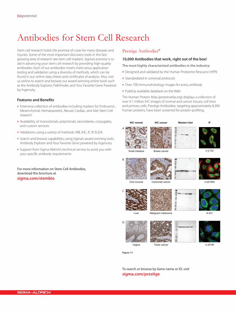
Prestige Antibodies®
10,000 Antibodies that work, right out of the box!
The most highly characterized antibodies in the industry:
•Designed and validated by the Human Proteome Resource (HPR)
•Standardized in universal protocols
•Over 700 immunohistology images for every antibody
•Publicly available database on the Web
The Human Protein Atlas (proteinatlas.org) displays a collection of over 9.1 million IHC images of normal and cancer tissues, cell lines and primary cells. Prestige Antibodies, targeting approximately 8,300 human proteins, have been screened for protein profiling.
IHC normal Western blot
Small intestine
Oral mucosa
Liver
Vagina
U-2 OS
U-251MG
A-431
U-251M
IF
Breast cancer
Colorectal cancer
Malignant melanoma
Testis cancer
IHC cancer
A
B
C
D
To search or browse by Gene name or ID, visit sigma.com/prestige
Figure 17.

Order 800-325-3010 Technical Service 800-325-5832 11
Figure 18. Cells derived from the culture of neural stem cells
The cells were expanded in Stemline Neural Stem Cell Expansion Medium (Cat. No. S3194) and then moved to conditions to allow them to differentiate. Differentiated cells were fixed and stained with an antibody for GFAP (an astrocyte marker in green, (Cat. No. G9269), actin is labeled with TRITC phalloidin (Cat. No. P1951), and the nuclei are labeled with DAPI (Cat. No. D8417).
Neural Stem Cell Markers
Ordering InformationCat. No. Product NameA1594 Anti-Activin AF3804 Anti-Frizzled-1A2052 Anti-GABAM3696 Anti-MAP2N5413 Anti-NestinN3537 Anti-NeurofibrominN5139 Anti-Neurofilament 68N4538 Anti-NogginN4788 Anti-NOTCH1O7139 Anti-Oligodendrocyte Marker 04P5117 Anti-PheripherinS2644 Anti-S-100S193 Anti-Synapsin IT6402 Anti-TauT3952 Anti-β-Tubulin IIIV4630 Anti-VimentinW2141 Anti-Wnt-1
Embryonic Stem Cell/iPS Cell MarkersEmbryonic stem cells and embryonic-like stem cells, such as induced pluripotent stem (iPS) cells, are the most versatile types of stem cells. Research in this area has the greatest potential in medical applica-tions because these cells are infinite if cultured in suitable environ-ment. They also have the ability to differentiate into a broad range of specialized cells.
Ordering InformationCat. No. Product NameC3956 Anti-c-MycM4439 Anti-c-MycD2569 Anti-DPPA5WH0002168M2 Anti-FABP1HPA002188 Anti-FABP4HPA011209 Anti-FGF4G6919 Anti-G9a MethyltransferaseG4165 Anti-gp130HPA007526 Anti-GREM1WH0009314M1 Anti-KLF4N4413 Anti-NanogN3038 Anti-NanogN5413 Anti-NestinWH0004838M1 Anti-NODALO8389 Anti-Oct 3/4P0873 Anti-POU5F1 (Oct4)P0082 Anti-POU5F1 (Oct4)HPA015791 Anti-SALL4S9072 Anti-SOX2S2931 Anti-Stem Cell FactorT3952 Anti-β-Tubulin III
Hematopoietic Stem Cell MarkersHematopoietic stem cells (HSC) give rise to all blood cell types and are found in the bone marrow of adults, umbilical cord blood, placenta, and mobilized peripherial blood. In the table you will find most commonly used antibodies that provide reliable means of identification in HSC research, such as isolation of these cells, their differentiation into a variety of specialized cells, their mobilization out of the bone marrow into circulating blood, apoptosis, and numerous other functions.
Ordering InformationCat. No. Product NameB9804 Anti-Bcl-2C0551 Anti-CD11bGW10811F Anti-CD34WH0000947M1 Anti-CD34WH0000952M2 Anti-CD38K0131 Anti-C-Kit/SCF ReceptorC9493 Anti-CD133C3116 Anti-CXCR4I8398 Anti-Interleukin-2I8026 Anti-Interleukin-8HPA004176 Anti-RUNX1T5567 Anti-TCF-1T5817 Anti-TCF-4WH0007070M1 Anti-THY1HPA003733 Anti-THY1

biopotential
Growth factors and cytokines are naturally occurring regulatory mole-cules that bind to receptors on the cell surface, and stimulate cell and tissue function by influencing differentiation of cells, changing their biochemical activity, cellular growth, and regulating their rate of proliferation. In the table, you will find some of the commonly used growth factors in stem cell research.
Ordering InformationCat. No. Product Description Stem Cells Affected A4941 Activin A human ESB1434 Bone Morphogenetic Protein 7 human ES, MES, NEURB2680 Bone Morphogenetic Protein 4 human ES, MES, NEURB3555 Bone Morphogenetic Protein 2 human ES, MES, NEURB3795 Brain-derived neurotrophic factor human ES, NEURC3835 Ciliary Neurotrophic Factor from rat NEURE4643 Heparin-Binding EGF-Like Growth
Factor human NEUR
E9530 Erythropoietin from mouse HEM, NEUE9644 Epidermal Growth Factor human ES, MES, NEURF0291 Fibroblast Growth Factor-Basic human ES, MES NEUR, PANF1168 Fibroblast Growth Factor-9 human ESF4537 Fibroblast Growth Factor-5 human ESF5392 Fibroblast Growth Factor-Basic from
bovine pituitaryES, MES, NEUR, PAN
F8924 Fibroblast Growth Factor-10 human ESF9786 Fibroblast Growth Factor-Basic Heparin
Stabilized humanES, MES, NEUR, PAN
G0282 Granulocyte-Macrophage Colony-Stimulating Factor mouse
MES, HEM
G0407 Granulocyte Colony-Stimulating Factor human
MES, HEM
G1401 Glial Cell Line-derived Neurotrophic Factor from rat
NEUR
G1777 Glial Cell Line-derived Neurotrophic Factor human
NEUR
G5035 Granulocyte-Macrophage Colony-Stimulating Factor human
MES, HEM
H1404 Hepatocyte Growth Factor human MES, NEURI0523 Interleukin-2 from mouse HEMI1020 Interleukin-4 from mouse HEMI1395 Interleukin-6 human HEMI1646 Interleukin-3 human ES, HEMI2276 Interleukin-12 human HEMI2406 Interleukin-11 human HEMI2644 Interleukin-2 human HEMI3019 Interleukin-10 from mouse HEMI3265 Interferon-γ human MES, HEMI3268 Interleukin-6 human HEMI3769 Insulin-like Growth Factor-I human MES, NEUR, HEMI4144 Interleukin-3 from mouse ES, HEMI4269 Interleukin-4 human HEMI4777 Interferon-γ from mouse MES, HEMI4892 Interleukin-7 from mouse HEM
Ordering InformationCat. No. Product Description Stem Cells Affected I5271 Interleukin-1β from mouse HEMI5896 Interleukin-7 human HEMI7389 Interleukin-3 human ES, HEMI8523 Interleukin-12 from mouse HEMI8648 Interleukin-15 human HEMI8779 Insulin-like Growth Factor-I from mouse MES, NEUR, HEMI9401 Interleukin-1β human HEMI9646 Interleukin-6 from mouse HEML5158 Leukemia Inhibitory Factor from mouse ES, MES, NEUR, HEML5283 Leukemia Inhibitory Factor human ES, MES, NEUR, HEMM6292 Macrophage Inflammatory
Protein-1α humanHEM
M6417 Macrophage Inflammatory Protein-1β human
HEM
M6667 Monocyte Chemotactic Protein-1 human HEMM9170 Macrophage Colony-Stimulating
Factor from mouseHEM
N0513 Nerve Growth Factor-7S from murine submaxillary gland
NEUR
N1408 Nerve Growth Factor-β human NEURN1780 Neurotrophin-4 human ES, NEURN1905 Neurotrophin-3 human ES, NEURN2513 Nerve Growth Factor-β from rat NEURN6009 Nerve Growth Factor-2.5S from murine
submaxillary glandNEUR
N8133 Nerve Growth Factor from Vipera lebetina venom
NEUR
O1637 Oncostatin M from mouse ESO9635 Oncostatin M human ESP3076 Platelet-Derived Growth Factor-AA human MES, NEUR, HEMP3201 Platelet-Derived Growth Factor-BB human MES, NEUR, HEMS7901 Stem Cell Factor human HEMS9915 Stem Cell Factor from mouse HEMT2815 Transforming Growth Factor-β2 human ES, MES, HEMT4184 Thrombopoietin from mouse HEMT5050 Transforming Growth Factor-β1 from
porcine plateletsES, MES, NEUR, HEM
T5300 Transforming Growth Factor-β2 from porcine platelets
ES, MES, HEM
T5425 Transforming Growth Factor-β3 human MEST5944 Tumor Necrosis Factor-α from rat HEMT6674 Tumor Necrosis Factor-α human HEMT7039 Transforming Growth Factor-β1 human ES, MES, NEUR, HEMT7924 Transforming Growth Factor-α human ES, MES, NEUR, HEMV7259 Vascular Endothelial Growth Factor human MES, NEUR, HEMAbbreviations: ES-Embryonic Stem Cell, NEUR-Neural Stem Cell, MES-Mesenchymal Stem Cell, HEM-Hematopoietic Stem Cell, PAN-Pancreatic Stem Cell.
For more information on Growth Factors for Stem Cells, visit sigma.com/stembio
Growth Factors and Cytokines in Stem Cell Biology

Order 800-325-3010 Technical Service 800-325-5832 13
Chemistry and biology are converging to transform the study of cellular processes and mechanisms of disease. As the understanding of cell fate determination has grown, so has the desire to manipulate
Ordering Information
Structure Cat. No. Molecule Name Stem Cell-related activity
O
OH
CH3CH3
CH3
CH3
CH3 R2625 All-trans retinoic acid Induces stem cell differentiation
O
N
O
NH
OHT8552 Trichostatin A Histone deacetylase inhibitor
N
N
HN
NH3CO
H3CO
N CH3
N
• 3HCl• xH2O
B9311 BIX 01294 Histone methyltransferase inhibitor
O
H3C CH3
OHCH3
HOH
O CH3
OCH3
OH
CH3CH2
O F6886 Forskolin Adenylyl cyclase activator
NH NH
O
NHO
BrB1686 BIO Maintains undifferentiated state in mouse ES cells
N
N
NH
NH
H3CO HCl
HO C4866 Cardiogenol C Cardiomyogenesis inducer in embryonic stem cells
O
OHHO
O
H
HOOH
A2218 Ascorbic acid Induces stem cell differentiation
CH3 O
OH
CH3 CH3
NH O
CH3
O
OCH3
O
NH2
O
OCH3
O
CH3 G3381 Geldanamycin HSP90 inhibitor
O
OH
HO
N
N
N
NH2
O
OH
A2385 5-Azacytidine Induces stem cell differentiation
and transform stem cell phenotypes using chemical tools. Sigma® is committed to offering the newest and most useful bioactive small molecules with activities that influence stem cell fate.
Small Molecules for Stem Cell Biology

biopotential
Ordering Information
Structure Cat. No. Molecule Name Stem Cell-related activity
N
N N
N
HN
O
OH• xH2O
Q4972 QS11 ARFGAP1 inhibitor; activates Wnt/β-catenin signaling
O
HN
H3C
HO
H3C H
HCH3
H H
H
H3CC4116 Cyclopamine Hedgehog signaling pathway inhibitor
D0160 16,16-Dimethyl prostaglandin E2
Signaling molecule
H3C
H3C
F H
H
O
HO CH3
OHO
OH D1756 Dexamethasone Induces stem cell differentiation
O
O
NO
HCl
L9908 LY-294,002 Specific cell permeable phosphatidylinositol 3-kinase inhibitor
N
N N
N
H3C CH3
NH
HN
CH3O
CH3O
M3191 Myoseverin Reversible inhibitor of tubulin polymerization
N
N NH
N
NH
NH
NO
R3904 Reversine Reverses differentiation toward multipotency in some cell types
N
HN
O
CH3
NH2H
• H2O
• 2HCl
Y0503 Y-27632 ATP-competitive ROCK inhibitor
Small Molecules for Stem Cell Biology continued

Order 800-325-3010 Technical Service 800-325-5832 15
Recent advances in the field of regenerative medicine include the production of induced pluripotent stem (iPS) cells. iPS cells are embry-onic-like stem cells, derived from reprogrammed somatic cells, typi-
cally by transfection of certain stem cell-associated genes into adult cells. Induced pluripotent stem cell technology is showing enormous possibilities for safe and ethical treatments of numerous diseases.
Figure 19. Induced Pluripotent Stem Cell (iPS) Pathway
Cardiac Muscle
Mesoderm(Middle Layer)
Endoderm(Internal Layer)
Ectoderm(External Layer)
SkeletalMuscle Cells
Red BloodCells
Smooth Muscle(In Gut)
Tubule Cellof the Kidney
Lung Cell(Aveolar Cell)
ThyroidCell
PancreaticCell
Skin Cellsof Epidermis
PigmentCell
NeuronCell
iPS Cells
iPS reprogramming factors
Adult Cell
Induced Pluripotent Stem (iPS) Cell Technology

biopotential
Stemgent® Reprogramming Lentiviruses for Induced Pluripotent Stem CellsSigma® is pleased to offer Stemgent Reprogramming Lentiviruses for the creation of induced pluripotent stem cells. Sigma Life Science and Stemgent have partnered to bring you proven reprogramming viruses for iPS cell generation. Available in Dox-inducible, polycistronic and noninducible formats for both human and mouse cells, Stemgent reprogramming viruses have been application tested and approved by leaders in the stem cell community. All six reprogramming factors are available as individual viruses and in functional sets.
Qualified by leaders in stem cell biology—Sigma’s Stemgent lentiviruses have been developed and qualified by experts and leaders in stem cell biology. With proven lentiviruses, you can increase your chances for successful iPSC creation and advance your research faster.
Lentiviral format with Dox-inducible option—By choosing a lentiviral format, you can maximize your chances for successful incorporation and increase your yields of iPS cells. By selecting the Dox-inducible promoter, you will have the control to suppress expression after reprogramming, allowing new IPS cells to differentiate.
Experienced reprogramming scientists ready to assist—Sigma’s reprogramming lentivirus customers have access to a team of experienced reprogramming experts ready to provide assistance with any reprogramming challenge. With support from reprogramming experts, scientists will be free to focus on their research rather than tedious troubleshooting.
Broad assortment of formats, concentrations, and species Sigma’s broad offering enables you to select the viruses optimal to your research, so there’s no need to compromise.
•OKSM Set includes Oct4, Klf4, Sox2 and cMyc
•OSLN Set includes Oct4, Sox2, Lin28 and Nanog
Reprogramming Lentivirus Selection GuideHuman Non-inducible Human Dox-inducible Mouse Dox-inducible Mouse Polycistronic Dox-inducible
OKSM Set ST000044 ST000036 ST000037 (conc)
ST000021ST000014 (conc)
ST000043*
Sox2 ST070012 ST070032ST070036 (conc)
ST070007ST070002 (conc)
ST000043*
Oct4 ST070013 ST070031ST070035 (conc)
ST070008ST070003 (conc)
ST000043*
cMyc ST070014 ST070034ST070038 (conc)
ST070009ST070004 (conc)
ST000043*
Klf4 ST070015 ST070033ST070037 (conc)
ST070010ST070005 (conc)
ST000043*
OSLN Set ST000005
Lin28 ST070016
Nanog ST070017(conc) indicates concentrated* ST000043 is a polycistronic lentivirus containing the four reprogramming factors: Oct4, Sox2, Klf4, and cMyc

Order 800-325-3010 Technical Service 800-325-5832 17
Figure 20. Pluripotency marker analysis (200x) of iPS cell colonies generated using the Stemgent Dox-Inducible Reprogramming Lentivirus Set: mOKSM (Cat. No. ST000021). Image overlay of SSEA-1-specific ICC staining (yellow) with DAPI (blue) shown to visualize nuclei.
Ordering InformationCat. No. Description SpeciesST000014 - 1 Set Stemgent Dox Lentivirus Set: mOKSM conc Mouse
ST000021 - 1 Set Stemgent Dox Lentivirus Set: mOKSM Mouse
ST070002 - 100 µl Stemgent Dox Lentivirus mSox2 conc Mouse
ST070003 - 100 µl Stemgent Dox Lentivirus mOct4 conc Mouse
ST070004 - 100 µl Stemgent Dox Lentivirus mcMyc conc Mouse
ST070005 - 100 µl Stemgent Dox Lentivirus mKlf4 conc Mouse
ST070007 - 1 mL Stemgent Dox Lentivirus mSox2 Mouse
ST070008 - 1 mL Stemgent Dox Lentivirus mOct4 Mouse
ST070009 - 1 mL Stemgent Dox Lentivirus mcMyc Mouse
ST070010 - 1 mL Stemgent Dox Lentivirus mKlf4 Mouse
ST000043 - 1 Set Stemgent Dox Lentivirus Set: m4F2A Mouse
ST000005 - 1 Set Stemgent Lentivirus Set: hOSLN Human
ST000037 - 1 Set Stemgent Dox Lentivirus Set: hOKSM conc Human
ST000036 - 1 Set Stemgent Dox Lentivirus Set: hOKSM Human
ST000044 - 1 mL Stemgent Lentivirus Set: hOKSM Human
ST070012 - 1 mL Stemgent Lentivirus hSox2 Human
ST070013 - 1 mL Stemgent Lentivirus hOct4 Human
ST070014 - 1 mL Stemgent Lentivirus hcMyc Human
ST070015 - 1 mL Stemgent Lentivirus hKlf4 Human
ST070016 - 1 mL Stemgent Lentivirus hLin28 Human
ST070017 - 1 mL Stemgent Lentivirus hNanog Human
ST070031 - 1 mL Stemgent Dox Lentivirus hOct4 Human
ST070032 - 1 mL Stemgent Dox Lentivirus hSox2 Human
ST070033 - 1 mL Stemgent Dox Lentivirus hKlf4 Human
ST070034 - 1 mL Stemgent Dox Lentivirus hcMyc Human
ST070035 - 100 µl Stemgent Dox Lentivirus hOct4 conc Human
ST070036 - 100 µl Stemgent Dox Lentivirus hSox2 conc Human
ST070037 - 100 µl Stemgent Dox Lentivirus hKlf4 conc Human
ST070038 - 100 µl Stemgent Dox Lentivirus hcMyc conc Human
To learn more about Sigma’s Stemgent Reprogramming Lentiviruses, visit sigma.com/ipsc

biopotential
Introducing CompoZr ZFN technology from Sigma® Life Science, a breakthrough that enables simple, efficient genomic editing. The power to insert, delete, and modify targeted genes in a single transfection experiment. A proven technology with unimaginable potential.
Benefits
•Rapid design, assembly, and validation of a ZFN pair targeting your gene of interest
•Rapid and permanent disruption of, or integration into, any genomic loci
Cell Line Engineering Made EasyCompoZr Zinc Finger Nucleases (ZFNs) are a class of engineered DNA-binding proteins that facilitate targeted editing of the genome by creating double-strand breaks in DNA at user-specified locations. Researchers use ZFNs to create modified cell lines with gene deletions, gene integrations, and gene corrections.
The CompoZr Custom ZFN Service provides researchers with a single pair of ZFNs that have been specifically designed, assembled, and validated to edit their target gene of interest.
CompoZr Custom ZFN DeliverablesEach Custom ZFN Project is managed by a dedicated Project Manager as your project moves through the three elements of the Custom ZFN service: ZFN Design, Assembly, and Validation.
Deliverables include:
•Best Performing ZFN Pair – 10 aliquots of ready-to-deliver ZFN pair in mRNA form – ZFN pair in plasmid form
• Forward and reverse primers that allow for determination of rate of mutation and for screening of individual clones
•Positive control DNA
FokI
FokI
DNA-Binding Domain
a)
DNA-Cleaving Domain
b)
Zinc Finger Nucleases: Highly-Specific Genomic Scissors
Figure 21. Zinc Finger Nucleases: Highly-Specific Genomic Scissors
A Zinc Finger Nuclease (ZFN) is comprised of two functional domains: a) A DNA-binding domain composed of an assembled chain of zinc finger proteins and b) A DNA-cleaving domain comprised of the nuclease domain of FokI. Due to the fact that Fokl must form a heterodimer in order to cut DNA, ZFNs must be delivered as a pair to be functional. When all ZFN components are properly assembled, a highly specific pair of ‘genomic scissors’ are created.
ZFN Candidate Design
ZFN Candidate Validation
ZFN Candidate Assembly a)
+
b)
c)
+
GOI
Double Strand Break at Target
break
FokI
Elements of the CompoZr Custom ZFN Service
Figure 22. Elements of the CompoZr Custom ZFN Service
A typical CompoZr ZFN-by-Design project consists of three steps, and takes a total of about 8 weeks.
CompoZr® Custom Zinc Finger Nuclease (ZFN) Technology
Functional Genomics/Target Validation
•Creation of gene knockouts in multiple cell lines
•Complete knockout of genes not amenable to RNAi
Cell-based Screening
•Creation of knock in cell lines with promoters, fusion tags, or reporters integrated into endogenous genes
Cell Line Optimization
•Creation of cell lines that produce higher yields of proteins or antibodies
•Rapid disruption of or integration into any genomic loci
•Mutations made are permanent and heritable
•Works in a variety of mammalian somatic cell types
•Edits induced through a single transfection experiment
•Knockout or knock in cell lines in as little as two months
•Single or biallelic edits occur in 1–20% of clone population
•No antibiotic selection required for screening
For more information, visit compozrzfn.com
Targeted Genomic Editing with CompoZr® ZFN Technology

Bioconnect.Making connections in bio logical.
Sigma and Sigma-Aldrich are registered trademarks belonging to Sigma-Aldrich Co. and its affiliate Sigma-Aldrich Biotechnology, L.P. Ingenuity is a registered trademark belonging to Ingenuity Systems.
Your Favorite Gene powered by Ingenuity has always allowed you to make connections to your biology through dynamic interaction networks and pathways. Based on your feedback, Your Favorite Gene now includes a biologically relevant literature search, new gene regulation viewers, expression study results, and identifies biochemical compounds related to your gene. Spend less time searching multiple sites and find it all on Your Favorite Gene, and best of all, it’s free.
sigma.com/yfg

LHG10859-5101781051
©2011 Sigma-Aldrich Co. All rights reserved. SIGMA, SAFC, SIGMA-ALDRICH, ALDRICH, FLUKA, and SUPELCO are trademarks belonging to Sigma-Aldrich Co. and its affiliate Sigma-Aldrich Biotechnology, L.P. Sigma brand products are sold through Sigma-Aldrich, Inc. Sigma-Aldrich, Inc. warrants that its products conform to the information contained in this and other Sigma-Aldrich publications. Purchaser must determine the suitability of the product(s) for their particular use. Additional terms and conditions may apply. Please see reverse side of the invoice or packing slip. MaxGel and HydroMatrix are trademarks of Sigma-Aldrich Biotechnology LP and Sigma-Aldrich Co. Stemline and CompoZr are registered trademarks of Sigma-Aldrich Biotechnology LP and Sigma-Aldrich Co. Stemgent is a registered trademark of Stemgent, Inc. HyStem is a trademark of Glycosan BioSystems, Inc.
Order/Customer Service (800) 325-3010 • Fax (800) 325-5052 Technical Service (800) 325-5832 • sigma-aldrich.com/techservice Development/Custom Manufacturing Inquiries (800) 244-1173 Safety-related Information sigma-aldrich.com/safetycenter
Sigma-Aldrich® Worldwide Offices
World Headquarters 3050 Spruce St.
St. Louis, MO 63103 (314) 771-5765
sigma-aldrich.com
Enabling Science to Improve the Quality of Life
ArgentinaFree Tel: 0810 888 7446 Tel: (+54) 11 4556 1472 Fax: (+54) 11 4552 1698
AustraliaFree Tel: 1800 800 097 Free Fax: 1800 800 096 Tel: (+61) 2 9841 0555 Fax: (+61) 2 9841 0500
AustriaTel: (+43) 1 605 81 10 Fax: (+43) 1 605 81 20
BelgiumFree Tel: 0800 14747 Free Fax: 0800 14745 Tel: (+32) 3 899 13 01 Fax: (+32) 3 899 13 11
BrazilFree Tel: 0800 701 7425 Tel: (+55) 11 3732 3100 Fax: (+55) 11 5522 9895
CanadaFree Tel: 1800 565 1400 Free Fax: 1800 265 3858 Tel: (+1) 905 829 9500 Fax: (+1) 905 829 9292
ChileTel: (+56) 2 495 7395 Fax: (+56) 2 495 7396
People’s Republic of ChinaFree Tel: 800 819 3336 Tel: (+86) 21 6141 5566 Fax: (+86) 21 6141 5567
Czech RepublicTel: (+420) 246 003 200 Fax: (+420) 246 003 291
DenmarkTel: (+45) 43 56 59 00 Fax: (+45) 43 56 59 05
FinlandTel: (+358) 9 350 9250 Fax: (+358) 9 350 92555
FranceFree Tel: 0800 211 408 Free Fax: 0800 031 052 Tel: (+33) 474 82 28 88 Fax: (+33) 474 95 68 08
GermanyFree Tel: 0800 51 55 000 Free Fax: 0800 64 90 000 Tel: (+49) 89 6513 0 Fax: (+49) 89 6513 1169
HungaryIngyenes telefonszám: 06 80 355 355 Ingyenes fax szám: 06 80 344 344 Tel: (+36) 1 235 9055 Fax: (+36) 1 269 6470
IndiaTelephone Bangalore: (+91) 80 6621 9400 New Delhi: (+91) 11 4358 8000 Mumbai: (+91) 22 4087 2364 Hyderabad: (+91) 40 4015 5488 Kolkata: (+91) 33 4013 8000
Fax Bangalore: (+91) 80 6621 9550 New Delhi: (+91) 11 4358 8001 Mumbai: (+91) 22 2579 7589 Hyderabad: (+91) 40 4015 5466 Kolkata: (+91) 33 4013 8016
IrelandFree Tel: 1800 200 888 Free Fax: 1800 600 222 Tel: (+353) 402 20370 Fax: (+ 353) 402 20375
IsraelFree Tel: 1 800 70 2222 Tel: (+972) 8 948 4222 Fax: (+972) 8 948 4200
Italy Free Tel: 800 827 018 Tel: (+39) 02 3341 7310 Fax: (+39) 02 3801 0737
JapanTel: (+81) 3 5796 7300 Fax: (+81) 3 5796 7315
KoreaFree Tel: (+82) 80 023 7111 Free Fax: (+82) 80 023 8111 Tel: (+82) 31 329 9000 Fax: (+82) 31 329 9090
LuxembourgTel: (+32) 3 899 1301 Fax: (+32) 3 899 1311
MalaysiaTel: (+60) 3 5635 3321 Fax: (+60) 3 5635 4116
MexicoFree Tel: 01 800 007 5300 Free Fax: 01 800 712 9920 Tel: (+52) 722 276 1600 Fax: (+52) 722 276 1601
The NetherlandsFree Tel: 0800 022 9088 Free Fax: 0800 022 9089 Tel: (+31) 78 620 5411 Fax: (+31) 78 620 5421
New ZealandFree Tel: 0800 936 666 Free Fax: 0800 937 777 Tel: (+61) 2 9841 0555 Fax: (+61) 2 9841 0500
NorwayTel: (+47) 23 17 60 00 Fax: (+47) 23 17 60 10
PolandTel: (+48) 61 829 01 00 Fax: (+48) 61 829 01 20
PortugalFree Tel: 800 202 180 Free Fax: 800 202 178 Tel: (+351) 21 924 2555 Fax: (+351) 21 924 2610
RussiaTel: (+7) 495 621 5828 Fax: (+7) 495 621 6037
SingaporeTel: (+65) 6779 1200 Fax: (+65) 6779 1822
SlovakiaTel: (+421) 255 571 562 Fax: (+421) 255 571 564
South AfricaFree Tel: 0800 1100 75 Free Fax: 0800 1100 79 Tel: (+27) 11 979 1188 Fax: (+27) 11 979 1119
SpainFree Tel: 900 101 376 Free Fax: 900 102 028 Tel: (+34) 91 661 99 77 Fax: (+34) 91 661 96 42
SwedenTel: (+46) 8 742 4200 Fax: (+46) 8 742 4243
SwitzerlandFree Tel: 0800 80 00 80 Free Fax: 0800 80 00 81 Tel: (+41) 81 755 2511 Fax: (+41) 81 756 5449
TaiwanSAFC Hitech Tel: (+886) 7 695 5066 Fax: (+886) 7 695 5088
ThailandTel: (+66) 2 126 8141 Fax: (+66) 2 126 8080
United KingdomFree Tel: 0800 717 181 Free Fax: 0800 378 785 Tel: (+44) 1747 833 000 Fax: (+44) 1747 833 313
United StatesToll-Free: 800 325 3010 Toll-Free Fax: 800 325 5052 Tel: (+1) 314 771 5765 Fax: (+1) 314 771 5757
VietnamTel: (+84) 3516 2810 Fax: (+84) 6258 4238
Internet sigma-aldrich.com