discovery and characterization of the ligands of nk-cell
TRANSCRIPT

Discovery and characterization of the ligands of NK-cell receptors implicated in human diseases
CitationGarcia Beltran, Wilfredo F. 2016. Discovery and characterization of the ligands of NK-cell receptors implicated in human diseases. Doctoral dissertation, Harvard University, Graduate School of Arts & Sciences.
Permanent linkhttp://nrs.harvard.edu/urn-3:HUL.InstRepos:33840690
Terms of UseThis article was downloaded from Harvard University’s DASH repository, and is made available under the terms and conditions applicable to Other Posted Material, as set forth at http://nrs.harvard.edu/urn-3:HUL.InstRepos:dash.current.terms-of-use#LAA
Share Your StoryThe Harvard community has made this article openly available.Please share how this access benefits you. Submit a story .
Accessibility

Discovery and characterization of the ligands of NK-cell receptors
implicated in human diseases
A dissertation presented
by
Wilfredo F. Garcia Beltran
to
The Division of Medical Sciences
in partial fulfillment of the requirements
for the degree of
Doctor of Philosophy
in the subject of
Immunology
Harvard University
Cambridge, Massachusetts
May 2016

© 2016 Wilfredo F. Garcia Beltran
All rights reserved.

Dissertation Advisor: Dr. Marcus Altfeld Wilfredo F. Garcia Beltran
Discovery and characterization of the ligands of NK-cell receptors
implicated in human diseases
ABSTRACT
Natural killer (NK) cells are cytotoxic lymphocytes of the innate immune system
that act as first-line defenders against intracellular pathogens. Their functions are
dictated by germline-encoded activating and inhibitory receptors, of which the most
diverse family is the killer-cell immunoglobulin-like receptors (KIRs). Whereas many
KIRs bind well-described HLA class I (HLA-I) ligands, many remain “orphaned” despite
robust disease associations. KIR3DS1, in particular, is an activating receptor that has
been associated to delayed HIV-1 disease progression, as well as the outcome of
several other human diseases. However, despite knowing the ligands of its highly
homologous inhibitory counterpart KIR3DL1, a ligand that accounts for the biological
effects of KIR3DS1 remained unknown.
To identify HLA-I ligands of KIR3DS1, we screened 100 HLA-I proteins and
found that KIR3DS1 binds HLA-F, which was validated biochemically and functionally.
Primary human KIR3DS1+ NK cells exhibited a polyfunctional response upon
encountering HLA-F, and suppressed HIV-1 replication in vitro. Next, we probed the
cellular contexts for HLA-F expression, and found that CD4+ T-cell activation induced
HLA-F expression and binding of soluble KIR3DS1-Fc. Although HLA-F was expressed
intracellularly in transduced cell lines, HLA-F mobilization to the cell surface could be
iii

iv
achieved by cellular activation and inflammatory cytokines, suggesting that HLA-F is
expressed in the context of inflammation.
To ascertain non–HLA-I ligands of KIR3DS1, we found that cell lines of various
tissue origins bound KIR3DS1-Fc irrespective of HLA-F expression. Using a genome-
wide CRISPR/Cas9 knock-out screen, we discovered that KIR3DS1 bound to heparan
sulfate proteoglycans (HSPGs), which was validated biochemically and using cell lines
and primary cells. In addition, given the previously assumed binding of KIR3DS1 to
HLA-B*57:01, as well as the well-document binding of KIR3DL1 to HLA-B*57:01, we
investigated these interactions and found that binding of KIR3DL1 to HLA-B*57:01 is N-
glycan dependent, which has not been previously described and may play a modulatory
role in KIR:HLA-I interactions.
Thus, we established HLA-F and HSPGs as ligands of KIR3DS1, demonstrated
cell-context–dependent expression of HLA-F and HSPGs, and revealed dependency of
the HLA-I N-glycan in KIR:HLA-I interactions that may explain the widespread influence
of KIR3DS1 and other NK-cell receptors in human diseases.

v
TABLE OF CONTENTS
ABSTRACT ..................................................................................................................... iii
TABLE OF CONTENTS .................................................................................................. v
ACKNOWLEDGEMENTS ............................................................................................... x
DEDICATION .................................................................................................................xv
LIST OF ILLUSTRATIONS, FIGURES, AND TABLES .................................................. xvi
GLOSSARY OF TERMS ............................................................................................. xviii
EPIGRAPH .................................................................................................................... xix
CHAPTER 1: Introduction ............................................................................................... 1
1.1 Natural killer cells and their receptor families ......................................................... 2
1.2 Overview of the KIR family ..................................................................................... 4
1.3 Previously unrecognized role of NK cells in immune responses to HIV-1 .............. 5
1.4 Association between KIRs and HIV-1 acquisition and disease progression .......... 7
1.5 A definite but mechanistically controversial role for KIR3DS1 in HIV-1 infection ...... 9
1.6 The need to discover the ligand for KIR3DS1 ...................................................... 10
CHAPTER 2: Open conformers of HLA-F are high-affinity ligands of the activating NK-
cell receptor KIR3DS1 ................................................................................................... 13
2.1 SUMMARY ........................................................................................................... 14
2.2 BACKGROUND ................................................................................................... 15
2.3 RESULTS ............................................................................................................ 17

vi
2.3.1 Comprehensive HLA-I screening shows that KIR3DS1 binds to HLA-F OCs . 17
2.3.2 Surface plasmon resonance confirms KIR3DS1 binding to HLA-F OCs ....... 20
2.3.3 Functional activation of KIR3DS1ζ Jurkat reporter cells is triggered by target
cells expressing HLA-F OCs .................................................................................. 21
2.4 MATERIALS AND METHODS ............................................................................. 28
CHAPTER 3: The physiological impact and regulation of KIR3DS1:HLA-F interactions33
3.1 SUMMARY ........................................................................................................... 34
3.2 RESULTS ............................................................................................................ 35
3.2.1 HLA-F OCs potently trigger a polyfunctional response in primary KIR3DS1+
NK cells .................................................................................................................. 35
3.2.2 Activation of primary CD4+ T cells induces KIR3DS1 ligand expression at the
cell surface ............................................................................................................. 38
3.2.3 HIV-1 infection of activated CD4+ T cells increases HLA-F transcription but
partially decreases KIR3DS1 ligand expression, and is suppressed by KIR3DS1+
NK cells .................................................................................................................. 40
3.2.4 HLA-F is expressed intracellularly when transduced into T-cell, monocytic,
and myeloid cell lines. ............................................................................................ 42
3.3 MATERIALS AND METHODS ............................................................................. 45
CHAPTER 4: Heparan sulfate proteoglycans are ligands of KIR3DS1 and other NK-cell
receptors ....................................................................................................................... 53
4.1 SUMMARY ........................................................................................................... 54
vii
4.2 BACKGROUND ................................................................................................... 55
4.3 RESULTS ............................................................................................................ 59
4.3.1 KIR3DS1 ligands are expressed in several human cell lines of various tissue
origins, but are variably expressed on primary cells. .............................................. 59
4.3.2 Genome-wide CRISPR/Cas9 knock-out screen reveals heparan sulfate
biosynthesis enzymes are critical for KIR3DS1 ligand expression ......................... 61
4.3.3 Surface plasmon resonance confirms KIR3DS1 binding to heparan sulfate ... 63
4.3.4 Elimination of cell-surface heparan sulfate abrogates KIR3DS1-Fc binding ... 64
4.3.5 Other D0-domain containing KIRs exhibit HS binding ................................... 66
4.4 MATERIALS AND METHODS ............................................................................. 69
CHAPTER 5: Influence of glycosylation inhibition on the binding of KIR3DL1 to HLA-
B*57:01 ......................................................................................................................... 74
5.1 SUMMARY ........................................................................................................... 75
5.2 BACKGROUND ................................................................................................... 76
5.3 RESULTS ............................................................................................................ 78
5.3.1 Effects of glycosylation enzyme inhibitors on HLA class I expression and KIR-
Fc binding .............................................................................................................. 78
5.3.2 HLA class I N-glycan is necessary for KIR3DL1 binding to HLA-B*57:01 ..... 80
5.3.3 HLA class I N-glycan is necessary for functional signaling through KIR3DL1 in
KIR3DL1ζ+ Jurkat cells ........................................................................................... 82

viii
5.3.4 Primary human KIR3DL1+ NK cells are ‘de-repressed’ upon encountering
tunicamycin-treated HLA-B*57:01+ target cells. ..................................................... 83
5.4 MATERIALS AND METHODS ............................................................................. 86
CHAPTER 6: Discussion ............................................................................................... 92
6.1 KIR3DS1 binds HLA-F ......................................................................................... 93
6.1.1 KIR3DS1 binding to HLA-F is functionally and physiologically relevant ........ 93
6.1.2 KIR3DS1 binding to HLA-F is not unique but is evolutionarily conserved ..... 93
6.1.3 KIR3DS1:HLA-F axis may be a mode of detecting “stressed self” similar to
NKG2D and its ligands ........................................................................................... 94
6.1.4 Previous associations of KIR3DS1 to HLA-Bw4I80 are likely driven by
KIR3DL1 ................................................................................................................ 96
6.1.5 KIR3DS1:HLA-F interactions may play roles in both elimination of
pathologically altered cells and regulation of adaptive immunity ............................ 97
6.2 KIR3DS1 binds heparan sulfate ........................................................................... 98
6.2.1 KIR3DS1 binding to HS can be explained through electrostatic interactions
and may be applicable to other KIRs. .................................................................... 98
6.2.2 NK-cell “heparanosome” may regulate KIR3DS1 and other NK-cell receptors
in cis ....................................................................................................................... 99
6.2.3 KIR3DS1:HS interactions between NK and target cells may play a role in
cancer immunesurveillance .................................................................................. 100
6.3 KIR3DL1 binding to HLA-B*57:01 is N-glycosylation dependent ....................... 101

ix
6.3.1 Presence of N86 HLA-I N-glycan is necessary for KIR3DL1 to bind to and
signal upon engagement of HLA-B*57:01 ............................................................ 101
6.3.2 This study uniquely describes N-glycan dependency in KIR:HLA interactions,
and refutes prior studies ....................................................................................... 103
6.3.3 HIV-1 may alter KIR:HLA interactions by modifying N-glycans as an immune
evasion tactic ....................................................................................................... 104
APPENDIX: Supplementary Data ............................................................................... 106
REFERENCES ............................................................................................................ 119

x
ACKNOWLEDGEMENTS
When I first began my PhD in August 2012, I believed that the harder and longer
I worked, the more results I would get. Three months in, I realized that this was not the
case (at some point it even became the opposite), and I learned that a healthy balance
between lab work and personal recreation was essential to maintain mental stamina
and creativity. Fortunately, I was privileged to have my dissertation advisor Dr. Marcus
Altfeld frequently advising me to “take a day off” to recoup my mind and begin fresh
when he saw me hitting my head against a wall. This was of particular importance in my
PhD work, which under wise advice from Dr. Altfeld, was divided into two endeavors: (i)
my main, very risky thesis project, namely, finding a ligand no one had found in over a
decade; and (ii) a safer back-up project using humanized mice to model HIV-1 infection.
Although I fortuitously succeeded in my “ligand hunt,” as we often referred to it, this was
not without significant obstacles and moments of almost completely giving up. It was not
without the continuous encouragement and practical advice from Dr. Altfeld—as well as
the help from many members of the lab and within and outside the Ragon Institute of
MGH, MIT, and Harvard—that I was able to identify the ligand(s), which along the way
taught me one of the most important skills in science: collaboration.
Although I still have much to learn to become a fully independent scientist, I feel I
am miles closer to this career goal because of the invaluable advice and mentoring that
I received from Dr. Altfeld. In addition to his massive amount of field knowledge, his
ability to forming long-lasting networks of collaborations, write clearly and persuasively,
engage an audience in scientific presentations, and manage people are skills that I will
hold onto for the rest of my career and will try to emulate in my own way. I also truly

xi
admire his work-life balance and commitment to maintain integrity and fairness amongst
a great amount of pressures. Very importantly, I cannot be grateful enough for his
commitment to meet with me regularly and adapt his mentoring style to my personality
and flaws; in particular, focusing me when I became derailed, teaching me time-
management skills, uplifting my spirits when experiments were failing, and unwaveringly
sharing my enthusiasm for my work. I also appreciate his generosity, allowing me to
attend and present at two scientific conferences every year, putting me in contact with
renowned scientists, and encouraging me to form new collaborations. I could not have
hoped for a better PhD experience—something that I hear is uncommon to say—and I
owe this to Dr. Altfeld.
Another wonderful mentor that I was extremely fortunate to meet early in my PhD
and became very close to is Dr. Mary Carrington, the scientific pioneer of KIR3DS1
involvement in human disease. I deeply thank her for many enjoyable and rigorous
scientific discussions, meetings, and critical analyses of my data, as well as exciting
collaborative projects she invited me into. I also owe great gratitude to Dr. Stephanie
Jost, who mentored me extensively and taught me essential lab immunology
techniques, particularly for NK cells, and was always available for scientific, technical,
practical, and experiential advice. In addition, I would like to thank Dr. Galit Alter, who
co-sponsored me on my NIH F31 grant, and Dr. Todd Allen, who provided funding and
guidance, particularly helping me on presentation style and skills.
Two individuals that I will forever be indebted to are Dr. Angelique Hoelzemer
and Dr. Yovana Pacheco Nieva. The years we spent together in lab have been of the
happiest and productive times in our lives, where we were intertwined in all aspects of

xii
our personal, social, and research lives. We supported and learned from each other,
and together experienced moments of intense tears and joy when experiments would
fail and succeed. From the moment Dr. Hoelzemer began working as a post-MD PhD
student in the lab, she became my “other half”, with our favorite “acquired” talent being
working efficiently as a single operator at the bench (me pipetting with my left hand and
her with her right), a skill that is a testament to what her sister would call “one mind in
two bodies.” Dr. Hoelzemer and I developed and optimized basic molecular biology,
cloning, and lentiviral transduction techniques in the lab with the help from an esteemed
collaborator, Dr. Thomas Pertel, which opened the door to many of our subsequent
research endeavors. Dr. Pacheco Nieva was and continues being an overabounding
source of scientific and personal support and advice, and her friendship is invaluable.
There was not a single experiment I performed that I did not discuss with her and
modify in accordance to her recommendations, deeming her as my “lab mother,” as she
often referred to herself as. These two individuals will forever be my scientific
collaborators and lifelong friends, and have left a special handprint on my heart.
I would also like to specially acknowledge collaborators and researchers without
whose help my work would not be possible. Dr. Pedro A. Lamothe-Molina has provided
a wealth of ideas and resources for my project, and I am always fond of our intense and
fruitful brainstorming sessions. Dr. Gloria Martrus taught me many new experimental
techniques that she optimized, including flow cytometry-based fluorescent in situ
hybridization, and has provided significant advice and support. I would also like to
thank Dr. Marijana Rucevic, my “go to” mass spectrometry expert, who carried out
laborious experiments involving an immunoprecipitation and mass-spectrometry–based

xiii
approach to ligand discovery, and was also a great source of encouragement. Dr.
Eileen Scully is an post-doctoral clinical/research fellow in the laboratory from whom I
got a great wealth of research ideas and physician-scientist career advice, for which I
am very grateful. I have also been very fortunate to collaborate with Dr. Jodie
Goodridge, an HLA-F expert who provided vital scientific and experimental advice and
suggestions for my work. In addition, I am very grateful for the genome-wide
CRISPR/Cas9 knock-out screening studies performed by Tim Wang and Klara Klein. I
would also like to acknowledge Dr. Christian Korner and Angela Crespo for many
scientific discussions. Special thanks to Tae-Eun Kim, an undergraduate student that
worked for me for an entire year as a volunteer and catalyzed a faster pace in my
research. Additional special thanks to many technicians and master’s students that
worked in the lab and provided crucial technical help, particularly Camille R. Simoneau,
Haley Dugan, Suppreetha Gubbala, and Simon Gressens.
I would also like to thank Dr. Shiv Pillai, who has mentored and advised me since
I arrived to Boston in 2010 under many “hats” (i.e. as HST (MD) mentor, MD/PhD
mentor, and dissertation advisory committee chair), particularly in moments of struggle
and uncertainty. He was the key person that directed me to rotate in and ultimately join
the Altfeld lab, for which I am forever grateful. I would also like to thank the rest of my
dissertation advisory committee, Dr. Kai Wucherpfennig and Dr. Ulrich von Andrian, for
very constructive and valuable advice and recommendations throughout my PhD.
I also want to appreciate and acknowledge the support and assistance from the
Ragon Institute Flow Cytometry Core and Virology Core, as well as members of the
Altfeld lab and the Ragon Institute of MGH, MIT, and Harvard, many of which have

xiv
become great colleagues and friends. I would also like to thank the human blood
donors, which through their donations have allowed this human-based research to be
possible.
I would also like to give immense thanks to my classmates, professors, and
administrative staff in the Harvard Immunology Program and Harvard/MIT MD/PhD and
HST programs, and my close friends and family—in particular my mother E. Ivonne
Beltrán Silvagnoli, my sister Lyan García Beltrán, and my father Carlos Roldán Cortés—
for always being there for me.
This work would not be possible without funding sources. I and/or the projects
described were supported by the National Institute of General Medical Sciences
(T32GM007753), the National Institute of Health (R01-AI067031-08, P01-AI104715,
F31AI116366), and the Ragon Institute of MGH, MIT and Harvard. The content in this
body of work is solely the responsibility of the authors and does not necessarily
represent the official views of the National Institute of General Medical Sciences or the
National Institutes of Health.
And last but by no means least, I would like to thank God for blessing me with
wonderful mentors, friends, and family, and giving me the privilege to work in what I
love.

xv
DEDICATION
I would like to dedicate this to my mother, E. Ivonne Beltrán Silvagnoli, who has
given me truly unconditional love my whole existence and has dedicated her life to see
me grow as a person and succeed. Without her care, advice, and guidance, I would not
be where I am today.

xvi
LIST OF ILLUSTRATIONS, FIGURES, AND TABLES
Illustration 1.1: NK-cell receptors and ligands.
Illustration 1.2: Schematic of the structure, signaling, and ligands of KIRs studied.
Illustration 1.3: Immune response to HIV-1.
Table 1.1 Disease associations when KIR3DS1 is present
Figure 2.1: KIR-Fc binding to beads coated with classical and non-classical HLA-I proteins.
Figure 2.2: KIR3DS1-Fc binding to beads coated with non-classical HLA-I proteins.
Figure 2.3: Surface plasmon resonance of KIR-Fc binding to HLA-F OCs.
Table 2.1: Kinetic values of KIR binding to HLA-F OCs as determined by surface plasmon resonance.
Figure 2.4: Generation of KIRζ Jurkat reporter cell lines.
Figure 2.5: Functionality of KIRζ Jurkat reporter cell lines.
Figure 2.6: HLA-F-coated bead binding to and stimulation of KIRζ Jurkat reporter cell lines.
Figure 2.7: Functional triggering of KIR3DS1 on reporter cell lines by HLA-F OCs.
Figure 2.8: Comparison of KIRζ Jurkat reporter cell functional triggering by HLA-F OC-expressing target cells.
Figure 2.9: Anti-KIR antibody and KIR-Fc blockade of KIR3DS1hiζ Jurkat reporter cell triggering by HLA-F OC-expressing BCLs.
Figure 3.1: HLA-F OCs trigger a polyfunctonal response in KIR3DS1+ NK-cell clones.
Figure 3.2: HLA-F OCs trigger degranulation and antiviral cytokine production in primary NK cells via KIR3DS1.
Figure 3.3: Activated CD4+ T cells express HLA-F on the cell surface and bind KIR3DS1-Fc.
Figure 3.4: Effect of HIV-1 infection on HLA-F expression and KIR3DS1-Fc binding.

xvii
Figure 3.5: KIR3DS1+ NK cells efficiently suppress HIV-1 replication in autologous CD4+ T cell in vitro.
Figure 3.6: HLA-F is expressed intracellularly.
Figure 3.7: Cell-surface expression of HLA-F is induced by PMA and IFN-γ.
Figure 3.8: Low-temperature incubation potently mobilizes HLA-F to the cell surface.
Figure 4.1: Human cell lines of various tissue origins express KIR3DS1 ligands.
Figure 4.2: Peripheral blood lymphocytes show cell-type–dependent KIR3DS1 ligand expression.
Figure 4.3: Genome-wide CRISPR/Cas9 knock-out screen for KIR3DS1 ligands identified heparan sulfate biosynthesis genes.
Illustration 4.1: Heparan sulfate biosynthesis.
Figure 4.4: KIR3DS1 binds to heparan sulfate.
Figure 4.5: KIR3DS1 binding to heparan sulfate requires sulfation.
Figure 4.6: Calculated isoelectric point of KIR domains and regions.
Figure 4.7: KIR-Fc staining of B-cell lines with and without sulfation inhibition.
Figure 4.8: KIR3DL1-Fc staining of sulfation-inhibited cells results in significantly better detection of HLA-I ligands.
Illustration 5.1: N-glycan processing and glycosylation inhibitor.
Figure 5.1: Glycosylation inhibitors screening.
Figure 5.2: TUN treatment of 721.221-HLA-B*57:01 cells increases HLA-I expression.
Figure 5.3: N-glycosylation inhibition increases HLA-B*57:01 surface expression while abrogating KIR3DL1-Fc binding.
Figure 5.4: TUN treatment of 721.221-HLA-B*57:01 cells abrogates triggering of KIR3DL1ζ+ Jurkat reporter cells.
Figure 5.5: KIR3DL1+ NK-cell clones are disinhibited by TUN treatment of 721.221-HLA-B*57:01 cells.
Illustration 6.1: N86 site on crystal structure of KIR3DL1 binding to HLA-B*57:01.

xviii
GLOSSARY OF TERMS
KIR: killer-cell immunoglobulin-like receptor
HLA-I: human leukocyte antigen (HLA) class I
MHC-I: major histocompatibility complex (MHC) class I
HIV-1: human immunodeficiency virus type 1
NK cell: natural killer cell
KIR-Fc: soluble chimeric construct composed the extracellular domain of the indicated
KIR and the Fc region of human IgG1 (dimerized)
KIRζ: KIR-CD3ζ chimeric receptor composed the extracellular and transmembrane
domain of the indicated KIR and the cytoplasmic tail of CD3ζ
NKCL: natural killer-cell clone
IFN-γ: interferon γ
MIP-1β: macrophage inflammatory protein 1β
TNF-α: tumor necrosis factor α
PMA: phorbol 12-myristate 13-acetate
iono: ionomycin
GAG: glycosaminoglycan
HA: hyaluronic acid
HS: heparan sulfate
PG: proteoglycan
HSPG: heparan sulfate proteoglycan
NCR: natural cytotoxicity receptor
CRISPR: clustered regularly interspaced short palindromic repeats

xix
EPIGRAPH
During my 8th grade graduation, my science teacher, Dr. Sara Spurlock (now Dr.
Sara Gamby) said awkwardly but zealously to the audience,
I know that in the future, Wilfredo will be working in a lab for two weeks straight, and
one day suddenly realize, “Oh wait! I have a wife and kids at home I have to go to!”
I don’t know how, but in me she saw passion and potential that I did not see, and she
unlocked it in the two years I took class with her. Her comical statement stuck with me
strongly, and has become not far from the truth at all.

CHAPTER 1: Introduction

2
1.1 Natural killer cells and their receptor families
In spite of their undeserving origins as ‘unspecific assassins,’ natural killer (NK)
cells are proving to bear increasingly recognized roles in innate and adaptive immune
responses, adopting characteristics of both arms of the immune system. These innate
cytotoxic lymphocytes are most notoriously known for killing HLA class I-deficient cells
and exerting antibody-dependent cell-mediated cytotoxicity (ADCC). However, their
variegated expression of highly diverse germline-encoded activating and inhibitory
receptors bestows upon them the ability to act as a multi-specific, poly-functional army
of first-line defenders against intracellular pathogens as part of their many crucial
functions.
The major classes of NK-cell receptors are depicted in Illustration 1.11, all of
which allow NK cells to discriminate between healthy “self” and a variety of pathological
cell states, including “missing self,” “altered/stress-induced self,” and “infectious non-
self.”2 Of these, the most diverse family is the killer-cell immunoglobulin (Ig)-like
receptors (KIRs), exhibiting the most polymorphisms in the entire human genome
secondary only to the major histocompatibility complex (MHC)3. The KIR locus encodes
for a plethora of activating and inhibitory receptors that bind to HLA class Ia and Ib
molecules with variable specificity.

3
Illustration 1.1: NK-cell receptors and ligands
NK cell
CD482B4+–
IL-2, IL-21, IL-12,
IL-15, IL-18, IL-1β,
TNF-α, IFN-α/β, IL-10
TGF-β
cytokine receptors
+–
GITRLGITR –
CD80/86CD28 +
LILRBs – HLA class I
CD16 + surface-bound IgG
viral HA
heparan sulfate
…
hCMV pp65, PfEMP-1
NKp30 +
NKp44+
NKp46 +
NC
R
MIC-A/B, ULBPs
HLA-ENKG2A
NKG2D
+
–
+
NK
G2
NKG2C/E
FcRL6 –HLA class II
–LAG-3
HLA class I
KIR3DL
KIR2DL
KIR3DS
KIR2DS
–
–
+
+
KIR
Target cell
B7-H6
BAT3
CD2 LFA-3
–
LFA-1 ICAM-1
PVR, PVRL2DNAM-1
+
+
+
(FcγRIIIA)
(CD244)
(CD155) (CD112)

4
1.2 Overview of the KIR family
KIR family receptors are categorized by the number of Ig-like extracellular
domains (2D or 3D) they contain and by whether they have a long (L) or short (S)
cytoplasmic tail. Overall, KIR2D molecules bind HLA-C ligands, whereas KIR3D
molecules bind HLA-A and -B ligands4, although there are many exceptions. KIR-L
molecules are inhibitory, as they have long immune tyrosine inhibitory motif (ITIM)-
bearing cytoplasmic tails. KIR-S molecules, on the other hand, have a truncated
cytoplasmic tail and possess a positively-charged residue in their transmembrane
domain that allows association to immune tyrosine activating motif (ITAM)-bearing
adaptor molecules such as DAP125 (for detailed information about KIRs used in this
body of work, see Illustration 1.2). Although KIR binding to HLA ligands was previously
deemed non–peptide-specific, an increasing amount of studies have discovered
exquisite peptide sensitivity in this interaction6,7, a finding that has only begun to be
appreciated.
Illustration 1.2: Schematic of the structure, signaling, and ligands of KIRs studied.
D0
D1
D2
? HLA-Bw4
KIR3DS1 KIR3DL1
Ligands
KIR3DL2KIR2D
HLA-B27,
A3/11, FHLA-C
ITIM
ITIM
+
DAP12
ITA
M
ITA
M
––
ITIM
ITIM
ITIM
ITIM
S
L
Receptor
Structure
Signaling

5
The high polymorphicity of both KIR and HLA genes highlights a deep-seated
evolutionary interplay of receptor:ligand pairs driven by forces of reproduction and
infectious disease survival3. However, the role KIR:HLA interactions play in dictating
NK-cell function has been continually shown to be crucial in five major areas of
human health and disease: cancer, transplantation, autoimmunity, reproduction, and
infectious diseases. In the realm of infectious diseases, control of viral pathogen and
intracellular bacteria is an essential NK-cell function necessary for host survival, as
exemplified by the rare of cases of human NK-cell deficiency that result in death in
early life8, but of particular interest to us was the well-documented role of NK cells
and their receptors in HIV-1 infection.
1.3 Previously unrecognized role of NK cells in immune responses to HIV-1
HIV-1 first arose in Africa as a cross-species transmission event from simian
immunodefiency virus (SIV) in the 1930s9. As such, there has been no time for human
evolution to take place and establish a ‘truce’ between host and pathogen, as occurred
for other viral infections (e.g. herpes viruses). Today it affects 36.9 million adults and
children worldwide and in 2014 alone led to 2 million new infections and 1.2 million
deaths10. Amidst our efforts to combat HIV/AIDS, however, a great deal has been
learned about the immune system and how this virus has been able to cripple it. HIV
immunology and virology research has largely focused on understanding the HIV life
cycle, elucidating virion structure, aiding anti-retroviral drug and vaccine development,
and characterizing cellular and humoral immune responses throughout infection.

6
Illustration 1.3: Immune response to HIV-1 (published in 11
)
The interplay between HIV-1 and the human immune response is one of rapid
micro-evolution12. The virus employs an armamentarium of immune evasion strategies,
the most significant being its extraordinary ability to rapidly mutate and escape
recognition by the immune system. Immune pressure driving escape arises from
exhuberant anti-viral CD8+ T cell responses targeting HLA-presented viral epitopes as
well as B-cell antibody responses that attempt to neutralize HIV envelope protein (see
Illustration 1.3, diagram published in 11). However, one previously unexpected source
of immune control and immune pressure is NK cells. Studies from our laboratory have

7
shown that NK cells alone mediate viral inhibition in co-cultures with HIV-1–infected
CD4+ T-cells via in-vitro/ex-vivo assays13. Additionally, we have described the existence
of ‘KIR footprints’—that is, HIV-1 peptide variants found more commonly in individuals
bearing a specific KIR—that lead to increased NK cell inhibition and, potentially,
reduced elimination of HIV-1–infected cells14,15. As a consequence, these innate
cytotoxic effectors highlight a previously ignored but increasingly important role that NK
cells and their KIRs play in killing HIV-1–infected cells and influencing HIV-1 acquisition
and disease progression.
1.4 Association between KIRs and HIV-1 acquisition and disease progression
The first association between KIRs and HIV-1 was made in an epidemiologic
study in 2002 by Martin et al.16, where patients possessing both the activating NK-cell
receptor KIR3DS1 gene and one of various HLA-A or HLA-B alleles with a Bw4 motif
and an isoleucine at position 80 (HLA-Bw4I80) were found to progress more slowly to
AIDS, when compared to patients having either or neither alleles. KIR3DS1 is a
functionally divergent allele of the KIR3DL1/S1 gene, a unique KIR gene locus in that it
contains both inhibitory KIR3DL1 alleles and activating KIR3DS1 alleles3. KIR3DL1 had
previously been shown to bind HLA-Bw4 allotypes with sensitivity to polymorphisms in
position 8017, but this epistatic association between KIR3DS1 and HLA-B in HIV-1
disease progression seemed to have uncovered a new KIR:HLA interaction. More
importantly, this was the first time an association had ever been made between a KIR
and a viral infection. This opened the path for a multitude of investigators to seek out
and dissect further associations between KIR3DL1/S1 and HIV-1 infection.

8
Additional findings showed that individuals possessing particular KIR3DL1 alleles
and HLA-Bw4 progressed more slowly to AIDS when compared to individuals without
HLA-Bw418.Furthermore, it appeared that the protective effect with HLA-Bw4 was most
prominent in a KIR3DL1/S1 heterozygous state19. Additionally, two studies20,21 found a
significant overrepresentation of KIR3DS1 in HIV-1-exposed seronegative individuals
that possessed HLA-Bw4 allotypes. This supported the notion that although both
KIR3DL1 and KIR3DS1 protected from HIV-1 disease progression in patients bearing
HLA-Bw4 allotypes, KIR3DS1 also protected against HIV-1 acquisition. This finding
paralleled a study which observed a protective effect against HIV-1 acquisition in HIV-
1–discordant couples that were KIR:HLA mismatched for KIR2DL1/L3 and HLA-C2/C1
ligands22. These findings are in agreement with the known functions of KIRs in NK cells,
one of which is to mediate licensing. Licensing is a tolerance-ensuring phenomenon in
which NK cells acquire the ability to kill target cells deficient in a specific HLA ligand
only after having that HLA ligand engage an inhibitory KIR during development23. For
example, Martin et al.’s study18 supports the idea that KIR3DL1+ NK cells licensed by
HLA-Bw4 killed HIV-1–infected CD4+ T cells undergoing Nef-induced downregulation of
HLA-A and -B, whereas Jennes et al.22 proposed that KIR2DL1+ NK cells licensed by
HLA-C2 killed transmitted HIV-1–infected CD4+ T cells lacking the appropriate HLA-C
ligands. However, research attempting to characterize and dissect the protective effect
of KIR3DS1 in HIV-1 has yielded mixed results, despite it being the first described KIR–
virus association.

9
1.5 A definite but mechanistically controversial role for KIR3DS1 in HIV-1 infection
The major reason for controversy over the mechanism by which KIR3DS1
confers protection in HIV-1 infection is because there was no proven ligand.
Epidemiologic and functional studies have provided circumstantial evidence implicating
HLA-Bw4I80 as a likely ligand, but efforts to establish this interaction using techniques
used to identify KIR3DL1 ligands failed to do so. Even in light of the published crystal
structure of self-peptide:HLA-B*57:01 bound to KIR3DL1*00124, a prototypical allotype
of KIR3DL1 that has 97.5% protein sequence homology to the most common allotype of
KIR3DS1 (*013) in its extracellular domain, no significant advances were made. The
only exception is a single recent study that demonstrated KIR3DS1 binding to HLA-
B*57:01 refolded around two HIV-1 peptides that arose from an exhaustive screen of
various self and viral peptide databases25. However, it is not clear whether this
interaction has real functional consequences, or represents an exceptional case where
a peptide can overcome KIR3DS1 mutations that normally abrogate HLA-Bw4 binding.
Thus far, investigations attempting to elucidate the ligand have done so through
cellular immunology, functional studies, and cohort analyses, the results of which are
not always in agreement. Increased frequencies of KIR3DS1+ NK cells have been
reported in HLA-Bw4I80–positive individuals when compared to HLA-Bw4–negative
individuals26. Also, KIR3DS1+ NK cells specifically expanded in acute HIV-1 infection
and persisted throughout chronic infection in HLA-Bw4I80–positive versus HLA-Bw4–
negative individuals27. Indeed, many studies have cogently demonstrated the ability of
KIR3DS1+ NK cells, but not KIR3DL1+ NK cells, to effectively inhibit HIV-1 viral
replication in co-culture with autologous HIV-1–infected CD4+ T cells from HLA-Bw4I80–

10
positive individuals13,28. However, a study by Long et al.29 revealed that while KIR3DS1
positivity correlated with increased CD107a and IFN-γ expression by bulk NK cells in
acute HIV-1 infection, there was no association to the co-presence of HLA-Bw4I80.
Morvan et al.26 also described a preferential expansion of KIR3DS1+ NK cells in
response to various non-specific stimuli such as HLA-deficient targets, interleukin
treatment, and Toll-like receptor agonists, but found that these cells responded
indiscriminately to HLA-Bw4 and HLA-Bw6–transduced cells. One controversial study
by Barbour et al.30 analyzed a cohort of 255 HIV-1–infected individuals and showed that
KIR3DS1 and HLA-Bw4I80 were independently associated with higher CD4+ T cells
counts and lower viral loads, respectively, when compared to subjects without these
allotypes, thus contesting the argument of synergy and favoring a phenomenon of
additive independence between KIR3DS1 and HLA-Bw4I80. Overall, investigators have
been limited in proving a direct interaction between KIR3DS1 and any ligand, leaving a
gap in our understanding of KIR3DS1 biology.
1.6 The need to discover the ligand for KIR3DS1
It has been over a decade since the first association between KIR and a viral
infection was made between KIR3DS1 and HIV-1 disease progression, and yet there
has been no ligand discovered to understand the underlying mechanism of this
phenomenon. Since then, numerous associations between KIR3DS1 and various
clinical outcomes have arisen—highlighted in Table 1.131–42—but with no known
mechanism for further study. As there is compelling evidence that NK cells and their

11
receptors play a major role in anti-viral and anti-tumor immunity not only in their innate
immune functions but also in their ability to act as ‘rheostats’ to adaptive immune
Viral Infections
↓ HIV-1 disease progression (HLA-Bw4I80+) ↑ spontaneous recovery from hepatitis B infection ↑ severe pandemic H1N1 influenza A infections ↑ HCV clearance after treatment in HIV/HCV co-infection (HLA-Bw4+) ↓ BK virus infection/nephropathy after kidney transplant
Malignancies
↓ HCV-related hepatocellular carcinoma (HLA-Bw4I80+) ↓ Hodgkin’s lymphoma ↓ respiratory papillomatosis ↑ risk of cervical neoplasia
BMT/HSCT ↓ progression-free survival in multiple myeloma (autologous) ↓ acute GvHD (donor allogeneic) ↓ overall survival (HLA-Bw4–)
Autoimmunity ↑ susceptibility to ankylosing spondylitis (HLA-B27+)
Table 1.1 Disease associations when KIR3DS1 is present
responses43,44 and adopt characteristics such as antigen specificity and possibly
memory45, it is imperative that the elusive KIR3DS1 ligand be found. The implications of
finding such ligand and unraveling KIR3DS1 biology would offer potential new targets to
manipulate in future preventative and therapeutic HIV-1 vaccine strategies and bolster
the use of NK-cell-receptor–directed immunotherapeutics, as has already been done in
a phase I clinical trial targeting KIR2DLs in multiple myeloma patients46. Consequently,
the goal of this work was to discover the ligand of KIR3DS1 by designing and
employing unbiased biochemical, molecular biological, and cellular
immunological strategies, in the hopes of elucidating the mechanism by which
KIR3DS1 confers protection in HIV-1 infection and alters the course of other
human diseases.

12
Undertaking this endeavor, multiple lines of investigation arose concerning other
NK-cell receptors, particularly KIRs, and their ligands. In Chapter 2 and Chapter 3, the
discovery and biological implications of HLA-F binding to KIR3DS1 are explored, with a
summary of background information between presented in the section 2.1 Background.
In Chapter 4, heparan sulfate proteoglycans are described as ligands for KIR3DS1 and
other NK-cell receptors; information concerning NK-cell receptor:carbohydrate
interactions and heparan sulfate biology can be found in the section 4.1 Background.
In Chapter 5, where HLA-I N-glycosylation is studied and manipulated in the context of
KIR:HLA-I binding, information regarding HLA-I N-glycans can be found in the section
5.1 Background section. Conclusions and discussion of the implications of all each of
these findings are discussed in Chapter 6 (Discussion).

CHAPTER 2: Open conformers of HLA-F are high-affinity ligands of the
activating NK-cell receptor KIR3DS1

14
2.1 SUMMARY
The activating NK-cell receptor KIR3DS1 has been implicated in the outcome of
various human diseases, including delayed HIV-1 disease progression, yet for over a
decade, a ligand that accounts for its biological effects remained unknown. We
screened 100 classical and non-classical HLA-I proteins as complexes and open
conformers (OCs) and found that KIR3DS1—along with its functionally divergent
allotype KIR3DL1 and the phylogenetically related KIR3DL2—bound to HLA-F OCs.
KIR-Fc binding to HLA-F OCs was validated by surface plasmon resonance, which
showed the highest affinity binding between KIR3DS1 and HLA-F OCs, followed by
KIR3DL2 and KIR3DL1. These interactions were assessed at the cellular level using
Jurkat cells stably expressing KIR-CD3ζ chimeric receptors. These confirmed that
KIR3DS1 strongly signaled upon engagement of HLA-F OCs on target cells, while
KIR3DL2 and KIR3DL1 also signaled although to a lesser extent. These findings
establish that HLA-F OCs are functionally relevant, high-affinity ligands of KIR3DS1 and
open the door to future studies seeking to understand the wide-spread biological roles
of KIR3DS1 in human disease.

15
2.2 BACKGROUND
Killer-cell immunoglobulin (Ig)-like receptors (KIRs) are a family of HLA class I-
binding receptors expressed on natural killer (NK) cells that are implicated in various
areas of human health and disease. The KIR locus contains some of the most highly
polymorphic human genes, a diversity comparable to HLA genes in the major
histocompatibility complex (MHC) locus3. This highlights a deep-seated evolutionary
interplay of receptor:ligand pairs driven mainly by forces of reproduction and infectious
disease survival3. Through variegated expression of KIRs and other highly diverse
germline-encoded receptors, NK cells are able to discriminate between healthy “self”
and a variety of pathological cell states2. As such, it is unsurprising that NK cells and
KIRs have a broad involvement in human disease47, especially in light of their
increasingly recognized roles in innate and adaptive immune responses43,48.
KIR family receptors are categorized by the number of Ig-like extracellular
domains they contain (2D or 3D) and by whether they have a long (L) or short (S)
cytoplasmic tail. KIR-L receptors are inhibitory, as they have immune tyrosine inhibitory
motif (ITIM)-bearing cytoplasmic tails, whereas KIR-S receptors have truncated
cytoplasmic tails and possess a transmembrane positive charge that allows association
to immune tyrosine activating motif (ITAM)-bearing adaptor molecules such as DAP12.
In general, KIR2D receptors bind HLA-C, whereas KIR3D receptors bind HLA-A and -B.
Nevertheless, while many HLA class I (HLA-I) ligands have been identified for multiple
KIRs, several have remained more elusive, particularly for activating KIRs.
KIR3DS1 was the first KIR to be associated with the outcome of a viral infection,
namely, delayed human immunodeficiency virus (HIV)-1 disease progression in patients

16
with certain HLA-B alleles16. Since then, it has been linked to other viral
infections16,19,31,42,49, autoimmune disorders41, cancer development/clearance33,34,36,37,
and transplantation outcomes38–40, and therefore has become one of the most studied
KIRs. KIR3DS1 is an activating receptor that stimulates cytotoxicity and IFN-γ
production in NK cells5. It is encoded in the KIR3DL1/S1 gene locus, a unique KIR locus
because it encodes for functionally divergent alleles3. Remarkably, while sharing >95%
homology in their extracellular domain, KIR3DS1 and its inhibitory counterpart KIR3DL1
have different ligand binding profiles. KIR3DL1 has conclusively been shown to bind
HLA-A and -B allotypes with a Bw4 motif, with variable sensitivity to C-terminal residues
of HLA-Bw4-bound peptides and to residues at position 80 of HLA-I17. However,
attempts to identify a KIR3DS1 ligand by various groups have repeatedly failed50,51,
save for a single recent study demonstrating peptide-dependent binding of KIR3DS1 to
HLA-B*57:01 in vitro25. Both KIR3DL1 and KIR3DS1 have been strongly linked to HLA-
Bw4 allotypes in disease association studies, including HIV-1 pathogenesis16,18, but
direct KIR3DS1:HLA-Bw4 interactions are a major point of controversy. Indeed, some
studies report an independence between KIR3DS1 and HLA-Bw429,30. Furthermore,
even in studies linking KIR3DS1 and HLA-Bw4, the vast majority of KIR3DS1+ subjects
also possessed KIR3DL1, imposing a confounding variable since KIR3DL1 binds HLA-
Bw4 proteins. Of note, while KIR3DL1 exhibits significant sequence polymorphisms that
have been shown to alter KIR3DL1 protein surface expression52 and ligand binding53,54,
KIR3DS1 is remarkably conserved and virtually monomorphic. This is despite KIR3DS1
having arisen in the human genome >3 million years ago along with various alleles of
KIR3DL1, and being present in almost all human populations worldwide3.

17
2.3 RESULTS
2.3.1 Comprehensive HLA-I screening shows that KIR3DS1 binds to HLA-F OCs
We aimed to systematically assess KIR3DS1 binding to a panel of HLA-I proteins
in two biologically relevant conformation states: as HLA-I complexes, which are folded
heavy chains bound to β2-microglobulin (β2m) and peptide, and as HLA-I open
conformers (OCs), which are HLA-I heavy chains without bound β2m or peptide
(reviewed in 55). To accomplish this, we tested binding of soluble KIR3DS1-Fc and other
KIR-Fc fusion constructs to HLA-I-coated beads individually bearing 97 allotypes of
classical HLA-I (i.e. HLA-A, -B, and -C) that were either untreated or acid pulsed. Acid
pulsing is a well-established method of stripping away β2m and HLA-I-bound peptides to
rapidly generate HLA-I OCs. KIR3DS1-Fc did not bind appreciably to any classical HLA-
I alleles tested either as complexes (untreated) or OCs (acid pulsed) (Fig. 2.1a).
However, KIR3DL1-Fc bound preferentially to HLA-Bw4 complexes, and KIR2DL3-Fc
and KIR2DS4-Fc bound to HLA-C complexes (Fig. 2.1a), as expected. KIR3DL2-Fc did
not have any preferential binding to specific HLA-I proteins. Of note, HLA-I complexes
on these beads presented a diverse repertoire of peptides derived from the human cell
line the HLA-I proteins were produced in, which precludes any peptide specificity
analysis. Thus, KIR3DS1 does not bind to classical HLA-I complexes (presenting a
diverse immunopeptidome) or HLA-I OCs.

18
Figure 2.1: KIR-Fc binding to beads coated with classical and non-classical HLA-I proteins. (a) HLA-I-coated beads (from One Lambda) were untreated or acid pulsed and stained with the indicated KIR-Fc constructs (200 μg/mL each) to assess binding to complexes (untreated) versus open conformers (acid treated) of each classical HLA-I gene allotype; negative control beads (red star) were used to assess background staining. For KIR3DL1-Fc and KIR3DS1-Fc, similar results were achieved with two other KIR-Fc concentrations (100 μg/mL and 20 μg/mL) done in parallel and also when repeated independently with a flow cytometry-based HLA-I-coated bead kit (data not shown). (b) HLA-E, -F, and -G-coated beads (generated in this study) were untreated or acid pulsed and stained with the indicated KIR-Fc constructs (25 μg/mL each). Negative control beads (neg. control) were used to assess background staining; ‘HLA-F (OC)’ denotes HLA-F in open conformation on both untreated and acid-pulsed beads.
HLA-A
HLA-Bw4I80
HLA-Bw4T80
HLA-Bw6
HLA-C1HLA-C2
neg. control
untreated (complex) (MdFI)
ac
id p
uls
ed
(o
pe
n c
on
form
er)
(M
dF
I)
untreated (complex) (MdFI)
acid
pu
lsed
(o
pen
co
nfo
rmer)
(M
dF
I)
a
b

19
Because non-classical HLA-I proteins (i.e. HLA-E, -F, and -G) were not included
in the original HLA-I-coated bead panel, we separately produced these. We coated
streptavidin-beads with biotinylated HLA-E, -F, or -G monomers refolded around β2m
and peptide (no peptide in the case of HLA-F given that it does not present peptide56;
details in Materials and Methods). In order to confirm that HLA-I proteins were in
complex conformation after loading them onto beads, we assessed anti-pan-HLA-I
complex (clone: W6/32) antibody binding to HLA-I-coated beads before and after acid
pulsing by flow cytometry; this antibody can recognize all HLA-A, -B, -C, -E, -F, and -G
proteins but only when they are bound to β2m. As expected, all acid-pulsed beads did
not bind anti-pan-HLA-I complex antibody due to stripping of β2m (Supplementary Fig.
S2.1a). However, we found that for untreated beads, while HLA-E and -G readily bound
anti-pan-HLA-I complex antibody, HLA-F did not (Supplementary Fig. S2.1a). We
assessed β2m content on the untreated HLA-I-coated beads by anti-β2m antibody
staining, and found that HLA-F-coated beads had ~60% less β2m content than HLA-E-
and HLA-G-coated beads (Supplementary Fig. S2.1b). This indicated that unlike other
HLA-I proteins, HLA-F readily dissociates from β2m and spontaneously forms HLA-F
OCs on artificial surfaces, which is in line with its unique property of being stable as an
open conformer57.
Upon testing KIR-Fc binding to non-classical HLA-I-coated beads, we found that
KIR3DS1-Fc strongly bound to HLA-F-coated beads; this was independent of acid
treatment, consistent with the fact that both untreated and acid-pulsed beads contained
HLA-F OCs (Fig. 2.1b and Fig. 2.2). KIR3DL1-Fc and KIR3DL2-Fc were similarly able
to bind HLA-F-coated beads (both acid pulsed and untreated); however, KIR2DL3-Fc

20
and KIR2DS4-Fc did not bind to any non-classical HLA-I-coated beads (Fig. 2.1b).
Consequently, we determined from this assay that KIR3DS1, along with its functionally
divergent allotype KIR3DL1 and the phylogenetically related KIR3DL23, bind HLA-F
OCs.
2.3.2 Surface plasmon resonance confirms KIR3DS1 binding to HLA-F OCs
To confirm our findings, we performed surface plasmon resonance to
quantitatively assess the affinities between HLA-F OCs and various KIRs. KIR3DS1 had
the highest affinity to HLA-F OCs of the tested KIRs (KD = (25 ± 1) nM), followed by
KIR3DL2 (KD = (118 ± 1) nM) and KIR3DL1 (KD = (157 ± 2) nM), while KIR2DS4 and
KIR2DL3-Fc did not exhibit any binding even at the highest tested concentrations (Fig.
2.3 and Table 2.1). The kinetic data showed that the affinity of KIR3DS1 to HLA-F OCs
is mainly driven by a relatively small dissociation rate (kd = (7.0 ± 0.3) × 10–4 s–1) as
compared to KIR3DL1 (kd = (6.91 ± 0.09) × 10–3 s–1) and KIR3DL2 (kd = (5.08 ± 0.04) ×
10–3 s–1), reflecting a higher stability of the interaction once formed. Together, these
data demonstrated that all ‘lineage II KIRs’ (i.e. KIR3DL1, KIR3DS1, and KIR3DL2) can
bind HLA-F OCs, although with varying affinities and in a manner that is opposite to that
(-)
E
F
G
KIR3DS1-Fc
Figure 2.2: KIR3DS1-Fc binding to beads coated with non-classical HLA-I proteins. Flow cytometry histograms showing results for binding of KIR3DS1-Fc (25 μg/mL) to untreated non-classical HLA-I-coated beads; ‘(-)’ denotes negative control beads, and ‘E’, ‘F’, and ‘G’ denote HLA-E-, -F-, and -G-coated beads, respectively.

21
involving binding to classical HLA-I molecules, where inhibitory KIRs normally bind with
significantly higher affinity.
Figure 2.3: Surface plasmon resonance of KIR-Fc binding to HLA-F OCs. Each panel represents flow sensograms of the indicated KIR-Fc constructs at the indicated concentrations flowed over a flow cell with approximately 1000 response units of immobilized HLA-F OCs. Colored lines legends apply for all graphs. Similar results were achieved in two independent experiments (data not shown).
Constant ka (M–1s–1) (×104) kd (s
–1) (×10–4) KD (nM)
KIR3DL1 4.40 ± 0.04 69.1 ± 0.9 157 ± 2
KIR3DS1 2.84 ± 0.02 7.0 ± 0.3 25 ± 1
KIR3DL2 4.30 ± 0.02 50.8 ± 0.4 118 ± 1
KIR2DL3 –* –* –*
KIR2DS4 –* –* –*
Table 2.1: Kinetic values of KIR binding to HLA-F OCs as determined by surface plasmon resonance. All values represent monomeric interaction kinetic values calculated from the ‘bivalent analyte’ fitting model on BIAevaluation software and are presented as mean ± standard error of the mean. Asterisk (*) indicates there was no binding detected at highest tested concentration (0.5 μM) of KIR-Fc. Kinetic constants: ka, association rate constant (a.k.a. on-rate); kd, dissociation rate constant (a.k.a. off-rate); KD, equilibrium dissociation constant.
2.3.3 Functional activation of KIR3DS1ζ Jurkat reporter cells is triggered by
target cells expressing HLA-F OCs
To determine whether cell-expressed KIR3DS1 can bind to and be activated by
HLA-F OC-expressing target cells, we developed a reporter cell assay using Jurkat
cells. Jurkat cells are a human CD4+ T-cell leukemia line that is HLA-F- and -G-deficient
and does not have HLA-Bw4. We furthermore performed CRISPR/Cas9-mediated
KIR3DL1-Fc KIR3DS1-Fc KIR3DL2-FcKIR2Ds-Fc
BCL

22
Figure 2.4: Generation of KIRζ Jurkat reporter cell lines. (a) KIRζ chimeric receptors for inhibitory and activating KIRs were designed by fusing the extracellular domain (ECD) and transmembrane domain (TMD) of the indicated KIRs with the triple immune tyrosine activating motif (ITAM)-containing cytoplasmic tail (CYT) of CD3ζ. (b) KIRζ-Jurkat reporter cell lines were labelled with fluorescent membrane dyes to allow for distinction and anti-KIR antibody staining was done for confirmation of KIR expression. PKH26 and PKH67 are red and green fluorescence membrane dyes, respectively; Z27 and DX9 are anti-KIR3DS1/L1 and anti-KIR3DL1 antibodies, respectively (c) CRISPR/Cas9-mediated β2m knockout in Jurkat cells was confirmed by absent expression of HLA-I as measured by staining with anti-pan-HLA-I antibody (clone: W6/32). (d) Jurkat-β2m-KO cells were stably transduced with indicated KIRζ chimeric receptors and staining with anti-KIR antibodies was performed to assess KIR expression; DX27, Z27, and 5.133 are anti-KIR2DL2/L3, anti-KIR3DS1/L1, and anti-KIR3DL2/S1/L1 antibodies, respectively.
FSC
SS
C
PKH67
PK
H2
6
Z2
7
DX9
N– KIR3DS1ECD KIR3DL1TMD CD3ζCYT –C
N– KIR2DL3/3DL1/3DL2ECD+TMD CD3ζCYT –C
DX27 Z27 5.133
KIR3DL2ζ
KIR3DS1hiζ
KIR3DL1ζ
KIR2DL3ζ
untransduced
Jurkat-β2m-KO
cell line:
KIR3DS1hiζ
KIR3DL1ζ
untransduced
Jurkat cell line:
a
b c
d

23
knock-out of β2m in Jurkat cells to eliminate surface HLA-I expression (Fig. 2.4c) and
prevent potential self-activation. Jurkat cells were then stably transduced with chimeric
receptors containing the extracellular and transmembrane domain of the KIR of interest
and the cytoplasmic domain of CD3ζ, from herein called ‘KIRζ’ (Fig. 2.4a). For
KIR3DS1, the transmembrane domain of KIR3DL1 was used instead to ensure cell
surface expression in the absence of its adaptor DAP12, as has been previously
shown58; we denoted this construct ‘KIR3DS1hiζ’. Surface expression of KIRζ on Jurkat
cells was confirmed by staining with the relevant anti-KIR antibodies (Fig. 2.4b and Fig.
2.4d), and CD69 expression was used as a reporter cell output, which was validated by
anti-KIR antibody-mediated crosslinking (Fig. 2.5).
Initially, the ability of KIRζ-Jurkat reporter cells to bind to and be triggered by
HLA-I-coated cell-sized beads was tested, and this assay showed that KIR3DS1ζ-Jurkat
reporter cells avidly bound to and were potently triggered by HLA-F-coated beads, as
were KIR3DL2ζ-Jurkat cells and KIR3DL1ζ-Jurkat cells to a much lesser extent (Fig.
2.6a and Fig. 2.6b). As expected, no binding or triggering by HLA-F-coated beads was
observed for KIR2DL3ζ-expressing or untransduced Jurkat reporter cells. Next, KIRζ-
Jurkat reporter cell activity was tested against cell lines that were untreated or acid
Jurkat reporter cell:
untransduced
KIR3DL1ζ
KIR3DS1hiζ
Figure 2.5: Functionality of KIRζ Jurkat reporter cell lines. Cell-sized Dynabeads covalently conjugated to anti-KIR3DL1/S1 (Z27) antibody or irrelevant mouse IgG (mIgG) were used for antibody-mediated crosslinking and activation of KIRζ Jurkat reporter cells as a functional positive control.

24
Figure 2.6: HLA-F-coated bead binding to and stimulation of KIRζ Jurkat reporter cell lines. Streptavidin beads were coated with free biotin (neg. ctrl. beads) or biotinylated HLA-F monomers (HLA-F beads), and these were co-incubated with the indicated KIRζ Jurkat reporter cells at a reporter:bead ratio of 1:50 for 8 h. The percentage of cells that bound beads (measured as a positive shift in side-scatter) (in a) and the reporter cell activity (percentage of cells that highly expressed CD69) (in b) was determined by flow cytometry.
pulsed to generate HLA-I OCs at the cell-surface, which was confirmed by anti-HLA-I
OC antibody staining (Supplemental Fig. S2.2). 721.221 cells are a highly-mutated
EBV-transformed B-cell line commonly used as NK-cell targets that are deficient in
classical HLA-I genes and only express HLA-E and -F (i.e. HLA-A–B–C–E+F+G–). KIRζ-
Jurkat reporter activity against 721.221 cells revealed that acid-pulsed 721.221 cells
potently stimulated KIR3DS1hiζ-Jurkat cells (Fig. 2.7a), a finding that occurred
independent of other expressed HLA-I allotypes (Supplemental Fig. S2.3). KIR3DL2ζ-
and KIR3DL1ζ-Jurkat cells, but not KIR2DL3ζ-Jurkat cells, were also triggered by acid-
pulsed 721.221 cells, but less potently, especially in the case of KIR3DL1ζ-Jurkat cells,
in line with binding data (Fig. 2.7a). These data indicated that KIR3DS1 (as well as
KIR3DL2 and to a lesser extent KIR3DL1) binds HLA-F OCs independent of classical
HLA-I.
In assessing KIRζ-Jurkat reporter activity to other cell lines, we found that acid-
pulsed EBV-transformed B cell lines (BCLs) derived from HLA-typed patients also
a b binding stimulation

25
stimulated KIR3DS1hiζ-Jurkat cells, while other cell lines not encoding for HLA-F did not
(i.e. Jurkat, EL-4, K562, and THP-1 cells) (Fig. 2.7a and Supplementary Fig. S2.4). In
a separate experiment where KIRζ surface expression was accounted for, triggering of
KIRζ Jurkat reporter cells by acid-pulsed BCLs revealed the highest level of reporter
activity by KIR3DS1hiζ, followed by KIR3DL2ζ and then KIR3DL1ζ (Fig. 2.8), which
Figure 2.7: Functional triggering of KIR3DS1 on reporter cell lines by HLA-F OCs. (a) KIR3DL1ζ (second panel), KIR3DS1
hiζ (third panel), KIR3DL2ζ (fourth panel), and untransduced (first panel) Jurkat
reporter cells were co-incubated with untreated (–; black bars) or acid-pulsed (+; gray bars) cell lines (2.5-h incubation and reporter:target cell ratio of 1:10); reporter activity was measured as the percentage of CD69
+hi Jurkat reporter cells. Target cell lines used were an HLA-Bw4
– donor-derived EBV-transformed
B-cell line (BCL), 721.221 cells, and Jurkat cells. Data represent pooled data from n = 2 – 4 independent experiments. (b) KIR3DL1ζ (green bars), KIR3DS1
hiζ (red bars), KIR3DL2ζ (blue bars), and untransduced
(white bars) Jurkat reporter cells were co-incubated with an untreated HLA-Bw4+ BCL or an acid-pulsed
HLA-Bw4– BCLs in the presence of anti-KIR or anti-HLA-I antibodies (each at 25 μg/mL) and reporter
activity was measured. Antibodies used on graph are labeled with antibody clone and target antigen in parenthesis, and are the following: Z27, anti-KIR3DL1/S1 antibody; DX9, anti-KIR3DL1 antibody; HC10, anti-HLA-B,C OC (indirectly downregulates HLA-F); HC10, anti-HLA-A,G antibody (exhibits reactivity to HLA-F); W6/32, anti-pan-HLA-I complex antibody. Data represent n = 3 technical replicates.
Jurkat reporter cell:
KIR3DL1ζ KIR3DS1hiζ KIR3DL2ζuntransduceda
b

26
correlated to SPR-determined affinities. These data showed that compared to other
lineage II KIRs, KIR3DS1 exhibits the most potent functional signaling capacity upon
engagement of HLA-F OCs on target cells.
To further confirm the interaction between KIR3DS1 and HLA-F OCs, antibody
blockade experiments were performed. Of note, we did not have a specific anti-HLA-F
OC blocking antibody at our disposal. Instead, two anti-HLA-I OC antibodies were used:
HC10, which binds HLA-B, -C, and -E OCs but has been shown to indirectly down-
regulate HLA-F OCs from the cell surface of target cells via endocytosis57, and HCA2,
which binds HLA-A and -G OCs but we determined exhibits reactivity to HLA-F OCs
(data not shown). Accordingly, KIR3DS1hiζ Jurkat reporter cell activity induced by acid-
pulsed BCLs could be blocked by anti-KIR3DS1/L1 and both anti-HLA-I OC antibodies,
but not by anti-KIR3DL1 or anti-pan-HLA-I complex antibodies (Fig. 2.7b). In addition,
adding soluble KIR3DS1-Fc to block ligands on target cells also abrogated KIR3DS1hiζ
Jurkat reporter cell activity induced by acid-pulsed BCLs (Fig. 2.9). The results of these
antibody blockade experiments are consistent with the finding that KIR3DS1 interacts
with HLA-F OCs. On the other hand, KIR3DL1ζ Jurkat reporter cell activity triggered by
Jurkat reporter cell:
BCLalone
Figure 2.8: Comparison of KIRζ Jurkat reporter cell functional triggering by HLA-F OC-expressing target cells. KIR3DL1ζ (green bars), KIR3DS1
hiζ (red bars),
KIR3DL2ζ (blue bars), and untransduced (white bars) Jurkat reporter cells were co-incubated with untreated (-) or acid-pulsed (+) HLA-Bw4
– BCLs and reporter activity
was measured. KIRζ expression was tightly controlled for when assessing percentage of CD69
+hi cells. Data
represent n = 3 technical replicates.
27
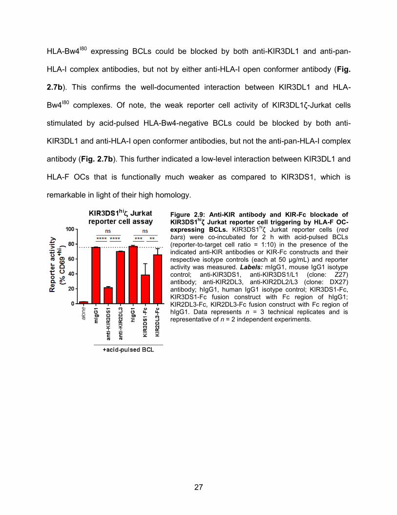
HLA-Bw4I80 expressing BCLs could be blocked by both anti-KIR3DL1 and anti-pan-
HLA-I complex antibodies, but not by either anti-HLA-I open conformer antibody (Fig.
2.7b). This confirms the well-documented interaction between KIR3DL1 and HLA-
Bw4I80 complexes. Of note, the weak reporter cell activity of KIR3DL1ζ-Jurkat cells
stimulated by acid-pulsed HLA-Bw4-negative BCLs could be blocked by both anti-
KIR3DL1 and anti-HLA-I open conformer antibodies, but not the anti-pan-HLA-I complex
antibody (Fig. 2.7b). This further indicated a low-level interaction between KIR3DL1 and
HLA-F OCs that is functionally much weaker as compared to KIR3DS1, which is
remarkable in light of their high homology.
Figure 2.9: Anti-KIR antibody and KIR-Fc blockade of KIR3DS1
hiζ Jurkat reporter cell triggering by HLA-F OC-
expressing BCLs. KIR3DS1hiζ Jurkat reporter cells (red
bars) were co-incubated for 2 h with acid-pulsed BCLs (reporter-to-target cell ratio = 1:10) in the presence of the indicated anti-KIR antibodies or KIR-Fc constructs and their respective isotype controls (each at 50 μg/mL) and reporter activity was measured. Labels: mIgG1, mouse IgG1 isotype control; anti-KIR3DS1, anti-KIR3DS1/L1 (clone: Z27) antibody; anti-KIR2DL3, anti-KIR2DL2/L3 (clone: DX27) antibody; hIgG1, human IgG1 isotype control; KIR3DS1-Fc, KIR3DS1-Fc fusion construct with Fc region of hIgG1; KIR2DL3-Fc, KIR2DL3-Fc fusion construct with Fc region of hIgG1. Data represents n = 3 technical replicates and is representative of n = 2 independent experiments.

28
2.4 MATERIALS AND METHODS
Cell lines and antibodies
721.221 and Jurkat (clone E6.1; ATCC) cell lines (including transductants) were
grown in RPMI-1640 supplemented with 10% fetal bovine serum (Sigma-Aldrich), 2 mM
L-glutamine (Gibco), 100 U/mL penicillin (Gibco), and 100 U/mL streptomycin (Gibco) at
37°C/5% CO2. EBV-transformed B-cell lines (BCLs) were generated from peripheral
blood mononuclear cells from donors bearing specific HLA genotypes; BCLs were also
grown in the same media and conditions as 721.221 and Jurkat cells. The following
purified antibodies were used for cell line-based staining and/or blocking assays: anti-
KIR3DS1/L1 (clone: Z27.3.7, Beckman Coulter), anti-KIR3DL1 (clone: DX9,
BioLegend), anti-KIR3DL1/L2/S1+2DS2/S4/S5/L2 (clone: 5.133, Miltenyi), anti-
KIR2DL2/L3/S2 (clone: DX27, Miltenyi), anti-pan-HLA-I complex (i.e. anti-HLA-
A,B,C,E,F,G +β2m) (clone: W6/32, BioLegend), anti-HLA-B,C open conformers (clone:
HC10; tebu-bio), and anti-HLA-A,G open conformers (clone: HCA2, tebu-bio), anti-CD3
(clone: HIT3a, BioLegend), anti-CD4 (clone: RPA-T4, BioLegend), and anti-CD69
(clone: FN50, BioLegend).
KIR-Fc binding to HLA-I-coated beads
Classical HLA-I-coated beads used were LABScreen single HLA-I beads (One
Lambda). Acid pulsing of HLA-I-coated beads was performed by resuspending beads in
50 μL of 300 mM glycine (pH 2.4), incubating at room temperature for exactly 2 min,
and washing three times with 1 mL of HBSS. Untreated and acid-pulsed beads were
stained according to the manufacturer’s instructions with KIR-Fc constructs (R&D

29
Systems) diluted to the indicated concentrations in PBS. Binding to beads was
measured on a Bio-Plex 3D Suspension Array system using Luminex xMAP technology
(Bio-Rad). To generate non-classical HLA-I-coated beads, biotinylated monomers of
HLA-E/β2m/VMAPRTLVL, HLA-F/β2m, and HLA-G/β2m/KGPPAALTL were purchased
from Immune Monitoring Lab at Fred Hutchinson Cancer Research Center, Seattle, WA,
and loaded onto streptavidin-coated beads (Life Technologies). Acid pulsing was
performed as before. KIR-Fc staining was performed at the indicated concentrations for
45 min at 4°C while shaking. Beads were then washed and stained with goat anti-
human IgG(Fc) F(ab’)2 PE-conjugated antibody (Life Technologies) diluted 1:50 for 30
min at 4°C while shaking. Beads were subsequently washed and fixed with 4%
paraformaldehyde in PBS (Affymetrix) before flow cytometric analysis.
Lentiviral transduction/transfection
Jurkat cells stably expressing genes of interest were generated via lentiviral
transduction. Gene constructs were designed accordingly and ordered from GeneArt
(Life Technologies). Constructs were cloned into a lentiviral transfer vector containing
an SFFV promoter and IRES-driven puromycin resistance. This backbone vector was
generated by cloning the SFFV promoter from pAPM59 into pLVX-EF1α-IRES-Puro
(Clontech). HEK293T cells (ATCC) were transfected with a VSV-G envelope vector
(pHEF-VSVG, obtained from NIH AIDS Reagent Program), HIV-1 gag-pol packaging
vector (psPAX2, obtained from NIH AIDS Reagent Program), and the transfer vector of
interest. Lentivirus-containing supernatants were harvested 3 d after transfection and
used to transduce Jurkat cells, which were subsequently selected in 1 μg/mL puromycin

30
and sorted for gene expression by fluorescence-activated cell sorting (FACS). 721.221
HLA transductants were previously generated by retroviral transduction (by Christian
Brander, Ragon Institute of MGH, MIT, and Harvard, Cambridge, MA) or generated via
lentiviral transduction for this study, with the exception of HLA-G-expressing 721.221
cells, which were a kind gift from Jack Strominger (Department of Stem Cell and
Regenerative Biology, Harvard University, Cambridge, MA). β2m-knockout (β2m-KO)
Jurkat cells were generated KIRζ Jurkat reporter cell lines. For this, Jurkat cells were
stably transduced with S. pyogenes Cas9 (lentiCas9-Blast was a gift from Feng
Zhang60; Addgene plasmid # 52962) and selected in 5 μg/mL blasticidin S (Gibco).
Jurkat-Cas9 cells were electroporated with a β2m-targeting gRNA CRISPR vector
(kindly provided by Leonardo Ferreira, Department of Stem Cell and Regenerative
Biology and Department of Molecular and Cellular Biology, Harvard University,
Cambridge, MA, and Thorsten Meissner, Department of Stem Cell and Regenerative
Biology, Harvard University, Cambridge, MA; published in 61) and sorted for loss of HLA
expression. Jurkat-β2m-KO cells were subsequently transduced with KIRζ chimeric
constructs.
KIRζ Jurkat reporter cell assay
KIRζ-Jurkat reporter cells were incubated with target cells at a reporter:target cell
ratio of 1:10 at 37°C/5% CO2 for 2–2.5 h for acid-pulse experiments or for 8 h for
experiments where acid pulsing was not a condition. For antibody blockade
experiments, antibodies were pre-incubated with relevant cells (reporters or targets) for
30 min at 4°C before co-incubation of reporter and target cells; the antibodies remained

31
during the co-incubation. After the co-incubation, cells were stained with anti-CD3 and
anti-CD69 antibodies and CD69 expression of reporter cells relative to negative and
positive controls was assessed and used as a measure of reporter activity.
Surface Plasmon Resonance (SPR)
SPR measurements were conducted in HBS-EP buffer using a Biacore 3000
system (Biacore AB). To assess binding of various KIR-Fc constructs to HLA-F open
conformers, biotinylated HLA-F monomers were immobilized onto a SA (streptavidin)
sensor chip (GE Healthcare) to approximately 1000 response units (RU). A blank flow
cell with no immobilized ligand was used as a reference flow cell. Injections of 60 μL of
KIR-Fc constructs diluted in PBS to the indicated concentrations were performed at a
flow rate of 20 μL/min, with a subsequent 10 min run of buffer to allow sufficient
dissociation. Regeneration after each injection was done with two pulses of 100 μL of
10 mM glycine∙HCl, pH 2.5, at a flow rate of 100 μL/min. Raw sensograms were
corrected by double referencing (subtracting from the reference flow cell response and
from PBS injection response). All experiments were done at standard temperature
(25°C).
Data Acquisition and Analysis
Flow cytometry data were analyzed using FlowJo software version 7.6 (Tree
Star) and statistical analyses were performed using GraphPad Prism 6 (GraphPad
Software). KIR-Fc binding and CD69 reporter cell expression values are shown as
mean values with error bars representing one standard deviation (SD). SPR data was

32
analyzed using BIAevaluation Software (GE Healthcare); given the dimeric nature of the
KIR-Fc analyte, the bivalent analyte model was used to obtain proper fit of kinetic
curves.

CHAPTER 3: The physiological impact and regulation of
KIR3DS1:HLA-F interactions

34
3.1 SUMMARY
The activating NK-cell receptor KIR3DS1 has been implicated in the outcome of
various human diseases, including delayed HIV-1 disease progression, yet a ligand that
accounts for its biological effects remained unknown. A previous screen revealed that
KIR3DS1 binds HLA-F OCs, which was validated biochemically and functionally (see
Chapter 2). In this study, we found that primary human KIR3DS1+ NK cells
degranulated and produced antiviral cytokines IFN-γ, MIP-1β, and TNF-α upon
encountering HLA-F OCs. KIR3DS1+ NK cells also exhibited a superior activity at
suppressing HIV-1 replication in vitro, as has been previously reported. Exploring the
cellular context of HLA-F expression, we found that CD4+ T-cell activation triggered
HLA-F transcription and expression and induced binding of soluble KIR3DS1-Fc. HIV-1
infection of activated CD4+ T cells further increased HLA-F transcription, but partially
decreased KIR3DS1 ligand expression, indicating an immune-evasion mechanism. In
monocytic and T-cell lines transduced with FLAG-tagged HLA-F, we find that HLA-F is
expressed intracellularly. However, mobilization of HLA-F to the cell surface could be
achieved with cellular activation, IFN-γ treatment, and most potently by low-temperature
incubation. Altogether, we established that KIR3DS1:HLA-F interactions are functionally
relevant in primary human NK cells and demonstrate tight and complex regulation of
HLA-F expression that may explain the widespread influence of KIR3DS1 in human
diseases.

35
3.2 RESULTS
3.2.1 HLA-F OCs potently trigger a polyfunctional response in primary
KIR3DS1+ NK cells
To assess the functional impact of HLA-F OC binding to KIR3DS1 in primary
human NK cells, KIR3DS1+ and KIR3DS1– NK-cell clones (NKCLs) were generated via
limiting dilution cloning from peripheral blood NK cells from a healthy KIR3DS1-
homozygous donor. NKCLs were confirmed to be CD56+KIR3DL1–LILRB1– to ensure
proper functionality, and had variable expression of KIR2Ds (Supplementary Fig.
S3.1). These NKCLs were seeded into individual well plates coated with human IgG (for
CD16 crosslinking), anti-KIR3DS1/L1 antibody (for KIR3DS1 crosslinking), or
monomers of HLA-E, -F, or -G. To assess NKCL responses to plate-bound ligands, we
measured cytotoxic granule exocytosis (i.e. degranulation), as determined by surface
expression of CD107a, and production of antiviral cytokines IFN-γ, TNF-α, and MIP-1β
by intracellular staining. Plate-bound HLA-F strongly and significantly triggered
degranulation in KIR3DS1+ NKCLs (Fig. 3.1) but not KIR3DS1– NKCLs (Fig. 3.2). The
magnitude of this response was comparable to crosslinking of KIR3DS1 and CD16, and
was able to be significantly reduced by addition of soluble anti-KIR3DS1/L1 antibody
(Fig. 3.2). Plate-bound HLA-E and HLA-G produced a weak response in some but not
all NKCLs, and this activation could not be blocked with soluble anti-KIR3DS1/L1
antibody. The observed effects were not due to intrinsic differences in response
capacity, as all NKCLs (except for one that was CD16–) were similarly triggered by
CD16 crosslinking. In addition, HLA-F elicited production of antiviral cytokines IFN-γ,
TNF-α, and MIP-1β in KIR3DS1+ (but not KIR3DS1–) NKCLs, and these responses
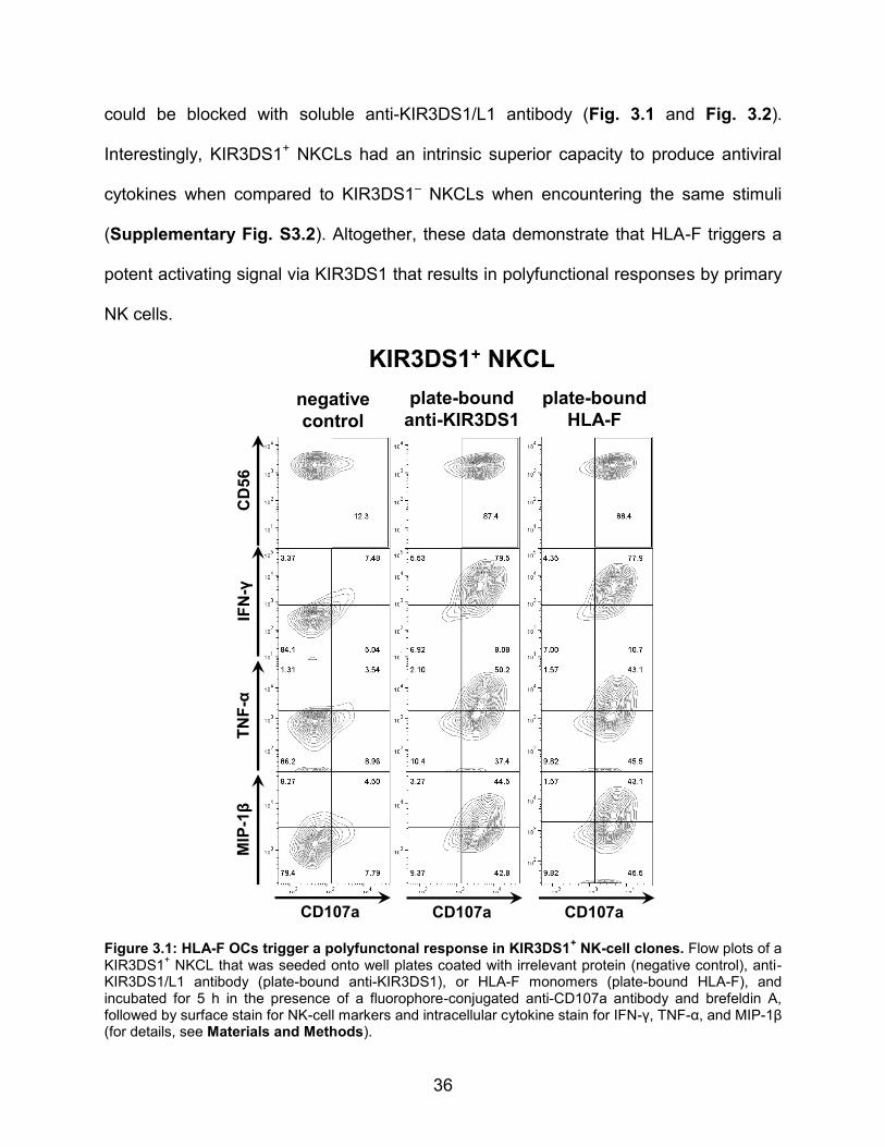
36
could be blocked with soluble anti-KIR3DS1/L1 antibody (Fig. 3.1 and Fig. 3.2).
Interestingly, KIR3DS1+ NKCLs had an intrinsic superior capacity to produce antiviral
cytokines when compared to KIR3DS1– NKCLs when encountering the same stimuli
(Supplementary Fig. S3.2). Altogether, these data demonstrate that HLA-F triggers a
potent activating signal via KIR3DS1 that results in polyfunctional responses by primary
NK cells.
Figure 3.1: HLA-F OCs trigger a polyfunctonal response in KIR3DS1+ NK-cell clones. Flow plots of a
KIR3DS1+ NKCL that was seeded onto well plates coated with irrelevant protein (negative control), anti-
KIR3DS1/L1 antibody (plate-bound anti-KIR3DS1), or HLA-F monomers (plate-bound HLA-F), and incubated for 5 h in the presence of a fluorophore-conjugated anti-CD107a antibody and brefeldin A, followed by surface stain for NK-cell markers and intracellular cytokine stain for IFN-γ, TNF-α, and MIP-1β (for details, see Materials and Methods).
CD107a CD107a CD107a
MIP
-1β
TN
F-α
IFN
-γC
D56
negative
control
plate-bound
anti-KIR3DS1
plate-bound
HLA-F
KIR3DS1+ NKCL

37
Figure 3.2: HLA-F OCs trigger degranulation and antiviral cytokine production in primary NK cells via KIR3DS1. KIR3DS1
+ (red dots and lines) and KIR3DS1
– NKCLs (black dots and lines) were seeded
onto well plates coated with irrelevant protein (fetal bovine serum; neg. ctrl.), human IgG (hIgG), anti-KIR3DS1/L1 antibody (clone: Z27; anti-KIR3DS1), or monomers of HLA-E, -F, or -G. For each plate-bound ligand condition, antibody-mediated blockade was performed by incubating NKCLs in the presence of mouse IgG1 isotype control antibody (+iso) or soluble anti-KIR3DS1/L1 antibody (+α-3DS1) (25 μg/mL each). NKCLs were incubated for 5 h with anti-CD107a fluorescent antibody and brefeldin A, followed by surface stain for NK-cell markers and intracellular cytokine stain for IFN-γ, TNF-α, and MIP-1β (for details, see Materials and Methods). The raw percentages of CD107a
+ NKCLs exposed to the indicated plate-
bound ligands are presented in the upper six panels, and the raw percentages of IFN-γ+, TNF-α
+, and
MIP-1β+ NKCLs exposed to plate-bound HLA-F are presented in the bottom three panels. One CD16
–
NKCL is indicated in hIgG. For each plate-bound ligand and marker, one-way ANOVA with Bonferroni multiple comparisons test comparing select columns was performed, and all statistically significant differences are presented (*, **, ***, and **** denote, p < 0.05, < 0.01, < 0.001, and < 0.0001, respectively).
CD16–
HLA-E HLA-F HLA-G
anti-KIR3DS1hIgGneg. ctrl.
HLA-F HLA-FHLA-F
blocking Ab
+iso +α-3DS1 +iso +α-3DS1 +iso +α-3DS1
blocking Ab blocking Ab
blocking Ab
+iso +α-3DS1 +iso +α-3DS1 +iso +α-3DS1
blocking Ab blocking Ab
blocking Ab
+iso +α-3DS1 +iso +α-3DS1 +iso +α-3DS1
blocking Ab blocking Ab
***** ****
**** * **
**** ***** **
KIR3DS1+ NKCL
KIR3DS1– NKCL
**

38
3.2.2 Activation of primary CD4+ T cells induces KIR3DS1 ligand expression at
the cell surface
A previous study demonstrated that HLA-F can be expressed on the surface of
activated lymphocytes62. To study KIR3DS1 ligand expression in a biologically relevant
context, HLA-F expression and KIR3DS1-Fc binding to primary resting CD4+ T cells and
CD4+ T cells stimulated with anti-CD3/28 beads and IL-2 was assessed. A Western
blot-suitable anti-HLA-F antibody (validated in Supplementary Fig. S3.3) was used to
assess protein levels, and resistance to endoglycosidase H (Endo H) digestion served
as a measure of cell-surface expression. Endo H is a glycosidase that removes
immature N-glycans from glycoproteins that have not undergone Golgi processing; thus,
glycoproteins shuttled to the cell surface through the Golgi are rendered Endo H-
resistant (i.e. there is no band shift on protein gel electrophoresis). Results showed that
whereas unstimulated CD4+ T cells do not have any detectable levels of HLA-F protein,
stimulated CD4+ T cells express HLA-F protein in a predominantly Endo H-resistant
form, consistent with cell-surface expression (Fig. 3.3a); similar results were obtained
with different stimulation conditions (48 h treatment with 12.5 ng/mL phorbol 12-
myristate 13-acetate and 0.335 μM ionomycin; data not shown).
To determine if the up-regulation of HLA-F protein in activated CD4+ T cells was
transcriptionally induced, HLA-F mRNA levels were assessed via flow cytometry-based
fluorescent in situ hybridization (FISH) (detailed in Materials and Methods). HLA-F
mRNA-specific probes were used after testing their sensitivity and specificity in HLA-F+
and HLA-F– cell lines (Supplementary Fig. S3.4). Activation of CD4+ T cells increased
the levels of HLA-F mRNA transcripts (Fig. 3.3b), indicating that HLA-F is
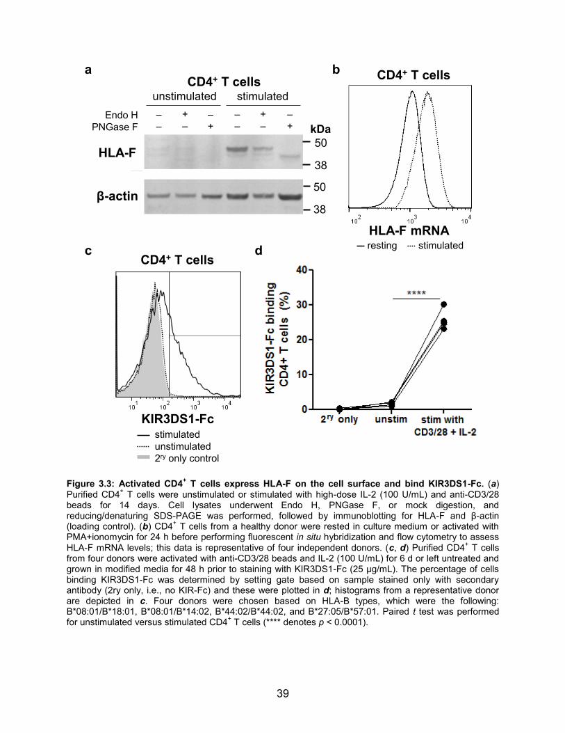
39
Figure 3.3: Activated CD4+ T cells express HLA-F on the cell surface and bind KIR3DS1-Fc. (a)
Purified CD4+ T cells were unstimulated or stimulated with high-dose IL-2 (100 U/mL) and anti-CD3/28
beads for 14 days. Cell lysates underwent Endo H, PNGase F, or mock digestion, and reducing/denaturing SDS-PAGE was performed, followed by immunoblotting for HLA-F and β-actin (loading control). (b) CD4
+ T cells from a healthy donor were rested in culture medium or activated with
PMA+ionomycin for 24 h before performing fluorescent in situ hybridization and flow cytometry to assess HLA-F mRNA levels; this data is representative of four independent donors. (c, d) Purified CD4
+ T cells
from four donors were activated with anti-CD3/28 beads and IL-2 (100 U/mL) for 6 d or left untreated and grown in modified media for 48 h prior to staining with KIR3DS1-Fc (25 μg/mL). The percentage of cells binding KIR3DS1-Fc was determined by setting gate based on sample stained only with secondary antibody (2ry only, i.e., no KIR-Fc) and these were plotted in d; histograms from a representative donor are depicted in c. Four donors were chosen based on HLA-B types, which were the following: B*08:01/B*18:01, B*08:01/B*14:02, B*44:02/B*44:02, and B*27:05/B*57:01. Paired t test was performed for unstimulated versus stimulated CD4
+ T cells (**** denotes p < 0.0001).
CD4+ T cells
stimulated
unstimulated
2ry only control
KIR3DS1-Fc
Figure 4
HLA-F mRNAresting
CD4+ T cells
38
50
38
50
kDa
HLA-F
β-actin
unstimulated stimulated
–
–Endo H
PNGase F
+
–
–
+
–
–
+
–
–
+
CD4+ T cells
stimulated
a b
c d
40
transcriptionally induced in CD4+ T cells upon activation. Furthermore, KIR3DS1-Fc
bound significantly to stimulated CD4+ T cells, but not to unstimulated CD4+ T cells (Fig.
3.3c and Fig. 3.3d, p < 0.0001). These results were consistent across donors with and
without HLA-Bw4 allotypes, demonstrating that KIR3DS1 ligands are expressed on
activated CD4+ T cells regardless of their HLA-I genotype, and strongly suggesting
KIR3DS1 binding to HLA-F expressed on activated CD4+ T cells.
3.2.3 HIV-1 infection of activated CD4+ T cells increases HLA-F transcription
but partially decreases KIR3DS1 ligand expression, and is suppressed by
KIR3DS1+ NK cells
To assess the influence of HIV-1 infection on expression of KIR3DS1 ligands in
CD4+ T cells, HLA-F mRNA levels and KIR3DS1-Fc binding were assessed using
activated CD4+ T cells that were uninfected or infected with HIV-1 NL4-3. HLA-F mRNA
levels were up-regulated in HIV-1–infected activated CD4+ T cells when compared to
uninfected activated CD4+ T cells (Fig. 3.4c), indicating that HIV-1 infection further
stimulated HLA-F mRNA transcription. In contrast, KIR3DS1-Fc binding was
significantly decreased upon HIV-1 infection (Fig. 3.4a and Fig. 3.4b). The decrease in
KIR3DS1-Fc binding was already observed in ‘early’ infected cells (defined as
p24dimCD4+HLA-I+tetherin+), but was more pronounced in ‘late’ infected cells (defined as
p24hiCD4dimHLA-Idimtetherindim), indicating an as-yet-unknown mechanism by which HIV-
1 might reduce KIR3DS1 ligand expression. This may be due to direct downregulation
of HLA-F protein by HIV-1; however, due to the absence of an available flow cytometry-

41
suitable antibody against HLA-F, it was not possible to directly quantify HLA-F surface
expression levels on HIV-1–infected cells.
Figure 3.4: Effect of HIV-1 infection on HLA-F expression and KIR3DS1-Fc binding. (a) CD4+ T cells
from healthy donors were stimulated with IL-2 (100 U/mL) and anti-CD3/28 beads for 3 d, and were then cultured or infected with HIV-1 NL4-3. At 3 d post-infection (infection rate ~50%), fluorescent in situ hybridization and flow cytometry was performed to assess HLA-F mRNA levels. This data is representative of two independent experiments on two different donors. (b, c) Purified CD4
+ T cells from
five healthy donors were cultured in IL-2 (100 U/mL) and were either unstimulated or stimulated by plate-bound anti-CD3/28 stimulation for 7 d. Stimulated cells were then either continually cultured or infected with HIV-1 NL4-3. At 3 d post-infection (infection rate among five donors was ~100%), cells were stained for KIR3DS1-Fc (25 μg/mL) as well as intracellular HIV-1 p24 and additional markers to assess HIV-1 infection stage. Flow histogram in b depicts KIR3DS1-Fc binding to IL-2-cultured CD4
+ T cells, stimulated
CD4+ T cells, and stimulated CD4
+ T cells that are ‘early’ HIV-1–infected (p24
dimCD4
+HLA-I
+tetherin
+) or
‘late’ HIV-1–infected (p24hiCD4
dimHLA-I
dimtetherin
dim). In c, aggregate data for five healthy donors with
background-subtracted median fluorescence intensities (bsMdFI) are shown with black lines connecting data points from each individual donor.
To evaluate the antiviral capacity of KIR3DS1+ NK cells, HIV-1–infected
autologous CD4+ T cells were co-cultured for seven days with KIR3DS1+ and KIR3DS1–
NK-cell clones derived from a KIR3DS1 homozygous donor (for NK-cell receptor
phenotypes, see Supplementary Fig. S3.5). Intracellular staining for HIV-1 p24 was
performed to quantify the percentage of infected cells. Only KIR3DS1+ NK-cell clones
were effective at suppressing HIV-1 replication in autologous CD4+ T cells, as seen by
significantly less HIV-1 p24+ CD4+ T cells in the presence of KIR3DS1+ NK cells (5.48 %
KIR3DS1-Fc
CD4+ T cells
2ry only
+IL-2
stimulated
early HIV-1
late HIV-1
HLA-F mRNA
CD4+ T cells
neg. ctrl. probe
stimulated
stimulated + HIV-1
CD4+ T cellsa b c

42
± 1.60 %), relative to KIR3DS1– NK-cell clones (16.9 % ± 2.32 %; p < 0.01) or no NK
cells added (mean 16.0 % ± 2.62 %, p < 0.01) (Fig. 3.5a and Fig. 3.5b). Thus,
KIR3DS1+ NK cells have superior antiviral capacity towards HIV-1–infected autologous
CD4+ T cells, as previously shown13, and HIV-1–mediated downregulation of KIR3DS1
ligands on infected cells is not sufficient to enable complete immune evasion.
Figure 3.5: KIR3DS1+ NK cells efficiently suppress HIV-1 replication in autologous CD4
+ T cell in
vitro. (a, b) KIR3DS1+ and KIR3DS1
– NKCLs were co-incubated with autologous HIV-1–infected CD4
+ T
cells for 7 d, after which intracellular staining for HIV-1 p24 and flow cytometry was performed to quantify amount of remaining HIV-1–infected CD4
+ T cells. Aggregate results are presented a and representative
flow cytometry plots are presented in b. For a, one-way ANOVA was performed with Tukey multiple comparisons test comparing all columns (** denotes p < 0.01).
3.2.4 HLA-F is expressed intracellularly when transduced into T-cell,
monocytic, and myeloid cell lines.
To explore how HLA-F expression is regulated, various cell lines (i.e. Jurkat,
THP-1, and K562 cells) were transduced with N-terminally FLAG-tagged HLA-F (HLA-
FFLAG) to allow detection by anti-FLAG antibody staining. Consistent with previous
studies56, all cell lines did not express HLA-FFLAG on the cell surface, but rather
expressed it intracellularly (Fig. 3.6 and Fig. 3.7). To assess whether cellular activation
or inflammatory cytokine treatment would mobilize HLA-F to the cell surface, as has
been previously document in human immune and non-immune cells62,63, THP-1 cells
**
Viral Inhibition Assay(HIV-1 JR-CSF MOI 0.01, NK:CD4 ratio = 2.5:1, Day 7)
HIV
-1 p
24
+ c
ells
(% o
f C
D4
+ T
cells)
alone KIR3DS1+
KIR3DS1-
0
5
10
15
20
25
NKCLs
**
NKCLsalone
HIV-1 p24
CD
4
aloneKIR3DS1+
NKCLKIR3DS1–
NKCL+ +
HIV-1-infected CD4+ T cells
**
Viral Inhibition Assay(HIV-1 JR-CSF MOI 0.01, NK:CD4 ratio = 2.5:1, Day 7)
HIV
-1 p
24
+ c
ells
(% o
f C
D4
+ T
cells)
alone KIR3DS1+
KIR3DS1-
0
5
10
15
20
25
NKCLs
**
NKCLsalone
HIV-1 p24
CD
4
aloneKIR3DS1+
NKCLKIR3DS1–
NKCL+ +
HIV-1-infected CD4+ T cellsa b

43
Figure 3.6: HLA-F is expressed intracellularly. Jurkat and K562 cells transduced with HLA-FFLAG
were stained with APC-conjugated anti-FLAG antibody to assess surface expression of HLA-F
FLAG, and then
subsequently fix, permeabilized, and stained intracellularly with PE-conjugated anti-FLAG antibody to assess intracellular expression. Note: HLA-F
FLAG transduction efficiency differed by cell line.
were treated with PMA, IFN-γ, or both. PMA is a DAG analogue that activates protein
kinase C and other cellular factors to induce potent cellular activation, and IFN-γ is a
pro-inflammatory cytokine that upregulates MHC class I and II expression among many
pleiotropic functions. Indeed, both PMA and IFN-γ induced cell-surface expression of
HLA-FFLAG in THP-1 cells, with IFN-γ being more potent than PMA, and both exhibiting
synergy when combined (Fig. 3.7). Of note, IFN-γ treatment also induced expression of
endogenous HLA-DR and HLA-G (data not shown), as has been previously reported for
Figure 3.7: Cell-surface expression of HLA-F is induced by PMA and IFN-γ. THP-1 cells transduced with HLA-F
FLAG were either untreated or treated with 50 ng/mL PMA, 100 U/mL IFN-γ, or both for 48 h.
Cells were subsequently stained with anti-FLAG antibody to assess cell-surface expression of HLA-F. Untransduced THP-1 cells were used as a negative control for staining.
HLA-FFLAG (intracellular)
HL
A-F
FL
AG
(su
rfa
ce
)untransduced HLA-FFLAG
Jurkat
untransduced HLA-FFLAG
K562
THP-1(untransduced)
THP-1-HLA-FFLAG
untreated +PMA +IFN-γ +PMA +IFN-γ
HLA-FFLAG (surface)
pan
-HL
A-I
THP-1 (untransduced)
37 C
26 C
THP-1HLA-FFLAG
HLA-FFLAG
(surface)
Jurkat-HLA-FFLAG
37 C 26 C
HLA-FFLAG (surface)

44
THP-1 cells64. Of interest, we serendipitously discovered that incubation of THP-1-HLA-
FFLAG cells at a low temperature (26°C) potently mobilizes HLA-FFLAG to the cell surface,
resulting in very high surface expression (Fig. 3.8); this also occurred in Jurkat cells,
although to a lesser extent (Supplementary Fig. S3.6). This finding, however, is not
unprecedented given that lower temperatures increase the stability of HLA-I OCs and
result in increased cell surface expression65; whether this increased stability is leading
to increasing trafficking to the cell surface or decreasing endocytosis in the case of
HLA-F remains to be determined. Altogether, our data shown that HLA-F is a
predominantly intracellular HLA-I gene whose surface expression is tightly regulated
and induced upon cellular activation or exposure to pro-inflammatory cytokines.
THP-1(untransduced)
THP-1-HLA-FFLAG
untreated +PMA +IFN-γ +PMA +IFN-γ
HLA-FFLAG (surface)
pan
-HL
A-I
THP-1 (untransduced)
37 C
26 C
THP-1HLA-FFLAG
HLA-FFLAG
(surface)
Jurkat-HLA-FFLAG
37 C 26 C
HLA-FFLAG (surface)
Figure 3.8: Low-temperature incubation potently mobilizes HLA-F to the cell surface. THP-1-HLA-F
FLAG cells were cultured under normal
conditions (37°C and 5% CO2) or incubated at 26°C (and 5% CO2) for 18 h. Cells were subsequently stained with anti-FLAG antibody to assess cell-surface expression of HLA-F. Untransduced THP-1 cells were used as a negative control for staining.

45
3.3 MATERIALS AND METHODS
Cell lines
Jurkat (clone E6.1; ATCC), K562, and THP-1 cell lines (including transductants)
were grown in RPMI-1640 supplemented with 10% fetal bovine serum (Sigma-Aldrich),
2 mM L-glutamine (Gibco), 100 U/mL penicillin (Gibco), and 100 U/mL streptomycin
(Gibco) at 37°C/5% CO2. The following purified antibodies were used for blocking or
plate-bound ligand assays: anti-KIR3DS1/L1 (clone: Z27.3.7, Beckman Coulter),
human.
Lentiviral transduction/transfection
Cell lines stably expressing genes of interest were generated via lentiviral
transduction. Gene constructs were designed accordingly and ordered from GeneArt
(Life Technologies). HLA-F sequence was obtained from NCBI (accession: P30511),
and FLAG sequence was added after the signal peptide sequence. Constructs were
cloned into a lentiviral transfer vector containing an SFFV promoter and IRES-driven
puromycin resistance. This backbone vector was generated by cloning the SFFV
promoter from pAPM59 into pLVX-EF1α-IRES-Puro (Clontech). HEK293T cells (ATCC)
were transfected with a VSV-G envelope vector (pHEF-VSVG, obtained from NIH AIDS
Reagent Program), HIV-1 gag-pol packaging vector (psPAX2, obtained from NIH AIDS
Reagent Program), and the transfer vector of interest. Lentivirus-containing
supernatants were harvested 3 d after transfection and used to transduce cell lines,
which were subsequently selected in 1 μg/mL puromycin and in some cases sorted for
gene expression by fluorescence-activated cell sorting (FACS).

46
Primary human CD4+ T-cell isolation, stimulation, infection, and staining with KIR-Fc
and HLA-F mRNA probes
CD4+ T cells were isolated from donor PBMCs by indirect magnetic labeling and
negative selection, using the CD4+ T cell isolation kit II (Miltenyi) according to the
manufacturer’s protocol. Stimulations were done differently depending on the
experiment. Stimulations for Western Blots were done with anti-CD3/28 Dynabeads
(Life Technologies) were done at a bead:cell ratio of 2:1 and culturing in supplemented
RPMI with IL-2 (100 U/mL) for the indicated amounts of time. Stimulations for flow
cytometry-based fluorescent in situ hybdridization were done with phorbol 12-myristate
13-acetate (12.5 ng/mL) and ionocymin (0.335 μM) (Cell Stimulation Cocktail used at
0.25X; eBioscience) for the indicated amounts of time. Stimulations for KIR-Fc staining
and HIV-1 infection were done for the indicated amounts of time in supplemented RPMI
with IL-2 (100 U/mL) on non-tissue culture-treated flat-bottom 48-well plates previously
pre-coated with anti-CD3 (clone: OKT3, BioLegend) and anti-CD28 (clone: CD28.2,
BioLegend) antibodies each at 10 μg/mL. HIV-1 infection was done by resuspending ~1
× 106 activated CD4+ T cells in 1 mL of supplemented RPMI containing 100 U/mL of IL-
2 and adding 1 × 10⁵ TCID50 of replication-competent HIV-1 NL4-3. Cell were
immediately spinfected (centrifuging for 2 h at 1,500 × g at 37°C) and incubated at
37°C/5% CO2 for 72 to allow for viral replication. For KIR-Fc staining of cells, cells were
first stained with LIVE/DEAD® Fixable Blue Dead Cell Staining Kit (Life Technologies)
following manufacturer’s instructions, and then stained with 25 μg/mL of KIR3DS1-Fc
for 45 min at 4°C while shaking, after which the cells were washed and stained with
anti-CD3 (clone: UCHT1, BioLegend), anti-CD4 (clone: RPA-T4, BioLegend), anti-pan-

47
HLA-I complex (clone: W6/32, BioLegend), anti-tetherin (clone: RS38E), and goat anti-
human IgG(Fc) F(ab’)2 PE-conjugated antibody (Life Technologies) for 30 min at 4°C
while shaking. Cells were subsequently washed and subjected to fixation and
permeabilization with the FIX & PERM ® Cell Fixation and Cell Permeabilization Kit
(Life Technologies) to stain with anti-HIV-1 p24 (clone: KC57, BD Biosciences). Cells
were subsequently washed and resuspended in PBS, except in assays where fixation
and permeabilizationn was not done, in which case cells were fixed with 4%
paraformaldehyde in PBS (Affymetrix) before flow cytometric analysis. Fluorescent in
situ hybdridization was done with the PrimeFlow™ RNA Assay from Affymetrix with
custom designed HLA-F mRNA specific probes following manufacturer’s intructions.
Flow cytometric analyses of samples were performed on a BD LSRFortessa.
Western Blot
2 × 10⁶ Cells were lysed in 100 μL of lysis buffer (20 mM Tris∙HCl, 100 mM NaCl,
1 mM EDTA, 0.5% Triton X-100, pH 8.0, all reagents from Sigma-Aldrich) containing 1X
Halt™ protease and phosphatase inhibitor cocktail (Life Technologies). Nuclei were
pelleted supernatants containing cytoplasmic and plasma membrane proteins were
collected. Lysates were treated with Endo Hf (New England Biolabs), PNGase F (New
England Biolabs), or mock treated according to manufacturer’s instructions. Reaction
products were denatured and reduced, and run on NuPAGE Novex 4-12% Bis-Tris
protein gels (Life Technologies). Gel products were transferred over to a PVDF
membrane, and dual immunoblotting was done with mouse anti-β-actin (clone: mAbcam
8224, Abcam) and rabbit anti-HLA-F (clone: EPR6803, Abcam) primary antibodies and

48
IRDye 800CW goat anti-human IgG(H+L) (LI-COR) and IRDye 680RD goat anti-rabbit
IgG(H+L) (LI-COR) secondary antibodies. The blots were visualized using an Odyssey
imaging system (LI-COR) and images were analyzed using Image Studio 3.1 software
(LI-COR).
Generation of primary NK-cell clones
Primary human NK cells isolated from peripheral blood mononuclear cells
(PBMCs) of healthy human donors were subcloned by limiting dilution in two manners.
For the HIV-1 replication inhibition assay, NK cells were subcloned in the presence of
feeders and maintained in NK-cell cloning medium consisting of supplement RPMI
additionally supplemented with 5% human serum (Sigma-Aldrich), 1X MEM-NEAA
(Gibco), 1X sodium pyruvate (Gibco), 100 μg/mL kanamycin, 450 U/mL IL-2 (AIDS
Reagent Program, NIH) using a protocol adapted from a previously reported method66.
Briefly, NK cells were isolated from peripheral blood mononuclear cells (PBMCs) from a
KIR3DS1+/+(homozygous) donor via magnetic negative selection (NK-cell isolation kit
from Miltenyi), added to a mix of irradiated feeders consisting of freshly isolated
allogeneic (PBMCs) combined with log-phase-growth RPMI 8866 cells (Sigma-Aldrich)
at a 10:1 ratio in cloning medium containing 1 μg/mL phytohaemagglutinin (PHA;
Fisher) and mixed thoroughly before plating at 100 µL/well (1 NK cell/well) in 96-well
plates and incubated for 14 days at 37°C/5% CO2. After 14 days, wells that had
outgrowth of cells were transferred to 48-well plates and maintained in NK-cell medium
with frequent media exchange (approximately every 3 days). For plate-bound ligand
assays, NK cells were subcloned in the presence of irradiated K562 cells expressing

49
mbIL-15 and CD137L (kind gift from Dario Campana, published in 67) and modified NK-
cell cloning media consisting of RPMI supplemented with 15% fetal bovine serum
(Sigma-Aldrich), 5% human serum (Sigma-Aldrich), 2 mM L-glutamine (Gibco), 1X
MEM-NEAA (Gibco), 1X sodium pyruvate (Gibco), 100 μg/mL Primocin™ (Invivogen),
500 U/mL IL-2 (AIDS Reagent Program, NIH), with the addition of the following four
cytokines to supplant the need for PBMC feeders: 5 ng/mL IL-15 (Peprotech), 10 ng/mL
IL-12 (Peprotech), 40 ng/mL IL-18 (Peprotech), and 20 ng/mL IL-21 (Peprotech). NK-
cell clones that grew out were continually cultured in modified NK-cell cloning media
without additional cytokines. Cells were phenotyped by flow cytometry with the following
antibodies: anti-CD56-BV421 (BioLegend), anti-CD16-BV785 (BioLegend), anti-
NKG2A-APC (Beckman Coulter), anti-LILRB1-PE-Cy5 (BD Biosciences), anti-
KIR3DL1/S1-PE (Beckman Coulter), anti-KIR2D1/S1-PerCP-Cy5.5 (clone: HP-MA4,
BioLegend), and anti-KIR2DL2/L3 (clone: DX27, BioLegend). Only NK-cell clones that
were CD3–CD56+ (rarely CD16–) and LILRB1– were used for subsequent assays to
ensure proper functionality.
NK-cell degranulation and intracellular cytokine staining assay
Non-tissue culture-treated flat-bottom 96-well plates were coated overnight at
4 C with 50 μL the following ligands diluted in PBS: 1 mg/mL human γ-globulins
(Thermo Scientific), 1 μg/mL of both anti-CD335 (a.k.a. NKp46) (clone: 9E2, BioLegend)
and anti-CD2 (clone: TS1/8, BioLegend) antibody, 1 μg/mL of anti-KIR3DS1/L1 (clone:
Z27.3.7, Beckman Coulter), 10 μg/mL of HLA-E, -F, or -G monomer, or 0.5% bovine
serum albumin (Miltenyi). ~1 × 10⁴ NK cells in 100 µL of supplemented RPMI containing

50
3 µL of anti-CD107a-PE-Cy7 antibody (BioLegend) and 5 μg/mL brefeldin A (Biolegend)
and in the presence of anti-KIR3DS1/L1 antibody or isotype control antibody (mouse
IgG1, clone: MG1-45, BioLegend) were seeded onto ligand-coated plate wells. After a
5-h incubation at 37°C/5% CO2, cells were first stained with LIVE/DEAD® Fixable Blue
Dead Cell Staining Kit (Life Technologies) following manufacturer’s instructions, and
then stained with anti-CD56-BV421 (clone: HCD56, BioLegend) and anti-CD16-BV785
(clone: 3G8, BioLegend) antibodies for 15 min at 4°C. Cells were then fixed with BD
Cytofix/Cytoperm solution (BD Biosciences) and permeabilized with BD Perm/Wash
solution (BD Biosciences) following manufacturer’s instructions, after which intracellular
cytokine staining was carried out using the following antibodies: anti-IFN-γ-Alexa Fluor
647 (clone: 4S.B3, BioLegend), anti-TNF-α-BV650 (clone: Mab11, BioLegend), and
anti-MIP-1β-PerCP-eFluor710 (clone: FL34Z3L, eBioscience). After washing, flow
cytometric analysis was performed on a BD LSR Fortessa.
NK-cell co-incubation with HIV-1–infected autologous CD4+ T cells
PBMCs from the same donor from which NK-cell clones were generated were
thawed and CD4+ T cells were enriched via magnetic negative selection with the
EasySep™ Human CD4+ T Cell Enrichment Kit (StemCell). CD4+ T cells were cultured
overnight in supplemented RPMI containing 50 U/mL of IL-2 and 1 μg/mL PHA. The
next day, cells were washed and resuspended at 1 × 10⁶ cells/mL in supplemented
RPMI containing 50 U/mL of IL-2. HIV-1 JR-CSF was added to a final concentration of 1
× 104 TCID50/mL, and cells were incubated for 4 h at 37°C/5% CO2. Afterwards, infected
CD4+ T cells were washed, resuspended in NK cell medium, and plated in a round-
51
bottom 96-well plate at 5 × 10⁴ cells/well. NK-cell clones from the same donor were
added to individuals wells at 1.25 × 10⁵ cells/well (NK cell:CD4+ T cell ratio = 2.5:1).
Cells were co-cultured for 7 days, after which surface staining for CD3 and CD4 and
intracellular staining for HIV-1 p24 was performed for flow cytometry assessment.
Human Samples and Viruses
Frozen peripheral blood mononuclear cells (PBMCs) from healthy donors were
used in this study in accordance to protocols approved by Partners Human Research
Committee and Institutional Review Board of Massachusetts General Hospital. HLA/KIR
genotypes of selected human samples were determined prior to this study by high-
resolution HLA typing performed at the HLA-typing laboratory of the National Cancer
Institute, National Institutes of Health, and Sanger Sequencing-based KIR genotyping
performed by Mary Carrington’s laboratory. HIV-1 NL4-3 and HIV-1 JR-CSF were
purchased from the Virology Core of the Ragon Institute of MGH, MIT, and Harvard,
and are quality controlled for infectivity and titered on stimulated human PBMCs by
standard endpoint TCID50 assay.
HLA-FFLAG–transduced cell lines assays
For FLAG staining, PE- and APC-conjugated anti-FLAG antibodies (clone: L5,
BioLegend) were used. In experiments that assessed cell-surface versus intracellular
HLA-FFLAG expression, cells were stained with APC-conjugated anti-FLAG antibody, and
then fixed with BD Cytofix/Cytoperm solution (BD Biosciences) and permeabilized with
BD Perm/Wash solution (BD Biosciences) following manufacturer’s instructions, after

52
which intracellular staining with PE-conjugated anti-FLAG antibody was performed.
Flow cytometric analysis followed.
Data Acquisition and Analysis
Flow cytometry data was acquired on BD LSR Fortessa and analyzed using
FlowJo software version 7.6 (Tree Star) and statistical analyses were performed using
GraphPad Prism 6 (GraphPad Software).
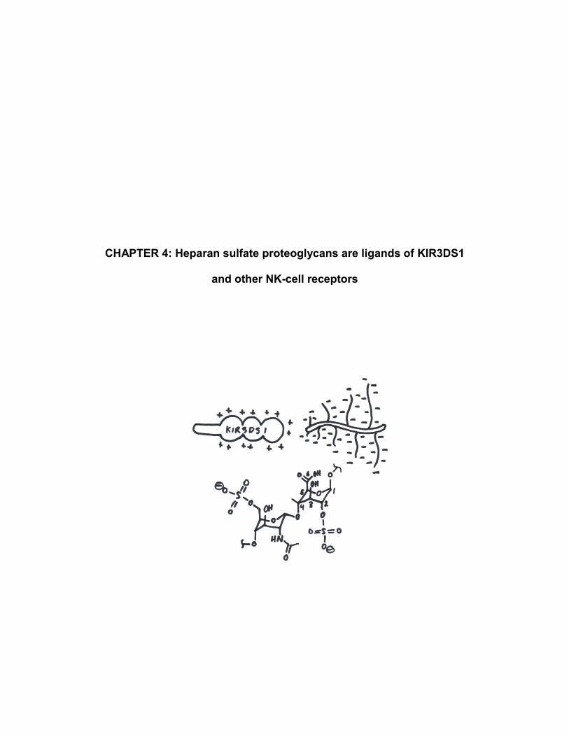
CHAPTER 4: Heparan sulfate proteoglycans are ligands of KIR3DS1
and other NK-cell receptors

54
4.1 SUMMARY
While an overwhelming majority of NK-cell receptor:ligand interactions studied
are protein:protein in nature, the role of protein:carbohydrate interactions in NK-cell
function exists and could be more fully explored. In this study, we performed a genome-
wide CRISPR/Cas9 knock-out screen for KIR3DS1 ligands and identified heparan
sulfate (HS) as a KIR3DS1 ligand. This was validated biochemically by surface plasmon
resonance and on cell lines, and was shown to be completely dependent on the
presence of HS sulfate moieties. Further studies revealed that KIR3DL1, a highly
homologous inhibitory allotype of KIR3DS1, also bound HS. As binding of other NK-cell
receptors to HS has been described (for NKp30, NKp44, NKp46, and KIR2DL4), these
data indicate that NK-cell receptor:HS interactions are widespread and could play
important roles in NK-cell–receptor regulation and target-cell recognition that need to be
further investigated.

55
4.2 BACKGROUND
Unlike T cells, which rely primarily on the T-cell receptor to recognize specific
MHC complexes presenting peptides sampled from intracellular and/or extracellular
compartments, NK-cell function is dictated by germline-encoded activating and
inhibitory receptors that interact with a plethora of ligands expressed on target cells. Of
all the receptor:ligand interactions studied, an overwhelming number are protein:protein
in nature, leaving a major gap in our knowledge of the role of non-protein NK-cell–
receptor ligands.
A major group of non-protein NK-cell–receptor ligands are carbohydrates
expressed on target cells. Indeed, all cells in the body bear a glycocalyx, a term used to
refer to the entire array of glycans coating the cell surface68. However, this term is highly
misleading because it grossly underestimates the complexity and heterogeneity of these
glycans. Cell surface glycans can be (i) non-covalently adsorbed to the cell surface, (ii)
covalently linked to lipids, called glycolipids, or (iii) covalently linked to proteins in the
form of glycoproteins or proteoglycans. Proteoglycans (PGs), in particular, consist of a
core protein with one or more covalently linked glycosaminoglycans (GAGs), which
usually dictate most or all of the PG’s function. GAGs are long, unbranched
polysaccharides whose disaccharide building blocks consist of an amino sugar and a
uronic acid or galactose and can be modified extensively by acetyl and sulfate
substitutions. These modifications are exquisitely intricate and diverse, and tightly
regulated on individual cells.
There are five types of GAGs: hyaluronic acid (HA), keratan sulfate (KS),
dermatan sulfate (DS), chondroitin sulfate (CS), and heparan sulfate (HS). The most

56
abundant GAGs found on most cell surfaces are HA and HS69. HA is a unique and
relatively simple GAG that is unmodified (i.e. non-sulfated and non-acetylated) and does
not exist as a PG. HS, on the other hand, is a GAG made as a PG by every cell in the
body and plays a multitude of biological roles (reviewed in 70) including: barrier
function71; coagulation72; wound repair73,74; development75,76; endocytosis77 and
transcellular transport78; virus attachment and entry79–84; tumorigenesis85; cell adhesion
and migration, including leukocyte homing86; signaling of growth factors87,88,
morphogens, cytokines89, and chemokines90; and host defense91. HS biosynthesis
begins in the endoplasmic reticulum (ER) with serial addition of specific glycosyl groups
to generate a β-linked tetrasaccharide primer—Xyl(β1-4)Gal(β1-3)Gal(β1-3)Glc—on a
serine residue of an HSPG protein core, a reaction catalyzed in series by XYLT1 and/or
XYLT2, B4GALT7, B4GALT6, and B3GAT3. The possible HSPG protein cores are
syndecans (1 through 4), glypicans (1 through 6), betaglycan, serglycin, and
occasionally, neuropilin-1 and certain isoforms of CD44; all of these contain at least one
Ser-Gly motif near a patch of acidic residues onto which HS is polymerized. Subsequent
reactions by exostosin glycosyltransferases (EXT1 and EXT2) and exostosin-like
glycosyltransferases (EXTL1, EXTL2, and EXTL3) add repeating β1-4-linked
disaccharides of N-acetyl glucosamine and glucuronic acid. The extended
polysaccharide can be subsequently modified in the Golgi by epimerization of
glucuronic acid to iduronic acid catalyzed by GLCE, and by a complex array of
sulfotransferase reactions. These sulfotransferase reactions are (i) N-deacetylation/N-
sulfation of GlcNAc by any of four enzymes (NDST1–4); (ii) 6-O-sulfation by any of
three enzymes (HS6ST1–3); (iii) 2-O-sulfation by a single enzyme (HS2ST1); and (iv)
57
more infrequently92, 3-O-sulfation by any of several enzymes (HS3ST1–6). Fully formed
HSPGs can be secreted or displayed on the cell surface, where they may be edited by
sulfatases (SULF1 and SULF2). While the most well-known form of HS is heparin, a
highly sulfated form that is made and secreted only by connective-tissue mast cells and
used as an anti-coagulant, by far the most prevalent form is HSPGs found on cell
surfaces. Much like DNA or RNA, HS does not have a single structure or sequence, but
rather, the basic polysaccharide components that make HS vary depending on the cell
type and cellular context under which they are made. Thus, HS has been referred to as
“the most information-dense biopolymers found in nature”93.
Among its biological functions, HS has been shown to interact with numerous
immune receptors. The natural cytotoxicity receptors (NCRs) NKp30, NKp44, and
NKp46, which are widely expressed activating NK-cell receptors94, have been shown to
bind to specific HS motifs. NKp30 and NKp46 bind to epitopes containing of 2-O-
sulfated iduronic acid and 6-O- and N-sulfated glucosamine, whereas NKp44 binds to
epitopes containing 2-O-sulfated iduronic acid and N-acetylated glucosamine95. In
addition, a genome-wide siRNA screen discovered that another activating NK-cell
receptor, KIR2DL4, bound to HS in a manner that depended on 3-O-sulfation96 and that
directly influenced KIR2DL4 signaling and NK-cell function.
In this study, while investigating possible non-HLA-I ligands for the activating NK-
cell receptor KIR3DS1 through a genome-wide CRISPR/Cas9 knock-out screen, we
found that KIR3DS1 binds HS. KIR3DS1 binding to HS was completely dependent on
HS sulfation and upon further testing of other KIRs, we found that the functionally
divergent inhibitory allotype KIR3DL1 also bound to HS, although to a lesser extent.

58
Similar to other described NK-cell receptor:HS interactions, which occur in cis and in
trans, these findings uncover a novel KIR3DS1 ligand that may play influential roles in
NK-cell tuning of receptor signaling and recognition of targets cells.

59
4.3 RESULTS
4.3.1 KIR3DS1 ligands are expressed in several human cell lines of various
tissue origins, but are variably expressed on primary cells.
In order to screen cell lines for KIR3DS1 ligand expression, we used a KIR3DS1-
Fc fusion chimera consisting of the extracellular domain of KIR3DS1 attached to the Fc
region of human IgG1 (KIR3DS1-Fc), which confers increased avidity to KIR3DS1
ligands due to Fc dimerization. Surprisingly, KIR3DS1-Fc bound significantly to all
human cell lines of T-cell origin (Jurkat), B-cell origin (721.221 and RAJI), monocytic
origin (THP-1), and erythro-myeloid origin (K562) tested (Fig. 4.1a). To assess
variability across human donors in transformed primary cells, we assessed KIR3DS1
ligand expression in various EBV-transformed B-cell lines (BCLs), all of which
expressed KIR3DS1 ligands although at variable levels (Fig. 4.1b). In addition, upon
staining peripheral blood lymphocytes isolated from healthy donors, we found that while
B cells stained positively for KIR3DS1-Fc across donors, although variably, NK cells
and CD4+ and CD8+ T cells did not (Fig. 4.2a and Fig. 4.2b).

60
Figure 4.1: Human cell lines of various tissue origins express KIR3DS1 ligands. Human cell lines (in a) and EBV-transformed B-cell lines (BCLs) from different donors (in b) were stained with KIR3DS1-Fc to screen for KIR3DS1 ligands. (Note: Additional peaks in ‘2ry only’ histograms in b are due to variable frequencies of IgG
+ BCL cells recognized by anti-hIgG(Fc)-PE secondary antibody)
Figure 4.2: Peripheral blood lymphocytes show cell-type–dependent KIR3DS1 ligand expression. Peripheral blood mononuclear cells were isolated form four different donors, stained with KIR3DS1-Fc and lineage markers (anti-CD3, anti-CD4, anti-CD8, anti-CD19, anti-CD56, and anti-CD16 markers). Median fluorescence intensity (MdFI) of secondary only (i.e. anti-hIgG(Fc)-PE) staining and KIR3DS1-Fc staining is plotted in a, and flow histograms for B cells and NK cells are ploted in b.
721.221 Jurkat K562 THP-1 RAJI
KIR3DS1-Fc2ry only
KIR-Fc (25 μg/mL)
Donor 1 Donor 2 Donor 3 Donor 4 Donor 5
EBV-BCLs
KIR3DS1-Fc2ry only
KIR-Fc (25 μg/mL)
2ry only
Md
FI
CD4+ CD8+ B
cells
NK
cellsT cells
KIR3DS1-Fc
CD4+ CD8+ B
cells
NK
cellsT cells
B cells NK cells
KIR3DS1-Fc
2ry only Donors
a b
a b

61
4.3.2 Genome-wide CRISPR/Cas9 knock-out screen reveals heparan sulfate
biosynthesis enzymes are critical for KIR3DS1 ligand expression
Given the widespread distribution of KIR3DS1 ligand expression across various
human cell lines, we decided to perform a genome-wide CRISPR/Cas9 knock-out
(GeCKO) screen in the highest expressing cell line—namely, K562 cells—as previously
described97 (Fig. 4.3a; see details in Materials and Methods). Briefly, ~1.8 × 10⁸ K562
cells expressing Cas9 were transduced with a single-guide RNA library that individually
targeted every human gene in individual cells, and were then cultured under selection to
allow for gene knock-out to occur. 5 × 10⁷ K562-GeCKO cells were then stained with
KIR3DS1-Fc, followed by a secondary stain with a PE-conjugated anti-human IgG (Fc-
specific) antibody, and a tertiary stain with a magnetic-bead–conjugated anti-PE
antibody. The stained K562-GeCKO cells were then passed through a magnetic
column to bind cells still bearing KIR3DS1 ligand, and flow-through cells were cultured,
expanded, and then analyzed by flow cytometry. As Fig. 4.3b shows, while almost all
pre-enriched library cells bound to KIR3DS1-Fc, magnetically depleted flow-through
cultured cells had ~50% of cells staining very dimly for KIR3DS1-Fc binding.
In order to determine the genes knocked-out in K562 cells that resulted in loss of
KIR3DS1-Fc binding, the enriched K562-GeCKO cells were deep sequenced to assess
for enrichment of specific gRNA sequences (for details, see Materials and Methods).
Remarkably, sequencing showed that the top nine enriched target genes were enzymes
involved in HS biosynthesis (Fig. 4.3c and Supplementary Table S4.1). These
enzymes were directly involved in either HS polysaccharide backbone polymerization
(i.e. XYLT2, B4GALT7, B3GAT3, EXTL3, EXT1, EXT2), or in the synthesis of precursor

62
molecules needed for HS biosynthesis (i.e. UXS1, UGDH, and PAPSS1) (see
Illustration 4.1). These results were highly suggestive of HS being necessary for
KIR3DS1-Fc binding to K562 cells.
Figure 4.3: Genome-wide CRISPR/Cas9 knock-out screen for KIR3DS1 ligands identified heparan sulfate biosynthesis genes. (a) Strategy used to magnetically enrich for K562-GeCKO cells that lost KIR3DS1 ligand expression. (b) Flow cytometry histograms of K562-GeCKO cells before (top) and after (bottom) magnetic enrichment. (c) Graph depicting enrichment of gRNA-targeted genes calculated from sequencing analysis of enriched K562-GeCKO cells. CRISPR score calculation is described in Material and Methods. Each dot indicates a particular gene, and each alternating section of black and gray dots indicates individual chromosomes (chromosomes 1 – 22 and X+Y). Large red dots indicate top nine enriched genes (i.e. hits), all of which were involved in HS biosynthesis.
PE
MB
KIR3DS1-Fc
anti-hIgG(Fc)-PE
anti-PE
magnetic
bead
K562
GeCKO
library
magnetic
column
culture KIR3DS1-Fc
enriched
KIR3DS1-Fc
pre-enrichment
2ry only
KIR3DS1-Fc
genomic position
CR
ISP
R s
co
rea b
c

63
Illustration 4.1: Heparan sulfate biosynthesis. Diagram of HS structure and enzymes involved in its biosynthesis. GeCKO screening top nine hits are in red bolded text.
4.3.3 Surface plasmon resonance confirms KIR3DS1 binding to heparan sulfate
To confirm our findings, we performed surface plasmon resonance to
comparatively assess the affinities of KIR3DS1 to different GAGs. Biotinylated
hyaluronic acid (HA), chondroitin sulfate (CS), and heparan sulfate (HS) were
immobilized onto individual flow cells of a streptavidin sensor chip at saturating
concentrations, and KIR3DS1-Fc was flowed over. In concordance with our previous
results, KIR3DS1-Fc bound significantly to HS, while exhibiting minimal to no binding to
CS or HA (Fig. 4.4a), thus confirming the interaction between KIR3DS1 and HS.
UXS1 PAPSS1
PAPSS2
UGDH
S sulfate
B4GALT7
B3GALT6
B3GAT3
EXTL2EXTL3
core
protein
Gal Xyl GlcA IdoA GlcNAc
n
α1-4 β1-4 α1-4 β1-4 α1-4 α1-4 α1-4 β1-4 α1-4 α1-4 α1-4 β1-4 α1-4 α1-4 α1-4 β1-46S
NS
6S
NS
6S
2S 2S NS
6S
NSS3
2S
HS6ST1–3
NDST1–4
GLCE
HS3ST1–6
HS2ST
EXT1EXT2 XYLT1
XYLT2
Serα1-4 β1-3 β1-3 β1-4 β

64
Figure 4.4: KIR3DS1 binds to heparan sulfate. (a) Surface plasmon resonance (SPR) sensogram of KIR3DS1-Fc (25 μg/mL) flowed over immobilized heparan sulfate, chondroitin sulfate, or hyaluronic acid for 3 min, and then allowed to dissociate by flowing over buffer for 10 min. (b) Flow histogram of 721.221 cells treated with heparinase II, proteinase K, or no enzyme and stained with KIR3DS1-Fc (25 μg/mL).
4.3.4 Elimination of cell-surface heparan sulfate abrogates KIR3DS1-Fc binding
Next, we investigated whether eliminating cell-surface HS reduced KIR3DS1-Fc
binding to cell lines that exhibited high KIR3DS1-Fc binding. To accomplish this,
721.221 cells were enzymatically treated with heparinase II, proteinase K, or no
enzyme. Heparinase II cleaves HS at glycosidic bonds between N-sulfated and
glucuronic or iduronic acid residues, whereas proteinase K (a serine protease) will
cleave cell-surface proteins including HS proteoglycan protein cores without affecting
cell integrity. Indeed, enzymatic treatment with both heparinase II and proteinase K
resulted in a dramatic decrease in KIR3DS1-Fc binding, proving that KIR3DS1-Fc is
indeed binding to HSPGs (Fig 4.4b).
To determine whether KIR3DS1 binding to cell-surface HS was dependent on HS
sulfate moieties, BCLs were cultured in sulfate-free media containing 50 mM sodium
chlorate (NaClO3). Chlorate (ClO3–) is an inhibitor of 3'-phosphoadenosine-5'-
+HepII
+ProtK
KIR3DS1-Fc
2ry
only
UT
a b

65
phosphosulfate (PAPS) synthase, an enzyme required to generate PAPS, which is the
universal sulfate donor for all cellular sulfotransferase reactions. In order to assess
KIR3DS1-Fc binding and HS sulfation content after sulfation inhibition treatment, cells
were stained at various time points with KIR3DS1-Fc and an anti-HS antibody (clone:
10E4) that recognizes N-sulfated GlcN residues98. Continuous culture in sulfate-
free/NaClO3-containing media resulted in loss of cell-surface HS sulfation and
concomitant decrease in KIR3DS1-Fc binding (Fig. 4.5a). This demonstrated that
KIR3DS1 binding to HS is dependent on the presence of sulfate moieties, which has
been shown to be the case for other HS-binding proteins93. A separate experiment
evaluating KIR3DS1 binding after culturing cells in sulfate-free media containing
different concentrations of ClO3– for 48 h revealed that KIR3DS1-Fc binding was lost
even at low concentrations of ClO3– (Fig. 4.5b). One report demonstrated that ClO3
–
concentrations of 5–20 mM resulted in selective loss of HS 6-O-sulfate moieties92,
which may implicate these specifically in KIR3DS1 binding to HS.
Figure 4.5: KIR3DS1 binding to heparan sulfate requires sulfation. (a) Human donor-derived BCL cells were cultured for the indicated amounts of time with sulfate-free media containing 50 mM NaClO3 or regular media and separately stained with anti-HS antibody (clone: 10E4) and KIR3DS1-Fc (25 μg/mL). (b) BCL cells were cultures for 48 h in sulfate-free media containing the indicated concentrations (in mM) of NaClO3, and then stained with KIR3DS1-Fc (25 μg/mL). For a and b, flow histograms are presented, with gray filled histograms indicating isotype control or secondary only staining (iso).
heparan sulfate(sulfated)
su
lfate
-fre
e
+ N
aC
lO3
reg.media
2ry only
24 h
12 h
48 h
KIR3DS1-Fc
su
lfate
-fre
e
+ N
aC
lO3
reg. media
40
25
50
KIR3DS1-Fc
10
0
2ry only
mM
a b

66
4.3.5 Other D0-domain containing KIRs exhibit HS binding
Given that protein:HS binding is dominated by electrostatic interactions between
positively-charged basic residues on proteins and negatively-charged sulfate residues
on HS, we computationally determined the isoelectric point (pI) of various domains and
regions of the extracellular portion of several KIRs to roughly assess their potential for
interacting with HS. We found that the D0 domain of all known KIRs have an pI above
physiological pH (Fig. 4.6), which signifies that they are net charge positive in the body
and are more likely binding to HS. However, the D1 domain of all KIRs exhibited an pI
below physiological pH and thus were net charge negative, with the exception of
KIR3DS1, which was the only KIR with a D1-domain pI above physiological pH driven
mainly by a L166R change (relative to KIR3DL1) that is critical to preventing binding to
HLA-Bw4I80 proteins24. Indeed, a prior study showed that the D0 domain of KIR2DL4
(which along with KIR2DL5 is the only KIR2D that bears a D0-D2 domain structure) is
responsible for binding of KIR2DL4 to HS. Thus, we proceeded to assess staining with
other KIR-Fc constructs on B-cell lines there were or were not cultured in sulfate-
free/NaClO3-containing media. Sulfation inhibition did not affect KIR2DL3-Fc or
KIR3DL2-Fc staining, but it did significantly decreased KIR3DL1-Fc staining (Fig. 4.7).
This indicates that KIR3DL1—a highly homologous but functionally divergent allotype of
KIR3DS1—also binds to HS, although to a lesser extent. Lack of KIR2DL3-Fc staining
was expected, given the D1-D2 domain structure. However, the lack of KIR3DL2
binding to HS was surprising, but this may be due to (i) lack of optimal HS
motifs/moieties necessary for KIR3DS1 binding on the tested B-cell lines, or

67
Figure 4.6: Calculated isoelectric point of KIR domains and regions. The isoelectric point (pI) of individual domains and regions of the indicated KIRs was calculated using the ExPASy Compute pI/Mw tool
99. This was used as a rough estimate of the net charge status of each domain/region.
Figure 4.7: KIR-Fc staining of B-cell lines with and without sulfation inhibition. BCL cells from two donors (HLA-types indicated, C1* denotes non-KIR2DL3-binding C1 allotype) were cultured for 48 h in sulfate-free media containing 50 mM NaClO3 (solid red line histograms) or regular media (solid black line histograms), and stained with the indicated KIR-Fc constructs (25 μg/mL each) and anti-hIgG(Fc)-PE secondary antibody. Controls were stained with anti-hIgG(Fc)-PE secondary antibody alone (2
ry only; gray
filled histograms). (Note: Additional peaks in ‘2ry only’ histograms are due to presence of IgG+ BCL cells
recognized by anti-hIgG(Fc)-PE).
pI
3.0 11.87.4
KIR pre D0 link D1 link D2 hinge
2DL1 7.03 - - 6.02 4.31 8.05 6.27
2DL2 8.61 - - 6.54 4.31 7.00 6.02
2DL3 7.03 - - 7.14 4.31 8.05 4.87
2DS1 8.61 - - 6.44 4.31 8.82 5.39
2DS4 6.93 - - 6.54 4.31 8.06 6.02
2DL4 8.24 9.51 8.60 - - 5.34 5.76
2DL5A 6.92 9.19 10.74 - - 7.01 5.77
3DL1 10.84 8.82 11.72 7.01 4.75 9.69 6.27
3DS1 10.84 8.82 11.72 8.05 4.75 9.69 6.27
3DL2 10.84 9.30 11.72 5.79 4.31 8.82 8.08
3DL3 6.01 8.86 9.31 6.04 4.31 8.06 6.92
KIR-Fc2ry only
untreated
+NaClO3
KIR-Fc (25 μg/mL)
BCL(HLA type)
Donor 2Bw4I80/Bw4I80
C2/C1*
Donor 1Bw6/Bw6
C1/C1
KIR2DL3-Fc KIR3DL1-Fc KIR3DS1-Fc KIR3DL2-Fc
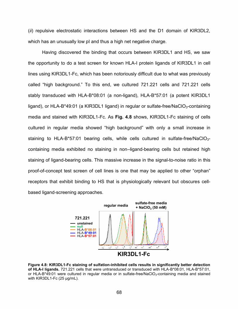
68
(ii) repulsive electrostatic interactions between HS and the D1 domain of KIR3DL2,
which has an unusually low pI and thus a high net negative charge.
Having discovered the binding that occurs between KIR3DL1 and HS, we saw
the opportunity to do a test screen for known HLA-I protein ligands of KIR3DL1 in cell
lines using KIR3DL1-Fc, which has been notoriously difficult due to what was previously
called “high background.” To this end, we cultured 721.221 cells and 721.221 cells
stably transduced with HLA-B*08:01 (a non-ligand), HLA-B*57:01 (a potent KIR3DL1
ligand), or HLA-B*49:01 (a KIR3DL1 ligand) in regular or sulfate-free/NaClO3-containing
media and stained with KIR3DL1-Fc. As Fig. 4.8 shows, KIR3DL1-Fc staining of cells
cultured in regular media showed “high background” with only a small increase in
staining to HLA-B*57:01 bearing cells, while cells cultured in sulfate-free/NaClO3-
containing media exhibited no staining in non–ligand-bearing cells but retained high
staining of ligand-bearing cells. This massive increase in the signal-to-noise ratio in this
proof-of-concept test screen of cell lines is one that may be applied to other “orphan”
receptors that exhibit binding to HS that is physiologically relevant but obscures cell-
based ligand-screening approaches.
Figure 4.8: KIR3DL1-Fc staining of sulfation-inhibited cells results in significantly better detection of HLA-I ligands. 721.221 cells that were untransduced or transduced with HLA-B*08:01, HLA-B*57:01, or HLA-B*49:01 were cultured in regular media or in sulfate-free/NaClO3-containing media and stained with KIR3DL1-Fc (25 μg/mL).
unstainednullHLA-B*08:01HLA-B*49:01HLA-B*57:01
KIR3DL1-Fc
sulfate-free media
+ NaClO3 (50 mM)regular media
721.221

69
4.4 MATERIALS AND METHODS
Cell lines and antibodies
721.221 cell lines (including HLA transductants) were grown in RPMI-1640
supplemented with 10% fetal bovine serum (Sigma-Aldrich), 2 mM L-glutamine (Gibco),
100 U/mL penicillin (Gibco), and 100 U/mL streptomycin (Gibco) at 37°C/5% CO2. EBV-
transformed B-cell lines (BCLs) were generated from peripheral blood mononuclear
cells from donors bearing specific HLA genotypes; BCLs were also grown in the same
media and conditions as 721.221 cells. The following purified antibodies were used:
anti-HS-FITC (clone: 10E4, US Biological; used at 1:10 dilution), anti-hIgG(Fc)-PE (Life
Technologies), anti-CD3 (BioLegend), anti-CD4 (BioLegend), anti-CD8 (BioLegend),
anti-CD19 (BioLegend), anti-CD56 (clone: HCD56, BioLegend), and anti-CD16 (clone:
3G8, BioLegend).
KIR-Fc staining
Cells were stained with KIR-Fc by washing extensively with PBS, incubating for
30–45 min with 25 μg/mL KIR3DS1-Fc (R&D) diluted in PBS at 4°C while shaking,
followed by a secondary staining with anti-hlgG(Fc)-PE (Life Technologies) for 30 min at
4°C while shaking. After staining, cells were washed and fixed using 4%
paraformaldehyde/PBS (Affymetrix) and flow cytometry analysis was performed on an
BD LSR II or LSR Fortessa.

70
Genome-wide CRISPR/Cas9 knock-out (GeCKO) screen
Screen was performed as previously described in 97. However, methods will be
briefly described. sgRNA Library Design. The optimized sgRNA library was designed by
first computationally filtering out potential off-target matches (except for sgRNAs that
targeted multiple homologs), and also to have high cleavage activity (based on rules
experimentally determined in another set of experiments). In total, we constructed a
novel library, containing 178,896 sgRNAs targeting 18,166 protein-coding genes in the
human consensus CDS (CCDS) and 1,004 non-targeting control sgRNAs. Screen
procedure. K562 cells were lentivirally transduced with Cas9/eGFP vector. 2.40 × 10⁸
Cas9-transduced cells were transduced with the viral pool containing the sgRNA library
to achieve an average 1000-fold coverage of the library after selection with puromycin
for 7 d. 5 × 10⁷ K562-GeCKO cells were washed in PBS and stained in 1 mL of 25
μg/mL KIR3DS1-Fc (R&D) for 15 min at 4°C. Cells were then washed with PBS and
stained with 1 mL of anti-hIgG(Fc)-PE (Life Technologies) diluted 1:50 in PBS.
Fluorescently labeled cells were washed with PBS and stained with anti-PE Microbeads
(Miltenyi) following manufacturer’s instructions, except for using twice the recommended
volumes. Magnetically labelled cells were then depleted in an LD column (Miltenyi)
following manufacturer’s instructions, and flow-through cells were collected and
cultured. Screen deconvolution. sgRNA inserts were PCR amplified from 5 million
genome equivalents of DNA from flow-through cells. The resultant PCR products were
purified and sequenced on a HiSeq 2500 (Illumina); primer sequences are as follows:
sgRNA quantification primers:
F: AATGATACGGCGACCACCGAGATCTAGAATACTGCCATTTGTCTCAAG

71
R: CAAGCAGAAGACGGCATACGAGATCnnnnnnTTTCTTGGGTAGTTTGCAGT
TTT
(nnnnn denotes the sample barcode)
Illumina sequencing primer:
CGGTGCCACTTTTTCAAGTTGATAACGGACTAGCCTTATTTTAACTTGCTATTT
CTAGCTCTAAAAC
Illumina indexing primer:
TTTCAAGTTACGGTAAGCATATGATAGTCCATTTTAAAACATAATTTTAAAACT
GCAAACTACCCAAGAAA
Data analysis. Sequencing reads were aligned to the sgRNA library and the abundance
of each sgRNA was calculated. Gene-based CRISPR scores (CS) were defined as the
average log2 fold change of all sgRNAs targeting a given gene and calculated for the
entire screen. To identify enriched genes, the CS distribution was mean-normalized to
zero.
Sulfation inhibition treatment
Cells were grown for 48 h (or other time period if specified) in sulfate-
free/NaClO3-containing media, which consisted of custom-made Advanced RPMI-1640
(Life Technologies) deficient in magnesium sulfate, zinc sulfate, and copper (II) sulfate,
supplemented with 10% dialyzed FBS (Gibco), 2 mM L-glutamine (Gibco), 100 U/mL
penicillin (Gibco), 100 U/mL streptomycin (Gibco), 407 μM magnesium chloride (Sigma-
Aldrich), 3.03 μM zinc chloride (Sigma-Aldrich), and 5 nM copper (II) chloride (Sigma-

72
Aldrich), and 50 mM sodium chlorate (NaClO3; Sigma-Aldrich) (or other NaClO3
concentration if specified).
Enzymatic treatment of cell lines
For surface enzymatic treatment of cells, 2.5 × 10⁵ cells were washed three times
with PBS, and then incubated in PBS containing 2 U/mL heparinase II (New England
BioLabs), 1 U/mL proteinase K (New England BioLabs), or no enzyme for 1 h at
37°C/5% CO2. Cells were then washed in ice-cold PBS and stained as indicated.
Computational analysis of pI
Isoelectric point analysis was determined by inputing Ig-domain sequences (from
Cys to Cys) into ExPASy Compute pI/Mw tool, which calculates pI using pKa values of
amino acids99. The KIR sequences analyzed were obtained from Immuno
Polymorphism Database (IPD)100,101 and were the following: KIR2DL1*001,
KIR2DL2*001, KIR2DL3*001, KIR2DL4*001, KIR2DL5A*001, KIR2DS1*002,
KIR2DS4*001, KIR3DL1*001, KIR3DS1*013, KIR3DL2*001, and KIR3DL3*001.
Surface Plasmon Resonance (SPR)
SPR measurements were conducted in phosphate-buffered saline (PBS;
Corning) containing 0.005% v/v surfactant P20 (GE Healthcare) using a Biacore 3000
system (Biacore AB). To assess binding of various KIR-Fc constructs to various GAGs,
biotinylated hyaluronate (molecular weight = 29 kDa), biotinylated heparin (molecular
weight = 18 kDa) and biotinylated chondroitin sulfate (molecular weight = 50 kDa) (all

73
from Creative PEGWorks), were immobilized onto individual flow cells of an SA
(streptavidin) sensor chip (GE Healthcare) until saturation. A blank flow cell with no
immobilized ligand was used as a reference flow cell. Injections of 60 μL of KIR-Fc
constructs diluted in PBS to 25 μg/mL were performed at a flow rate of 20 μL/min, with a
subsequent 10 min run of buffer to allow sufficient dissociation. Although not presented
here, regeneration after each injection was achieved with two pulses of 100 μL of 0.2 M
sodium hydroxide (NaOH) (GE Healthcare) at a flow rate of 100 μL/min. Raw
sensograms were corrected by double referencing (subtracting from the reference flow
cell response and from PBS injection response). All experiments were done at standard
temperature (25°C).
Human Samples and Viruses
Frozen peripheral blood mononuclear cells (PBMCs) from healthy donors were
used in this study in accordance to protocols approved by Partners Human Research
Committee and Institutional Review Board of Massachusetts General Hospital.
Data Acquisition and Analysis
Flow cytometry data was acquired on BD LSR II or LSR Fortessa and analyzed
using FlowJo software version 10.1 (FlowJo) and statistical analyses were performed
using GraphPad Prism 6 (GraphPad Software).

CHAPTER 5: Influence of glycosylation inhibition on the binding
of KIR3DL1 to HLA-B*57:01

75
5.1 SUMMARY
Viral infections can affect the glycosylation pattern of glycoproteins involved in
antiviral immunity. Given the importance of protein glycosylation in immune function, we
investigated the effect that modulation of the highly conserved HLA class I (HLA-I) N-
glycan has on KIR:HLA-I interactions and NK-cell function. We focused on the
interaction between HLA-B*57:01 and KIR3DL1, which has been shown to play an
influential role on the outcome of several human diseases, including HIV-1 infection.
721.221 cells stably expressing HLA-B*57:01 were treated with a panel of glycosylation
enzyme inhibitors, and HLA-I expression and KIR3DL1 binding was quantified. In
addition, the functional outcomes of HLA-B*57:01 N-glycan disruption/modulation on
KIR3DL1ζ+ Jurkat reporter cells and primary human KIR3DL1+ NK cells was assessed.
Different glycosylation enzyme inhibitors had varying effects on HLA-B*57:01
expression and KIR3DL1-Fc binding. The most remarkable effect was that of
tunicamycin, an inhibitor of the first step of N-glycosylation, which resulted in
significantly reduced KIR3DL1-Fc binding despite sustained expression of HLA-B*57:01
on 721.221 cells. This effect was paralleled by decreased activation of KIR3DL1ζ+
Jurkat reporter cells, as well as increased degranulation of primary human KIR3DL1+
NK cells when encountering tunicamycin-treated HLA-B*57:01-expressing 721.221
cells. Overall, these results demonstrate that N-glycosylation of HLA-I is important for
KIR:HLA-I binding and has an impact on NK-cell function.

76
5.2 BACKGROUND
Natural killer (NK) cells are part of the innate immune system, and serve as a first
line of defense against intracellular pathogens and malignantly transformed cells. NK
cells can lyse target cells by secreting perforin and granzyme and by inducing
FasL:Fas-mediated cell death, but can also activate and recruit other immune cells via
secretion of pro-inflammatory cytokines and chemokines. The importance of NK-cell
function is highlighted in the setting of human NK-cell deficiency syndromes, which
cause increase risk of death in early life due to increased susceptibility to infections with
commonly encountered viruses and intracellular bacteria (reviewed in 8,102). NK-cell
activity is regulated via a number of activating and inhibitory receptors (reviewed in 103),
including killer-cell immunoglobulin-like receptors (KIRs), which recognize human
leukocyte antigen class I (HLA-I) ligands. Binding of KIRs to HLA-I is determined by the
respective HLA-I and KIR allotypes, as well as the sequence of the HLA-I presented
peptides7,104. Whereas binding of inhibitory KIRs to HLA-I molecules results in NK-cell
inhibition, engagement of activating KIRs or loss of inhibitory KIR binding can lead to
NK-cell activation105,106. The KIR-HLA interaction has been demonstrated to have an
important impact on viral infections, including human immunodeficiency virus-1 (HIV-1).
In HIV-1 positive individuals, KIR3DL1+ NK cells expanded preferentially in the
presence of HLA-Bw4-80I, the ligand class for KIR3DL1 14,27.
Protein glycosylation is a post-translational modification occurring in the
endoplasmatic reticulum (ER) and Golgi apparatus. In multiple enzymatic steps, a
complex oligosaccharide (i.e. glycan) is synthesized in the ER, transferred to a specific
receptor sequence on its target protein, and subsequently cropped and remodeled in

77
the ER and Golgi107. In the case of asparagine N-glycosylation, the target amino acid
sequence on the respective proteins is Asn-X-Ser/Thr, where the glycan is bound to the
asparagine. It is estimated that over 7,000 distinct glycan structures can be generated in
mammals108, suggesting a wide range of functional properties, which can be modulated
by differential glycosylation. The HLA-I glycan at position N86, which is located close to
the Bw4 motif that is critical for KIR3DL1 binding, is highly conserved and present on all
classical and non-classical HLA-I genes and allotypes100,109,110. Here we use a panel of
glycosylation inhibitors to examine the effects of N-glycan modifications on KIR3DL1
binding to HLA-B*57:01 and their potential effect on NK-cell function, with a more in-
depth characterization of the effect tunicamycin (TUN)—an antibiotic that can
completely block N-glycosylation in eukaryotic cells by preventing the linkage of the
glycan to the asparagine111,112—and castanospermine (CSP)—an α-glucosidase I and II
inhibitor able to inhibit ER glycan trimming that is necessary for the assembly of a fully
functional glycan112,113.

78
5.3 RESULTS
5.3.1 Effects of glycosylation enzyme inhibitors on HLA class I expression and
KIR-Fc binding
In order to examine the effect of different commercially available glycosylation
inhibitors (see Illustration 5.1) on KIR:HLA interactions, we examined their effect on
HLA-B*57:01 expression on HLA-B*57:01-transduced 721.221 cells (also referred to as
221 cells) and on KIR3DL1-Fc binding to those cells. As shown in Fig. 5.1a and Fig.
5.1b, inhibitors that targeted Golgi-resident α-mannosidase enzymes (KIF, SWA)
increased HLA-B*57:01 surface expression, which resulted in increased KIR3DL1-Fc
binding. The Golgi-resident sialyl transferase inhibitor (STI) tested, however, did not
result in any appreciable changes in HLA-B*57:01 surface expression or KIR3DL1-Fc
binding. Inhibitors that targeted ER-resident α-glucosidase II (CSP, DNM), which is an
enzyme necessary for the last removal of glucose from N-glycans to allow anterograde
transport to the Golgi, dramatically reduced HLA-B*57:01 surface expression and
abrogated KIR3DL1-Fc binding. The ER-resident α-glucosidase I-specific inhibitor
(AUS) minimally increased HLA-B*57:01 surface expression and KIR3DL1-Fc binding.
However, in the case of TUN, we observed a major decrease in KIR3DL1-Fc binding
while HLA-B*57:01 surface expression was only slightly increased. Thus, after
preliminary testing of the effects that several different glycosylation inhibitors had on
HLA-I surface expression and KIR binding, we decided to focus on TUN, the only
inhibitor tested that did not decrease HLA-I surface expression but dramatically reduced
KIR3DL1-Fc binding, and that had the unique function of completely inhibiting addition
of N-glycans.

79
Illustration 5.1: N-glycan processing and glycosylation inhibitor. Diagram illustrating processing of N-glycans in endoplasmic reticulum (ER), Golgi, and their structure at the plasma membrane (PM) (based on
114). Glycosylation inhibitors used in this study and the enzymes they target are indicated in red text
and black blunt-head arrows (for full names of glycosylation inhibitors and target enzymes, see Materials and Methods).
Figure 5.1: Glycosylation inhibitors screening. Relative median fluorescence intensities (RMdFI) of anti-HLA-Bw4 antibody staining (in a) and KIR3DL1-Fc binding (in b) of untransduced 721.221 cells and HLA-B*57:01–transduced 721.221 cells treated with a panel of glycosylation inhibitors (n = 2 for each) (for details, see Materials and Methods). Black bars indicate glycosylation inhibitors that were used in subsequent experiments.
We subsequently performed a titration assay in order to identify the minimal
amount of glycosylation inhibitors required to observe a strong functional effect on the
binding of KIR3DL1ζ+ Jurkat cells to HLA-B*57:01-expressing target 721.221 cells (221-
HLA-B*57:01 cells) while keeping target cell death to a minimum. We observed a dose-
dependent effect of both TUN and CSP on KIR3DL1ζ Jurkat cell signaling, with the
strongest measured effect (i.e. decreased activation of KIR3DL1ζ+ Jurkat cells as
measured by CD69 expression) corresponding to the highest treatment dose used in
TUNCSP
AUS
CSP
DNM
KIF SWA
STI
ER Golgi PM
HLA-Bw4 surface staining KIR3DL1-Fc bindinga b

80
previous studies: 0.5 µg/mL of TUN (Supplementary Fig. S5.1a) or 2 mM of CSP
(Supplementary Fig. S5.1b). Higher doses of glycosylation inhibitors increased cell
toxicity to a degree that interfered with our assay by causing cell death, while lower
doses of inhibitors increased signaling of KIR3DL1ζ Jurkat cells after co-incubation,
indicating a loss of glycosylation inhibition in target cells. Thus, we focused on the
glycosylation inhibitor TUN at 0.5 µg/mL, the concentration used in previous studies to
investigating the effects of TUN on HLA-I glycosylation115,116.
5.3.2 HLA class I N-glycan is necessary for KIR3DL1 binding to HLA-B*57:01
We next sought to quantitatively assess the effects of TUN on HLA-B*57:01 surface
expression and KIR3DL1-Fc binding. 221-HLA-B*57:01 cells (and wildtype 221 cells as
control) were treated PBS, CSP or TUN for 24 h, and then stained with anti-Bw4
antibody, anti-pan-HLA-I antibodies, and KIR3DL1-Fc for flow cytometric assessment.
TUN treatment of 221-HLA-B*57:01 cells significantly increased HLA-B*57:01 surface
expression as measured by anti-HLA-Bw4 and anti-pan-HLA-I antibody staining (1.5-
fold HLA-Bw4 MdFI increase, p < 0.02; 2.0-fold W6/32 MdFI increase, p < 0.001) (Fig.
5.2 and Fig. 5.3a). In contrast, co-incubation with CSP significantly decreased HLA-I
pan-HLA-I (W6/32)
surface stainingFigure 5.2: TUN treatment of 721.221-HLA-B*57:01 cells increases HLA-I surface expression. Relative median fluorescence intensity (RMdFI) of anti-pan-HLA-I (W6/32) staining was calculated for 721.221 cells and 721.221-HLA-B*57:01 cells treated with TUN, CSP, or untreated

81
expression (2.1-fold HLA-Bw4 MdFI decrease, p < 0.02; 1.7-fold W6/32 MdFI decrease,
p < 0.05). Since glycosylation inhibition similarly affected staining with anti-HLA-Bw4
and anti-pan-HLA-I antibody, which only recognizes folded HLA-I complexes bound to
β2m, we believe that HLA- B*57:01 proteins expressed at the cell surface were folded
complexes of HLA-B*57:01 and β2-miroglobulin and not free heavy chains.
Figure 5.3: N-glycosylation inhibition increases HLA-B*57:01 surface expression while abrogating KIR3DL1-Fc binding. Anti-HLA-Bw4 antibody staining (in a) and KIR3DL1-Fc staining (in b) was performed on 721.221-HLA-B*57:01 cells treated with TUN, CSP, or untreated, and untransduced 721.221 cells. Representative histograms are on left panels and data representing n = 5 technical replicates are presented as bar graphs on right panels. One-way ANOVA with Dunnette multiple comparisons test was performed (*, p < 0.05; ***, p < 0.001).
721.221
721.221-
HLA-
B*57:01
–
TUN
CSP
HLA-Bw4
–
TUN
CSP
721.221-
HLA-
B*57:01
721.221
KIR3DL1-Fc
HLA-Bw4 surface staining
KIR3DL1-Fc binding
a
b

82
Sustained expression of HLA-B*57:01 on 221-B*57:01 cells following TUN
treatment enabled assessment of the consequences of HLA-I de-glycosylation on
KIR3DL1 binding to HLA-B*57:01. Despite enhancement of overall HLA-B*57:01
expression, TUN treatment abrogated KIR3DL1-Fc binding. Cells treated with TUN
bound KIR3DL1-Fc significantly less than untreated cells (4.7-fold KIR3DL1-Fc MdFI
decrease, p < 0.001). 221-HLA-B*57:01 cells treated with CSP also exhibited reduced
KIR3DL1-Fc binding (6.9-fold KIR3DL1-Fc MdFI decrease, p < 0.001) (Fig. 5.3b), which
was expected given the reduced surface stating of HLA-B*57:01 on CSP-treated cells.
Taken together, these data demonstrate that the presence of N-glycosylation is critical
for KIR3DL1 binding to HLA-B*57:01.
5.3.3 HLA class I N-glycan is necessary for functional signaling through
KIR3DL1 in KIR3DL1ζ+ Jurkat cells
Jurkat reporter cells stably expressing a chimeric receptor of the extracellular and
transmembrane domain of KIR3DL1 fused to the cytoplasmic tail of CD3ζ, referred to as
‘KIR3DL1ζ’, was employed to assess the functional consequences of glycosylation
inhibition. KIR3DL1ζ engagement of its ligand HLA-B*57:01 results in an activating
signal that triggers CD69 expression. Jurkat cells were gated by size, CD3 expression,
and KIR3DL1ζ expression. Cells that were CD3+KIR3DL1ζ– were, by definition,
untransduced and thus were used as an internal negative control.
KIR3DL1ζ+ Jurkat reporter cells were potently stimulated by 221-HLA-B*57:01
cells. However, stimulation of KIR3DL1ζ+ Jurkat cells was abrogated by pre-treatment of
221-HLA-B*57:01 cells with TUN (4.4-fold CD69 MdFI decrease, p < 0.001) (Fig. 5.4a

83
and Fig. 5.4c). As expected, 221-HLA-B*08:01 cells and untransduced 221 cells did not
stimulate KIR3DL1ζ+ Jurkat cells, and treatment with TUN had no effect (Fig. 5.4a).
Untransduced Jurkat cells were not activated after co-incubation with any of the tested
target cells regardless of TUN treatment (Fig. 5.4b and Fig. 5.4c). These results
demonstrate that lack of N-glycosylation abrogates KIR3DL1:HLA-B*57:01 binding, and
this has direct functional consequences on KIR3DL1ζ+ Jurkat cell function.
Figure 5.4: TUN treatment of 721.221-HLA-B*57:01 cells abrogates triggering of KIR3DL1ζ+ Jurkat
reporter cells. Reporter activity of KIR3DL1ζ+ (in a) and untransduced (in b) Jurkat reporter cells was
measured by calculating relative median fluorescence intensity (RMdFI) of CD69 relative to unstimulated Jurkat cells. Reporter assay was carried out for 2 h at a repoter-to-target cell ratio = 1:1. Bar graphs in a and b show the mean ± standard deviation of n = 10 replicates, whereas representative flow histograms are presented in c.
5.3.4 Primary human KIR3DL1+ NK cells are ‘de-repressed’ upon encountering
tunicamycin-treated HLA-B*57:01+ target cells.
We subsequently examined the effect of TUN on the interaction between primary
human KIR3DL1+ NK cells and HLA-B*57:01+ target cells by assessing degranulation in
KIR3DL1+ NK-cell clones co-incubated with TUN-treated or untreated 221-HLA-B*57:01
cells. NK cell clones were gated for FSC, SSC and expression of CD56, CD16,
KIR3DL1 and CD107a (Fig. 5.5a). KIR3DL1+ NK cells exposed to 221 cells
KIR3DL1ζ+ + 721.221
untransduced + 721.221-B*57:01
untransduced + 721.221-B*57:01 + TUN
KIR3DL1ζ+ + 721.221-B*57:01
KIR3DL1ζ+ + 721.221-B*57:01 + TUN
CD69
a b c

84
degranulated extensively (74.47% ± 2.20% CD107a+), but were significantly suppressed
when exposed to 221-HLA-B*57:01 cells (27.20% ± 0.7% CD107a+, 2.7-fold decrease
compared to the 221 samples, p < 0.0001), which was still significantly higher than
unstimulated NK cells (0.46% ± 0.11% CD107a+) (Fig. 5.5b). However, TUN pre-
treatment of 221-HLA-B*57:01 cells resulted in a significant increase in degranulation
(38.53% ± 1.37% CD107a+, 1.4-fold increase compared to 221-HLA-B*57:01, p <
0.0001) compared to untreated 221-HLA-B*57:01 cells. KIR3DL1– NK cells exposed to
221 cells (80.57% ± 1.87%) degranulated significantly more than when exposed to 221-
B*57:01 cells (66.7% ± 1.05%, 1.2-fold decrease compared to 221, p = 0.0007) (Fig.
5.5c). Co-incubation of target cells with TUN had no significant effect on KIR3DL1– NK
cells (221: p = 0.6874; 221-B57: p = 0.1629). Thus, TUN treatment of 221-HLA-B*57:01
cells ‘de-repressed’ KIR3DL1+ NK cells that would normally have been suppressed by
KIR3DL1 engagement of N-glycosylated HLA-B*57:01 proteins, suggesting a critical
and functional role for HLA-I N-glycans in KIR binding and primary NK-cell function
modulation.

85
Figure 5.5: KIR3DL1+ NK-cell clones are disinhibited by TUN treatment of 721.221-HLA-B*57:01
cells. (a) Representative flow plots of a KIR3DL1+ NK-cell clone degranulating in response to TUN-pre-
treated and untreated 721.221 and 721.221-HLA-B*57:01 cells. Degranulation was carried out for 2 h in the presence of fluorophore-conjugated anti-CD107a antibody before staining for surface NK-cell receptors. (b, c) Bar graphs show frequency of degranulating KIR3DL1
+ (in b) and KIR3DL1
– (in c) NK-
cell clones co-incubated with the indicated target cells. Data represented mean ± standard deviation of n = 3 technical replicates. One-way ANOVE with Dunnett multiple comparisons test was performed (ns, non-significant; ***, p < 0.001; *, p < 0.05).
CD107a
KIR
3D
L1
no targets– +TUN – +TUN
721.221-HLA-B*57:01721.221
KIR3DL1+ NK-cell clonea
b c

86
5.4 MATERIALS AND METHODS
Target Cells
HLA-I-deficient 721.221 cells (also referred to as ‘221 cells’) stably expressing
HLA-B*08:01 or HLA-B*57:01 were provided by Christian Brander (Ragon Institute of
MGH, MIT and Harvard, Cambridge, MA). All cells were cultured at 37°C with 5% CO2
in RPMI 1640 medium (Sigma-Aldrich) supplemented with 10% heat-inactivated fetal
bovine serum (FBS) (Biochrom AG), 2500 U/mL penicillin and 2500 µg/mL streptomycin
(Sigma-Aldrich) unless stated otherwise; this media was referred to as ‘R10’.
Glycosylation inhibition screening and titration
Glycosylation inhibition was performed by incubating target cells for 24 h at
37°C/5%CO2 in R10 supplemented with different glycosylation inhibitors: 5 mM of 1-
deoxynojirimycin (DNM; Tocris Bioscience), an α-glucosidase II > I inhibitor; 500 µM of
australine (AUS; Santa Cruz Biotechnology), an α-glucosidase I inhibitor; 2 mM of
castanospermine (CSP; Tocris Bioscience), an α-glucosidase I and II inhibitor; 100 µM
of kifunensine (KIF; Tocris Bioscience), an α-mannosidase I inhibitor; 500 µM of 3Fax-
peracetyl-Neu5Ac (STI; EMD Millipore), a sialyltransferase inhibitor; 100 µM of
swainsonine (SWA; Tocris Bioscience), an α-mannosidase II and lysosomal α-
mannosidase inhibitor; 0.5 µg/mL of tunicamycin (TUN; Sigma-Aldrich), a GlcNAc
phosphotransferase inhibitor; or phosphate buffered saline (PBS) as negative control.
Optimal concentrations of glycosylation inhibitors were determined by using effective
concentrations reported in the literature as well as lower and higher doses. Readouts
measured were HLA expression, KIR3DL1-Fc binding, CD69 expression of KIR3DL1ζ+

87
Jurkat cells, or degranulation of KIR3DL1+ NK cells after staining or co-incubating with
glycosylation-inhibitor–pre-treated untransduced or HLA-transduced 721.221 target
cells.
Quantification of HLA-I expression and KIR3DL1 binding
For HLA-I cell surface expression, target 221 cells (1.5 × 10⁵ per well) were
treated with TUN, CSP, or PBS for 24 h. After washing, cells were stained with either
biotinylated anti-HLA-Bw4 antibody (One Lambda) followed by secondary staining with
streptavidin-BV421 (BioLegend), or anti-pan-HLA-I-PE antibody (clone: W6/32)
(BioLegend). For KIR-Fc binding assessment, target cells (1.5 × 10⁵ per well) were
grown for 48 h in modified media prior to KIR-Fc staining to reduce background staining.
Modified media consisted of custom-made Advanced RPMI-1640 (Life Technologies)
deficient in magnesium sulfate, zinc sulfate, and copper (II) sulfate supplemented with
10% dialyzed FBS (Gibco), 2 mM L-glutamine (Gibco), 100 U/mL penicillin (Gibco), 100
U/mL streptomycin (Gibco), 407 μM magnesium chloride (Sigma-Aldrich), 3.03 μM zinc
chloride (Sigma-Aldrich), and 5 nM copper (II) chloride (Sigma-Aldrich), which was
mixed with 150 mM sodium chlorate (Sigma-Aldrich) at a 5:1 volume ratio. During the
last 24 h, TUN, CSP or PBS were added to the medium. After washing, cells were
stained for 45 min with 25 μg/mL KIR3DL1-Fc (R&D) at 4°C while shaking, followed by
a secondary staining with anti-hlgG(Fc)-PE (Life Technologies) for 30 min at 4°C while
shaking. After staining, cells were fixed using 4% paraformaldehyde/PBS (Affymetrix)
and flow cytometry analysis was performed on an LSR-II (BD).

88
Jurkat reporter cell assay
Jurkat cells (clone E6.1; ATCC) were cultivated in R10 medium. 24 h before
performing experiments, 10% fetal bovine serum was added to the culture to reduce
background activation. Jurkat cells stably expressing KIR3DL1ζ, a chimeric receptor
composed of the extracellular and transmembrane domains of KIR3DL1 linked to the
cytoplasmic tail of CD3ζ, which we denote as ‘KIR3DL1ζ‘, were produced via lentiviral
transduction/transfection. Briefly, gene constructs were designed and ordered via
GeneArt (Life Technologies), and cloned into a lentiviral transfer vector containing an
SFFV promotor and IRES-driven puromycin resistance. HEK293T cells (ATCC) were
transfected with a VSV-G envelope vector (pHEF-VSVG, NIH AIDS Reagent Program),
HIV-1 gag-pol packaging vector (psPAX2, NIH AIDS Reagent Program), and the
transfer vector containing KIR3DL1ζ. Lentivirus-containing supernatants were harvested
72 h after transfection and used to transduce Jurkat cells, which were subsequently
selected in R10 containing 1 μg/mL puromycin and sorted for gene expression by flow
cytometry. In these KIR3DL1ζ+ Jurkat cells, ligand engagement by KIR3DL1ζ results in
an activating signal that triggers CD69 expression, making them a suitable reporter cell
system. Prior to co-incubation with target cells, Jurkat cells were cultured for 24 h in
R10 medium supplemented with an additional 10% fetal bovine serum. 221 cells
expressing HLA-B*08:01, HLA-B*57:01, or no HLA-I underwent glycosylation inhibition
and were washed twice. Target cells were washed to remove glycosylation inhibitors
and then co-incubated with either KIR3DL1ζ+ or untransduced Jurkat cells at a reporter-
to-target cell ratio of 1:1 (1 × 10⁵ each) for 2.5 h at 37°C/5%CO2. Cells were then
washed, stained with anti-CD3-PerCP-Cy5.5, anti-CD69-BV421, and anti-KIR3DL1-

89
APC (all from BD) for 30 min at 4°C, fixed with 4% paraformaldehyde/PBS, and
analyzed on an LSR-Fortessa (BD). KIR3DL1ζ-transduced Jurkat cells were gated for
KIR3DL1 expression, classifying them either as KIR3DL1ζlow, KIR3DL1ζdim or
KIR3DL1ζbright.
NK cell cloning and degranulation assay
Primary human NK cells isolated from peripheral blood mononuclear cells
(PBMCs) of healthy donors were cloned by limiting dilution in the presence of feeders
and maintained in NK cell cloning medium consisting of R10 supplemented with 5%
human serum (Sigma-Aldrich), 1X MEM-NEAA (Gibco), 1X sodium pyruvate (Gibco),
100ug/mL Kanamycin, 200 U/mL IL-2 (AIDS Reagent Program, NIH) using a protocol
adapted from a previously reported method (22). Briefly, NK cells were isolated from
peripheral blood mononuclear cells (PBMCs) from a KIR3DL1+ HLA-Bw4+ donor via
magnetic negative selection (NK cell isolation kit from Miltenyi), added to a mix of
irradiated feeders consisting of freshly isolated allogeneic (PBMCs) combined with log-
phase-growth RPMI 8866 cells (Sigma-Aldrich) at a 10:1 ratio in cloning medium
containing 1 μg/mL phytohaemagglutinin (PHA; Fisher) and mixed thoroughly before
plating at 100 µL/well (0.5 NK cell/well) in 96-well plates and incubated for 14 days at
37°C/5% CO2. After 14 days, wells that had outgrowth of cells were transferred to 48-
well plates and maintained in NK cell cloning medium with frequent media exchange
(approximately every 3 days). Cells were phenotyped by flow cytometry to assess NK
cell marker (e.g. CD56 and CD16) and KIR3DL1 expression. Degranulation assays
were performed by co-incubating 5 × 10⁴ NK cells with 2.5 × 10⁵ target cells (effector-to-
90
target cell ratio of 1:5) in 200 µL of R10 containing 3 µL of anti-CD107a-PE-Cy7
antibody (BioLegend) for 2 h at 37°C/5% CO2, and subsequently staining with anti-
CD56-BV605 and anti-CD16-BV785 antibodies (both from BioLegend) for 30 min at
4°C, and then fixed with 4% paraformaldehyde/PBS. Flow cytometric analysis was
performed on a BD LSRFortessa.
Ethics
PBMCs from healthy donors were obtained and frozen after obtaining written
informed consent following procedures approved by the Partners Human Research
Committee (ethics committee) and the Institutional Review Board of Massachusetts
General Hospital. HLA-I types of selected human samples were determined prior to this
study by high-resolution HLA-I typing performed at the HLA-typing laboratory of the
National Cancer Institute, National Institutes of Health. KIR types were determined prior
to this study by Sanger sequencing in the laboratory of Mary Carrington.
Statistics
Flow data was analyzed in FlowJo v10 (FlowJo LLC, Ashland, Oregon).
Statistical analyses were performed using GraphPad Prism 6.0c (GraphPad Software
Inc., La Jolla, California, USA). Normality tests (Kolmogorov-Smirnov test with Dallal-
Wilkinson-Lilliefor P value) were performed on all data sets, statistical significance was
calculated via one-way ANOVA, and multiple comparisons were corrected using
Tukeys’s test if not stated otherwise. All graphs show mean ± SD if not stated otherwise

91
in the figure legends. Significance levels were defined as the following: *, p < 0.05; **, p
< 0.01; ***, p < 0.001; and ****, p < 0.0001.

CHAPTER 6: Discussion

93
6.1 KIR3DS1 binds HLA-F
6.1.1 KIR3DS1 binding to HLA-F is functionally and physiologically relevant
The activating NK-cell receptor KIR3DS1 has been associated with the outcome
of viral infections31,42,49 including HIV-116,19, autoimmune disorders41, transplantation38–
40, and cancer development/treatment33,34,36,37. However, the precise nature of a ligand
that can account for such broad biological effects has remained—although extensively
studied—unknown. Here, we identified HLA-F OCs as ligands for KIR3DS1, and
showed that HLA-F OCs trigger polyfunctional responses in primary human NK cells
through KIR3DS1. It was further demonstrated that KIR3DS1 ligands, in particular HLA-
F, are expressed on the surface of activated CD4+ T cells. However, upon HIV-1
infection of activated CD4+ T cells, KIR3DS1 ligand surface expression was
downregulated, yet this did not abrogate efficient suppression of viral replication by co-
cultured KIR3DS1+ NK cells in vitro. Our findings provide novel and significant insight
into the protective effect of KIR3DS1 in HIV-1 infection, and provide an explanation for
the widespread influence of KIR3DS1 in human disease.
6.1.2 KIR3DS1 binding to HLA-F is not unique but is evolutionarily conserved
Differential binding of KIR3DS1 and the other lineage II KIRs3 to HLA-F is
remarkable from a structural homology perspective, and is shared with other related
receptors. The leukocyte immunoglobulin-like receptor (LILR) family is another group of
HLA-I-binding receptors found near the KIR locus in the leukocyte receptor complex
(LRC)117. The LILR family has inhibitory members (i.e. LILRBs) that bind HLA-I
complexes and activating members (i.e. LILRAs) that bind HLA-I OCs55,118, a property

94
that our data suggests is partially shared with KIRs; two members in particular, LILRB1
and LILRB2, exhibit robust binding to HLA-F119. From this present study, we find that all
lineage II KIRs bind HLA-F OCs, with KIR3DS1 exhibiting the highest affinity binding to
and the most potent functional signaling capacity upon engagement of HLA-F OCs on
target cells. KIR3DL2, which has been previously shown to bind HLA-F OCs120, shares
~86% extracellular domain sequence identity with KIR3DS1, which readily explains
differences in binding affinity. However, it is remarkable that despite >97% extracellular
domain sequence identity between KIR3DS1 and KIR3DL1, KIR3DS1 exhibits
significantly higher-affinity binding to HLA-F OCs than KIR3DL1, whereas KIR3DL1 (but
not KIR3DS1) bears high-affinity binding to HLA-Bw4 ligands. Structural studies indicate
that the few amino acid differences between KIR3DS1 and KIR3DL1 occur at sites
critical for HLA binding24. From an evolutionary standpoint, these different ligand-binding
profiles of KIR3DL1 towards various HLA-Bw4 allotypes and KIR3DS1 towards HLA-F,
which is also monomorphic with one predominant allele (HLA-F*01:01 at >95%
frequency)121, provide an explanation for the high polymorphicity of KIR3DL1 and the
relative monomorphicity of KIR3DS1.
6.1.3 KIR3DS1:HLA-F axis may be a mode of detecting “stressed self” similar
to NKG2D and its ligands
This newly identified KIR3DS1:HLA-F axis has many similarities to the well-
known stress-induced NKG2D:MIC-A/B axis, even in the context of HIV-1 infection.
Although it is less well-studied, HLA-F bears unique and distinguishing characteristics
that separates it from all other HLA-I genes. According to a study examining the genesis

95
and architectural evolution of the MHC locus, HLA-F acted as the ancestral progenitor
to today’s HLA-I and MIC-A/B genes122. HLA-F retains features that are similar to MIC-
A/B genes, including being peptide-devoid and being able to refold in vitro with and
without β2m, indicating its unique ability to be stable as an OC57, in line with data
presented here. In agreement with previous studies62,123, our data show that HLA-F
OCs are expressed on the surface of activated CD4+ T cells. Also, although only
partially assessed in this study with B-cell lines, several studies have indicated that
cancers of various tissue origins aberrantly express HLA-F123,124. Additionally, HIV-1
infection of CD4+ T cells increased transcription of HLA-F but reduced KIR3DS1 ligand
expression, particularly in late-infected cells, which might suggest the employment of an
immune-evasion strategy, potentially through the downregulation of HLA-F by HIV-1
accessory proteins, similar to what has been described for HIV-1 Nef for HLA-A and
HLA-B125–127 and NKG2D ligands including MIC-A128, but also HIV-1 Vpu for HLA-G129
and HLA-C130. Our findings are therefore reminiscent of the up-regulation of the NKG2D
ligands MIC-A and MIC-B on the surface of activated T cells, virally infected cells, and
transformed cells, which are downregulated in the context of HIV-1 by the actions of the
accessory protein Nef128.
HLA-F expression is tightly regulated131, with restricted tissue expression119,123
and predominant localization to the endoplasmic reticulum. This indicates that HLA-F
expression might serve as a marker for specific kinds of cell stress, such as
endoplasmic reticulum stress. Similar to the phenomenon seen for MIC-A132,133, HLA-F
can be a target for humoral immune responses, as was shown in one study that
reported the presence of anti-HLA-F antibodies in the sera of cancer patients but not

96
healthy controls124. Collectively, these studies underscore a critical role of HLA-F
expression in human diseases, and our data demonstrating that KIR3DS1 recognizes
HLA-F OCs uncovers a previously unknown mechanism of innate immune surveillance
of stressed cells.
6.1.4 Previous associations of KIR3DS1 to HLA-Bw4I80 are likely driven by
KIR3DL1
KIR3DS1 binding to HLA-F expounds its widespread influence on human
diseases, but it does not fully explain the well-known association of KIR3DS1 to HIV-1
disease control described only for HLA-Bw4+ patients. Several studies have confirmed,
however, that in the context of HIV-1 infection, presence of KIR3DS1 and KIR3DL1
together associate with decreased viremia, delayed progression to AIDS, and better in-
vitro viral inhibition by NK cells in patient bearing HLA-Bw4I8019,134. This protective
compound genotype can be explained by independent but functionally synergistic
effects of KIR3DS1:HLA-F and KIR3DL1:HLA-Bw4 interactions, the latter of which has
independently been linked to delayed HIV-1 disease progression18. Of note, a single
recent study demonstrated KIR3DS1 binding to HLA-B*57:01 presenting two HIV-1
peptides that arose from an exhaustive screen of various viral peptide databases25.
However, it is not clear whether this interaction has real functional consequences, or
represents an exceptional case where a peptide can overcome KIR3DS1 mutations that
normally abrogate HLA-Bw4 binding. Indeed, KIR3DS1 has been linked to many other
human diseases without association to HLA-Bw431,34,36,40,42, suggesting that KIR3DS1
exerts disease modulatory effects independent of HLA-Bw4.

97
6.1.5 KIR3DS1:HLA-F interactions may play roles in both elimination of
pathologically altered cells and regulation of adaptive immunity
KIR3DS1:HLA-F interactions between NK cells and pathologically altered target
cells would incur the well-known innate function of NK cells to recognize and eliminate
target cells expressing “stressed self” ligands. This is supported by our in-vitro co-
culture assay, which showed that NK cells singly expressing KIR3DS1 are more
effective at suppressing HIV-1 replication in autologous CD4+ T cells as compared to
KIR3DS1– NK cells. In addition, our data show that KIR3DS1:HLA-F interactions elicit
NK-cell production of antiviral and pro-inflammatory cytokines such as IFN-γ, TNF-α,
and MIP-1β, which would have pleiotropic effects on immune responses. Furthermore,
KIR3DS1:HLA-F interactions between NK cells and activated CD4+ T cells would also
suggest a means of NK cell-mediated adaptive immune regulation. This notion is
warranted by previous work in LCMV infection mouse models, which show that NK cells
regulate adaptive immunity by killing activated CD4+ T cells, resulting in decreased
immunopathology43. Thus, KIR3DS1 recognition of HLA-F OCs expressed on activated
immune cells and/or infected target cells provides a mechanistic link between KIR3DS1
and HIV-1 disease progression, and will also have relevance for the pathogenesis of
other infectious diseases, autoimmune disorders, and tumor immune-surveillance.
In conclusion, the novel interaction between the activating NK-cell receptor
KIR3DS1 and HLA-F OCs described here uncovers a previously unknown mechanism
of target cell recognition by NK cells. Further studies will be required to determine how
KIR3DS1:HLA-F interactions play a role in NK cell-mediated regulation of immunity

98
and/or elimination of pathologically altered target cells. Each of the clinical diseases
KIR3DS1 has been implicated in, including autoimmune disorders41, transplantation
outcomes38–40, cancer development/clearance33,34,36,37, and viral infections16,19,31,42,49,
offers a variety of prospects to exploit KIR3DS1:HLA-F interactions therapeutically.
6.2 KIR3DS1 binds heparan sulfate
6.2.1 KIR3DS1 binding to HS can be explained through electrostatic
interactions and may be applicable to other KIRs.
In this study, we found through a genome-wide CRISPR/Cas9 knock-out screen
that HS is a ligand for KIR3DS1, which we confirmed biochemically and on primary cells
and cell lines. KIR3DS1 domain analysis revealed that in contrast to other KIRs, each of
its domains is predicted to be net charge positive at physiological pH. This is consistent
with the interaction between KIR3DS1 and HS, which is usually electrostatically driven
for other HS-binding proteins. The fact that KIR3DL1, a highly homologous inhibitory
allotype with >97% extracellular domain protein sequence identity, also bound to HS
although to a lesser extent is also unsurprising, as the critical L166R mutation that
abrogates KIR3DS1 binding to HLA-Bw4I80 proteins24 is also critical for conferring a net
positive charge to its D1 domain. Indeed, whereas KIR2DL4 was previously shown to
interact with HS via its net positively charged D0 domain96, other potential HS-
interacting receptors not tested here are the “orphan” inhibitory NK-cell receptors
KIR2DL5 and KIR3DL3, both of which contain a net positively charged D0 domain.
Interestingly, in the case of KIR2DL5, KIR2DL5-Fc fusion proteins have been described
to exhibit “dull staining of, essentially, every human cell line…independent of the cells

99
HLA allotypes”135. To explain this, we believe it is very likely that KIR2DL5 also binds to
HS, although this needs to be formally tested. Of particular practical interest was the
finding that with sulfation inhibition treatment, the ability to detect binding of KIRs to
HLA-I ligands was significantly increased due to the elimination of HS-interactions that
obscure detection of binding (i.e. reduce signal-to-noise ratio). This may also prove to
be a very useful adjunctive strategy for investigators seeking to perform cell-based
screens for protein ligands of HS-interacting proteins.
6.2.2 NK-cell “heparanosome” may regulate KIR3DS1 and other NK-cell
receptors in cis
Cis interactions between HS and NK-cell receptors like KIR2DL4 have been
shown to cause clustering and block trans interactions, allowing for another level of
tunable control over NK-cell–receptor signaling96. Interestingly, KIR3DS1-Fc did not
bind to primary human NK cells from healthy donors, which may suggest that at
baseline, cis interactions between KIR3DS1 and HS do not occur. However, it is
conceivable that upon cellular activation, NK cells alter the composition of cell-surface
HS—also referred to as the “heparanosome”—to alter receptor functionality. This is
particularly interesting in light of the altered functionality in NK cells that express CD57,
which is an epitope comprising a surface-expressed glucuronic acid 3-O-sulfate moiety
produced by induction of B3GAT1. Expression of CD57 has been shown to correlate
with a more “mature” NK-cell phenotype characterized by poor proliferative capacity,
poor cytokine responsiveness, and increased sensitivity to CD16-mediated activation.
The effect of induction or modulation of heparan sulfate biosynthetic enzymes on NK-
100
cell function, however, has not been appreciably studied, but in light of these data, may
be highly influential on the signaling capacity of receptors such as KIR3DS1 and overall
NK-cell function.
6.2.3 KIR3DS1:HS interactions between NK and target cells may play a role in
cancer immunesurveillance
The binding of D0-domain–containing KIRs to HS can influence NK-cell function
depending on whether these interactions occurs in cis and/or in trans. Interactions of
KIRs and other NK-cell receptors in trans with target-cell–expressed HS would incur an
understudied mode of target cell recognition mediated by protein-carbohydrate
interactions. Indeed, specific cell types (and cellular contexts) exhibit precise
heparanosome signatures that encode for a variety of motifs for HS-binding proteins to
interact with. Cancer cells of various tissue origins significantly alter heparan sulfate
biosynthesis genes to alter cell-surface HS, which has been shown to play a role in
tumorigenesis. One mode of modulation that is widely found in cancer cells is
methylation-associated silencing of HS sulfotransferase genes136, which has been found
to correlate with cancer progression and poor prognosis137. Although various biological
processes that are hallmarks of cancer138 (such as tissue invasion and altered growth
factor signaling) are undoubtedly affected, it is conceivable that this may also contribute
to immune evasion via decreased recognition by HS-binding NK-cell receptors.
The role of KIR3DS1:HS interactions in cancer immunosurveillance is of
particular interest because it is an activating receptor associated to the outcome of
various cancers33–36. Similar to NCR binding to specific HS motifs, it is possible that
101
KIR3DS1 and other KIRs have specific motifs or binding domains on HS they can dock
onto, although a more careful analysis over-expressing and knocking-out specific
sulfotransferase genes will be required for this kind of study. Understanding target-cell–
expressed HS motifs recognized by NK-cell receptors, as well as their potential to be
eliminated in cancer cells through the silencing of HS biosynthetic enzymes to evade
NK cell recognition, is an area of study warranting further investigation. In the realm of
autoimmunity, the use of HS mimetics that block these interactions may also be very
useful, such as in the case of ankylosing spondylitis, which has been shown to be
strongly associated to particular alleles of KIR3DS1 and KIR3DL1.
Ultimately, it is clear that protein-carbohydrate interactions play a crucial role in
NK-cell function, and studies that further explore the extent and specificity of these
interactions will allow for the development of therapies that modulate these interactions,
particularly in the context of cancer and autoimmunity.
6.3 KIR3DL1 binding to HLA-B*57:01 is N-glycosylation dependent
6.3.1 Presence of N86 HLA-I N-glycan is necessary for KIR3DL1 to bind to and
signal upon engagement of HLA-B*57:01
N-glycosylation is a critical step in post-translational protein modification with
functional importance that is not yet fully understood. In the case of all HLA-I allotypes,
a single, highly conserved N-glycosylation site is located at position N86100,109,110, in
close proximity to the highly polymorphic motif spanning from amino acids 77 to 83,
which constitutes the Bw4 motif in HLA-Bw4 allotypes and is critical for KIR3DL1
binding to these24. We therefore hypothesized that HLA-I N-glycosylation can modulate

102
binding between HLA-Bw4 proteins and KIR3DL1. To test this hypothesis, we employed
a panel of glycosylation enzyme inhibitors to modify or eliminate N-glycan structures in
HLA-B*57:01-expressing cells and assessed HLA-B*57:01 expression and binding to
KIR3DL1. We demonstrate that presence of the N-glycan on HLA-B*57:01 is critical for
binding to KIR3DL1, and that inhibition of N-glycosylation in target cells can have
functional consequences on KIR3DL1+ cell lines and primary KIR3DL1+ NK cells.
Inhibition of different glycosylation enzymes led to different effects in terms of
HLA-B*57:01 expression and KIR3DL1 binding. The most striking effect was observed
for TUN-induced inhibition of N-glycosylation, which increased cell surface expression
of HLA-B*57:01 while abrogating KIR3DL1 binding. This was in contrast to all other
inhibitors, which either increased (for KIF, SWA), decreased (for CSP, DNM), or did not
significantly alter (for AUS, STI) HLA-B*57:01 surface expression in a manner that
correlated with increased, decreased, or unchanged KIR3DL1 binding, respectively.
TUN-induced increase in HLA-B*57:01 surface expression was measured using two
different antibodies—anti-HLA-Bw4 (clone: Bw4) and anti-pan-HLA-I (clone: W6/32)—
demonstrating that the Bw4 motif, which is situated next to the N-glycosylation site, is
still recognized, and that the HLA-I molecule is expressed as a complex bound to β2-
microglobulin. We further demonstrated that loss of KIR3DL1-Fc binding to TUN-treated
HLA-B*57:01+ target cells matched functional readouts using KIR3DL1ζ+ Jurkat reporter
cells and primary human KIR3DL1+ NK cells. KIR3DL1ζ+ Jurkat cells were stimulated by
co-incubation with 221-HLA:B*57:01 cells, yet this stimulation was almost completely
abrogated by treating target cells with TUN prior to co-incubation. In line with this,
primary KIR3DL1+ NK cells were potently inhibited by 221-HLA-B*57:01 cells, but were

103
‘de-repressed’ when 221-HLA-B*57:01 cells were pre-treated with TUN. These data
indicates that N-glycosylation of HLA-B*57:01 plays a critical and functional role in
KIR3DL1 binding and can modulate NK cell function, a finding that may extend to other
KIR:HLA interactions that have not yet been studied in the context of N-glycolysation.
6.3.2 This study uniquely describes N-glycan dependency in KIR:HLA
interactions, and refutes prior studies
Reports of the importance of N-glycosylation for KIR:HLA binding are scarce,
save for one study suggesting that the HLA-I N-glycan does not influence KIR
binding139. In contrast, several studies reported the consequences of HLA-I N-
glycosylation modifications on T-cell receptor (TCR) binding115,140–142. Those reports all
concluded that TCR binding to HLA-I (and MHC class I in mice) is independent of HLA-I
glycosylation, given the relatively long distance between the TCR binding site and the
HLA-I N86 glycan. KIR:HLA crystal structures, however, reveal a very close proximity
between the KIR binding site on HLA-I and position N86, which in these structures is
aglycosylated due to the production of these proteins in E. coli (Illustration 6.1). This
suggests that the HLA-I N86 glycan may be contacting KIR and influencing binding
avidity. Of note, the one study that concluded that HLA-I glycosylation was not
necessary for KIR binding was based on a generally assumed interaction between HLA-
B*08:01 and an undiscovered inhibitory KIR, which later was found to be the effects of
the interaction between the inhibitory receptor NKG2A and HLA-E, which was not
discovered at the time of the study143. Thus, to the best of our knowledge, our study is
the first to implicate the HLA-I N-glycan as being critical for KIR:HLA binding, which may

104
serve as another means of modulating the interaction between NK cell receptors and
target cell ligands.
Illustration 6.1: N86 site on crystal structure of KIR3DL1 binding to HLA-B*57:01. KIR3DL1 (red) is shown binding to HLA-B*57 (green) complexed to β2-microglobulin (β2m; blue-gray) and presenting the LF9 peptide (magenta). Asparagine at position 86 (N86), the site of HLA-I N-glycosylation, is indicated from side view (a) and top view (b). Images were generated using PyMOL (DeLano Scientific LLC) based on the structure published in
24.
6.3.3 HIV-1 may alter KIR:HLA interactions by modifying N-glycans as an
immune evasion tactic
It has been demonstrated that the glycosylation pattern of several immune
receptor-ligand pairs can be influenced in the setting of infection. In HIV-1 infection, a
global shift in the glycosylation pattern of IgG has been observed, with HIV-1-specific
antibodies displaying the most distinct glycosylation patterns144,145. This shift in IgG
glycosylation patterns can alter Fc receptor binding and is associated with improved
antiviral activity and control of HIV-1, but has also been described for other viral and
HLA-B*57:01
KIR3DL1*001
β2m
N86LF9
N86
a b
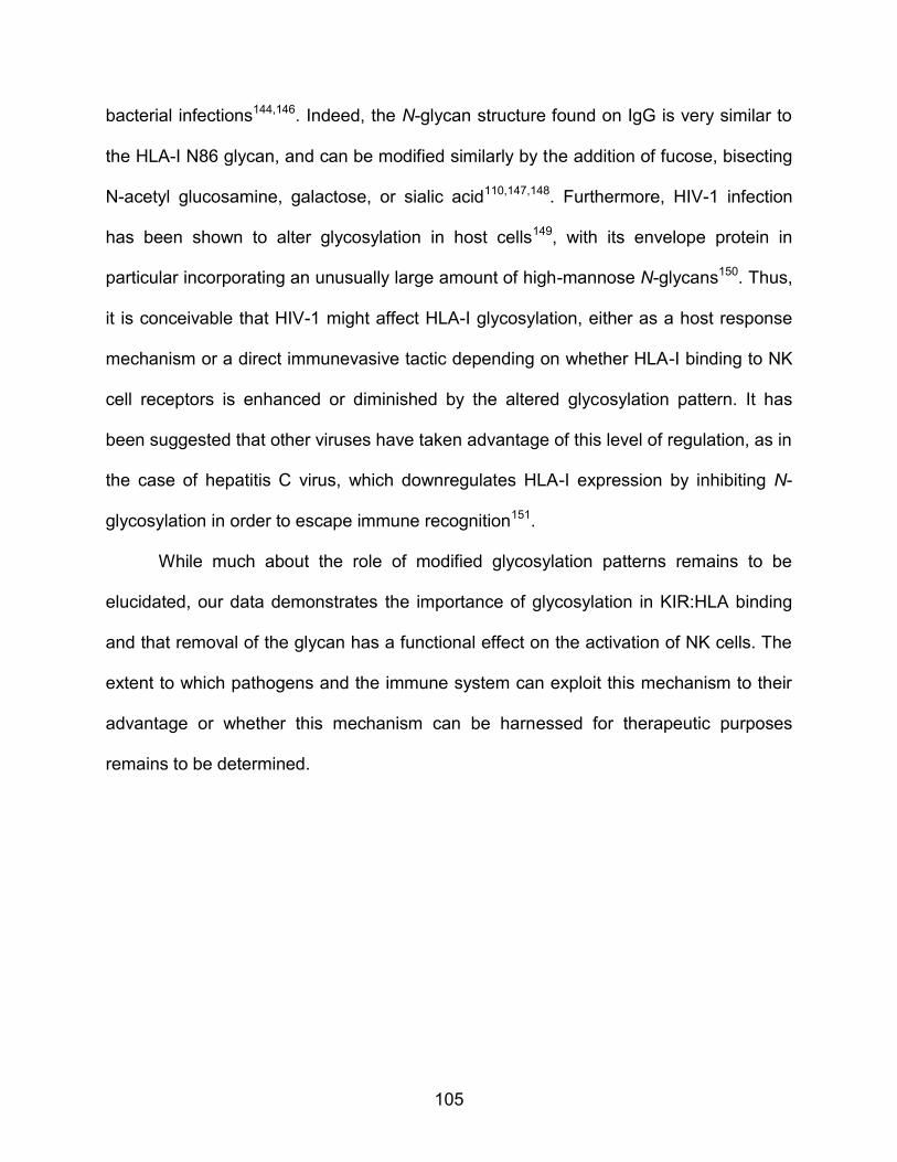
105
bacterial infections144,146. Indeed, the N-glycan structure found on IgG is very similar to
the HLA-I N86 glycan, and can be modified similarly by the addition of fucose, bisecting
N-acetyl glucosamine, galactose, or sialic acid110,147,148. Furthermore, HIV-1 infection
has been shown to alter glycosylation in host cells149, with its envelope protein in
particular incorporating an unusually large amount of high-mannose N-glycans150. Thus,
it is conceivable that HIV-1 might affect HLA-I glycosylation, either as a host response
mechanism or a direct immunevasive tactic depending on whether HLA-I binding to NK
cell receptors is enhanced or diminished by the altered glycosylation pattern. It has
been suggested that other viruses have taken advantage of this level of regulation, as in
the case of hepatitis C virus, which downregulates HLA-I expression by inhibiting N-
glycosylation in order to escape immune recognition151.
While much about the role of modified glycosylation patterns remains to be
elucidated, our data demonstrates the importance of glycosylation in KIR:HLA binding
and that removal of the glycan has a functional effect on the activation of NK cells. The
extent to which pathogens and the immune system can exploit this mechanism to their
advantage or whether this mechanism can be harnessed for therapeutic purposes
remains to be determined.

APPENDIX: Supplementary Data

107
Supplementary Figure S2.1: Complex conformation and β2m content assessment of non-classical HLA-I-coated beads. Non-classical HLA-I-coated beads (generated in this study) were untreated or acid-pulsed and stained with (a) anti-pan-HLA-I complex antibody (clone: W6/32) to assess for complex conformation on beads and (b) anti-β2m antibody to quantify β2m content.
a b
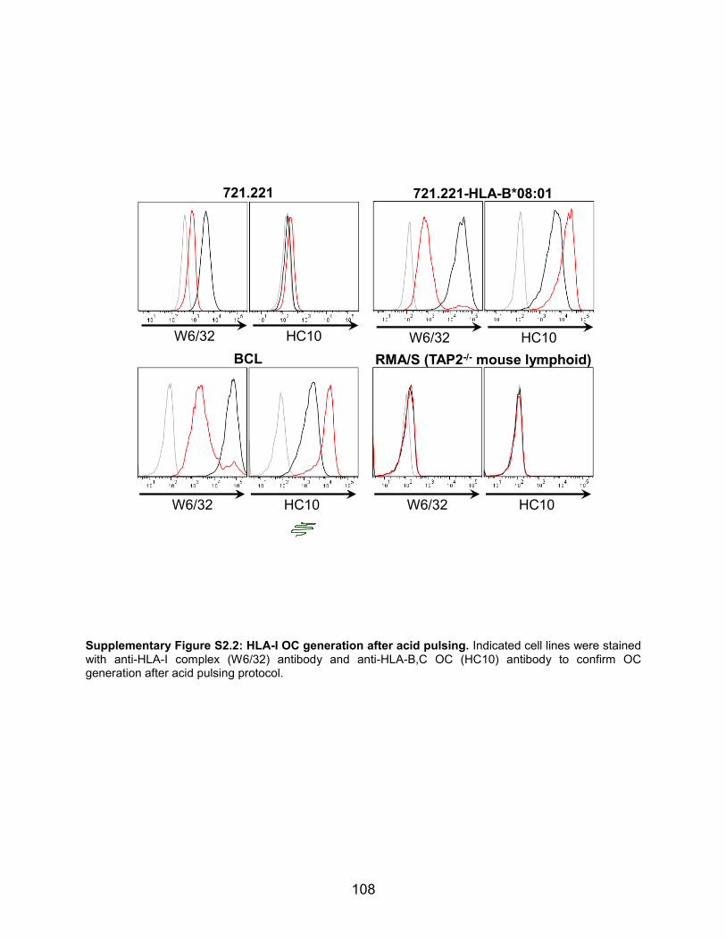
108
Supplementary Figure S2.2: HLA-I OC generation after acid pulsing. Indicated cell lines were stained with anti-HLA-I complex (W6/32) antibody and anti-HLA-B,C OC (HC10) antibody to confirm OC generation after acid pulsing protocol.
BCL
W6/32 HC10
721.221-HLA-B*08:01
W6/32 HC10
721.221
W6/32 HC10
RMA/S (TAP2-/- mouse lymphoid)
W6/32 HC10

109
Supplementary Figure S2.3: 721.221 HLA transductants triggering of KIRζ Jurkat reporter cells. KIR3DL1ζ (green bars), KIR3DS1
hiζ (red bars), and untransduced (white bars) Jurkat reporter cells were
co-incubated with untreated (-) or acid pulsed (+) 721.221 cells expressing the indicated HLA-I proteins (2.5-h incubation and reporter:target cell ratio of 1:10); reporter activity was measured as the percentage of CD69
+hi Jurkat reporter cells. Data represent n = 3 technical replicates; independent experiments were
performed for each 721.221 HLA transductant co-incubated with each Jurkat reporter cell line (n = 2 – 5; data not shown).
Jurkat reporter cell: untransduced KIR3DL1ζ KIR3DS1hiζ
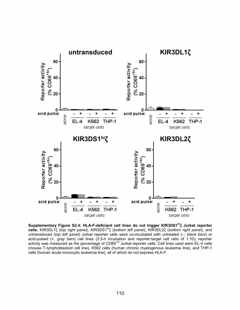
110
Supplementary Figure S2.4: HLA-F-deficient cell lines do not trigger KIR3DS1hi
ζ Jurkat reporter cells. KIR3DL1ζ (top right panel), KIR3DS1
hiζ (bottom left panel), KIR3DL2ζ (bottom right panel), and
untransduced (top left panel) Jurkat reporter cells were co-incubated with untreated (–; black bars) or acid-pulsed (+, gray bars) cell lines (2.5-h incubation and reporter:target cell ratio of 1:10); reporter activity was measured as the percentage of CD69
+hi Jurkat reporter cells. Cell lines used were EL-4 cells
(mouse T-lymphoblastoid cell line), K562 cells (human chronic myelogenous leukemia line), and THP-1 cells (human acute monocytic leukemia line), all of which do not express HLA-F.
KIR3DL1ζ
KIR3DS1hiζ KIR3DL2ζ
untransduced

111
Supplementary Figure S3.1: NK-cell clone phenotypes for plate-bound ligand assay. NK-cell clones used for plate-bound ligand assays were stained for various NK-cell receptors, as indicated; KIR2Ds was measured by staining with anti-KIR2DL2/L3 (clone: DX27) and anti-KIR2DL1/S1 (clone: HP-MA4) having the same fluorophore.
KIR3DS1+ NKCL
KIR3DS1– NKCL
NKCL
#36
#48
#49
#10
#12
#42
#11
#16
KIR3DS1 CD16 KIR2Ds
NKG2A KIR3DL1 LILRB1
NKCL
#36
#48
#49
#10
#12
#42
#11
#16
KIR3DS1+ NKCL
KIR3DS1– NKCL
NKCL
#36
#48
#49
#10
#12
#42
#11
#16
KIR3DS1 CD16 KIR2Ds
NKG2A KIR3DL1 LILRB1
NKCL
#36
#48
#49
#10
#12
#42
#11
#16

112
Supplementary Figure S3.2: KIR3DS1+ NKCLs have superior intrinsic antiviral cytokine production
capacity. KIR3DS1+ (red dots) and KIR3DS1
– NKCLs (black dots) were seeded onto well plates coated
with irrelevant protein (neg. ctrl), human IgG (hIgG), or anti-NKp46 + anti-CD2 antibodies (α-NKp46+α-CD2). NKCLs were incubated for 5 h with anti-CD107a fluorescent antibody and brefeldin A, followed by surface stain for NK-cell markers and intracellular cytokine stain for IFN-γ, TNF-α, and MIP-1β (for details, see Materials and Methods). The raw percentages of CD107a
+ (top left panel), IFN-γ
+ (top right panel),
TNF-α+ (bottom left panel), and MIP-1β
+ (bottom right panel) NKCLs are presented. One-way ANOVA
with Sidak multiple comparisons test comparing select columns was performed, and all statistically significant differences are presented (*, **, and *** denotes, p < 0.05, < 0.01, and < 0.001, respectively).
KIR3DS1+ NKCL
KIR3DS1– NKCL
CD16–
CD16–CD16–
hIgG hIgG
hIgG hIgG
CD16–
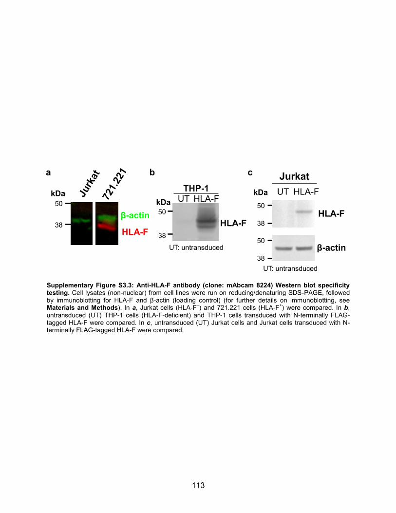
113
Supplementary Figure S3.3: Anti-HLA-F antibody (clone: mAbcam 8224) Western blot specificity testing. Cell lysates (non-nuclear) from cell lines were run on reducing/denaturing SDS-PAGE, followed by immunoblotting for HLA-F and β-actin (loading control) (for further details on immunoblotting, see Materials and Methods). In a, Jurkat cells (HLA-F
–) and 721.221 cells (HLA-F
+) were compared. In b,
untransduced (UT) THP-1 cells (HLA-F-deficient) and THP-1 cells transduced with N-terminally FLAG-tagged HLA-F were compared. In c, untransduced (UT) Jurkat cells and Jurkat cells transduced with N-terminally FLAG-tagged HLA-F were compared.
50
38
kDa HLA-FTHP-1
UT
UT: untransduced
HLA-F
kDa
HLA-F
β-actin
38
50
HLA-FUT
Jurkat
38
50
UT: untransduced
50
38
β-actin
HLA-F
kDa
a b c

114
Supplementary Figure S3.4: Quality control testing of HLA-F mRNA probes in HLA-F+ and HLA-F
–
cell lines. Fluorescent in situ hybridization and flow cytometry was done on an HLA-F+ cell line (BCL) and
an HLA-F– cell line (Jurkat cells) with probes for HLA-F mRNA and also RPL13a mRNA (a housekeeping
gene). Dotted histograms indicate staining without probes (‘– probe’; negative control) and solid-line, filled histograms are staining with probe (‘+ probe’).
RPL13a mRNA(housekeeping gene)
HLA-F mRNA
+ probes
– probes
+ probes
– probes
BCL
Jurkat cells (HLA-F–)
(HLA-F+)

115
Supplementary Figure S3.5: NK-cell clone phenotypes for co-culture assay. NK-cell clones used for HIV-1 replication inhibition assays were stained for various NK-cell receptors. ‘+’ denotes >90% expression of the indicated receptor, ‘–’ denotes a frequency of <5%, and ‘+/–’ denotes a frequency >5% (in both cases for #0, ~10%).
NKCL #33 #23 #36 #40 #27 #0
KIR
3DS1 + + + - - -
3DL2 - - - + - -
2DL2/L3 - + - - - +/-
2DL1/S1 + - - + - +/-
NKG2A + + + + + +
LILRB1 - - - - - -

116
Supplementary Figure S3.6: Low-temperature incubation potently mobilizes HLA-F to the cell surface. Jurkat-HLA-F
FLAG cells (transduction efficiency ~10%) were cultured under normal conditions
(37°C and 5% CO2) or incubated at 26°C (and 5% CO2) for 18 h. Cells were subsequently stained with anti-FLAG antibody to assess cell-surface expression of HLA-F.
THP-1(untransduced)
THP-1-HLA-FFLAG
untreated +PMA +IFN-γ +PMA +IFN-γ
HLA-FFLAG (surface)
pan
-HL
A-I
THP-1 (untransduced)
37 C
26 C
THP-1HLA-FFLAG
HLA-FFLAG
(surface)
Jurkat-HLA-FFLAG
37 C 26 C
HLA-FFLAG (surface)

117
Gene Symbol
Description CRISPR Score
EXT1 exostosin glycosyltransferase 1 7.047
UXS1 UDP-glucuronate decarboxylase 1 (a.k.a. UDP-xylose synthase 1) 4.9493
EXTL3 exostosin-like glycosyltransferase 3 4.394
UGDH UDP-glucose 6-dehydrogenase 4.0326
PAPSS1 3'-phosphoadenosine 5'-phosphosulfate synthase 1.1497
EXT2 exostosin glycosyltransferase 2 0.7124
B3GAT3 beta-1,3-glucuronyltransferase 3 0.6418
XYLT2 xylosyltransferase 2 0.4728
B4GALT7 xylosylprotein beta 1,4-galactosyltransferase 7 -0.4949
Supplementary Table S4.1: Genome-wide CRISPR/Cas9 knock-out screen top hits. Top nine enriched genes (symbol and description) are presented with their determined CRISPR score.

118
Supplemental Figure S5.1: Titration of glycosylation inhibitors. The effect of co-incubating 721.221-HLA-B*57:01 cells with different concentrations of TUN (in a) and CSP (in b) on the expression of CD69 in KIR3DL1ζ
+ Jurkat cells (n = 1 for each). Reporter assay was carried out for 2 h at a reporter-to-target
cell ratio = 10:1.
119
REFERENCES
1. Horowitz, A., Stegmann, K. A. & Riley, E. M. Activation of natural killer cells during microbial infections. Front. Immunol. 2, 1–13 (2012).
2. Vivier, E. & Ugolini, S. Natural killer cells: From basic research to treatments. Front. Immunol. 2, 2–5 (2011).
3. Parham, P., Norman, P. J., Abi-Rached, L. & Guethlein, L. A. Variable NK cell receptors exemplified by human KIR3DL1/S1. J. Immunol. 187, 11–19 (2011).
4. Guethlein, L. A., Older Aguilar, A. M., Abi-Rached, L. & Parham, P. Evolution of killer cell Ig-like receptor (KIR) genes: definition of an orangutan KIR haplotype reveals expansion of lineage III KIR associated with the emergence of MHC-C. J. Immunol. 179, 491–504 (2007).
5. Carr, W. H. et al. Cutting Edge: KIR3DS1, a gene implicated in resistance to progression to AIDS, encodes a DAP12-associated receptor expressed on NK cells that triggers NK cell activation. J. Immunol. 178, 647–651 (2007).
6. Fadda, L. et al. Peptide antagonism as a mechanism for NK cell activation. Proc. Natl. Acad. Sci. 107, 10160–10165 (2010).
7. Fadda, L. et al. Common HIV-1 peptide variants mediate differential binding of KIR3DL1 to HLA-Bw4 molecules. J. Virol. 85, 5970–5974 (2011).
8. Orange, J. S. Natural killer cell deficiency. J. Allergy Clin. Immunol. 132, 515–525 (2013).
9. Hillis, D. M. AIDS: Origins of HIV. Science 288, 1757–1759 (2000).
10. UNAIDS. Fact Sheet 2015. UNAIDS Rep. 1–8 (2015).
11. Walker, B. D. & Yu, X. G. Unravelling the mechanisms of durable control of HIV-1. Nat. Rev. Immunol. 13, 487–98 (2013).
12. Keele, B. F. et al. Identification and characterization of transmitted and early founder virus envelopes in primary HIV-1 infection. Proc. Natl. Acad. Sci. 105, 7552–7557 (2008).
13. Alter, G. et al. Differential natural killer cell-mediated inhibition of HIV-1 replication based on distinct KIR/HLA subtypes. J. Exp. Med. 204, 3027–3036 (2007).
14. Alter, G. et al. HIV-1 adaptation to NK-cell-mediated immune pressure. Nature 476, 96–100 (2011).
15. Hölzemer, A. et al. Selection of an HLA-C*03:04-restricted HIV-1 p24 Gag sequence variant is associated with viral escape from KIR2DL3+ natural killer

120
cells: data from an observational cohort in South Africa. PLoS Med. 12, 1–27 (2015).
16. Martin, M. P. et al. Epistatic interaction between KIR3DS1 and HLA-B delays the progression to AIDS. Nat. Genet. 31, 429–434 (2002).
17. Cella, M., Longo, a, Ferrara, G. B., Strominger, J. L. & Colonna, M. NK3-specific natural killer cells are selectively inhibited by Bw4-positive HLA alleles with isoleucine 80. J. Exp. Med. 180, 1235–1242 (1994).
18. Martin, M. P. et al. Innate partnership of HLA-B and KIR3DL1 subtypes against HIV-1. Nat. Genet. 39, 733–740 (2007).
19. Jiang, Y. et al. KIR3DS1/L1 and HLA-Bw4-80I are associated with HIV disease progression among HIV typical progressors and long-term nonprogressors. BMC Infect. Dis. 13, 405 (2013).
20. Guerini, F. R. et al. Under representation of the inhibitory KIR3DL1 molecule and the KIR3DL1+/BW4+ complex in HIV exposed seronegative individuals. J. Infect. Dis. 203, 1235–1239 (2011).
21. Boulet, S. et al. Increased proportion of KIR3DS1 homozygotes in HIV-exposed uninfected individuals. AIDS 22, 595–599 (2008).
22. Jennes, W. et al. Inhibitory KIR/HLA incompatibility between sexual partners confers protection against HIV-1 transmission. Blood 121, 1157–1164 (2013).
23. Kim, S. et al. HLA alleles determine differences in human natural killer cell responsiveness and potency. Proc. Natl. Acad. Sci. 105, 3053–3058 (2008).
24. Vivian, J. P. et al. Killer cell immunoglobulin-like receptor 3DL1-mediated recognition of human leukocyte antigen B. Nature 479, 401–405 (2011).
25. O’Connor, G. M. et al. Peptide-dependent recognition of HLA-B*57:01 by KIR3DS1. J. Virol. 89, 5213–5221 (2015).
26. Morvan, M. et al. Phenotypic and functional analyses of KIR3DL1+ and KIR3DS1+ NK cell subsets demonstrate differential regulation by Bw4 molecules and induced KIR3DS1 expression on stimulated NK cells. J. Immunol. 182, 6727–6735 (2009).
27. Alter, G. et al. HLA class I subtype-dependent expansion of KIR3DS1+ and KIR3DL1+ NK cells during acute human immunodeficiency virus type 1 infection. J. Virol. 83, 6798–6805 (2009).
28. Song, R. et al. HIV Protective KIR3DL1/S1-HLA-B Genotypes Influence NK Cell-Mediated Inhibition of HIV Replication in Autologous CD4 Targets. PLoS Pathog. 10, 1–12 (2014).

121
29. Long, B. R. et al. Conferral of enhanced natural killer cell function by KIR3DS1 in early human immunodeficiency virus type 1 infection. J. Virol. 82, 4785–4792 (2008).
30. Barbour, J. D. et al. Synergy or independence? Deciphering the interaction of HLA class I and NK cell KIR alleles in early HIV-1 disease progression. PLoS Pathog. 3, 1540 (2007).
31. Zhi-ming, L. et al. Polymorphisms of killer cell immunoglobulin-like receptor gene: possible association with susceptibility to or clearance of hepatitis B virus infection in Chinese Han population. Croat. Med. J. 48, 800–806 (2007).
32. Aranda-Romo, S. et al. Killer-cell immunoglobulin-like receptors (KIR) in severe A (H1N1) 2009 influenza infections. Immunogenetics 64, 653–662 (2012).
33. López-Vázquez, A. et al. Protective effect of the HLA-Bw4I80 epitope and the killer cell immunoglobulin-like receptor 3DS1 gene against the development of hepatocellular carcinoma in patients with hepatitis C virus infection. J. Infect. Dis. 192, 162–165 (2005).
34. Besson, C. et al. Association of killer cell immunoglobulin-like receptor genes with Hogkin’s lymphoma in a familial study. PLoS One 2, 1–10 (2007).
35. Cariani, E. et al. HLA and killer immunoglobulin-like receptor genes as outcome predictors of hepatitis C virus-related hepatocellular carcinoma. Clin. Cancer Res. 19, 5465–5473 (2013).
36. Bonagura, V. R. et al. Activating killer cell immunoglobulin-like receptors 3DS1 and 2DS1 protect against developing the severe form of recurrent respiratory papillomatosis. Hum. Immunol. 71, 212–219 (2010).
37. Carrington, M. et al. Hierarchy of resistance to cervical neoplasia mediated by combinations of killer immunoglobulin-like receptor and human leukocyte antigen loci. J. Exp. Med. 201, 1069–1075 (2005).
38. Gabriel, I. H. et al. Interaction between KIR3DS1 and HLA-Bw4 predicts for progression-free survival after autologous stem cell transplantation in patients with multiple myeloma. Blood 116, 2033–2039 (2010).
39. Gagne, K. et al. Donor KIR3DL1/3DS1 gene and recipient Bw4 KIR ligand as prognostic markers for outcome in unrelated hematopoietic stem cell transplantation. Biol. Blood Marrow Transplant. 15, 1366–1375 (2009).
40. Venstrom, J. M. et al. Donor activating KIR3DS1 is associated with decreased acute GVHD in unrelated allogeneic hematopoietic stem cell transplantation. Blood 115, 3162–3165 (2010).
41. Díaz-Peña, R. et al. Association of the KIR3DS1*013 and KIR3DL1*004 alleles

122
with susceptibility to ankylosing Spondylitis. Arthritis Rheum. 62, 1000–1006 (2010).
42. Trydzenskaya, H. et al. The genetic predisposition of natural killer cell to BK virus-associated nephropathy in renal transplant patients. Kidney Int. 84, 359–65 (2013).
43. Waggoner, S. N., Cornberg, M., Selin, L. K. & Welsh, R. M. Natural killer cells act as rheostats modulating antiviral T cells. Nature 481, 394–398 (2011).
44. Alter, G. & Altfeld, M. Mutiny or scrutiny: NK cell modulation of DC function in HIV-1 infection. Trends Immunol. 32, 219–224 (2011).
45. Paust, S. & von Andrian, U. H. Natural killer cell memory. Nat. Immunol. 12, 500–508 (2011).
46. Benson, D. M. et al. Aphase 1 trial of the anti-KIR antibody IPH2101 in patients with relapsed/refractory multiple myeloma. Blood 120, 4324–4333 (2012).
47. Rajalingam, R. Human diversity of killer cell immunoglobulin-like receptors and disease. Korean J. Hematol. 46, 216 (2011).
48. Min-Oo, G., Kamimura, Y., Hendricks, D. W., Nabekura, T. & Lanier, L. L. Natural killer cells: Walking three paths down memory lane. Trends Immunol. 34, 251–258 (2013).
49. Rivero-Juarez, A. et al. Natural killer KIR3DS1 is closely associated with HCV viral clearance and sustained virological response in HIV/HCV patients. PLoS One 8, 8–13 (2013).
50. O’Connor, G. M. et al. Functional polymorphism of the KIR3DL1/S1 receptor on human NK cells. J. Immunol. 178, 235–241 (2010).
51. Gillespie, G. M. a et al. Lack of KIR3DS1 binding to MHC class I Bw4 tetramers in complex with CD8+ T cell epitopes. AIDS Res. Hum. Retroviruses 23, 451–455 (2007).
52. Gardiner, C. M. et al. Different NK cell surface phenotypes defined by the DX9 antibody are due to KIR3DL1 gene polymorphism. J. Immunol. 166, 2992–3001 (2001).
53. Thananchai, H. et al. Cutting Edge: Allele-specific and peptide-dependent interactions between KIR3DL1 and HLA-A and HLA-B. J. Immunol. 178, 33–37 (2007).
54. Sharma, D. et al. Dimorphic motifs in D0 and D1+D2 domains of killer cell Ig-like receptor 3DL1 combine to form receptors with high, moderate, and no avidity for the complex of a peptide derived from HIV and HLA-A*2402. J. Immunol. 183,

123
4569–4582 (2009).
55. Arosa, F. A., Santos, S. G. & Powis, S. J. Open conformers: the hidden face of MHC-I molecules. Trends Immunol. 28, 115–123 (2007).
56. Wainwright, S. D., Biro, P. a & Holmes, C. H. HLA-F is a predominantly empty, intracellular, TAP-associated MHC class Ib protein with a restricted expression pattern. J. Immunol. 164, 319–328 (2000).
57. Goodridge, J. P., Burian, A., Lee, N. & Geraghty, D. E. HLA-F complex without peptide binds to MHC class I protein in the open conformer form. J. Immunol. 184, 6199–6208 (2010).
58. Trundley, A., Frebel, H., Jones, D., Chang, C. & Trowsdale, J. Allelic expression patterns of KIR3DS1 and 3DL1 using the Z27 and DX9 antibodies. Eur. J. Immunol. 37, 780–787 (2007).
59. Pertel, T. et al. TRIM5 is an innate immune sensor for the retrovirus capsid lattice. Nature 472, 361–365 (2011).
60. Sanjana, N. E., Shalem, O. & Zhang, F. Improved vectors and genome-wide libraries for CRISPR screening. Nat. Methods 11, 783–784 (2014).
61. Mandal, P. K. et al. Efficient Ablation of Genes in Human Hematopoietic Stem and Effector Cells using CRISPR/Cas9. Cell Stem Cell 15, 643–652 (2014).
62. Lee, N., Ishitani, A. & Geraghty, D. E. HLA-F is a surface marker on activated lymphocytes. Eur. J. Immunol. 40, 2308–2318 (2010).
63. Kumar, G., Date, O. S., Kim, K. S. & Manjunath, R. Infection of human amniotic and endothelial cells by Japanese encephalitis virus: Increased expression of HLA-F. Virology 471-473, 29–37 (2014).
64. Yang, Y., Chu, W., Geraghty, D. E. & Hunt, J. S. Expression of HLA-G in human mononuclear phagocytes and selective induction by IFN-gamma. J. Immunol. 156, 4224–4231 (1996).
65. Ljunggren, H.-G. et al. Empty MHC class I molecules come out in the cold. Nature 346, 476–480 (1990).
66. Cella, M. & Colonna, M. in Methods in Molecular Biology: Natural Killer Cell Protocols: Cellular and Molecular Methods (eds. Campbell, K. S. & Colonna, M.) 121, 1–4 (Humana Press, 1999).
67. Imai C., Iwamoto S., C. D. Genetic modification of primary natural killer cells overcomes inhibitory signals and induces specific killing of leukemic cells. Leukemia 106, 376–383 (2005).

124
68. Tarbell, J. M. & Cancel, L. M. The glycocalyx and its significance in human medicine. J. Intern. Med. 280, 97–113 (2016).
69. Esko, J. D., Kimata, K. & Lindahl, U. in Essentials of Glycobiology (2009). doi:10:0-87969-559-5
70. Sarrazin, S., Lamanna, W. C. & Esko, J. D. Heparan sulfate proteoglycans. Cold Spring Harb. Perspect. Biol. 3, 1–33 (2011).
71. Bode, L. et al. Heparan sulfate and syndecan-1 are essential in maintaining murine and human intestinal epithelial barrier function. J. Clin. Invest. 118, 229–238 (2008).
72. O’Brien, J. R. The effect of heparin on the early stages of blood coagulation. J. Clin. Path. 13, 93–98 (1960).
73. Olczyk, P., Mencner, Ł. & Komosinska-Vassev, K. Diverse Roles of Heparan Sulfate and Heparin in Wound Repair. BioMed Res. Int. 2015, 1–7 (2015).
74. Echtermeyer, F. et al. Delayed wound repair and impaired angiogenesis in mice lacking syndecan-4. J. Clin. Invest. 107, R9–R14 (2001).
75. Inatani, M., Irie, F., Plump, A. S., Tessier-Lavigne, M. & Yamaguchi, Y. Mammalian brain morphogenesis and midline axon guidance require heparan sulfate. Science 302, 1044–1046 (2003).
76. Crawford, B. E. et al. Loss of the heparan sulfate sulfotransferase, Ndst1, in mammary epithelial cells selectively blocks lobuloalveolar development in mice. PLoS One 5, 1–10 (2010).
77. Zimmermann, P. et al. Syndecan recyling is controlled by syntenin-PIP2 interaction and Arf6. Dev. Cell 9, 377–388 (2005).
78. Wang, L., Fuster, M., Sriramarao, P. & Esko, J. D. Endothelial heparan sulfate deficiency impairs L-selectin- and chemokine-mediated neutrophil trafficking during inflammatory responses. Nat. Immunol. 6, 902–10 (2005).
79. Qiu, J., Handa, a, Kirby, M. & Brown, K. E. The interaction of heparin sulfate and adeno-associated virus 2. Virology 269, 137–147 (2000).
80. Jones, K. S., Petrow-sadowski, C., Bertolette, D. C., Huang, Y. & Ruscetti, F. W. Heparan Sulfate Proteoglycans Mediate Attachment and Entry of Human T-Cell Leukemia Virus Type 1 Virions into. J. Virol. 79, 12692–12702 (2005).
81. Kalia, M., Chandra, V., Rahman, S. A., Sehgal, D. & Jameel, S. Heparan sulfate proteoglycans are required for cellular binding of the hepatitis E virus ORF2 capsid protein and for viral infection. J. Virol. 83, 12714–12724 (2009).

125
82. Xu, D. et al. Characterization of heparan sulphate 3-O-sulphotransferase isoform 6 and its role in assisting the entry of herpes simplex virus type 1. Biochem. J. 385, 451–9 (2005).
83. Zhang, Z. et al. Heparin sulphate d-glucosaminyl 3-O-sulfotransferase 3B1 plays a role in HBV replication. Virology 406, 280–285 (2010).
84. Shukla, D. & Spear, P. G. Herpesviruses and heparan sulfate: an intimate relationship in aid of viral entry. J. Clin. Invest. 108, 503–510 (2001).
85. McDermott, S. P. et al. Juvenile syndecan-1 null mice are protected from carcinogen-induced tumor development. Oncogene 26, 1407–1416 (2007).
86. Parish, C. R. Heparan sulfate and inflammation. Nat. Immunol. 6, 861–862 (2005).
87. Jakobsson, L. et al. Heparan Sulfate in trans Potentiates VEGFR-Mediated Angiogenesis. Dev. Cell 10, 625–634 (2006).
88. Yayon, A., Klagsbrun, M., Esko, J. D., Leder, P. & Ornitz, D. M. Cell-surface, heparin-like molecules are required for binding of basic fibroblast growth-factor to its high-affinity receptor. Cell 64, 841–848 (1991).
89. Lortat-Jacob, H., Turnbull, J. E. & Grimaud, J. a. Molecular organization of the interferon gamma-binding domain in heparan sulphate. Biochem. J. 310 ( Pt 2, 497–505 (1995).
90. Lortat-Jacob, H., Grosdidier, A. & Imberty, A. Structural diversity of heparan sulfate binding domains in chemokines. Proc. Natl. Acad. Sci. 99, 1229–1234 (2002).
91. Parish, C. R. The role of heparan sulphate in inflammation. Nat. Rev. Immunol. 6, 633–643 (2006).
92. Safaiyan, F. et al. Selective effects of sodium chlorate treatment on the sulfation of heparan sulfate. J. Biol. Chem. 274, 36267–36273 (1999).
93. Sasisekharan, R. & Venkataraman, G. Heparin and heparan sulfate: biosynthesis , structure and function. Biopolymers 4, 626–631 (2000).
94. Hudspeth, K., Silva-Santos, B. & Mavilio, D. Natural Cytotoxicity Receptors: Broader Expression Patterns and Functions in Innate and Adaptive Immune Cells. Front. Immunol. 4, 1–15 (2013).
95. Hecht, M. et al. Natural Cytotoxicity Receptors NKp30 , NKp44 and NKp46 Bind to Different Heparan Sulfate / Heparin Sequences Natural Cytotoxicity Receptors NKp30 , NKp44 and NKp46 Bind to Different Heparan Sulfate / Heparin Sequences. J. Proteome Res. 712–720 (2009). doi:10.1021/pr800747c

126
96. Brusilovsky, M. et al. Genome-wide siRNA screen reveals a new cellular partner of NK cell receptor KIR2DL4: heparan sulfate directly modulates KIR2DL4-mediated responses. J. Immunol. 191, 5256–5267 (2013).
97. Wang, T. et al. Identification and characterization of essential genes in the human genome. Science 350, 1096–1101 (2015).
98. van den Born, J. et al. Novel Heparan Sulfate Structures Revealed by Monoclonal Antibodies. J. Biol. Chem. 280, 20516–20523 (2005).
99. Gasteiger, E. et al. in The Proteomics Protocols Handbook (ed. Walker, J. M.) 571–607 (Humana Press Inc., 2005). at <http://web.expasy.org/protparam>
100. Robinson, J. et al. The IPD and IMGT/HLA database: Allele variant databases. Nucleic Acids Res. 43, D423–D431 (2015).
101. Robinson, J., Halliwell, J. A., McWilliam, H., Lopez, R. & Marsh, S. G. E. IPD - The Immuno Polymorphism Database. Nucleic Acids Res. 41, 1234–1240 (2013).
102. Orange, J. S. Unraveling human natural killer cell deficiency. J. Clin. Invest. 122, 798–801 (2012).
103. Long, E. O., Sik Kim, H., Liu, D., Peterson, M. E. & Rajagopalan, S. Controlling Natural Killer Cell Responses: Integration of Signals for Activation and Inhibition. Annu. Rev. Immunol. 31, 227–258 (2013).
104. Fadda, L. et al. HLA-Cw*0102-restricted HIV-1 p24 epitope variants can modulate the binding of the inhibitory KIR2DL2 receptor and primary NK cell function. PLoS Pathog. 8, 40 (2012).
105. Körner, C. & Altfeld, M. Role of KIR3DS1 in human diseases. Front. Immunol. 3, 1–11 (2012).
106. Bashirova, A. a, Martin, M. P., McVicar, D. W. & Carrington, M. The killer immunoglobulin-like receptor gene cluster: tuning the genome for defense. Annu. Rev. Genomics Hum. Genet. 7, 277–300 (2006).
107. Bieberich, E. Synthesis, processing, and function of N-glycans in N-glycoproteins. Adv. Neurobiol. 9, 47–70 (2014).
108. Moremen, K. W., Tiemeyer, M. & Nairn, A. V. Vertebrate protein glycosylation: diversity, synthesis and function. Nat. Rev. Mol. Cell Biol. 13, 4484–62 (2012).
109. Barbosa, J. a et al. Site-directed mutagenesis of class I HLA genes: role of glycosylation in surface expression and functional recognition. J. Exp. Med. 166, 1329–1350 (1987).
110. Barber, L. D. et al. Unusual uniformity of the N-linked oligosaccharides of HLA-A,

127
-B, and -C glycoproteins. J. Immunol. 156, 3275–3284 (1996).
111. Schönthal, A. H. Endoplasmic reticulum stress: its role in disease and novel prospects for therapy. Scientifica (Cairo) 2012, 857516 (2012).
112. Esko, J. D. & Bertozzi, C. R. in Essential of Glycobiology (2009).
113. van Kemenade, F. J. et al. Glucosidase trimming inhibitors preferentially perturb T cell activation induced by CD2 mAb. J. Leukoc. Biol. 56, 159–165 (1994).
114. Wolfert, M. a & Boons, G.-J. Adaptive immune activation: glycosylation does matter. Nat. Chem. Biol. 9, 776–84 (2013).
115. Bagriacik, E. U., Kirkpatrick, A. & Miller, K. S. Glycosylation of native MHC class la molecules is required for recognition by allogeneic cytotoxic T lymphocytes. Glycobiology 6, 413–421 (1996).
116. Ploegh, H. L., Orr, H. T. & Strominger, J. L. Biosynthesis and cell surface localization of nonglycosylated human histocompatibility antigens. J. Immunol. 126, 270–275 (1981).
117. Allen, R. L., Raine, T., Haude, a, Trowsdale, J. & Wilson, M. J. Leukocyte receptor complex-encoded immunomodulatory receptors show differing specificity for alternative HLA-B27 structures. J. Immunol. 167, 5543–5547 (2001).
118. Jones, D. C. et al. HLA class I allelic sequence and conformation regulate leukocyte Ig-like receptor binding. J. Immunol. 186, 2990–2997 (2011).
119. Lepin, E. J. M. et al. Functional characterization of HLA-F and binding of HLA-F tetramers to ILT2 and ILT4 receptors. Eur. J. Immunol. 30, 3552–3561 (2000).
120. Goodridge, J. P., Burian, A., Lee, N. & Geraghty, D. E. HLA-F and MHC class I open conformers are ligands for NK cell Ig-like receptors. J. Immunol. 191, 3553–62 (2013).
121. Pan, F. H., Liu, X. X. & Tian, W. Characterization of HLA-F polymorphism in four distinct populations in Mainland China. Int. J. Immunogenet. 40, 369–376 (2013).
122. Shiina, T. et al. Molecular dynamics of MHC genesis unraveled by sequence analysis of the 1,796,938-bp HLA class I region. Proc. Natl. Acad. Sci. 96, 13282–13287 (1999).
123. Lee, N. & Geraghty, D. E. HLA-F surface expression on B cell and monocyte cell lines is partially independent from tapasin and completely independent from TAP. J. Immunol. 171, 5264–5271 (2003).
124. Noguchi, K. et al. Detection of anti-HLA-F antibodies in sera from cancer patients. Anticancer Res. 42, 3387–3392 (2004).

128
125. Le Gall, S. et al. Nef Interacts with the μ Subunit of Clathrin Adaptor Complexes and Reveals a Cryptic Sorting Signal in MHC I Molecules. Immunity 8, 483–495 (1998).
126. Collins, K. L., Chen, B. K., Kalams, S. A., Walker, B. D. & Baltimore, D. HIV-1 Nef protein protects infected primary cells against killing by cytotoxic T lymphocytes. Nature 391, 397–401 (1998).
127. Cohen, G. B. et al. The selective downregulation of class I major histocompatibility complex proteins by HIV-1 protects HIV-infected cells from NK cells. Immunity 10, 661–671 (1999).
128. Cerboni, C. et al. Human immunodeficiency virus 1 Nef protein downmodulates the ligands of the activating receptor NKG2D and inhibits natural killer cell-mediated cytotoxicity. J. Gen. Virol. 88, 242–50 (2007).
129. Derrien, M. et al. Human immunodeficiency virus 1 downregulates cell surface expression of the non-classical major histocompatibility class I molecule HLA-G1. J. Gen. Virol. 85, 1945–1954 (2004).
130. Apps, R. et al. HIV-1 Vpu mediates HLA-C downregulation. Cell Host Microbe 19, 686–695 (2016).
131. Boyle, L. H., Gillingham, A. K., Munro, S. & Trowsdale, J. Selective export of HLA-F by its cytoplasmic tail. J. Immunol. 176, 6464–6472 (2006).
132. Jinushi, M., Hodi, F. S. & Dranoff, G. Therapy-induced antibodies to MHC class I chain-related protein A antagonize immune suppression and stimulate antitumor cytotoxicity. Proc. Natl. Acad. Sci. 103, 9190–9195 (2006).
133. Jinushi, M. et al. MHC class I chain-related protein A antibodies and shedding are associated with the progression of multiple myeloma. Proc. Natl. Acad. Sci. 105, 1285–1290 (2008).
134. Pelak, K. et al. Copy number variation of KIR genes influences HIV-1 control. PLoS Biol. 9, (2011).
135. Cisneros, E., Moraru, M., Gómez-Lozano, N., López-Botet, M. & Vilches, C. KIR2DL5: An orphan inhibitory receptor displaying complex patterns of polymorphism and expression. Front. Immunol. 3, 1–8 (2012).
136. Miyamoto, K. et al. Methylation-associated silencing of heparan sulfate D-glucosaminyl 3-O-sulfotransferase-2 (3-OST-2) in human breast, colon, lung and pancreatic cancers. Oncogene 22, 274–280 (2003).
137. Bui, C. et al. Epigenetics: methylation-associated repression of heparan sulfate 3-O-sulfotransferase gene expression contributes to the invasive phenotype of H-EMC-SS chondrosarcoma cells. FASEB J. 24, 436–450 (2010).
129
138. Hanahan, D. & Weinberg, R. A. Hallmarks of cancer: the next generation. Cell 144, 646–674 (2011).
139. Storkus, W. J., Alexander, J., Payne, J. A., Cresswell, P. & Dawson, J. R. The alpha1/alpha2 domains of class I HLA molecules confer resistance to natural killing. J. Immunol. 143, 3853–3857 (1989).
140. Goldstein, S. A. N. & Mescher, M. F. Carbohydrate moieties of major histocompatibility complex class I alloantigens are not required for their recognition by T lymphocytes. J. Exp. Med. 162, 1381–1386 (1985).
141. Hart, G. W. The role of asparagine-linked oligosaccharides in cellular recognition by thymic lymphocytes. J. Biol. Chem. 257, 151–158 (1982).
142. Miyazaki, J.-I., Appella, E., Zhao, H., Forman, J. & Ozato, K. Expression and function of a nonglycosylated major histocompatibility class I antigen. J. Exp. Med. 163, 856–871 (1986).
143. Braud, V. M. et al. HLA-E binds to natural killer cell receptors CD94/NKG2A, B and C. Nature 391, 795–799 (1998).
144. Ackerman, M. E. et al. Natural variation in Fc glycosylation of HIV-specific antibodies impacts antiviral activity. J. Clin. Invest. 123, 2183–2192 (2013).
145. Chung, A. W. et al. Identification of antibody glycosylation structures that predict monoclonal antibody Fc-effector function. AIDS 28, 2523–2530 (2014).
146. Vestrheim, A. C. et al. A pilot study showing differences in glycosylation patterns of IgG subclasses induced by pneumococcal, meningococcal, and two types of influenza vaccines. Immunity, Inflamm. Dis. 2, 76–91 (2014).
147. Parham, P., Alpert, B. N., Orr, H. T. & Strominger, J. L. Carbohydrate moiety of HLA antigens: antigenic properties and amino acid sequences around the site of glycosylation. J. Biol. Chem. 252, 7555–7567 (1977).
148. Nagae, M. & Yamaguchi, Y. Function and 3D structure of the N-glycans on glycoproteins. Int. J. Mol. Sci. 13, 8398–8429 (2012).
149. Lantéri, M. et al. Altered T cell surface glycosylation in HIV-1 infection results in increased susceptibility to galectin-1-induced cell death. Glycobiology 13, 909–918 (2003).
150. Raska, M. et al. Glycosylation patterns of HIV-1 gp120 depend on the type of expressing cells and affect antibody recognition. J. Biol. Chem. 285, 20860–20869 (2010).
151. Tardif, K. D. & Siddiqui, A. Cell surface expression of major histocompatibility complex class I molecules is reduced in hepatitis C virus subgenomic replicon-

130
expressing cells. J. Virol. 77, 11644–11650 (2003).