disorder drives cooperative folding in a multidomain … · disorder drives cooperative folding in...
TRANSCRIPT

Disorder drives cooperative folding in amultidomain proteinDominika T. Gruszkaa,1, Carolina A. T. F. Mendonçaa, Emanuele Pacib, Fiona Whelanc, Judith Hawkheadc,Jennifer R. Pottsc, and Jane Clarkea,2
aDepartment of Chemistry, University of Cambridge, Cambridge CB2 1EW, United Kingdom; bAstbury Centre for Structural Molecular Biology, University ofLeeds, Leeds LS2 9JT, United Kingdom; and cDepartment of Biology, University of York, York YO10 5DD, United Kingdom
Edited by G. Marius Clore, National Institutes of Health, Bethesda, MD, and approved August 23, 2016 (received for review May 31, 2016)
Many human proteins contain intrinsically disordered regions, anddisorder in these proteins can be fundamental to their function—forexample, facilitating transient but specific binding, promoting allo-stery, or allowing efficient posttranslational modification. SasG, amultidomain protein implicated in host colonization and biofilmformation in Staphylococcus aureus, provides another example ofhow disorder can play an important role. Approximately one-half ofthe domains in the extracellular repetitive region of SasG are intrin-sically unfolded in isolation, but these E domains fold in the contextof their neighboring folded G5 domains. We have previously shownthat the intrinsic disorder of the E domains mediates long-rangecooperativity between nonneighboring G5 domains, allowing SasGto form a long, rod-like, mechanically strong structure. Here, weshow that the disorder of the E domains coupled with the remark-able stability of the interdomain interface result in cooperative fold-ing kinetics across long distances. Formation of a small structuralnucleus at one end of the molecule results in rapid structure forma-tion over a distance of 10 nm, which is likely to be important for themaintenance of the structural integrity of SasG. Moreover, if thisnormal folding nucleus is disrupted by mutation, the interdomaininterface is sufficiently stable to drive the folding of adjacent E andG5 domains along a parallel folding pathway, thus maintainingcooperative folding.
IDP | protein folding | parallel pathways | protein engineering |cooperativity
It has been suggested that as much as 20% of the proteome maybe intrinsically disordered (1), mainly manifested as intrinsically
disordered regions within multidomain proteins, although a fewproteins are apparently entirely disordered. Some proteins func-tion as a consequence of disorder: for example, disordered PEVKregions of titin act as an entropic spring (2), whereas in the nuclearpore complex, disordered nucleoporins provide a thick selectivebarrier controlling nuclear import (3). Disorder can also play otherroles: it facilitates posttranslational modification and may promoteallostery (4, 5). SasG (Staphylococcus aureus surface protein G) is acell wall-attached protein from S. aureus that promotes intercel-lular adhesion during the accumulation phase of biofilm formationvia its C-terminal repetitive region (6–8). We previously showed thatthis part of SasG contains alternating E and G5 domains (Fig. 1A)and that E folds when it is N-terminal of a G5 domain. The disorderof E domains in isolation is essential for formation of a long, stiff,mechanically strong, rod-like structure (9) capable of projecting theN-terminal A domain, which is involved in host colonization (6).Here, we combine biophysical measurements, protein engineer-
ing, and simulation to show that the disorder in the E domains ofSasG also promotes cooperative folding and unfolding pathways. Wefind that SasG domains have a highly polarized transition-state (TS)structure, where formation of a small portion of a three-strandedsheet in the far C-terminal region of a SasG G5 domain is suffi-cient to drive the folding of structure over a distance of 10 nm. Ourstudies reveal the importance of the E–G5 interface in driving thiscooperativity. Furthermore, when the usual folding nucleus is dis-rupted by mutation in the multidomain protein, then this interface
is sufficiently stable to drive folding of the two adjacent domains.Thus, we propose that disorder can play a key role in ensuring co-operative folding over long distances in multidomain proteins.
ResultsSasG Domains Fold Cooperatively at Equilibrium. SasG domains arehighly homologous: the sequence identity between G5 domains(except for the first and last) and between E domains is>97%. Here,we investigated the first E domain and the second G5 domain (G52)either alone or in tandem (E–G52) (Fig. 1). We have previouslyshown that the E domain is fully unfolded in isolation (10). BecauseSasG domains have no tryptophans, (un)folding was followed bymonitoring intrinsic tyrosine fluorescence. We have shown that urea-induced equilibrium denaturation curves of E–G52 monitored byfluorescence coincide with those recorded by ellipticity at 235 nm (7)and domain-specific FRET probes (9), showing that equilibriumunfolding of the two-domain construct is fully cooperative: two-statewith concerted disruption of both domains and secondary and ter-tiary structure with no accumulation of intermediates (Fig. 1C). Thestability of E–G52 is around 3.5 kcal mol−1 greater than that of anisolated G52 domain (6.3 vs. 2.8 kcal mol−1, respectively).
Kinetic Experiments Reveal That SasG Domains Fold and UnfoldCooperatively. The refolding kinetics of G52 and E–G52 can bothbe described by a sum of two exponential phases, with a fast foldingphase (accounting for at least 30% of the amplitude) and a slower
Significance
Understanding the role played by disorder in biology is becomingincreasingly important. Disordered proteins are central to sig-naling, development, initiation of transcription, and other vitalcellular processes. How and why disordered proteins are used isnot entirely clear, but disorder can be important in allostery, facili-tate regulatory posttranslational modification, and allow rapid andspecific but promiscuous binding. Here, our investigations of bio-film-promoting protein SasG illustrate that disorder can play an-other role. We show that the intrinsic disorder of one-half of thedomains is important for imparting long-range cooperativity infolding of a large multidomain protein—allowing formation of asmall local element of structure to precipitate cooperative folding ofadjacent disordered domains across a length scale of ∼10 nm.
Author contributions: D.T.G., C.A.T.F.M., J.R.P., and J.C. designed research; D.T.G., C.A.T.F.M.,E.P., and F.W. performed research; J.H. contributed new reagents/analytic tools; D.T.G.,C.A.T.F.M., E.P., F.W., and J.C. analyzed data; and D.T.G. and J.C. wrote the paper.
The authors declare no conflict of interest.
This article is a PNAS Direct Submission.
Data deposition: The crystallography, atomic coordinates, and structure factors have beendeposited in the Protein Data Bank, www.pdb.org (PDB ID code 5DBL).1Present address: Clare Hall Laboratory, The Francis Crick Institute, South Mimms EN6 3LD,United Kingdom.
2To whom correspondence should be addressed. Email: [email protected].
This article contains supporting information online at www.pnas.org/lookup/suppl/doi:10.1073/pnas.1608762113/-/DCSupplemental.
www.pnas.org/cgi/doi/10.1073/pnas.1608762113 PNAS | October 18, 2016 | vol. 113 | no. 42 | 11841–11846
BIOPH
YSICSAND
COMPU
TATIONALBIOLO
GY
CHEM
ISTR
Y
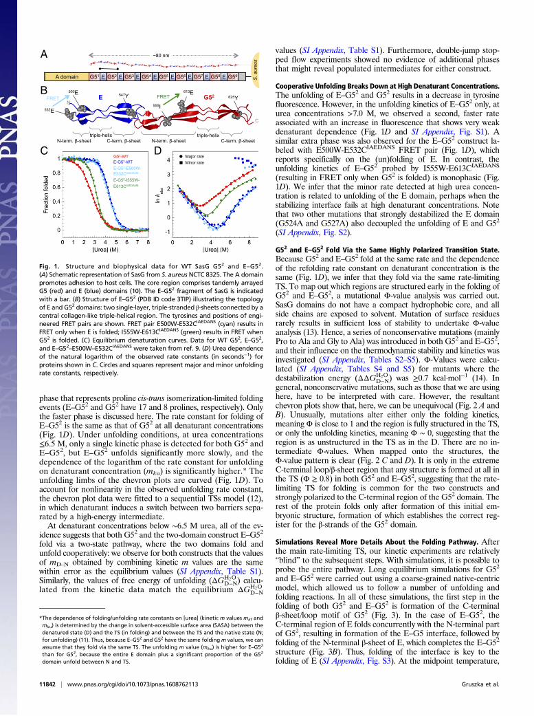
phase that represents proline cis-trans isomerization-limited foldingevents (E–G52 and G52 have 17 and 8 prolines, respectively). Onlythe faster phase is discussed here. The rate constant for folding ofE–G52 is the same as that of G52 at all denaturant concentrations(Fig. 1D). Under unfolding conditions, at urea concentrations≤6.5 M, only a single kinetic phase is detected for both G52 andE–G52, but E–G52 unfolds significantly more slowly, and thedependence of the logarithm of the rate constant for unfoldingon denaturant concentration (mku) is significantly higher.* Theunfolding limbs of the chevron plots are curved (Fig. 1D). Toaccount for nonlinearity in the observed unfolding rate constant,the chevron plot data were fitted to a sequential TSs model (12),in which denaturant induces a switch between two barriers sepa-rated by a high-energy intermediate.At denaturant concentrations below ∼6.5 M urea, all of the ev-
idence suggests that both G52 and the two-domain construct E–G52
fold via a two-state pathway, where the two domains fold andunfold cooperatively: we observe for both constructs that the valuesof mD–N obtained by combining kinetic m values are the samewithin error as the equilibrium values (SI Appendix, Table S1).Similarly, the values of free energy of unfolding (ΔGH2O
D−N) calcu-lated from the kinetic data match the equilibrium ΔGH2O
D−N
values (SI Appendix, Table S1). Furthermore, double-jump stop-ped flow experiments showed no evidence of additional phasesthat might reveal populated intermediates for either construct.
Cooperative Unfolding Breaks Down at High Denaturant Concentrations.The unfolding of E–G52 and G52 results in a decrease in tyrosinefluorescence. However, in the unfolding kinetics of E–G52 only, aturea concentrations >7.0 M, we observed a second, faster rateassociated with an increase in fluorescence that shows very weakdenaturant dependence (Fig. 1D and SI Appendix, Fig. S1). Asimilar extra phase was also observed for the E–G52 construct la-beled with E500W-E532CIAEDANS FRET pair (Fig. 1D), whichreports specifically on the (un)folding of E. In contrast, theunfolding kinetics of E–G52 probed by I555W-E613CIAEDANS
(resulting in FRET only when G52 is folded) is monophasic (Fig.1D). We infer that the minor rate detected at high urea concen-tration is related to unfolding of the E domain, perhaps when thestabilizing interface fails at high denaturant concentrations. Notethat two other mutations that strongly destabilized the E domain(G524A and G527A) also decoupled the unfolding of E and G52
(SI Appendix, Fig. S2).
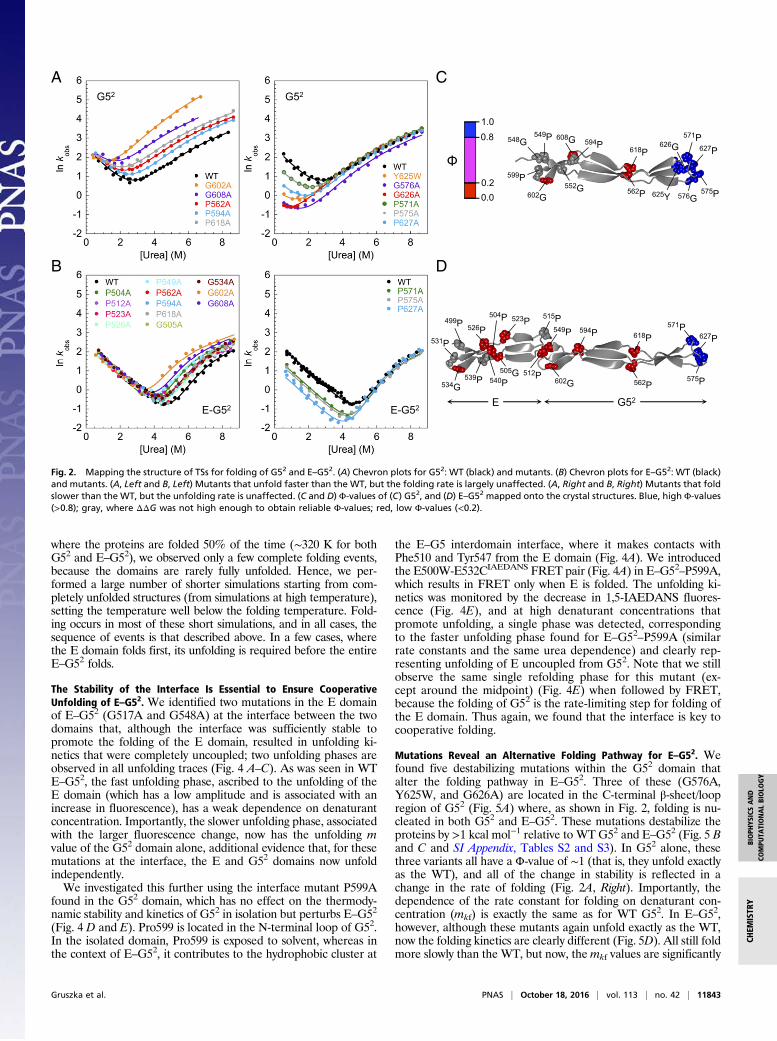
G52 and E–G52 Fold Via the Same Highly Polarized Transition State.Because G52 and E–G52 fold at the same rate and the dependenceof the refolding rate constant on denaturant concentration is thesame (Fig. 1D), we infer that they fold via the same rate-limitingTS. To map out which regions are structured early in the folding ofG52 and E–G52, a mutational Φ-value analysis was carried out.SasG domains do not have a compact hydrophobic core, and allside chains are exposed to solvent. Mutation of surface residuesrarely results in sufficient loss of stability to undertake Φ-valueanalysis (13). Hence, a series of nonconservative mutations (mainlyPro to Ala and Gly to Ala) was introduced in both G52 and E–G52,and their influence on the thermodynamic stability and kinetics wasinvestigated (SI Appendix, Tables S2–S5). Φ-Values were calcu-lated (SI Appendix, Tables S4 and S5) for mutants where thedestabilization energy (ΔΔGH2O
D−N) was ≥0.7 kcal·mol−1 (14). Ingeneral, nonconservative mutations, such as those that we are usinghere, have to be interpreted with care. However, the resultantchevron plots show that, here, we can be unequivocal (Fig. 2 A andB). Unusually, mutations alter either only the folding kinetics,meaning Φ is close to 1 and the region is fully structured in the TS,or only the unfolding kinetics, meaning Φ ∼ 0, suggesting that theregion is as unstructured in the TS as in the D. There are no in-termediate Φ-values. When mapped onto the structures, theΦ-value pattern is clear (Fig. 2 C and D). It is only in the extremeC-terminal loop/β-sheet region that any structure is formed at all inthe TS (Φ ≥ 0.8) in both G52 and E–G52, suggesting that the rate-limiting TS for folding is common for the two constructs andstrongly polarized to the C-terminal region of the G52 domain. Therest of the protein folds only after formation of this initial em-bryonic structure, formation of which establishes the correct reg-ister for the β-strands of the G52 domain.
Simulations Reveal More Details About the Folding Pathway. Afterthe main rate-limiting TS, our kinetic experiments are relatively“blind” to the subsequent steps. With simulations, it is possible toprobe the entire pathway. Long equilibrium simulations for G52
and E–G52 were carried out using a coarse-grained native-centricmodel, which allowed us to follow a number of unfolding andfolding reactions. In all of these simulations, the first step in thefolding of both G52 and E–G52 is formation of the C-terminalβ-sheet/loop motif of G52 (Fig. 3). In the case of E–G52, theC-terminal region of E folds concurrently with the N-terminal partof G52, resulting in formation of the E–G5 interface, followed byfolding of the N-terminal β-sheet of E, which completes the E–G52
structure (Fig. 3B). Thus, folding of the interface is key to thefolding of E (SI Appendix, Fig. S3). At the midpoint temperature,
613E 547Y 555I
625Y 500E
532E
FRET FRETE G52
triple-helix triple-helixN-term. β-sheet N-term. β-sheetC-term. β-sheet C-term. β-sheet
C
N
G52-WT E-G52-WT E-G52-E500W- E532CIAEDANS
E-G52-I555W- E613CIAEDANS
Major rateMinor rate
S. a
ureu
s
A domain G51 E G52 E G53 E G54 E G55 E G56 E G57 E G58 E G59
~80 nmA
B
C D
Fig. 1. Structure and biophysical data for WT SasG G52 and E–G52.(A) Schematic representation of SasG from S. aureus NCTC 8325. The A domainpromotes adhesion to host cells. The core region comprises tandemly arrayedG5 (red) and E (blue) domains (10). The E–G52 fragment of SasG is indicatedwith a bar. (B) Structure of E–G52 (PDB ID code 3TIP) illustrating the topologyof E and G52 domains: two single-layer, triple-stranded β-sheets connected by acentral collagen-like triple-helical region. The tyrosines and positions of engi-neered FRET pairs are shown. FRET pair E500W-E532CIAEDANS (cyan) results inFRET only when E is folded; I555W-E613CIAEDANS (green) results in FRET whenG52 is folded. (C) Equilibrium denaturation curves. Data for WT G52, E–G52,and E–G52–E500W–E532CIAEDANS were taken from ref. 9. (D) Urea dependenceof the natural logarithm of the observed rate constants (in seconds−1) forproteins shown in C. Circles and squares represent major and minor unfoldingrate constants, respectively.
*The dependence of folding/unfolding rate constants on [urea] (kinetic m values mkf andmku) is determined by the change in solvent-accessible surface area (SASA) between thedenatured state (D) and the TS (in folding) and between the TS and the native state (N;for unfolding) (11). Thus, because E–G52 and G52 have the same foldingm values, we canassume that they fold via the same TS. The unfolding m value (mku) is higher for E–G5
2
than for G52, because the entire E domain plus a significant proportion of the G52
domain unfold between N and TS.
11842 | www.pnas.org/cgi/doi/10.1073/pnas.1608762113 Gruszka et al.
where the proteins are folded 50% of the time (∼320 K for bothG52 and E–G52), we observed only a few complete folding events,because the domains are rarely fully unfolded. Hence, we per-formed a large number of shorter simulations starting from com-pletely unfolded structures (from simulations at high temperature),setting the temperature well below the folding temperature. Fold-ing occurs in most of these short simulations, and in all cases, thesequence of events is that described above. In a few cases, wherethe E domain folds first, its unfolding is required before the entireE–G52 folds.
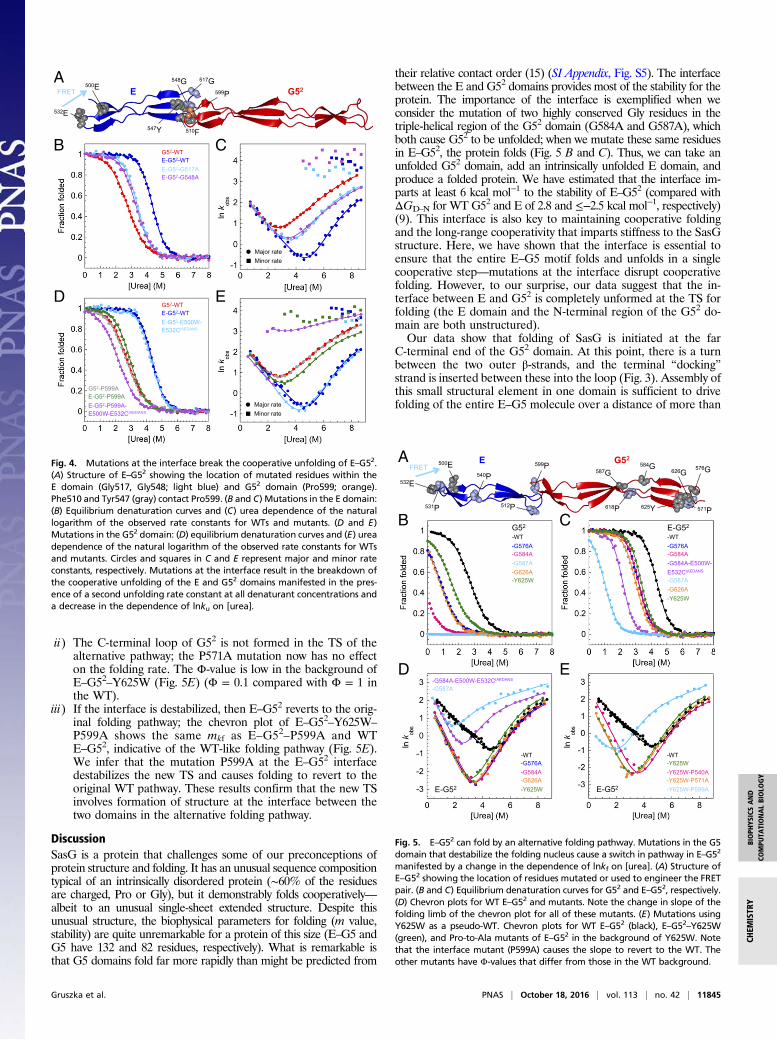
The Stability of the Interface Is Essential to Ensure CooperativeUnfolding of E–G52. We identified two mutations in the E domainof E–G52 (G517A and G548A) at the interface between the twodomains that, although the interface was sufficiently stable topromote the folding of the E domain, resulted in unfolding ki-netics that were completely uncoupled; two unfolding phases areobserved in all unfolding traces (Fig. 4 A–C). As was seen in WTE–G52, the fast unfolding phase, ascribed to the unfolding of theE domain (which has a low amplitude and is associated with anincrease in fluorescence), has a weak dependence on denaturantconcentration. Importantly, the slower unfolding phase, associatedwith the larger fluorescence change, now has the unfolding mvalue of the G52 domain alone, additional evidence that, for thesemutations at the interface, the E and G52 domains now unfoldindependently.We investigated this further using the interface mutant P599A
found in the G52 domain, which has no effect on the thermody-namic stability and kinetics of G52 in isolation but perturbs E–G52
(Fig. 4 D and E). Pro599 is located in the N-terminal loop of G52.In the isolated domain, Pro599 is exposed to solvent, whereas inthe context of E–G52, it contributes to the hydrophobic cluster at
the E–G5 interdomain interface, where it makes contacts withPhe510 and Tyr547 from the E domain (Fig. 4A). We introducedthe E500W-E532CIAEDANS FRET pair (Fig. 4A) in E–G52–P599A,which results in FRET only when E is folded. The unfolding ki-netics was monitored by the decrease in 1,5-IAEDANS fluores-cence (Fig. 4E), and at high denaturant concentrations thatpromote unfolding, a single phase was detected, correspondingto the faster unfolding phase found for E–G52–P599A (similarrate constants and the same urea dependence) and clearly rep-resenting unfolding of E uncoupled from G52. Note that we stillobserve the same single refolding phase for this mutant (ex-cept around the midpoint) (Fig. 4E) when followed by FRET,because the folding of G52 is the rate-limiting step for folding ofthe E domain. Thus again, we found that the interface is key tocooperative folding.
Mutations Reveal an Alternative Folding Pathway for E–G52. Wefound five destabilizing mutations within the G52 domain thatalter the folding pathway in E–G52. Three of these (G576A,Y625W, and G626A) are located in the C-terminal β-sheet/loopregion of G52 (Fig. 5A) where, as shown in Fig. 2, folding is nu-cleated in both G52 and E–G52. These mutations destabilize theproteins by >1 kcal mol−1 relative to WT G52 and E–G52 (Fig. 5 Band C and SI Appendix, Tables S2 and S3). In G52 alone, thesethree variants all have a Φ-value of ∼1 (that is, they unfold exactlyas the WT), and all of the change in stability is reflected in achange in the rate of folding (Fig. 2A, Right). Importantly, thedependence of the rate constant for folding on denaturant con-centration (mkf) is exactly the same as for WT G52. In E–G52,however, although these mutants again unfold exactly as the WT,now the folding kinetics are clearly different (Fig. 5D). All still foldmore slowly than the WT, but now, themkf values are significantly
1.0 0.8
0.2 0.0
A
594P
512P
549P
515P
602G
618P
562P
627P 571P
575P
523P 526P
504P 499P
531P
539P 540P
608G 549P
599P
618P
562P
627P
571P
575P
Φ
B
C
D
534G
505G
E G52
548G 594P
552G 602G
626G
576G 625Y
G52 G52
E-G52 E-G52
Fig. 2. Mapping the structure of TSs for folding of G52 and E–G52. (A) Chevron plots for G52: WT (black) and mutants. (B) Chevron plots for E–G52: WT (black)and mutants. (A, Left and B, Left) Mutants that unfold faster than the WT, but the folding rate is largely unaffected. (A, Right and B, Right) Mutants that foldslower than the WT, but the unfolding rate is unaffected. (C and D) Φ-values of (C) G52, and (D) E–G52 mapped onto the crystal structures. Blue, high Φ-values(>0.8); gray, where ΔΔG was not high enough to obtain reliable Φ-values; red, low Φ-values (<0.2).
Gruszka et al. PNAS | October 18, 2016 | vol. 113 | no. 42 | 11843
BIOPH
YSICSAND
COMPU
TATIONALBIOLO
GY
CHEM
ISTR
Y

increased compared with the WT, suggesting that these variantsare folding via a different, significantly more compact, TS with aβT = 0.53 (compared with 0.33 for WT E–G52).†
Two other Gly to Ala mutations within the triple-helical regionof G52 (G584A and G587A) (Fig. 5A) destabilized the domain sosignificantly that the mutants are largely disordered at 0 M urea(Fig. 5B and SI Appendix, Table S2). In E–G52, these mutationsare also destabilizing, but now, both E and G52 are folded (Fig. 5Cand SI Appendix, Table S3). Interestingly, the chevron plots ofboth E–G52–G584A and E–G52–G587A show the samemkf valuesas the mutants that destabilize the extreme C-terminal region ofE–G52 (Fig. 5D), suggesting that these variants also fold via a new,more compact TS (with a βT of 0.53). Note that folding is stillcooperative; in a control experiment, the kinetics of E–G52–G584Arecorded using the E500W-E532CIAEDANS FRET pair (report-ing specifically on folding of E) was characterized by an identi-cal mkf to the one measured by intrinsic tyrosine fluorescence(Fig. 5D).Thus, if we make mutations that significantly destabilize the
folding nucleus at the extreme C-terminal end of the G52 domainor mutations that are essential for formation of the triple helixconnecting the nucleus to the rest of the protein, we apparentlyalter the folding pathway—but only when the E domain is present.
Formation of the Interface Is Key to Driving Folding Along theAlternative Pathway. Crucially, for some of these mutations inthe G52 domain (e.g., Y625W and G576A), the folding pathway ofisolated G52 does not change; the new pathway is only accessible
when the E domain is present, and yet we know that E does notfold in isolation. Given the importance of the interface betweenthe two domains in imparting stability and cooperativity, we hy-pothesized that the alternative TS (characterized by βT of 0.53)involves formation of a structured E–G52 interface as an early stepin this alternative pathway.If this hypothesis is correct, then residues close to the E–G52
interface, in the E and G52 domains, which all originally have aΦ-value ∼ 0, should have increased Φ-values in this new pathway,and residues in the region with high Φ-values in the WT wouldhave low Φ-values in this alternative pathway. We would alsopredict that a mutation that destabilized the interface could switchthe new pathway back to the original polarized TS in E–G52. Thus,we performed a mutational analysis based on Φ-values, in whichE–G52–Y625W was treated as a pseudo-WT (Fig. 5 A and E and SIAppendix, Table S6). [A crystal structure of the protein at 1.6-Åresolution reveals that this substitution does not disrupt the struc-ture of G52 (SI Appendix, Fig. S4 and Table S7).] In that back-ground, we introduced a number of Pro-to-Ala mutations, most ofwhich originally hadΦ-values = 0 in the background of WT E–G52.P531A and P540A in E and P618A in G52 (all Φ ∼ 0) weredesigned to probe the folding of the individual domains, andP512A and P599A (also Φ ∼ 0) were designed to weaken the in-terface. P571A, which originally had Φ ∼ 1, is found in the C-terminal loop at the center of the nucleation site for the WTpathway. Although one-half of the mutants (P512A, P531A, andP618A) were insufficiently destabilizing to determine Φ-values inthe background of E–G52–Y625W, three of the mutants gave usinformation.
i) The E domain is partly structured in the TS of the alternativepathway; the P540A mutation resulted in a fractionalΦ (0.7) inthe context of E–G52–Y625W (compared withΦ-values = 0 forGly to Ala mutations in the same region of the WT E domain).Folding is more affected than unfolding, implying that the tri-ple helix of the E domain is now significantly structured in theTS (Fig. 5E).
B
332 ns
336 ns
344 ns
349 ns
992 ns
994 ns
1004 ns
1005 ns
A
992 996 1000 1004 1008 Time (ns)
5
25
35
15 RM
SD
(Å)
10
30
40
20 RM
SD
(Å)
330 335 340 345 350 Time (ns)
E G52 G52
E G52 G52
Fig. 3. Probing the folding pathways of SasG using simulations. Simulations of (A) G52 and (B) E–G52 by coarse-grained native-centric model simulations at320 K. A, Upper and B, Upper show the rmsd as a function of simulation time for a typical refolding event. (A) For G52, rmsd values were calculated for allatoms (black), the C-terminal β-sheet/loop region (cyan), and the N-terminal β-sheet/loop region (red). (B) For E–G52, rmsd values were calculated for all atoms(black), the C-terminal β-sheet/loop region of G52 (cyan), the N-terminal β-sheet/loop region of G52 together with the C-terminal β-sheet/loop region ofE (red), and the N-terminal β-sheet/loop region of E (orange). A, Lower and B, Lower illustrate corresponding sequential snapshots from the refolding tra-jectory and the related schematic topology representation. The G52 domain is shown in red, except for the C-terminal β-sheet/loop region (cyan) and itscentral C-terminal docking strand (green). The E domain is shown in blue. Additional details from the same trajectory are illustrated in SI Appendix, Fig. S3.
†The Tanford β-value, βT = ðmkf=mkf +mkuÞ, is a measure of the position of the TS (in termsof SASA or compactness) between D and N (11). An alternative explanation for a switchin mkf is that a mutation results in destabilization of a TS that falls later on the samesingle pathway. Several lines of evidence suggest that this is a less reasonable explana-tion than parallel pathways. Only mutations that destabilize the WT pathway (with Φ∼1) are affected; the same mutations in G52 alone do not result in a change in mkf; aresidue with Φ ∼ 1 in the WT hasΦ ∼ 0 in Y625W (see Formation of the Interface Is Key toDriving Folding Along the Alternative Pathway, point ii).
11844 | www.pnas.org/cgi/doi/10.1073/pnas.1608762113 Gruszka et al.
ii) The C-terminal loop of G52 is not formed in the TS of thealternative pathway; the P571A mutation now has no effecton the folding rate. The Φ-value is low in the background ofE–G52–Y625W (Fig. 5E) (Φ = 0.1 compared with Φ = 1 inthe WT).
iii) If the interface is destabilized, then E–G52 reverts to the orig-inal folding pathway; the chevron plot of E–G52–Y625W–
P599A shows the same mkf as E–G52–P599A and WTE–G52, indicative of the WT-like folding pathway (Fig. 5E).We infer that the mutation P599A at the E–G52 interfacedestabilizes the new TS and causes folding to revert to theoriginal WT pathway. These results confirm that the new TSinvolves formation of structure at the interface between thetwo domains in the alternative folding pathway.
DiscussionSasG is a protein that challenges some of our preconceptions ofprotein structure and folding. It has an unusual sequence compositiontypical of an intrinsically disordered protein (∼60% of the residuesare charged, Pro or Gly), but it demonstrably folds cooperatively—albeit to an unusual single-sheet extended structure. Despite thisunusual structure, the biophysical parameters for folding (m value,stability) are quite unremarkable for a protein of this size (E–G5 andG5 have 132 and 82 residues, respectively). What is remarkable isthat G5 domains fold far more rapidly than might be predicted from
their relative contact order (15) (SI Appendix, Fig. S5). The interfacebetween the E and G52 domains provides most of the stability for theprotein. The importance of the interface is exemplified when weconsider the mutation of two highly conserved Gly residues in thetriple-helical region of the G52 domain (G584A and G587A), whichboth cause G52 to be unfolded; when we mutate these same residuesin E–G52, the protein folds (Fig. 5 B and C). Thus, we can take anunfolded G52 domain, add an intrinsically unfolded E domain, andproduce a folded protein. We have estimated that the interface im-parts at least 6 kcal mol−1 to the stability of E–G52 (compared withΔGD–N for WT G52 and E of 2.8 and ≤−2.5 kcal mol−1, respectively)(9). This interface is also key to maintaining cooperative foldingand the long-range cooperativity that imparts stiffness to the SasGstructure. Here, we have shown that the interface is essential toensure that the entire E–G5 motif folds and unfolds in a singlecooperative step—mutations at the interface disrupt cooperativefolding. However, to our surprise, our data suggest that the in-terface between E and G52 is completely unformed at the TS forfolding (the E domain and the N-terminal region of the G52 do-main are both unstructured).Our data show that folding of SasG is initiated at the far
C-terminal end of the G52 domain. At this point, there is a turnbetween the two outer β-strands, and the terminal “docking”strand is inserted between these into the loop (Fig. 3). Assembly ofthis small structural element in one domain is sufficient to drivefolding of the entire E–G5 molecule over a distance of more than
599P
547Y
517G
510F
A
B C
E G52
G52-WT E-G52-WTE-G52-G517AE-G52-G548A
Major rateMinor rate
548G
D E G52-WT E-G52-WT E-G52-E500W-E532CIAEDANS
G52-P599AE-G52-P599AE-G52-P599A- E500W-E532CIAEDANS
Major rateMinor rate
500E
532E
FRET
Fig. 4. Mutations at the interface break the cooperative unfolding of E–G52.(A) Structure of E–G52 showing the location of mutated residues within theE domain (Gly517, Gly548; light blue) and G52 domain (Pro599; orange).Phe510 and Tyr547 (gray) contact Pro599. (B and C) Mutations in the E domain:(B) Equilibrium denaturation curves and (C) urea dependence of the naturallogarithm of the observed rate constants for WTs and mutants. (D and E)Mutations in the G52 domain: (D) equilibrium denaturation curves and (E) ureadependence of the natural logarithm of the observed rate constants for WTsand mutants. Circles and squares in C and E represent major and minor rateconstants, respectively. Mutations at the interface result in the breakdown ofthe cooperative unfolding of the E and G52 domains manifested in the pres-ence of a second unfolding rate constant at all denaturant concentrations anda decrease in the dependence of lnku on [urea].
B C G52
-WT -G576A-G584A-G587A-G626A-Y625W
D E
E-G52
-WT -G576A-G584A-G584A-E500W- E532CIAEDANS
-G587A-G626A-Y625W
-WT -G576A-G584A-G626A-Y625W
-WT -Y625W-Y625W-P540A-Y625W-P571A-Y625W-P599A
584G 576G 587G 626G 500E
532E
FRETA E G52
531P
599P 540P
512P 618P 625Y 571P
E-G52 E-G52
-G584A-E500W-E532CIAEDANS
-G587A
Fig. 5. E–G52 can fold by an alternative folding pathway. Mutations in the G5domain that destabilize the folding nucleus cause a switch in pathway in E–G52
manifested by a change in the dependence of lnkf on [urea]. (A) Structure ofE–G52 showing the location of residues mutated or used to engineer the FRETpair. (B and C) Equilibrium denaturation curves for G52 and E–G52, respectively.(D) Chevron plots for WT E–G52 and mutants. Note the change in slope of thefolding limb of the chevron plot for all of these mutants. (E) Mutations usingY625W as a pseudo-WT. Chevron plots for WT E–G52 (black), E–G52–Y625W(green), and Pro-to-Ala mutants of E–G52 in the background of Y625W. Notethat the interface mutant (P599A) causes the slope to revert to the WT. Theother mutants have Φ-values that differ from those in the WT background.
Gruszka et al. PNAS | October 18, 2016 | vol. 113 | no. 42 | 11845
BIOPH
YSICSAND
COMPU
TATIONALBIOLO
GY
CHEM
ISTR
Y

10 nm. However, folding at the interface is clearly an option, be-cause destabilization of the C-terminal nucleation site allows foldingvia a higher-energy TS, where formation of the interface is key.E–G52 can thus fold via parallel pathways, but the lowest-energypathway involves formation of the C-terminal nucleus. It is unclearwhy this WT pathway should be lower in energy than a pathwayinvolving formation of the interface, the most stable region of thestructure and essentially, the only region where there is any signifi-cant burial of hydrophobic residues. It may be because the entropiccost of forming the interface is larger; it involves bringing togetherloops from the E and the G5 domains that are distant in sequence(∼85 residues apart), although the interactions in the C-terminalnucleus are by no means short range (∼50 residues between theC-terminal residues of the final strand and the turn). Alternatively, theintrinsic disorder of the E domain may again be key. The formationof the interface involves the folding, at least in part, of the E domain,a process that is inherently costly in terms of free energy. Impor-tantly, however, cooperative folding is a feature of both pathways,because the E domain cannot fold in the absence of G5.In WT protein (except under very destabilizing conditions as
described), the protein folds and unfolds as a single unit; no in-termediates are populated in folding, in unfolding, or at equilib-rium, which is, by definition, cooperative folding. Such tight androbust cooperativity in folding has not been seen previously inmultidomain proteins. Even where there are significant interfacesbetween domains, kinetics reveals that the domains fold in a non–two-state manner, with each domain behaving as an independentfolding unit (16, 17). The “obligate” cooperativity of SasG arises,because E can only fold in the presence of folded G5, but oncefolded, the entire domain is very significantly more stable than thesum of the stability of the two domains individually.The kind of cooperativity that we are observing in the SasG
protein (obligate folding cooperativity) is reminiscent of the foldingof repeat proteins. These proteins comprise tandem arrays of smallrepeats (20–40 residues) that are unstable on their own and fold,apparently cooperatively, through formation of interfaces betweenthe repeats (18–25). However, tandem repeats are very different toSasG, where contacts within the domains themselves and betweendomains are very long range, whereas contacts in repeat proteinsare very local (SI Appendix, Fig. S5). Although there is a dominantfolding pathway in SasG, parallel pathways are a key feature ofrepeat proteins, in particular as the number of repeats increases.
Despite each subunit being intrinsically unstable alone, kineticcooperativity is not generally maintained beyond three to foursubunits in repeat proteins, but SasG is able to maintain co-operative folding across a distance of ∼12 nm.
ConclusionThe importance of intrinsic disorder in biology is becoming in-creasingly apparent; however, why would nature choose disordereddomains to form a multidomain protein? We had previously shownthat disorder-mediated thermodynamic cooperativity allows SasGto adopt long, mechanically strong, rod-like structures (9). Now, wehave shown how this disorder coupled with the remarkable stabilityof the interdomain interface can result in cooperative folding ki-netics, with no populated intermediates, across long distances. Thefolding of classic multidomain proteins is highly cooperative butonly within the relatively local confines of a single domain. In re-peat proteins, short-range cooperativity is apparent between threeand four individually unstable repeats. SasG provides a para-digm for much longer-range cooperative folding—by the oblig-atory folding of alternate intrinsically disordered domains withtheir folded neighbors.
Materials and MethodsAll experimental procedures are described in detail in SI Appendix.
Analysis of Kinetic Data. For somemutants, kinetic data were fitted to amodelallowing for parallel pathways (details are in SI Appendix, Fig. S6).
Simulations. Simulations were performed using a coarse-grained model whereonly Cα atoms are represented and interactions depend on the native refer-ence structure and the residue type. Details are given in SI Appendix.
Determination of the Structure of E–G52–Y625W. Details of the crystallizationand structure determination of E–G52–Y625W can be found in SI Appendix.The coordinates and structure factors have been deposited in the Protein DataBank (PDB) with ID code 5DBL.
ACKNOWLEDGMENTS. This research was supported by Biotechnology andBiological Sciences Research Council Grants BB/J006459/1 (to D.T.G. and J.C.)and BB/J005029/1 (to F.W. and J.R.P). Also, C.A.T.F.M is supported by theCambridge Trust and CAPES (Coordenação de Aperfeiçoamento de Pessoalde Nível Superior) Science Without Borders Cambridge Scholarship. J.R.P holdsBritish Heart Foundation Senior Basic Science Fellowship FS/12/36/29588. J.C. isa Wellcome Trust Senior Research Fellow WT/095195.
1. Ward JJ, Sodhi JS, McGuffin LJ, Buxton BF, Jones DT (2004) Prediction and functionalanalysis of native disorder in proteins from the three kingdoms of life. J Mol Biol337(3):635–645.
2. Tskhovrebova L, Trinick J (2003) Titin: Properties and family relationships. Nat Rev MolCell Biol 4(9):679–689.
3. Milles S, et al. (2015) Plasticity of an ultrafast interaction between nucleoporins andnuclear transport receptors. Cell 163(3):734–745.
4. Shammas SL, Travis AJ, Clarke J (2014) Allostery within a transcription coactivator ispredominantly mediated through dissociation rate constants. Proc Natl Acad Sci USA111(33):12055–12060.
5. Law SM, Gagnon JK, Mapp AK, Brooks CL 3rd (2014) Prepaying the entropic cost forallosteric regulation in KIX. Proc Natl Acad Sci USA 111(33):12067–12072.
6. Corrigan RM, Rigby D, Handley P, Foster TJ (2007) The role of Staphylococcus aureus sur-face protein SasG in adherence and biofilm formation. Microbiology 153(Pt 8):2435–2446.
7. Geoghegan JA, et al. (2010) Role of surface protein SasG in biofilm formation byStaphylococcus aureus. J Bacteriol 192(21):5663–5673.
8. Formosa-Dague C, Speziale P, Foster TJ, Geoghegan JA, Dufrêne YF (2016) Zinc-dependent mechanical properties of Staphylococcus aureus biofilm-forming surfaceprotein SasG. Proc Natl Acad Sci USA 113(2):410–415.
9. Gruszka DT, et al. (2015) Cooperative folding of intrinsically disordered domainsdrives assembly of a strong elongated protein. Nat Commun 6:7271.
10. Gruszka DT, et al. (2012) Staphylococcal biofilm-forming protein has a contiguousrod-like structure. Proc Natl Acad Sci USA 109(17):E1011–E1018.
11. Fersht AR (1999) Structure and Mechanism in Protein Science: A Guide to EnzymeCatalysis and Protein Folding (Freeman, New York).
12. Bachmann A, Kiefhaber T (2001) Apparent two-state tendamistat folding is a se-quential process along a defined route. J Mol Biol 306(2):375–386.
13. Fersht AR, Matouschek A, Serrano L (1992) The folding of an enzyme. I. Theory ofprotein engineering analysis of stability and pathway of protein folding. J Mol Biol224(3):771–782.
14. Fersht AR, Sato S (2004) Phi-value analysis and the nature of protein-folding transi-tion states. Proc Natl Acad Sci USA 101(21):7976–7981.
15. Plaxco KW, Simons KT, Baker D (1998) Contact order, transition state placement andthe refolding rates of single domain proteins. J Mol Biol 277(4):985–994.
16. Batey S, Nickson AA, Clarke J (2008) Studying the folding of multidomain proteins.HFSP J 2(6):365–377.
17. Batey S, Clarke J (2006) Apparent cooperativity in the folding of multidomain pro-teins depends on the relative rates of folding of the constituent domains. Proc NatlAcad Sci USA 103(48):18113–18118.
18. Werbeck ND, Rowling PJ, Chellamuthu VR, Itzhaki LS (2008) Shifting transition states in theunfolding of a large ankyrin repeat protein. Proc Natl Acad Sci USA 105(29):9982–9987.
19. Werbeck ND, Itzhaki LS (2007) Probing a moving target with a plastic unfolding in-termediate of an ankyrin-repeat protein. Proc Natl Acad Sci USA 104(19):7863–7868.
20. Lowe AR, Itzhaki LS (2007) Biophysical characterisation of the small ankyrin repeatprotein myotrophin. J Mol Biol 365(4):1245–1255.
21. Tang KS, Fersht AR, Itzhaki LS (2003) Sequential unfolding of ankyrin repeats in tu-mor suppressor p16. Structure 11(1):67–73.
22. Tripp KW, Barrick D (2008) Rerouting the folding pathway of the Notch ankyrindomain by reshaping the energy landscape. J Am Chem Soc 130(17):5681–5688.
23. Barrick D, Ferreiro DU, Komives EA (2008) Folding landscapes of ankyrin repeatproteins: Experiments meet theory. Curr Opin Struct Biol 18(1):27–34.
24. Bradley CM, Barrick D (2006) The notch ankyrin domain folds via a discrete, central-ized pathway. Structure 14(8):1303–1312.
25. Mello CC, Bradley CM, Tripp KW, Barrick D (2005) Experimental characterization ofthe folding kinetics of the notch ankyrin domain. J Mol Biol 352(2):266–281.
11846 | www.pnas.org/cgi/doi/10.1073/pnas.1608762113 Gruszka et al.