disorders of ventilation university of san francisco dr. m. maag ©2003 margaret maag
TRANSCRIPT
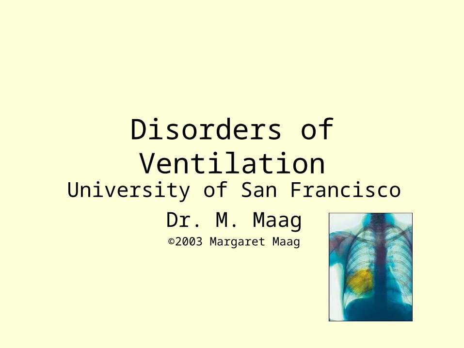
Disorders of VentilationUniversity of San Francisco
Dr. M. Maag©2003 Margaret Maag
Class 10 Objectives
Upon completion of this lesson, the student will be able to• design nursing interventions for patients
presenting with atelectasis, pneumonia, TB, COPD, SARS
• recognize clinical manifestations of respiratory disease for different age groups.
• predict major symptoms seen in patients experiencing respiratory difficulty.
• analyze arterial blood gases associated with various respiratory disorders.
Respiratory Failure• Dynamics: alveolar hypoventialtion, V/Q
disequilibrium, decreased FiO2 & inadequate exchange of gases between alveoli & blood
• Results in hypoxia, hypercapnia, and acidosis• Work of breathing becomes difficult, exhaustion
occurs, and there is no energy to breathe.• Risk factors: pneumonia, Hantavirus, sepsis,
atelectasis, bronchospasms, CHF– atelectasis: areas of the lung where alveoli are
collapsed (alveoli are airless & no gas exchange)

Respiratory Failure Failure of Oxygenation: Etiology
• “Hypoxemia” = reduced PaO2 concentrations– PaO2 < 80 mm Hg (Horne & Derrico, 1999)– Hypoventilation can reduce the PaO2 & PAO2– Factors leading to hypoventilation are drugs
(opiods), neurological disorders (e.g. IICP) & COPD• Ventialtion/ Perfusion Mismatching: V/Q ratio
– Perfusion exceeds ventilation at the lung base• 4:5 = 0.8 = normal V/Q
– “In respiratory failure, the V/Q mismatching is the most common cause of hypoxemia” (Hartshorn, 1997, p. 325)

Respiratory Failure Clinical S & S
• Severe dyspnea (difficulty breathing)– > RR & HR– use of accessory muscles of ventilation
• Cyanosis: indicative of unoxygenated hemoglobin – check nailbeds & mucous membranes
• Clubbing: check fingers and toes• Multi-organ failure leading to death• Tx: O2 & ventilation support
See p. 1108 of McCance
See p. 1108 of McCance

Pulmonary Embolism
Image borrowed from http://www.benlovejoy.com/pulmonary_embolism_main.html

Pulmonary Embolism
Image borrowed from the online NHS encyclopedia

Pulmonary Embolism: “PE”A complication of a venous thromboembolism
– d/t immobility or leg injuries– The most common preventable cause of hospital deaths– 60-80% of fatal PE cases are not suspected
• S & S– Pleuritic chest pain (> with inspiration)– Dyspnea: check ABGs– tachycardia– Dry cough & low grade fever – Hemoptysis & syncope– Abnormal ECG (up to 85% of PE patients)

Pulmonary Embolism: “PE”
• A common and challenging diagnosis– Diagnostic procedures:
• Presenting symptoms assessed by medical team• Pulmonary angiography: ? Accuracy• V-P lung scan: not real definitive• CT angiography: accurate non-invasive tool
– Can diagnose other intrathorasic disease as well
• MRI, Echocardiography, CT angiography & venography
• Treatment– Anticoagulation: heparin drip then coumadin– Streptokinase– Surgery
Chronic Obstructive Pulmonary Disease(COPD)
• Refers to 3 diseases:
– Chronic bronchitis
– Emphysema
– Asthma
• Causes obstruction of airflow into the lungs

Chronic Bronchitis Pathophysiology
• Bronchial inflammation with hypertrophy and hypersecretion of the bronchial mucous glands
• Pulmonary “fibrosis” (scarring) occurs due to inflammatory response, leading to “stenosis” of airway passages and airway obstruction
• Causes: inhalation of chemical or physical irritants– tobacco smoke, smog, occupational hazards– viral or bacterial infections

Chronic Bronchitis• Clinical Sx:
– productive cough is the earliest symptom– chronic productive cough for 3 months each year for
2 consecutive years (excluding other causes)– > Work of breathing (WOB) to overcome obstruction
• > PaCO2 with < PaO2– stimulus to breathe is low level of oxygen
– breathlessness, rhonchi, cyanosis, increased susceptibility to infections
– cor pulmonale: right-sided heart failure due to pulmonary hypertension; pulmonary edema occurs
– “Blue Bloater:” appearance of arterial blood is poor

Chronic Bronchitis
• Tx:– Cigarette smoking cessation programs– Prophylactic antibiotic treatment– Bronchodilators– Anti-inflammatory medications– Expectorants & hydration– O2 therapy – Vaccine against pneumococcal pneumonia

Emphysema• A nonreversible obstructive disease
characterized by the destruction of alveolar walls & connective tissue
• Terminal airways collapse during expiration & secretions are retained– < forced expiratory volume (FVE)
• Causes: – inhalation of physical or chemical irritants
• (almost always cigarette smoke)
– genetic: very rare

Emphysema
• Clinical Sx:– tachypnea caused by hypoxia & hypercapnia– barrel chest configuration– non-productive cough– pursed-lip breathing
• use of accesory muscles to aid in exhalation
– respiratory acidosis– “Pink Puffers”

Emphysema• Tx:
– stop smoking and live in clean air– relaxation and energy conservation– breathing techniques to reduce air trapping– bronchodilators, antibiotics, hydration,– chest physiotherapy, – O2 therapy to assist with ADLs
• Prognosis: – poor for those who continue to smoke

Asthma• Intermittent airway obstruction due to bronchospasm,
bronchial edema, and > mucus secretions. • Hyper-responsiveness of the airways after exposure to
one or more irritating stimuli.• Immunologic (Allergic, Extrinsic)
– usually occurs in children– follows other allergic disorders (e.g.eczema)– IgE levels are elevated
• Nonimmunologic (Nonallergic,Intrinsic)– usually does not occur until adulthood– associated with recurrnet upper RTI– IgE levels are not generally elevated

AsthmaPathophysiology
Episode may be triggered by– physical exertion– change in temperature and humidity– emotional stress: PNS constricts broncioles– Animal dander– Strong fumes
• Mast cells degranulate releasing histamine, SRSA, and ECF-A
“Status Asthmaticus:”
• A life-threatening condition:– prolonged bronchiolar spasm that can’t be reversed
with medications – WOB greatly increased O2 demand increases– can’t meet the high O2 demands needed to inspire
and expire during prolonged bronchiolar spasm, bronchiolar edema, and thick mucous.
– Client is exhausted with effort to breathe• respiratory acidosis• respiratory failure• death can occur

Asthma
• Clinical S & S: – tachypnea (>RR)– wheezing– coughing at night– hyperventilate– < PaCO2– >WOB – anxiety– dyspnea
• Tx:is based on staging– mild to severe
• Prevent exposure to allergens
• Avoid cigarette smoke• Inhalation of steroids• Oral use of steroids• Bronchodilators• Relaxation techniques
Pneumonia• Inflammation of the respiratory unit tissue caused by a
microorganism that is inhaled, circulated, or aspirated• Common bacterial agents:
– gram positive: strep pneumonia, mycoplamsa, staph aureas– gram negative: E. coli, proteus, P. aeruginosa
• Non-bacterial agents:– pneumocystis carinii, fungi, viruses, Legionella
• Clinical Sx: fever, chills,productive or dry cough, malaise, pleural pain, dyspnea, hemoptysis, leukocytosis
• Tx: rest, hydration, decongestants, cough suppressants, antibiotics for bacterial, Vitamin C

Tuberculosis• A communicable infection of lung tissue
– Mycobacterium tuberculosis – Lower respiratory tract infection
• Inhalation of droplets: colonizes respiratory bronchioles or alveoli
• Risk factors: living in close quarters, immigrants, HIV, malnourished, homeless in shelters
• Primary: overt disease occurring within 2 yrs.after infection
• Reactivation: disease that occurs later
Tuberculosis• Patho: tubercles are formed in the lung• Primary: first TB infection
– About 5% or Americans infected with TB develop active clinical disease
– A CMI reaction occurs and sensitized T cells develop• Positive skin test indicates a CMI reaction and previous
exposure to the bacillus
– Sputum culture will reveal the bacillus of an active tuberculosis
– Chest x-ray demonstrates current or previous tubercle formation
Tuberculosis
• Clinical manifestations of active disease:– fevers (afternoon), malaise, night
sweats, anorexia, productive purulent cough with chest pain
• Prevention: education & screening to < risk of infection and transmission
• Tx: of active disease– INH, Rifampin, and other non-resistant
antibiotics

Lung Cancer• Defined as a malignant neoplasm arising in the epithelial
lining of the respiratory tract or any lung tissue• Four types:
– squamous cell: 30 % bronchogenic cancers– adenocarcinoma: 35-40% of all bronchogenic cancers
• arises from the glands of the lungs
– small cell (“oat cell”): 25% of all lung cancers– large-cell undifferentiated: 10-15% of all lung cancers
• rapid metastasis; comfort measures
• Local tumor growth is invasive & erodes blood vessels and adjacent structures

Lung CancerThe most common fatal malignancy in the US for
both males and females (2000)156,900 people will die d/t this disease
(2000)many are minorities: black men have >
incidence• Primary risk factor is tobacco
– Women have a higher risk of lung cancer from smoking then men. Why?
– Air pollution, asbestos, chemicals, and dusts
• Clinical Sx: persistent cough, hemoptysis, recurring lower respiratory tract infection, respiratory failure
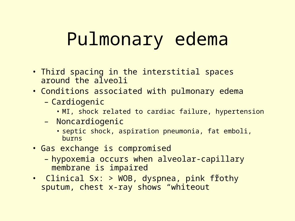
Pulmonary edema
• Third spacing in the interstitial spaces around the alveoli
• Conditions associated with pulmonary edema– Cardiogenic
• MI, shock related to cardiac failure, hypertension
– Noncardiogenic• septic shock, aspiration pneumonia, fat emboli, burns
• Gas exchange is compromised – hypoxemia occurs when alveolar-capillary
membrane is impaired• Clinical Sx: > WOB, dyspnea, pink frothy sputum,
chest x-ray shows “whiteout”

ARDS• A widespread breakdown of the alveolar and/or capillary
membranes• Patho: injury to lung results in hypoperfusion which
damages the alveolar epithelium– > mediators of inflammatory response causes injury– > Permeability of alveolar capillary membrane– > edema leads to a < production of surfactant – A major cause of severe respiratory failure in clients
with previously healthy lungs• Risk factors:
– Elderly and severe infections: > mortality rate– occurs after major pulmonary,cardiovascular, or
systemic insult

ARDS
• Clinical Sx: – Rapid, shallow breathing– Respiratory alkalosis– Dyspnea– Refractory hypoxemia– Diffuse alveolar infiltrates
• Tx: prevention of trauma to body– diuretics, digoxin, anti-inflammatory medications,
O2 & ventilator therapy

SARS Epidemic• Etiology
– Thought to be caused by a coronavirus– animal host: masked palm civet, raccoon dog, ferret
badger– First known case in Guangdong, China 11/02
• High percentage of victims were caterers in Southern China
– 22 countries, but China, Hong Kong, Canada reports the most cases
• WHO reported 8,200 cases as of May 30, 2003– 700 deaths
• http://www.nytimes.com/pages/multimedia/index.html• http://www.cdc.gov/ncidod/sars/qa/illness.htm - 2

Developmental differences
• CHILDREN: < surfactant until 28 weeks gestation
• lung tissue develops until ~ 8 years old
• airways are small• muscles underdeveloped• > exposure• < immune system• nasal breathers until 2
months
• ELDERLY: < muscle mass makes > WOB
• < immunity• > risk for pneumonia• > malnourisment• pathology may place
patient at risk for aspiration

References• Corwin, E.J.(2000).Handbook of pathophysiology. Baltimore:
Lippincott.• Hansen, M. (1998). Pathophysiology: Foundations of disease and
clinical intervention. Philadelphia: Saunders.• Hartshorn, J. C., Sole, M. L., & Lamborn, M. L. (1997).Introduction
to critical care nursing. Philadelphia: Saunders.• Horne, C., & Derrico, D. (1999). Mastering ABGs. The art of
arterial blood gas measurement. American Journal of Nursing, 8:26-32.
• http://www.pathoplus.com• Huether, S. E., & McCance, K. L. (2002). Pathophysiology. St.
Louis: Mosby.• Ryu, J.H., Swensen, J., Olson, EJ, & Pellikka, A. (2001).
Diagnosis of pulmonary embolism with use of computed tomographic angiography. Mayo Clin Proc.,76, 59-65.

Acid – Base Pure Water:
55 M 1 x 10⁻⁷ M 1 x 10⁻⁷ M
pH = 7 [neutral]
Highly Acidic 1 M pH = 0 1 x 10-14 M
Highly Basic 1 x 10-14 M pH = 14 1 M
HO
H HO
+
H
H
OH
+
pH = log = -log[H+]1[H+]
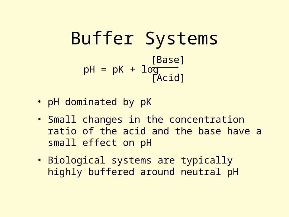
Buffer Systems
pH = pK + log[Base]
[Acid]
• pH dominated by pK
• Small changes in the concentration ratio of the acid and the base have a small effect on pH
• Biological systems are typically highly buffered around neutral pH

Carbonic Acid - Bicarbonate
CO2 + H2O H2CO3 H+ + HCO3- 2 H+ +
CO32- pK = 10.2pK = 6.1
pH = pK + log[Base]
[Acid]pH = 6.1 + log
[HCOHCO33--]
[HH22COCO33]
pH = 7.4 = 6.1 + log = 6.1 + 1.324 mEq/L
0.03 x PCO2

Partial Gas Pressures
Air (at sea level) Blood (Humans)
Total: 760 mmHg
pO2 (21%) 160 mmHg 80 mmHg
pCO2 (<1%) < 7 mmHg 40 mmHg
System greatly favors absorption of oxygen and elimination of carbon dioxide
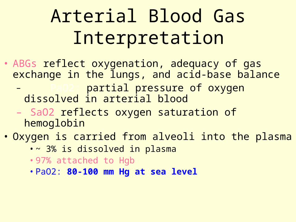
Arterial Blood Gas Interpretation
• ABGs reflect oxygenation, adequacy of gas exchange in the lungs, and acid-base balance– PaO2: partial pressure of oxygen dissolved in
arterial blood– SaO2 reflects oxygen saturation of hemoglobin
• Oxygen is carried from alveoli into the plasma• ~ 3% is dissolved in plasma • 97% attached to Hgb• PaO2: 80-100 mm Hg at sea level

Arterial Blood Gas Interpretation• PaO2 < 80 mm Hg = hypoxemia• PaO2 < 60 mm Hg may be seen in COPD• PaO2 < 40 mm Hg is life threatening• S & S of hypoxemia: pallor, dyspnea, use of accessory
muscles, anxiety, tachypnea• Hypoxia is decreased oxygen at the tissue level
– SaO2: amount of oxygen bound to Hgb• normal saturation is defined as 93 - 100%

Arterial Blood Gas Interpretation
• pH: negative log of H+ concentration
• In blood:
–Normal range: 7.35 - 7.45
–Acidosis = pH less than 7.35
–Alkalosis = pH greater than 7.45
–A pH < 7.0 or > 7.8 can cause death

Arterial Blood Gas Interpretation• PaCO2: partial pressure of carbon dioxide dissolved
in the arterial plasma– normal: 35 - 45 mm Hg– is regulated in the lungs– A primary respiratory problem is when PaCO2 is:– > 45 mm Hg = respiratory acidosis– < 35 mm Hg = respiratory alkalosis– HCO3 is normal (22 - 26 mEq/L)

Arterial Blood Gas Interpretation
• HCO3 (bicarbonate) normal is: 22 -26 mEq/L
• A primary metabolic or renal disorder is when the HCO3 is less than 22 (acidosis) or greater than 26 (alkalosis)– PaCo2 is normal

Arterial Blood Gas Interpretation
• Compensation:– body attempts to recover from primary problem
and return to homeostasis– Primary metabolic acidosis can cause the patient
to breathe faster to compensate (blow off CO2) by creating a respiratory alkalosis state
– This would be labeled as: Metabolic acidosis with a compensatory respiratory alkalosis
– pH 7.30; PaCO2 28; & HCO3 15• Are PaCo2 & HCO3 below normal? Yes, compensation

Metabolic Acidosis• Risk factors: >ingestion of acids or < production
of HCO3• Etiology: lactic acidosis, ketoacidosis, uremic
acidosis• Patho:compensatory hyperventilation
– hyperkalemia: shift of acid to ICF – <pH, <HCO3, PaCo2 normal; or low if
compensation is occurring– cardiac dysrhythmias & CNS dysfunction– headache, diarrhea, tremors

Metabolic Alkalosis• Risk factors: hypovolemia, excess aldosterone, iatrogenic base
administration• Etiology: acid loss or base gain
– renal excretion of HCO3 will fix the problem– prolonged vomiting (loss of HCL)
• Patho: respiratory compensation is limited – hypokalemia: Loop diuretics? NGT? Diarrhea?
cardiac dysrhythmias; seizures; confusion; muscle twitching, agitation
– > pH; >HCO3; normal PaCo2 or elevated if compensation occurs

Respiratory Acidosis• Risk factors: excess of acid in body fluids• Etiology: due to hypoventialtion; COPD; Cystic Fibrosis;
airway obstruction; spinal cord injury; CVA; depressant drugs; inadequate mechanical ventilation
• Patho: hypercapnia; CO2 diffuses easily across biological membranes
• Clinical: <pH; >PaCo2; HCO3 is normal or > in renal compensation– vasodilatation; cardiac arrhythmias, tachycardia,
somnolence, decreased ventilation

Respiratory Alkalosis
• Risk factors: relative excess of base in body fluids secondary to > ventilatory elimination of CO2; pneumonia; ARDS; shock; severe anemia
• Etiology: hypoxemia (<PaO2) causing rate & depth of ventilation to increase in an attempt to raise CO2
• Patho: buffer response is to shift acid from ICF to the blood by moving HCO3 into the cells in exchange of chloride– >pH; <PaC02; HCO3 normal or low due to compensation– nausea, vomiting, tingling of fingers