disruption of the phagosomal membrane and egress of ...iai.asm.org/content/72/7/4040.full.pdf ·...
TRANSCRIPT
INFECTION AND IMMUNITY, July 2004, p. 4040–4051 Vol. 72, No. 70019-9567/04/$08.00�0 DOI: 10.1128/IAI.72.7.4040–4051.2004Copyright © 2004, American Society for Microbiology. All Rights Reserved.
Disruption of the Phagosomal Membrane and Egress of Legionellapneumophila into the Cytoplasm during the Last Stages
of Intracellular Infection of Macrophages andAcanthamoeba polyphaga
Maelle Molmeret,1 Dina M. Bitar,2 Lihui Han,3 and Yousef Abu Kwaik1*Department of Microbiology and Immunology, University of Louisville College of Medicine, Louisville, Kentucky 402921;
Department of Microbiology and Department of Medical Microbiology and Immunology, Faculty of Medicine,Al-Quds University, Jerusalem 193562; and Department of Microbiology and Immunology, College
of Medicine, Shandong University, Jinan, Shandong, China 2500123
Received 29 October 2003/Returned for modification 15 December 2003/Accepted 14 March 2004
Although the early stages of intracellular infection by Legionella pneumophila are well established at theultrastructural level, a detailed ultrastructural analysis of late stages of intracellular replication has neverbeen done. Here we show that the membrane of the L. pneumophila-containing phagosome (LCP) is intact forup to 8 h postinfection of macrophages and Acanthamoeba polyphaga. At 12 h, 71 and 74% of the LCPs aredisrupted within macrophages and A. polyphaga, respectively, while the plasma membrane remains intact. At18 and 24 h postinfection, cytoplasmic elements such as mitochondria, lysosomes, vesicles, and amorphousmaterial are dispersed among the bacteria and these bacteria are considered cytoplasmic. At 18 h, 77% ofinfected macrophages and 32% of infected A. polyphaga amoebae harbor cytoplasmic bacteria. At 24 h, 99 and78% of infected macrophages and amoebae, respectively, contain cytoplasmic bacteria. On the basis of lyso-somal acid phosphatase staining of infected macrophages and A. polyphaga, the lysosomal enzyme is presentamong the bacteria when host vesicles are dispersed among bacteria. Our data indicate that bacterialreplication proceeds despite physical disruption of the phagosomal membrane. We also show that an lspGmutant that is defective in the type II secretion system and therefore does not secrete the hydrolytic enzymesmetalloprotease, p-nitrophenol phosphorylcholine hydrolase, lipase, phospholipase A, and lysophospholipaseA is as efficient as the wild-type strain in disruption of the LCP. Therefore, L. pneumophila disrupts thephagosomal membrane and becomes cytoplasmic at the last stages of infection in both macrophages and A.polyphaga. Lysosomal elements, mitochondria, cytoplasmic vesicles, and amorphous material are all dispersedamong the bacteria, after phagosomal disruption, within both human macrophages and A. polyphaga. Thedisruption of the LCP is independent of the hydrolytic enzymes exported by the type II secretion system.
The hallmark of Legionnaires’ disease is intracellular repli-cation of Legionella pneumophila in alveolar macrophages (10,11, 41). L. pneumophila also infects protozoa, which are con-sidered the environmental hosts for this intracellular pathogen(41, 42). The intracellular fates of L. pneumophila in both ofthese evolutionarily distant host cells are similar, where thebacteria evade fusion of the phagosome to lysosomes (2, 13,30–32). In addition, the L. pneumophila-containing phagosome(LCP) recruits the endoplasmic reticulum (ER), vesicles, andmitochondria within the first 15 min of infection (2, 13, 30, 45).Formation of the rough-ER-surrounded LCP is independentof autophagy (39, 45). After a lag phase of 4 h, bacterialreplication is initiated within both host cells (30, 41). Uponentry into the postexponential growth phase, L. pneumophilaexhibits several virulence traits, including a pore-forming ac-tivity that may allow the bacteria to lyse the host cell (6, 15, 21,36).
We have previously shown that upon termination of the
intracellular infection, the pore-forming activity of L. pneumo-phila facilitates lysis of the host cell and bacterial egress (6,34–36). It has been proposed that bacterial egress may occur intwo stages upon termination of intracellular bacterial replica-tion. First, the phagosomal membrane is disrupted allowing thebacteria to egress to the cytoplasm. The second stage is man-ifested by lysis of the plasma membrane and bacterial releaseinto the extracellular environment (34).
Although numerous studies have examined the early stagesof this intracellular infection at the ultrastructural level byelectron microscopy, there have been no studies to examinethe late stages of this intracellular infection at the ultrastruc-tural level. Therefore, we examined the integrity of the pha-gosomal membrane throughout intracellular infection of U937macrophages and Acanthamoeba polyphaga at the ultrastruc-tural level. In addition, we examined the presence of the lyso-somal enzyme acid phosphatase in the phagosome. Our datashow that the phagosomal membrane remains intact for up to8 h postinfection. While the plasma membrane remains intact,the LCP membrane is disrupted by 12 h. By 18 to 24 h, almostall of the infected cells harbored bacteria free in the cytoplasm,which coincided with the presence of host cell organelles dis-persed between the bacteria, as well as the lysosomal enzymeacid phosphatase. We show that the type II secretion system,
* Corresponding author. Mailing address: Department of Microbi-ology and Immunology, Room 316, University of Louisville College ofMedicine, 319 Abraham Flexner Way 55A, Louisville, KY 40202. Phone:(502) 852-4117. Fax: (502) 852-7531. E-mail [email protected].
4040
on June 19, 2018 by guesthttp://iai.asm
.org/D
ownloaded from

which is essential for secretion of many hydrolytic enzymes thatcan potentially disrupt membranes, is not involved in the dis-ruption of LCPs within macrophages.
MATERIALS AND METHODS
Bacterial and cell cultures. L. pneumophila serogroup I strain AA100 is aclinical isolate that has been described previously (4). L. pneumophila strainAA100, the isogenic lspG mutant NU259 (40), and the icmT null mutantAA100kmT (40) were grown on buffered charcoal-yeast extract plates or inbuffered yeast extract broth. U937 cells were cultured in RPMI 1640 mediumcontaining 10% fetal bovine serum (Gibco). U937 cells were differentiated withphorbol 12-myristate 13-acetate (Sigma) for 48 h before use, as previously de-scribed (25). A. polyphaga cells were maintained in peptone-yeast extract-glucosemedium.
Electron microscopy. For examination of infected amoebae or macrophages bytransmission electron microscopy, monolayers in six-well plates were infectedwith L. pneumophila strains at a multiplicity of infection of 10 for 1 h, followedby three washes. At 8, 12, 18, and 24 h postinfection, the infected monolayerswere washed with 0.1 M Sorenson’s phosphate buffer and incubated for 45 minin 0.1 M Sorenson’s phosphate buffer containing 3.5% glutaraldehyde, pH 7.4, at4°C. After four washes in Sorenson’s phosphate buffer for 5 min each time,infected cells were postfixed with 1% OsO4 in the same buffer for 45 min.Samples were dehydrated and processed as we described previously (23). Sec-tions were stained with uranyl acetate and lead citrate and examined with aHitachi H-7000/STEM electron microscope (Hitachi, Ltd., Tokyo, Japan) at 80kV as described previously (23).
Fusion between lysosomes of the LCP were determined by examination for thepresence of the lysosomal enzyme acid phosphatase as described previously (13,35). The monolayers were incubated with the acid phosphatase-specific substrate�-glycerophosphate (0.1 M acetate buffer, 2 mM �-glycerol phosphate [as thesubstrate], 1.2% lead nitrate [as the capture metal]) for 1 h at 37°C prior tofixation by osmium tetroxide as described above.
RESULTS
Egress of L. pneumophila into the cytoplasm during the latestages of intracellular infection of macrophages. We examinedthe integrity of the phagosomal membrane within U937 mac-rophages throughout the intracellular life cycle by transmissionelectronic microscopy. The phagosomal membrane was con-sidered intact when the membrane had no visible disruptionsof its integrity and no host cell elements, such as mitochondria,cytoplasmic vesicles, or amorphous material, were presentwithin the phagosome. The phagosomal membrane was con-sidered disrupted when apparent physical disruptions in theintegrity of the phagosomal membrane were present and amor-phous material was within the phagosome. The bacteria wereconsidered cytoplasmic when the phagosomal membrane wasnot continuous and host cell cytoplasmic elements such mito-chondria and cytoplasmic vesicles, in addition to amorphousmaterial, were dispersed among the bacteria. During the in-fection process, lysis of infected cell is initiated by 12 to 18 hand the released bacteria initiate secondary infections. To ex-clude secondary infection from our analysis at 12 h and there-after, we excluded cells with fewer than 5, 10, and 30 bacteriaat 12, 18, and 24 h, respectively. In order to rule out thepossibility of deterioration of the membranes of the host cellsby the fixation and staining procedures used, we observed theplasma membrane at 12 and 18 h postinfection of cells con-taining disrupted LCPs.
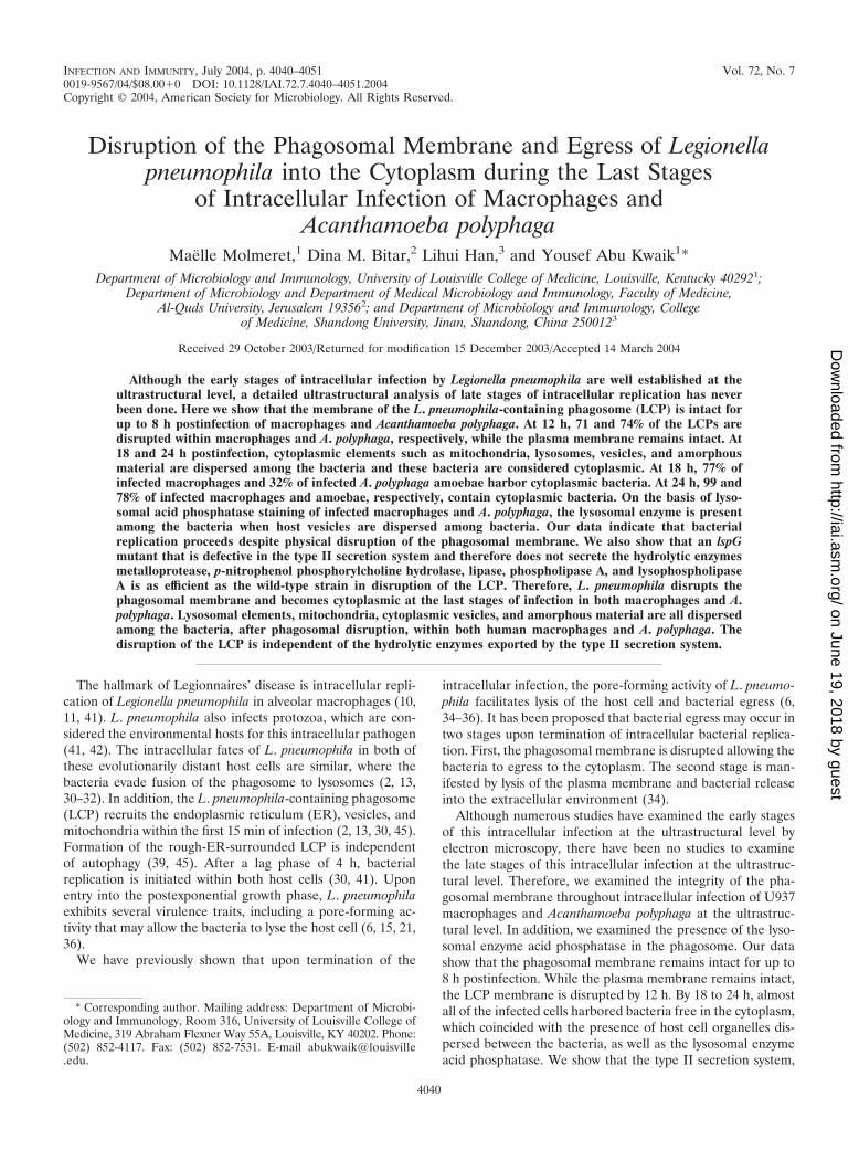
At 8 h postinfection, all of the phagosomal membranes werethick and did not show any sign of disruption of integrity (Fig.1 and data not shown) and the phagosomal membranes weretightly closed around the bacteria, similar to what has been
shown by numerous studies at 2 to 6 h. The lumen of thephagosomes did not contain any host cell cytoplasmic ele-ments. The phagosomal membranes were therefore consideredintact at 8 h postinfection. ER was also observed around mostphagosomes at 8 h (Fig. 1b and c).
A small portion of the cells had already been lysed by 12 hpostinfection. Only �2% of the phagosomes had intact mem-branes with no signs of disruption of integrity in this timeperiod (Fig. 2A; see also Fig. 8A). The lumen of the phago-somes was distended compared to that of 8-h-old phagosomes.Seventy percent of the LCP membranes were disrupted at 12 h(Fig. 2A, part b; see also Fig. 8A). These phagosomes were stillfree of host cell cytoplasmic elements such as mitochondriaand cytoplasmic vesicles. The rough ER was not visible anymore around some phagosomes, but few mitochondria werewithin the vicinity of the phagosomes (data not shown). Twen-ty-seven percent of the infected cells harbored cytoplasmicbacteria at 12 h (Fig. 8A). Importantly, the plasma membranewas completely intact in 95% of infected cells harboring dis-rupted LCPs or cytoplasmic bacteria.
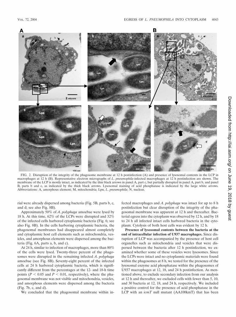
By 18 h, �50% of the cells in the monolayers were lysed.Among the remaining infected cells at 18 h, 22% of the phago-somes were disrupted (P � 0.01, Student t test) and 77% of theinfected cells harbored cytoplasmic bacteria (P � 0.01, Studentt test), which percentages are significantly different from thoseat the 12-h time point (Fig. 8A). Phagosomal membranes be-came very thin and were discontinuous in many parts (Fig. 3A,parts b and c). Vesicles and cytoplasmic amorphous elementsjuxtaposed the bacteria (Fig. 3A, part b). The chromatin in thenucleus was condensed and was located perinuclearly (Fig. 3Aand B, parts a), which is a characteristic of apoptosis (22, 29)that has been shown to be triggered during late stages ofinfection (37). The plasma membrane was intact in 80% of theinfected cells harboring disrupted LCPs or cytoplasmic bacte-ria.
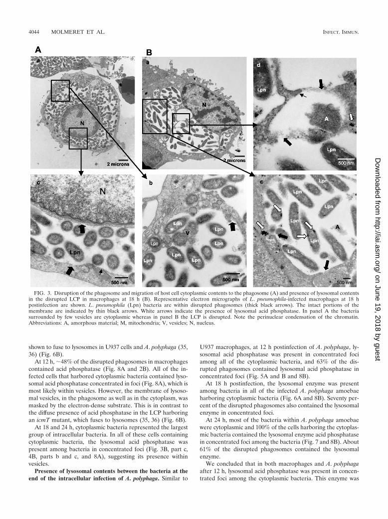
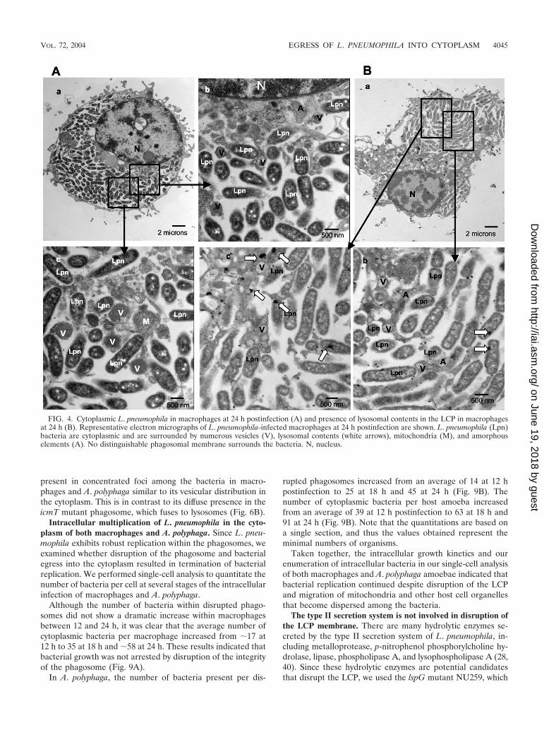
By 24 h postinfection, the majority of the cells had beenlysed. In 99% of the remaining infected cells, the bacteria werecytoplasmic, which is significantly different from the percent-ages at 12 and 18 h (P � 0.01, Student t test) (see Fig. 8A).Most of the phagosomal membranes disappeared (Fig. 4A,parts b and c). Cytoplasmic elements such as mitochondria,vesicles, and amorphous material were dispersed among thebacteria (Fig. 4A, parts c and d). The bacteria occupied most ofthe space within the host cell that did not include the nucleus(Fig. 4A, part a). Perinuclear condensed chromatin was de-tected in all cells, a clear sign of apoptosis (Fig. 4A, parts a andb).
Egress of L. pneumophila into the cytoplasm during the latestages of intracellular infection of A. polyphaga. Similar tohuman macrophages, at 8 h postinfection of A. polyphaga, mostof the LCPs were tight and intact with a thick phagosomalmembrane around the bacteria (data not shown). A smallportion of cells had already been lysed by 12 h postinfection.At this time, 74% of the LCPs were disrupted (Fig. 5A and B;see also Fig. 8B). The lumen of the LCPs was still tight (Fig. 5).Signs of nuclear apoptosis were not visible in A. polyphaga (Fig.5). Thirteen percent of the infected A. polyphaga cells con-tained cytoplasmic bacteria at 12 h postinfection, where cyto-plasmic elements such as mitochondria and amorphous mate-
VOL. 72, 2004 EGRESS OF L. PNEUMOPHILA INTO CYTOPLASM 4041
on June 19, 2018 by guesthttp://iai.asm
.org/D
ownloaded from
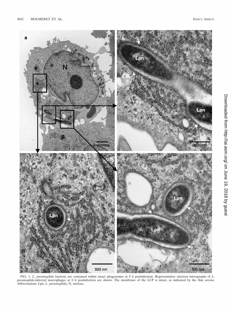
FIG. 1. L. pneumophila bacteria are contained within intact phagosomes at 8 h postinfection. Representative electron micrographs of L.pneumophila-infected macrophages at 8 h postinfection are shown. The membrane of the LCP is intact, as indicated by the thin arrows.Abbreviations: Lpn, L. pneumophila; N, nucleus.
4042 MOLMERET ET AL. INFECT. IMMUN.
on June 19, 2018 by guesthttp://iai.asm
.org/D
ownloaded from
rial were already dispersed among bacteria (Fig. 5B, parts b, c,and d; see also Fig. 8B).
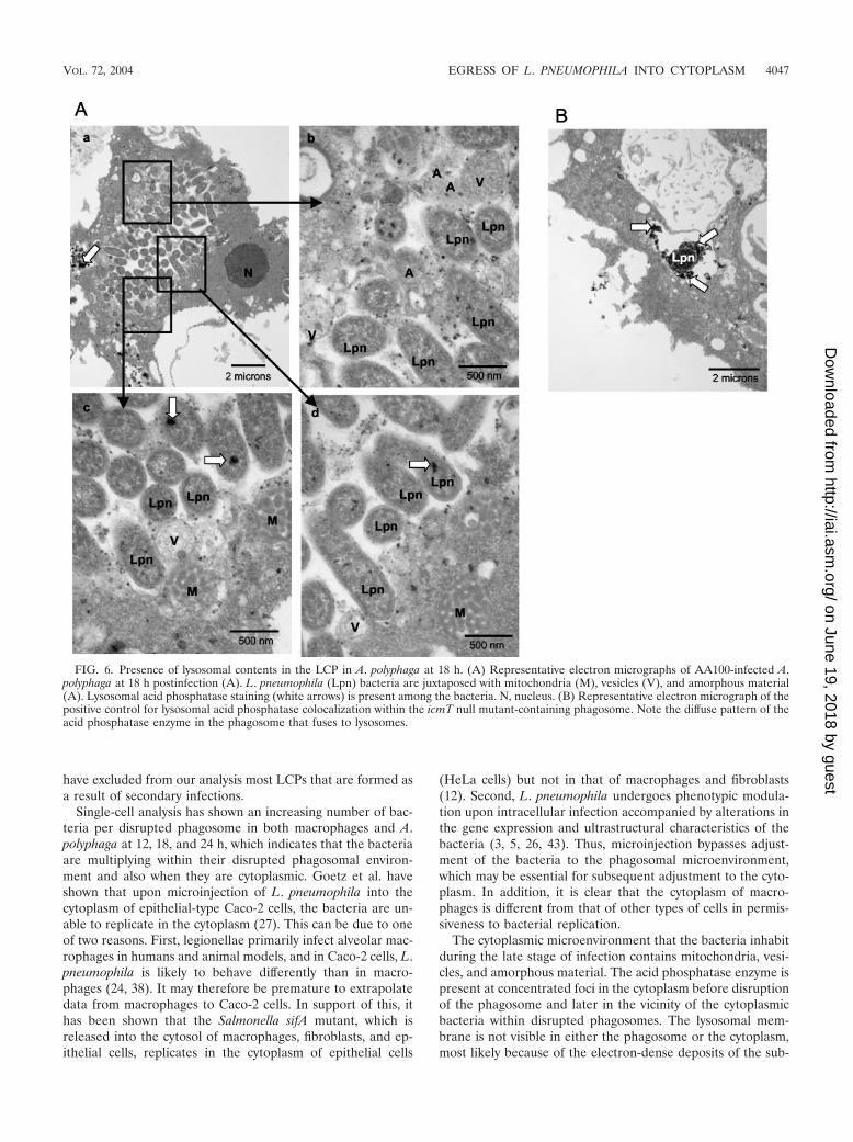
Approximately 50% of A. polyphaga amoebae were lysed by18 h. At this time, 62% of the LCPs were disrupted and 32%of the infected cells harbored cytoplasmic bacteria (Fig. 6; seealso Fig. 8B). In the cells harboring cytoplasmic bacteria, thephagosomal membranes had disappeared almost completelyand cytoplasmic host cell elements such as mitochondria, ves-icles, and amorphous elements were dispersed among the bac-teria (Fig. 6A, parts a, b, and c).
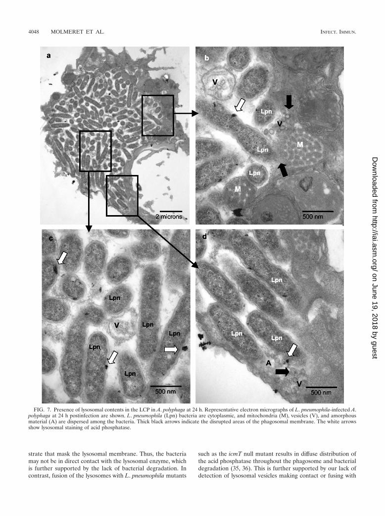
At 24 h, similar to infection of macrophages, more than 80%of the cells were lysed. Twenty-three percent of the phago-somes were disrupted in the remaining infected A. polyphagaamoebae (see Fig. 8B). Seventy-eight percent of the infectedcells at 24 h harbored cytoplasmic bacteria, which is signifi-cantly different from the percentages at the 12- and 18-h timepoints (P � 0.05 and P � 0.01, respectively), where the pha-gosomal membrane was not visible and mitochondria, vesicles,and amorphous elements were dispersed among the bacteria(Fig. 7b, c, and d).
We concluded that the phagosomal membrane within in-
fected macrophages and A. polyphaga was intact for up to 8 hpostinfection but clear disruption of the integrity of the pha-gosomal membrane was apparent at 12 h and thereafter. Bac-terial egress into the cytoplasm was observed by 12 h, and by 18to 24 h all infected intact cells harbored bacteria in the cyto-plasm. Cytolysis of both host cells was evident by 12 h.
Presence of lysosomal contents between the bacteria at theend of intracellular infection of U937 macrophages. Since dis-ruption of LCP was accompanied by the presence of host cellorganelles such as mitochondria and vesicles that were dis-persed between the bacteria after 12 h postinfection, we ex-amined whether some of these vesicles were lysosomes. Sincethe LCPs were intact and no cytoplasmic materials were foundwithin the phagosomes at 8 h, we tested for the presence of thelysosomal enzyme acid phosphatase within the phagosomes ofU937 macrophages at 12, 18, and 24 h postinfection. As men-tioned above, to exclude secondary infection from our analysisat 12 h and thereafter, we excluded cells with fewer than 5, 10,and 30 bacteria at 12, 18, and 24 h, respectively. We includeda positive control for the presence of acid phosphatase in theLCP with an icmT null mutant (AA100kmT) that has been
FIG. 2. Disruption of the integrity of the phagosome membrane at 12 h postinfection (A) and presence of lysosomal contents in the LCP inmacrophages at 12 h (B). Representative electron micrographs of L. pneumophila-infected macrophages at 12 h postinfection are shown. Themembrane of the LCP is mostly intact, as indicated by the thin black arrows in panel A, part c, but partially disrupted in panel A, part b, and panelB, parts b and c, as indicated by the thick black arrows. Lysosomal staining of acid phosphatase is indicated by the large white arrows.Abbreviations: A, amorphous element; M, mitochondria; Lpn, L. pneumophila; N, nucleus.
VOL. 72, 2004 EGRESS OF L. PNEUMOPHILA INTO CYTOPLASM 4043
on June 19, 2018 by guesthttp://iai.asm
.org/D
ownloaded from
shown to fuse to lysosomes in U937 cells and A. polyphaga (35,36) (Fig. 6B).
At 12 h, �48% of the disrupted phagosomes in macrophagescontained acid phosphatase (Fig. 8A and 2B). All of the in-fected cells that harbored cytoplasmic bacteria contained lyso-somal acid phosphatase concentrated in foci (Fig. 8A), which ismost likely within vesicles. However, the membrane of lysoso-mal vesicles, in the phagosome as well as in the cytoplasm, wasmasked by the electron-dense substrate. This is in contrast tothe diffuse presence of acid phosphatase in the LCP harboringan icmT mutant, which fuses to lysosomes (35, 36) (Fig. 6B).
At 18 and 24 h, cytoplasmic bacteria represented the largestgroup of intracellular bacteria. In all of these cells containingcytoplasmic bacteria, the lysosomal acid phosphatase waspresent among bacteria in concentrated foci (Fig. 3B, part c,4B, parts b and c, and 8A), suggesting its presence withinvesicles.
Presence of lysosomal contents between the bacteria at theend of the intracellular infection of A. polyphaga. Similar to
U937 macrophages, at 12 h postinfection of A. polyphaga, ly-sosomal acid phosphatase was present in concentrated fociamong all of the cytoplasmic bacteria, and 63% of the dis-rupted phagosomes contained lysosomal acid phosphatase inconcentrated foci (Fig. 5A and B and 8B).
At 18 h postinfection, the lysosomal enzyme was presentamong bacteria in all of the infected A. polyphaga amoebaeharboring cytoplasmic bacteria (Fig. 6A and 8B). Seventy per-cent of the disrupted phagosomes also contained the lysosomalenzyme in concentrated foci.
At 24 h, most of the bacteria within A. polyphaga amoebaewere cytoplasmic and 100% of the cells harboring the cytoplas-mic bacteria contained the lysosomal enzyme acid phosphatasein concentrated foci among the bacteria (Fig. 7 and 8B). About61% of the disrupted phagosomes contained the lysosomalenzyme.
We concluded that in both macrophages and A. polyphagaafter 12 h, lysosomal acid phosphatase was present in concen-trated foci among the cytoplasmic bacteria. This enzyme was
FIG. 3. Disruption of the phagosome and migration of host cell cytoplasmic contents to the phagosome (A) and presence of lysosomal contentsin the disrupted LCP in macrophages at 18 h (B). Representative electron micrographs of L. pneumophila-infected macrophages at 18 hpostinfection are shown. L. pneumophila (Lpn) bacteria are within disrupted phagosomes (thick black arrows). The intact portions of themembrane are indicated by thin black arrows. White arrows indicate the presence of lysosomal acid phosphatase. In panel A the bacteriasurrounded by few vesicles are cytoplasmic whereas in panel B the LCP is disrupted. Note the perinuclear condensation of the chromatin.Abbreviations: A, amorphous material; M, mitochondria; V, vesicles; N, nucleus.
4044 MOLMERET ET AL. INFECT. IMMUN.
on June 19, 2018 by guesthttp://iai.asm
.org/D
ownloaded from
present in concentrated foci among the bacteria in macro-phages and A. polyphaga similar to its vesicular distribution inthe cytoplasm. This is in contrast to its diffuse presence in theicmT mutant phagosome, which fuses to lysosomes (Fig. 6B).
Intracellular multiplication of L. pneumophila in the cyto-plasm of both macrophages and A. polyphaga. Since L. pneu-mophila exhibits robust replication within the phagosomes, weexamined whether disruption of the phagosome and bacterialegress into the cytoplasm resulted in termination of bacterialreplication. We performed single-cell analysis to quantitate thenumber of bacteria per cell at several stages of the intracellularinfection of macrophages and A. polyphaga.
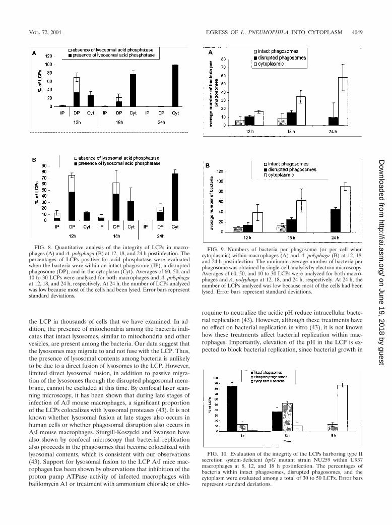
Although the number of bacteria within disrupted phago-somes did not show a dramatic increase within macrophagesbetween 12 and 24 h, it was clear that the average number ofcytoplasmic bacteria per macrophage increased from �17 at12 h to 35 at 18 h and �58 at 24 h. These results indicated thatbacterial growth was not arrested by disruption of the integrityof the phagosome (Fig. 9A).
In A. polyphaga, the number of bacteria present per dis-
rupted phagosomes increased from an average of 14 at 12 hpostinfection to 25 at 18 h and 45 at 24 h (Fig. 9B). Thenumber of cytoplasmic bacteria per host amoeba increasedfrom an average of 39 at 12 h postinfection to 63 at 18 h and91 at 24 h (Fig. 9B). Note that the quantitations are based ona single section, and thus the values obtained represent theminimal numbers of organisms.
Taken together, the intracellular growth kinetics and ourenumeration of intracellular bacteria in our single-cell analysisof both macrophages and A. polyphaga amoebae indicated thatbacterial replication continued despite disruption of the LCPand migration of mitochondria and other host cell organellesthat become dispersed among the bacteria.
The type II secretion system is not involved in disruption ofthe LCP membrane. There are many hydrolytic enzymes se-creted by the type II secretion system of L. pneumophila, in-cluding metalloprotease, p-nitrophenol phosphorylcholine hy-drolase, lipase, phospholipase A, and lysophospholipase A (28,40). Since these hydrolytic enzymes are potential candidatesthat disrupt the LCP, we used the lspG mutant NU259, which
FIG. 4. Cytoplasmic L. pneumophila in macrophages at 24 h postinfection (A) and presence of lysosomal contents in the LCP in macrophagesat 24 h (B). Representative electron micrographs of L. pneumophila-infected macrophages at 24 h postinfection are shown. L. pneumophila (Lpn)bacteria are cytoplasmic and are surrounded by numerous vesicles (V), lysosomal contents (white arrows), mitochondria (M), and amorphouselements (A). No distinguishable phagosomal membrane surrounds the bacteria. N, nucleus.
VOL. 72, 2004 EGRESS OF L. PNEUMOPHILA INTO CYTOPLASM 4045
on June 19, 2018 by guesthttp://iai.asm
.org/D
ownloaded from

is defective in the type II secretion system owing to a defect inan inner membrane structural protein component of the typeII secretion system (40). In order to determine if the secretionof theses exoproteins is responsible for the disruption of thewild-type LCP, U937 macrophage cell lines were infected withthe lspG mutant NU259 and examined at 8, 12, 18, and 24 hpostinfection by electron microscopy. About 85% of the LCPswere intact at 8 h postinfection (Fig. 10). At 12 h postinfection53% of the LCPs were disrupted, and at 18 h postinfection87% of the bacteria were cytoplasmic within U937 macro-phages (Fig. 10). At 24 h, most of cells were in a disintegratedstate and it was not possible to analyze the data. Thus, the typeII secretion system and the hydrolytic enzymes secretedthrough it are not involved in the disruption of the LCPs.
DISCUSSION
L. pneumophila invades both mammalian and protozoanhost cells and resides within a replicative vacuole, evadingphagolysosomal fusion. Late stages of the intracellular lifecycle and the mechanism of bacterial egress after cessation ofintracellular bacterial replication are not well understood. Thetwo-step egress model that we have previously proposed (34)predicts that L. pneumophila first egresses from the phagosome
into the cytoplasm, followed by lysis of the host cell plasmamembrane. Here we show for the first time in both human-derived macrophages and A. polyphaga amoebae that L. pneu-mophila is present in an intact phagosomal membrane for up to8 h postinfection. However, by 12 h and thereafter, physicaland structural disruptions of integrity of the phagosomal mem-brane are apparent and cytoplasmic organelles including mi-tochondria, vesicles, and lysosomes are dispersed among thebacteria.
While ultrastructural studies of early stages of the LCP arerather numerous, LCP integrity during later stages of intracel-lular infection has never been examined. Tilney et al. havedescribed an intact phagosomal membrane at 20 h postinfec-tion of U937 macrophages, but the phagosome described con-tains only two bacteria (45). It is well documented that thebacterial lag phase within U937 macrophages is �4 h and thegeneration time is �90 to 120 min (1, 41, 42). Therefore,phagosomes containing two or fewer bacteria at 20 h are mostlikely secondary infections or bacteria that fail to replicate andare not representative of a 20-h-old LCP. In our study, we haveconsidered that a 12-h LCP may harbor at least 5 bacteria, an18-h LCP may harbor at least 10 bacteria, and a 24-h LCP mayharbor at least 30 bacteria. In that manner, we believe that we
FIG. 5. Presence of lysosomal contents in the LCP in A. polyphaga at 12 h. Shown are representative electron micrographs of L. pneumophila-infected A. polyphaga at 12 h postinfection where the bacteria are in a disrupted phagosome (A) or cytoplasmic (B). Thick black arrows show siteswithout any visible phagosomal membrane. Thin black arrows show sites where the phagosomal membrane is still visible. Lysosomal acidphosphatase is indicated by the large white arrows. In panel A, the phagosomal membrane is disrupted (large black arrows) and amorphousmaterial (A) is present within the phagosome. In panel B, although parts of the phagosomal membrane are still visible (thin black arrows), L.pneumophila (Lpn) bacteria are mostly cytoplasmic, as shown by the presence of amorphous material (A) and the presence of mitochondria(M) dispersed among the bacteria.
4046 MOLMERET ET AL. INFECT. IMMUN.
on June 19, 2018 by guesthttp://iai.asm
.org/D
ownloaded from
have excluded from our analysis most LCPs that are formed asa result of secondary infections.
Single-cell analysis has shown an increasing number of bac-teria per disrupted phagosome in both macrophages and A.polyphaga at 12, 18, and 24 h, which indicates that the bacteriaare multiplying within their disrupted phagosomal environ-ment and also when they are cytoplasmic. Goetz et al. haveshown that upon microinjection of L. pneumophila into thecytoplasm of epithelial-type Caco-2 cells, the bacteria are un-able to replicate in the cytoplasm (27). This can be due to oneof two reasons. First, legionellae primarily infect alveolar mac-rophages in humans and animal models, and in Caco-2 cells, L.pneumophila is likely to behave differently than in macro-phages (24, 38). It may therefore be premature to extrapolatedata from macrophages to Caco-2 cells. In support of this, ithas been shown that the Salmonella sifA mutant, which isreleased into the cytosol of macrophages, fibroblasts, and ep-ithelial cells, replicates in the cytoplasm of epithelial cells
(HeLa cells) but not in that of macrophages and fibroblasts(12). Second, L. pneumophila undergoes phenotypic modula-tion upon intracellular infection accompanied by alterations inthe gene expression and ultrastructural characteristics of thebacteria (3, 5, 26, 43). Thus, microinjection bypasses adjust-ment of the bacteria to the phagosomal microenvironment,which may be essential for subsequent adjustment to the cyto-plasm. In addition, it is clear that the cytoplasm of macro-phages is different from that of other types of cells in permis-siveness to bacterial replication.
The cytoplasmic microenvironment that the bacteria inhabitduring the late stage of infection contains mitochondria, vesi-cles, and amorphous material. The acid phosphatase enzyme ispresent at concentrated foci in the cytoplasm before disruptionof the phagosome and later in the vicinity of the cytoplasmicbacteria within disrupted phagosomes. The lysosomal mem-brane is not visible in either the phagosome or the cytoplasm,most likely because of the electron-dense deposits of the sub-
FIG. 6. Presence of lysosomal contents in the LCP in A. polyphaga at 18 h. (A) Representative electron micrographs of AA100-infected A.polyphaga at 18 h postinfection (A). L. pneumophila (Lpn) bacteria are juxtaposed with mitochondria (M), vesicles (V), and amorphous material(A). Lysosomal acid phosphatase staining (white arrows) is present among the bacteria. N, nucleus. (B) Representative electron micrograph of thepositive control for lysosomal acid phosphatase colocalization within the icmT null mutant-containing phagosome. Note the diffuse pattern of theacid phosphatase enzyme in the phagosome that fuses to lysosomes.
VOL. 72, 2004 EGRESS OF L. PNEUMOPHILA INTO CYTOPLASM 4047
on June 19, 2018 by guesthttp://iai.asm
.org/D
ownloaded from
strate that mask the lysosomal membrane. Thus, the bacteriamay not be in direct contact with the lysosomal enzyme, whichis further supported by the lack of bacterial degradation. Incontrast, fusion of the lysosomes with L. pneumophila mutants
such as the icmT null mutant results in diffuse distribution ofthe acid phosphatase throughout the phagosome and bacterialdegradation (35, 36). This is further supported by our lack ofdetection of lysosomal vesicles making contact or fusing with
FIG. 7. Presence of lysosomal contents in the LCP in A. polyphaga at 24 h. Representative electron micrographs of L. pneumophila-infected A.polyphaga at 24 h postinfection are shown. L. pneumophila (Lpn) bacteria are cytoplasmic, and mitochondria (M), vesicles (V), and amorphousmaterial (A) are dispersed among the bacteria. Thick black arrows indicate the disrupted areas of the phagosomal membrane. The white arrowsshow lysosomal staining of acid phosphatase.
4048 MOLMERET ET AL. INFECT. IMMUN.
on June 19, 2018 by guesthttp://iai.asm
.org/D
ownloaded from
the LCP in thousands of cells that we have examined. In ad-dition, the presence of mitochondria among the bacteria indi-cates that intact lysosomes, similar to mitochondria and othervesicles, are present among the bacteria. Our data suggest thatthe lysosomes may migrate to and not fuse with the LCP. Thus,the presence of lysosomal contents among bacteria is unlikelyto be due to a direct fusion of lysosomes to the LCP. However,limited direct lysosomal fusion, in addition to passive migra-tion of the lysosomes through the disrupted phagosomal mem-brane, cannot be excluded at this time. By confocal laser scan-ning microscopy, it has been shown that during late stages ofinfection of A/J mouse macrophages, a significant proportionof the LCPs colocalizes with lysosomal proteases (43). It is notknown whether lysosomal fusion at late stages also occurs inhuman cells or whether phagosomal disruption also occurs inA/J mouse macrophages. Sturgill-Koszycki and Swanson havealso shown by confocal microscopy that bacterial replicationalso proceeds in the phagosomes that become colocalized withlysosomal contents, which is consistent with our observations(43). Support for lysosomal fusion to the LCP A/J mice mac-rophages has been shown by observations that inhibition of theproton pump ATPase activity of infected macrophages withbafilomycin A1 or treatment with ammonium chloride or chlo-
roquine to neutralize the acidic pH reduce intracellular bacte-rial replication (43). However, although these treatments haveno effect on bacterial replication in vitro (43), it is not knownhow these treatments affect bacterial replication within mac-rophages. Importantly, elevation of the pH in the LCP is ex-pected to block bacterial replication, since bacterial growth in
FIG. 8. Quantitative analysis of the integrity of LCPs in macro-phages (A) and A. polyphaga (B) at 12, 18, and 24 h postinfection. Thepercentages of LCPs positive for acid phosphatase were evaluatedwhen the bacteria were within an intact phagosome (IP), a disruptedphagosome (DP), and in the cytoplasm (Cyt). Averages of 60, 50, and10 to 30 LCPs were analyzed for both macrophages and A. polyphagaat 12, 18, and 24 h, respectively. At 24 h, the number of LCPs analyzedwas low because most of the cells had been lysed. Error bars representstandard deviations.
FIG. 9. Numbers of bacteria per phagosome (or per cell whencytoplasmic) within macrophages (A) and A. polyphaga (B) at 12, 18,and 24 h postinfection. The minimum average number of bacteria perphagosome was obtained by single-cell analysis by electron microscopy.Averages of 60, 50, and 10 to 30 LCPs were analyzed for both macro-phages and A. polyphaga at 12, 18, and 24 h, respectively. At 24 h, thenumber of LCPs analyzed was low because most of the cells had beenlysed. Error bars represent standard deviations.
FIG. 10. Evaluation of the integrity of the LCPs harboring type IIsecretion system-deficient lspG mutant strain NU259 within U937macrophages at 8, 12, and 18 h postinfection. The percentages ofbacteria within intact phagosomes, disrupted phagosomes, and thecytoplasm were evaluated among a total of 30 to 50 LCPs. Error barsrepresent standard deviations.
VOL. 72, 2004 EGRESS OF L. PNEUMOPHILA INTO CYTOPLASM 4049
on June 19, 2018 by guesthttp://iai.asm
.org/D
ownloaded from
vitro occurs within a very limited pH range around pH 6.9.Taken together, our data suggest that colocalization of theLCP with lysosomal contents during late stages of infectionmay be due to migration of lysosome vesicles to the disruptedLCPs. Our data do not exclude direct fusion and release oflysosomal contents to the LCP lumen.
L. pneumophila possesses a type II secretion system thatallows the secretion of many hydrolytic enzymes that have thepotential to degrade membranes, including a metalloprotease,p-nitrophenylphosphorylcholine hydrolase, lipase, phospho-lipase A, and lysophospholipase A. In this study, we have usedan lspG mutant that is defective in an inner membrane struc-tural protein component of the type II secretion system (40) inorder to determine whether the type II secretion system andthe enzymes secreted through it are involved in disruption ofthe LCP membrane. Our data show that the type II secretionsystem and its secreted enzymes are not required for disruptionof the LCPs. Thus, these hydrolytic enzymes do not play anydetectable role in disruption of the LCPs.
Necrosis is usually the cause of a severe cellular insult, suchas loss of membrane integrity caused by a toxin (33). L. pneu-mophila has been shown to have a cytopathogenic effect onhost cells leading to necrosis, which is thought to be mediatedby a pore-forming activity (6, 21, 35, 36). It is likely that thepore-forming activity during late stages of infection of macro-phages contributes to cytolysis of the host cell. However, manyother factors may be involved. First, apoptosis in mammaliancells is likely to be a contributing factor, since it is triggeredduring late stages of infection when the LCPs harbor morethan 20 bacteria (37). Second, whether other phospholipasessecreted by L. pneumophila (7–9, 16–20, 44) also contribute todismantling of the cell is not known. Third, it is possible thatdisruption of the phagosomal membrane may be partially me-diated by physical pressure from the increasing number ofbacteria in the phagosome. Fourth, it is also possible that,similar to S. enterica serovar Typhimurium (14), L. pneumo-phila is actively involved in maintaining the integrity of thephagosome during early stages of infection but loses that abil-ity later. Therefore, the mechanism of egress of L. pneumo-phila into the cytoplasm is likely to be complex and multifac-torial.
In summary, we have shown that L. pneumophila is within anintact phagosome for up to 8 h in both macrophages and A.polyphaga. The phagosomal membrane is disrupted at 12 h,and the bacteria egress into the cytoplasm between 12 and24 h, prior to disruption and lysis of the host cell plasmamembrane. Cytoplasmic elements such as mitochondria andvesicles are dispersed among the bacteria at the end of intra-cellular replication, and this phagosomal disruption does notseem to affect bacterial replication. Disruption of the LCP isnot mediated by hydrolytic enzymes secreted by the type IIsecretion system.
REFERENCES
1. Abu Kwaik, Y. 1998. Induced expression of the Legionella pneumophila geneencoding a 20-kilodalton protein during intracellular infection. Infect. Im-mun. 66:203–212.
2. Abu Kwaik, Y. 1996. The phagosome containing Legionella pneumophilawithin the protozoan Hartmanella vermiformis is surrounded by the roughendoplasmic reticulum. Appl. Environ. Microbiol. 62:2022–2028.
3. Abu Kwaik, Y., B. I. Eisenstein, and N. C. Engleberg. 1993. Phenotypic
modulation by Legionella pneumophila upon infection of macrophages. In-fect. Immun. 61:1320–1329.
4. Abu Kwaik, Y., and N. C. Engleberg. 1994. Cloning and molecular charac-terization of a Legionella pneumophila gene induced by intracellular infectionand by various in vitro stress stimuli. Mol. Microbiol. 13:243–251.
5. Abu Kwaik, Y., L.-Y. Gao, O. S. Harb, and B. J. Stone. 1997. Transcriptionalregulation of the macrophage-induced gene (gspA) of Legionella pneumo-phila and phenotypic characterization of a null mutant. Mol. Microbiol.24:629–642.
6. Alli, O. A. T., L.-Y. Gao, L. L. Pedersen, Zink. S., M. Radulic, M. Doric, andY. Abu Kwaik. 2000. Temporal pore formation-mediated egress from mac-rophages and alveolar epithelial cells by Legionella pneumophila. Infect.Immun. 68:6431–6440.
7. Aragon, V., S. Kurtz, and N. P. Cianciotto. 2001. Legionella pneumophilamajor acid phosphatase and its role in intracellular infection. Infect. Immun.69:177–185.
8. Aragon, V., O. Rossier, and N. P. Cianciotto. 2002. Legionella pneumophilagenes that encode lipase and phospholipase C activities. Microbiology 148:2223–2231.
9. Baine, W. B. 1985. Cytolytic and phospholipase C activity in Legionellaspecies. J. Gen. Microbiol. 131:1383–1391.
10. Baskerville, A., A. B. Dowsett, R. B. Fitzgeorge, P. Hambleton, and M.Broster. 1983. Ultrastructure of pulmonary alveoli and macrophages in ex-perimental Legionnaires’ disease. J. Pathol. 140:77–90.
11. Baskerville, A., R. B. Fitzgeorge, M. Broster, P. Hambleton, and P. J. Den-nis. 1981. Experimental transmission of Legionnaires’ disease by exposure toaerosols of Legionella pneumophila. Lancet ii:1389–1390.
12. Beuzon, C. R., S. P. Salcedo, and D. W. Holden. 2002. Growth and killing ofa Salmonella enterica serovar Typhimurium sifA mutant strain in the cytosolof different host cell lines. Microbiology 148:2705–2715.
13. Bozue, J. A., and W. Johnson. 1996. Interaction of Legionella pneumophilawith Acanthamoeba catellanii: uptake by coiling phagocytosis and inhibitionof phagosome-lysosome fusion. Infect. Immun. 64:668–673.
14. Brumell, J. H., P. Tang, M. L. Zaharik, and B. B. Finlay. 2002. Disruptionof the Salmonella-containing vacuole leads to increased replication of Sal-monella enterica serovar Typhimurium in the cytosol of epithelial cells. In-fect. Immun. 70:3264–3270.
15. Byrne, B., and M. S. Swanson. 1998. Expression of Legionella pneumophilavirulence traits in response to growth conditions. Infect. Immun. 66:3029–3034.
16. Flieger, A., S. Gong, M. Faigle, M. Deeg, P. Bartmann, and B. Neumeister.2000. Novel phospholipase A activity secreted by Legionella species. J. Bac-teriol. 182:1321–1327.
17. Flieger, A., S. Gong, M. Faigle, H. Northoff, and B. Neumeister. 2001. In vitrosecretion kinetics of proteins from Legionella pneumophila in comparison toproteins from non-pneumophila species. Microbiology 147:3127–3134.
18. Flieger, A., S. Gong, M. Faigle, S. Stevanovic, N. P. Cianciotto, and B.Neumeister. 2001. Novel lysophospholipase A secreted by Legionella pneu-mophila. J. Bacteriol. 183:2121–2124.
19. Flieger, A., S. Gongab, M. Faigle, H. A. Mayer, U. Kehrer, J. Mussotter, P.Bartmann, and B. Neumeister. 2000. Phospholipase A secreted by Legionellapneumophila destroys alveolar surfactant phospholipids. FEMS Microbiol.Lett. 188:129–133.
20. Flieger, A., B. Neumeister, and N. P. Cianciotto. 2002. Characterization ofthe gene encoding the major secreted lysophospholipase A of Legionellapneumophila and its role in detoxification of lysophosphatidylcholine. Infect.Immun. 70:6094–6106.
21. Gao, L.-Y., and Y. Abu Kwaik. 2000. The mechanism of killing and exitingthe protozoan host Acanthamoeba polyphaga by Legionella pneumophila.Environ. Microbiol. 2:79–90.
22. Gao, L.-Y., and Y. Abu Kwaik. 2000. The modulation of host cell apoptosisby intracellular bacterial pathogens. Trends. Microbiol. 8:306–313.
23. Gao, L.-Y., O. S. Harb, and Y. Abu Kwaik. 1998. Identification of macro-phage-specific infectivity loci (mil) of Legionella pneumophila that are notrequired for infectivity of protozoa. Infect. Immun. 66:883–892.
24. Gao, L.-Y., B. J. Stone, J. K. Brieland, and Y. Abu Kwaik. 1998. Differentfates of Legionella pneumophila pmi and mil mutants within human-derivedmacrophages and alveolar epithelial cells. Microb. Pathog. 25:291–306.
25. Gao, L.-Y., M. Susa, B. Ticac, and Y. Abu Kwaik. 1999. Heterogeneity inintracellular replication and cytopathogenicity of Legionella pneumophilaand Legionella micdadei in mammalian and protozoan cells. Microb. Pathog.27:273–287.
26. Garduno, R. A., E. Garduno, M. Hiltz, and P. S. Hoffman. 2002. Intracellulargrowth of Legionella pneumophila gives rise to a differentiated form dissim-ilar to stationary-phase forms. Infect. Immun. 70:6273–6283.
27. Goetz, M., A. Bubert, G. Wang, I. Chico-Calero, J. A. Vazquez-Boland, M.Beck, J. Slaghuis, A. A. Szalay, and W. Goebel. 2001. Microinjection andgrowth of bacteria in the cytosol of mammalian host cells. Proc. Natl. Acad.Sci. USA 98:12221–12226.
28. Hales, L. M., and H. A. Shuman. 1999. Legionella pneumophila contains atype II general secretion pathway required for growth in amoebae as well asfor secretion of the Msp protease. Infect. Immun. 67:3662–3666.
4050 MOLMERET ET AL. INFECT. IMMUN.
on June 19, 2018 by guesthttp://iai.asm
.org/D
ownloaded from
29. Hilbi, H., A. Zychlinsky, and P. J. Sansonetti. 1997. Macrophage apoptosisin microbial infections. Parasitology 115(Suppl.):S79–S87.
30. Horwitz, M. A. 1983. Formation of a novel phagosome by the Legionnaires’disease bacterium (Legionella pneumophila) in human monocytes. J. Exp.Med. 158:1319–1331.
31. Horwitz, M. A. 1983. The Legionnaires’ disease bacterium (Legionella pneu-mophila) inhibits phagosome-lysosome fusion in human monocytes. J. Exp.Med. 158:2108–2126.
32. Horwitz, M. A., and F. R. Maxfield. 1984. Legionella pneumophila inhibitsacidification of its phagosome in human monocytes. J. Cell Biol. 99:1936–1943.
33. Knodler, L. A., and B. B. Finlay. 2001. Salmonella and apoptosis: to live orlet die? Microb. Infect. 3:1321–1326.
34. Molmeret, M., and Y. Abu Kwaik. 2002. How does Legionella pneumophilaexit the host cell? Trends Microbiol. 10:258–260.
35. Molmeret, M., O. A. Alli, M. Radulic, M. Susa, M. Doric, and Y. Abu Kwaik.2002. The C-terminus of IcmT is essential for pore formation and for intra-cellular trafficking of Legionella pneumophila within Acanthamoebapolyphaga. Mol. Microbiol. 43:1139–1150.
36. Molmeret, M., O. A. Alli, S. Zink, A. Flieger, N. P. Cianciotto, and Y. AbuKwaik. 2002. icmT is essential for pore formation-mediated egress of Legio-nella pneumophila from mammalian and protozoan cells. Infect. Immun.70:69–78.
37. Molmeret, M., S. D. Zink, L. Han, A. Abu-Zant, R. Asari, D. M. Bitar, andY. Abu Kwaik. 2004. Activation of caspase-3 by the Dot/Icm virulence system
is essential for arrested biogenesis of the Legionella-containing phagosome.Cell. Microbiol. 6:33–48.
38. Neumeister, B., M. Faigle, K. Lauber, H. Northoff, and S. Wesselborg. 2002.Legionella pneumophila induces apoptosis via the mitochondrial death path-way. Microbiology 148:3639–3650.
39. Otto, G. P., M. Y. Wu, M. Clarke, H. Lu, O. R. Anderson, H. Hilbi, H. A.Shuman, and R. H. Kessin. 2004. Macroautophagy is dispensable for intra-cellular replication of Legionella pneumophila in Dictyostelium discoideum.Mol. Microbiol. 51:63–72.
40. Rossier, O., and N. P. Cianciotto. 2001. Type II protein secretion is a subsetof the PilD-dependent processes that facilitate intracellular infection byLegionella pneumophila. Infect. Immun. 69:2092–2098.
41. Rowbotham, T. J. 1986. Current views on the relationships between amoe-bae, legionellae and man. Isr. J. Med. Sci. 22:678–689.
42. Rowbotham, T. J. 1980. Preliminary report on the pathogenicity of Legionellapneumophila for freshwater and soil amoebae. J. Clin. Pathol. 33:1179–1183.
43. Sturgill-Koszycki, S., and M. S. Swanson. 2000. Legionella pneumophilareplication vacuoles mature into acidic, endocytic organelles. J. Exp. Med.192:1261–1272.
44. Thorpe, T. C., and R. D. Miller. 1981. Extracellular enzymes of Legionellapneumophila. Infect. Immun. 33:632–635.
45. Tilney, L. G., O. S. Harb, P. S. Connelly, C. G. Robinson, and C. R. Roy.2001. How the parasitic bacterium Legionella pneumophila modifies itsphagosome and transforms it into rough ER: implications for conversion ofplasma membrane to the ER membrane. J. Cell Sci. 114:4637–4650.
Editor: J. T. Barbieri
VOL. 72, 2004 EGRESS OF L. PNEUMOPHILA INTO CYTOPLASM 4051
on June 19, 2018 by guesthttp://iai.asm
.org/D
ownloaded from