diversité moléculaire des effecteurs antimicrobiens chez l...
TRANSCRIPT

UNIVERSITÉ DE MONTPELLIER 2
Faculté des Sciences et Techniques du Languedoc
Thèse
pour obtenir le grade de
DOCTEUR DE L’UNIVERSITÉ DE MONTPELLIER 2
Discipline: Microbiologie, Parasitologie
Ecole Doctorale: Systèmes Intégrés en Biologie, Agronomie, Géosciences, Hydrosciences,
Environnement
par
Paulina SCHMITT
Diversité moléculaire des effecteurs antimicrobiens chez l'huître
creuse Crassostrea gigas: mise en évidence et rôle dans la réponse
antimicrobienne.
JURY composé de Messieurs :
Prof. Guy CHARMANTIER, Université de Montpellier 2 Président Prof. Guillaume MITTA, Université de Perpignan Rapporteur Dr. Philippe BULET, CNRS, Grenoble Rapporteur Dr. Pierre BOUDRY, IFREMER, Brest Examinateur Dr. Evelyne BACHERE, IFREMER, Montpellier Examinatrice Dr. Delphine DESTOUMIEUX-GARZON, CNRS, Montpellier Examinatrice

Table of contents

1
Table of contents
TABLE OF CONTENTS .................................................................................................................... 1
LIST OF ABBREVIATIONS ............................................................................................................... 6
LIST OF FIGURES ........................................................................................................................... 7
LIST OF TABLES ............................................................................................................................. 8
RESUME ....................................................................................................................................... 9
GENERAL INTRODUCTION ........................................................................................................... 31
CHAPTER 1. STATE OF ART .......................................................................................................... 33
I. THE PACIFIC OYSTER CRASSOTREA GIGAS: AQUACULTURE, INFECTIOUS AGENTS AND
EPIZOOTIES ................................................................................................................................ 33
I.1. The aquaculture of C. gigas in France .............................................................................. 33 I.1.1. History ............................................................................................................................. 33 I.1.2. Oyster production and cultural practices ........................................................................ 35 I.1.3. Infectious agents and diseases in oyster species ............................................................ 36
I.1.3.1. Protozoans................................................................................................................... 36 I.1.3.2. Viruses ......................................................................................................................... 37 I.1.3.3. Bacteria ....................................................................................................................... 38
I.2. Principal phenomena of summer mortalities ................................................................... 40 I.2.1. The summer mortalities and the MOREST program ....................................................... 40 I.2.2. The recent summer mortalities ....................................................................................... 41
II. BIOLOGY AND IMMUNE DEFENSE OF OYSTER ......................................................................... 43
II.1. The biology of oysters .................................................................................................... 43 II.1.1. Anatomical and physiological characteristics .................................................................. 43 II.1.2. The hemocytes ................................................................................................................ 45
II.2. The immune defense of oysters ...................................................................................... 46 II.2.1. Overview .......................................................................................................................... 46 II.2.2. Non-self recognition ........................................................................................................ 47
II.2.2.1. Lipopolysaccharide (LPS)-binding proteins ............................................................. 47 II.2.2.2. Peptidoglycan recognition proteins (PGRP) ............................................................ 48
II.2.2.3. β-glucan-binding proteins (βGBP) .......................................................................... 48 II.2.2.4. Other lectins ............................................................................................................ 49
II.2.3. Cellular communication ................................................................................................... 49 II.2.3.1. β-Integrins ............................................................................................................... 49 II.2.3.2. Toll-like receptors ................................................................................................... 50 II.2.3.3. Cytokine-like proteins and receptors ...................................................................... 50

2
II.2.4. Signaling pathways .......................................................................................................... 51 II.2.5. Defense reactions in oysters ........................................................................................... 52
II.2.5.1. Infiltration, phagocytosis and encapsulation .......................................................... 52 II.2.5.2. Coagulation ............................................................................................................. 54 II.2.5.3. The phenoloxidase system ...................................................................................... 54 II.2.5.4. Protease inhibitors .................................................................................................. 55 II.2.5.5. Hydrolytic enzymes ................................................................................................. 56 II.2.5.6. Antimicrobial peptides and proteins ...................................................................... 57
III. ANTIMICROBIAL PEPTIDES IN THE INNATE IMMUNE RESPONSE ............................................. 59
III.1. Expression of AMPs in host cells and tissues ............................................................... 59 III.1.1. Gene-encoded AMPs. ...................................................................................................... 59 III.1.2. AMPs derived from large protein precursors. ................................................................. 60
III.2. Classification of AMPs ................................................................................................ 61 III.2.1. Amphipatic α-helical peptides ......................................................................................... 61 III.2.2. Cationic peptides that contain cysteines and form disulphide bonds ............................ 62 III.2.3. Peptides with one or two over-represented amino acids ............................................... 63 III.2.4. Anionic AMPs ................................................................................................................... 63
III.3. Mechanisms of action of AMPs ................................................................................... 64 III.3.1. Interaction with biological membranes........................................................................... 64 III.3.2. Membrane-disruptive mechanisms ................................................................................. 64
III.3.2.1. The Toroidal Pore model ........................................................................................ 65 III.3.2.2. The Barrel-Stave model .......................................................................................... 66 III.3.2.3. The Carpet-like Mechanism .................................................................................... 66
III.3.3. Non membrane-disruptive mechanisms ......................................................................... 66 III.3.3.1. Inhibition of DNA and protein synthesis ................................................................. 67 III.3.3.2. Inhibition of chaperone-assisted protein folding ................................................... 67 III.3.3.3. Inhibition of cell wall biosynthesis .......................................................................... 68
IV. MOLECULAR DIVERSITY IN IMMUNE SYSTEMS ....................................................................... 70
IV.1. Host-pathogen interactions and the co-evolution theory ............................................. 70
IV.2. Natural selection and molecular adaptation ................................................................ 70
IV.3. Mechanisms of genetic diversity ................................................................................. 71
IV.4. The adaptive immune response in vertebrates ............................................................ 73 IV.4.1. The paradigm shift between adaptive and innate immunity .......................................... 73
IV.5. Diversification of immune receptors ........................................................................... 74 IV.5.1. In the adaptive response of vertebrates ......................................................................... 74 IV.5.2. In the innate response of invertebrates .......................................................................... 75
IV.5.2.1. Germline-encoded diversity ................................................................................... 75 IV.5.2.2. Somatically-generated diversity ............................................................................. 76
IV.6. Diversification of immune effectors ............................................................................ 78 IV.6.1. Molecular evolution of AMPs and functional diversification .......................................... 78
IV.6.1.1. Antimicrobial activities ........................................................................................... 78

3
IV.6.1.2. Ecology .................................................................................................................... 80 IV.6.1.3. In invertebrate AMPs .............................................................................................. 81
CHAPTER 2. RESULTS .................................................................................................................. 83
I. SECTION 1. MOLECULAR DIVERSITY OF ANTIMICROBIALS FROM CRASSOSTREA GIGAS. ........... 83
Publication N°1. Molecular diversity of antimicrobial effectors in the oyster Crassostrea gigas. BMC
Evolutionary Biology 2010, 10: 23 ............................................................................................... 85
II. SECTION 2. MECHANISM OF ACTION OF OYSTER DEFENSINS. .................................................. 97
Publication N°2. Insight into invertebrate defensin mechanism of action: oyster defensins inhibit
peptidoglycan biosynthesis by binding to lipid II. The Journal of Biological Chemistry. 2010 in press.
.................................................................................................................................................. 99
III. SECTION 3. ROLE OF ANTIMICROBIALS IN CRASSOSTREA GIGAS IMMUNE RESPONSE. ............ 116
Publication N°3. Role of antimicrobial peptides and proteins in the immune response of the oyster
Crassostrea gigas. To be submitted. ........................................................................................... 118
Complementary results: Evidence of cationic antimicrobials in C. gigas gills ................................ 135
CHAPTER 3. GENERAL DISCUSSION AND FUTURE PROSPECTS ...................................................... 142
I. DIVERSIFICATION OF AMPS AND THE COEVOLUTIONARY “ARM RACE” BETWEEN HOST AND
PATHOGENS .............................................................................................................................. 142
II. BIOLOGICAL MEANING OF THE DIVERSITY IN OYSTER ANTIMICROBIALS ................................. 144
II.1. Antimicrobial spectra .......................................................................................................... 144
II.2. Oyster defensins: inhibitors of peptidoglycan biosynthesis ............................................. 145 II.2.1. Trapping of Lipid II: an ancient mechanism kept through evolution? ........................... 146
II.3. Synergistic activities between oyster antimicrobials ....................................................... 147
III. ROLE OF ANTIMICROBIALS IN C. GIGAS IMMUNE RESPONSE .................................................. 149
III.1. Chemotactic properties of hemocytes: a major element in the oyster antimicrobial response?
................................................................................................................................................. 149
III.2. Involvement of antimicrobials in oyster epithelial defense ......................................... 150
III.3. Antimicrobials and oyster endobiont microflora ........................................................ 151
III.4. Hypothesis on the role of AMPs in oyster antimicrobial defense ................................. 152
IV. FUTURE PROSPECTS .............................................................................................................. 154

4
CHAPTER 4. MATERIAL AND METHODS ...................................................................................... 157
I. BIOLOGICAL MATERIAL ......................................................................................................... 157
I.1. Bacterial strains and culture media ................................................................................ 157
I.2. Oysters ......................................................................................................................... 159 I.2.1. Oyster bacterial challenges ........................................................................................... 159 I.2.2. Hemocyte obtention for immunochemistry.................................................................. 160
II. METHODS IN MOLECULAR BIOLOGY ...................................................................................... 161
II.1. Primers and plasmids .................................................................................................... 161
II.2. Extraction and quantification of total RNA ..................................................................... 163
II.3. Extraction and quantification of genomic DNA ............................................................... 164
II.4. Complementary DNA (cDNA) synthesis .......................................................................... 164
II.5. Polymerase chain reaction (PCR) amplification ............................................................... 165
II.6. PCR product cloning ...................................................................................................... 165
II.7. Plasmid purification ...................................................................................................... 166
II.8. Plasmid sequencing ....................................................................................................... 166
II.9. 5‘ and 3‘ Rapid amplification of cDNA ends (RACE) ......................................................... 166
II.10. Gene copy number estimation .................................................................................. 167
II.11. quantitative PCR (qPCR) analysis of gene expression .................................................. 168
II.12. Agarose gels .............................................................................................................. 168
II.13. Bioinformatics analysis .............................................................................................. 169 II.13.1. In silico searches ............................................................................................................ 169 II.13.2. Sequence data analysis. ................................................................................................. 169
III. METHODS IN BIOCHEMISTRY ................................................................................................ 170
III.1. Characterization of peptides and proteins .................................................................. 170 III.1.1. Protein quantification .................................................................................................... 170
III.1.1.1. Micro BCA® Protein Assay Kit (Pierce) .................................................................. 170 III.1.1.2. Protein quantification by spectrometry............................................................... 170
III.2. Electrophoresis ......................................................................................................... 170 III.2.1. Sodium Dodecyl Sulfate PolyAcrylamide Gel Electrophoresis (SDS PAGE) ................... 170 III.2.2. Tris-tricine SDS PAGE ..................................................................................................... 171 III.2.3. Tris-glycine SDS PAGE .................................................................................................... 171 III.2.4. Acid–Urea PAGE............................................................................................................. 172

5
III.3. Edman Degradation Sequence analysis ...................................................................... 172 III.3.1. Transfer PVDF membrane ............................................................................................. 172 III.3.2. Edman degradation ....................................................................................................... 172
III.4. Matrix-assisted laser desorption ionization mass spectrometry .................................. 173
III.5. MALDI-TOF Liquid chromatography coupled to tandem MS ........................................ 174
III.6. Production of recombinant and synthetic antimicrobial peptides and protein ............. 175 III.6.1. Production and purification of Cg-BPI ........................................................................... 175 III.6.2. Chemical synthesis of Cg-Prp variants ........................................................................... 175 III.6.3. Production and purification of Cg-Defhs ....................................................................... 176
III.6.3.1. Recombinant construction of Cg-Defhs ............................................................... 176 III.6.3.2. Obtention of competent cells and transformation .............................................. 176 III.6.3.3. Expression of recombinant Cg-Defhs ................................................................... 177 III.6.3.4. Purification and folding of recombinant Cg-Defhs .............................................. 177 III.6.3.5. Reverse phase Sep-Pak® C18 ............................................................................... 178 III.6.3.6. Reverse Phase High Performance Liquid Chromatography ................................. 178
III.7. Purification of cationic peptides from gills.................................................................. 178
III.8. Surface Plasmon Resonance ...................................................................................... 179 III.8.1. Preparation of liposomes. ............................................................................................. 180
IV. METHODS IN MICROBIOLOGY ............................................................................................... 181
IV.1.1. Liquid growth inhibition assay ....................................................................................... 181 IV.1.2. Checkerboard microtiter assay ...................................................................................... 181 IV.1.3. Gel overlay antibacterial assay ...................................................................................... 182 IV.1.4. Antagonization assays ................................................................................................... 182
IV.2. Determination of bacterial membrane potential using TPP+ ....................................... 183
IV.3. Purification of cytoplasmic peptidoglycan precursor pool ........................................... 183
V. METHODS IN IMMUNOCHEMISTRY ....................................................................................... 184
V.1 Polyclonal antibodies .................................................................................................... 184
V.2 Immunofluorescence assays .......................................................................................... 184
REFERENCES .............................................................................................................................. 185
ANEXES ..................................................................................................................................... 217

List of Abbreviations

6
List of Abbreviations
ACN Acetonitrile
AMPs Antimicrobial Peptides
AU PAGE Acid Urea PolyAcrylamide Gel Electrophoresis
BCA Bicinchoninic Acid
bp base pair
BPI Bactericidal Permeability Increasing Protein
BSA Bovine Serum Albumin
C55P Undecaprenyl Phosphate
cDNA Complementary DNA
CFU Colony Forming Units
Cm Chloramphenicol
Cq quantification Cycle
C-ter Carboxy-terminus
Def Defensin
dNTP Deoxyribonucleotide
EDTA Ethylenediaminetetraacetic acid
EST Expressed Sequence Tag
FIC Fractional Inhibitory Concentration
gDNA Genomic Deoxyribonucleic Acid
IPTG Isopropyl β-D-1-thiogalactopyranoside
kDa Kilodalton
LC MS/MS Liquid Chromatography coupled to tandem Mass Spectrometry
LPS Lipopolysaccharide
MALDI-TOF Matrix Assisted Laser Desorption/Ionization Time-of-Flight
MIC Minimal Inhibitory Concentration
N-ter Amino-terminus
PCR Polymerase Chain Reaction
pDNA Plasmid Deoxyribonucleic Acid
Prp Proline rich peptide
qPCR quantitative Polymerase Chain Reaction
RACE Rapid Amplification of cDNA Ends
RNA Ribonucleic Acid
RP-HPLC Reverse Phase High Performance Liquid Chromatography
RU Resonance Units
SDS Sodium Dodecyl Sulfate
SDS PAGE SDS PolyAcrylamide Gel Electrophoresis
TEMED N,N,N’,N’-Tetramethylethylenediamine
TFA Trifluoroacetic Acid
TPP+ [3H]TetraPhenylPhosphonium bromide
UDP-GlcNAc UDP-N-acetyl glucosamine
UDP-MurNAc-pp UDP-N-acetylmuramyl-pentapeptide

List of Figures

7
Figure 1. Historical trends of French oyster production. Adapted from Buestel et al., 2009 ............... 34
Figure 2. Seven regions of oyster production along the French water coasts ...................................... 35
Figure 3. Interactions of factors involved in the 2001-2005 summer mortalities phenomena ............ 41
Figure 4. C. gigas anatomy. Adapted from Kennedy et al., 1996.. ....................................................... 44
Figure 5. Types of hemocytes in C. gigas. Adapted from Bachère et al., 2004. ................................... 46
Figure 6. Conservation of Rel/NF-UB pathways in vertebrates, Drosophila and oyster (from Montagnani et al., 2004) ....................................................................................................................... 52
Figure 7. Schematic representation of phagocytic process of hemocytes (from Stuart and Ezekowitz, 2008)...................................................................................................................................................... 53
Figure 8. Precursor structure of three different AMPs ......................................................................... 60
Figure 9. Structural classes of antimicrobial peptides .......................................................................... 62
Figure 10. Models for pore formation by AMPs. Adapted from Sahl et al., 2005 ................................ 65
Figure 11. Mode of action of intracellular AMPs by non membrane disruptive mechanisms (from Brogden, 2005) ...................................................................................................................................... 67
Figure 12. Schematic representation of cell wall biosynthesis in S. aureus (from Schneider and Sahl, 2010)...................................................................................................................................................... 68
Figure 13. Molecular phylogeny of preprodermaseptins. Adapted from Vanhoye et al., 2003 ........... 81
Figure 14. A. Acid-Urea PAGE of cationic extract from gills of non challenged (nc) and challenged (Vs) oysters with V. splendidus LGP32. B. Overlay antibacterial assay against S. aureus. ......................... 136
Figure 15 Alignment of aminoacid sequences of fragments obtained by LC MS/MS and Edman degradation from active bands from gills extract. A. C. gigas histone 2A [Genbank AM856086]. B. N-terminal sequence from band 5 obtained by Edman degradation with RPL29 aminoacid sequences from M. mulatta [Genbank XP_001091393] and M. Sexta [Genbank GU084325]. C. C. gigas ribosomal protein L35 [Genbank ABY27350].. ..................................................................................................... 139
Figure 16. Model of synergism between oyster antimicrobials against Gram-negative bacteria. Proposed mode of action between A. Cg-BPI and Cg-Defs. and B. Cg-BPI and Cg-Prp. ..................... 148

List of Tables

8
Table 1. Peptides identified by Edman sequencing and LC MS/MS from the 5 AU-PAGE bands of the cationic extract from oyster gill tissues presenting antimicrobial properties .................................... 141
Table 2. List of bacterial strains used in the antimicrobial assays, oyster challenges and study of mechanism of action of oyster defensins ........................................................................................... 157
Table 3. Primers used for 5‘ and 3’ rapid amplification of cDNA ends (RACE), PCR amplification from cDNA and gDNA, plasmid sequencing and construction of recombinants Cg-Defhs .......................... 161
Table 4. Primers used for gene copy number estimation and expression analysis by qPCR .............. 162
Table 5. List of plasmids used in cloning and expression. ................................................................... 162

Résumé

9
Contexte d’étude et objectifs de la thèse
L’huître creuse, Crassostrea gigas, est un mollusque bivalve d’importance majeure pour
l’aquaculture. Parmi les 15 espèces d’huîtres cultivées, C. gigas représente 98% de la production
mondiale, avec 4.23 millions de tonnes produites en 2008. Avec une production annuelle de 3.9
millions de tonnes, la Chine est le plus grand producteur mondial. Viennent ensuite, La Corée du Sud,
le Japon et la France, avec des productions entre 116000 et 314000 tonnes par an (FAO, 2009).
L’ostréiculture est l’activité aquacole principale de la France où l’espèce C. gigas a été introduite, à
partir de stocks en provenance du Canada et du Japon (Grizel and Heral, 1991) au début des années
1970, suite à la disparition de l’espèce alors cultivée, C. angulata, décimée par une maladie virale.
Depuis plusieurs décennies, les productions d’huître creuse connaissent des épisodes de
mortalités anormales de juvéniles survenant au cours de périodes estivales. Ces phénomènes de
mortalités récurrents sont rapportés dans différents pays au niveau mondial, incluant la France où les
élevages sont affectés avec plus ou moins d’intensité selon les années. Les phénomènes de
mortalités estivales ont fait notamment l’objet d’un programme de recherches pluridisciplinaires,
dénommé défi MOREST (MORtalités ESTivales) coordonné par l’IFREMER entre 2000 et 2005 (Samain
and McCombie, 2007). Ces études ont permis de montrer que les mortalités estivales reposaient sur
un modèle général d’interactions multifactorielles impliquant l’animal (statut physiologique et/ou
génétique), l’environnement (climatologie, hydrologie, zootechnie) et des facteurs biotiques incluant
des agents infectieux (OsHV1 herpes virus et bactéries, Vibrio splendidus et V. aestuarianus). Depuis
2008, les mortalités estivales qui frappent l’ostréiculture constituent la crise la plus grave jamais
observée depuis l’introduction de l’huître creuse en France. Ces mortalités sont caractérisées par (i)
l’ampleur du phénomène, où tous les bassins ostréicoles français sont concernés avec des pertes de
60 à plus de 80% de leurs stocks de juvéniles, (ii) des mortalités qui affectent principalement les
juvéniles jusqu’à 18 mois, toutes origines confondues, de captage naturel ou d’écloserie, et (iii) qui
surviennent de façon quasi simultanée sur l’ensemble des façades maritimes françaises et dans des
écosystèmes très variés. Enfin, le caractère infectieux de ces phénomènes est maintenant avéré,
faisant intervenir de façon prédominante un variant de l’Herpès virus OsHV1 (IFREMER, 2009) et la
bactérie Vibrio splendidus (Paillard et al., 2004; Garnier et al., 2008).
L'importance économique de l'huître a motivé les efforts de recherche sur les processus
moléculaires et cellulaires des mécanismes de défense de C. gigas. La réponse immunitaire chez les
huîtres, comme chez tous les invertébrés, est dite innée ou non spécifique, par opposition à
l’immunité dite acquise (à mémoire cellulaire) des vertébrés. Ce type de système immunitaire,
dépourvu d’immunoglobulines, ne permet donc pas d’envisager chez les mollusques des traitements
de masse telle que la vaccination pour limiter les infections. Les stratégies visant à améliorer la

10
résistance de ces animaux doivent alors passer par d’autres voies telles que la sélection génétique et
des mesures prophylactiques d’élevage, nécessitant l’acquisition des connaissances sur les bases
moléculaires de l’immunité. Ces connaissances sont de prime importance pour comprendre la
susceptibilité des huîtres vis-à-vis de microorganismes agresseurs. Dans ce cadre, les projets de
recherche de l’équipe « Réponse Immunitaire, Aquaculture, Environnement » (RIAE) concernent la
compréhension à l’échelle moléculaire et cellulaire de la réponse immunitaire comme grande
fonction physiologique des huîtres, en considérant les interactions avec les communautés
biologiques qui constituent l’environnement des ces animaux en élevage. Ces travaux ont pour
objectifs d’améliorer les connaissances sur les bases physiologiques et génétiques qui gouvernent la
plus grande résistance des huîtres aux mortalités et une meilleure adaptation aux conditions
d’élevage. Dans ce contexte, l'équipe de recherche d'Evelyne Bachère (RIAE) a identifié depuis
plusieurs années par des approches de génomique et biochimique divers effecteurs de la réponse
immunitaire de l'huître, tels que des peptides et protéines antimicrobiens (AMPs pour «AntiMicrobial
Peptides»).
Dans ce contexte, le sujet de travail de ma thèse a concerné l’étude de la diversité des
effecteurs de l’immunité de l’huître C. gigas avec une attention particulière aux peptides/protéines
antimicrobiens. Les objectifs de cette thèse ont été (1) d’appréhender et caractériser la diversité de
séquences observées chez les effecteurs antimicrobiens de l’huître C. gigas et (2) de déterminer le
rôle de cette diversité dans les mécanismes de l’immunité innée chez cette espèce. Pour cela, j’ai
étudié 3 familles des peptides et protéines antimicrobiens identifiées précédemment au laboratoire:
des défensines (Gueguen et al., 2006b; Gonzalez et al., 2007a), un peptide riche en proline (Gueguen
et al., 2009), ainsi qu’une protéine appeée BPI pour«Bactericidal/Permeability-Increasing Protein»
(Gonzalez et al., 2007b). Les travaux exposés dans cette thèse ont été entrepris au sein de l’Unité
Mixte de Recherche UMR 5119 Ecosystèmes Lagunaires IFREMER / CNRS / Université de Montpellier
2 / IRD.
Le premier chapitre de cette thèse est organisé en 4 parties et présente une revue
bibliographique de données existantes sur le modèle d’étude de l’huître creuse C. gigas. Dans une
première partie, l’aquaculture de C. gigas en France, les principaux agents pathogènes et les aspects
généraux des mortalités estivales sont décrits. Dans une seconde partie sont présentés, les
caractéristiques anatomiques et physiologiques de l’huître, et un état de l’art sur son système
immunitaire incluant les effecteurs antimicrobiens. Dans une troisième partie, sont présentés le rôle
des peptides antimicrobiens dans la réponse immunitaire, leur classification, et leurs différents
mécanismes d’action. La quatrième partie concerne l’évolution des systèmes immunitaires, avec une
attention particulière à la sélection naturelle, à la théorie de la coévolution et aux processus
génétiques impliqués dans la diversification moléculaire. Pour finir cette partie, une comparaison

11
entre les réponses immunitaires acquise et innée est proposée, avec plusieurs exemples de
diversification de récepteurs et d’effecteurs de l’immunité, tout en considérant le sens biologique de
cette diversité.
Le deuxième chapitre présente les résultats obtenus dans le cadre du travail de cette thèse.
Ceux-ci sont organisés en trois sections : (i) la mise en évidence de la grande diversité des peptides
antimicrobiens chez C. gigas et les processus génétiques impliqués dans cette diversification (cf.
Section 1), (ii) la détermination du mécanisme d’action de certains de ces peptides, les défensines (cf.
Section 2) et (iii) l’approfondissement des connaissances sur l’implication des effecteurs
antimicrobiens dans la réponse antimicrobienne de l’huître suite à une infection par une bactérie
pathogène (cf. Section 3).
Le troisième chapitre synthétise l’ensemble de ces résultats qui font l’objet d’une discussion
générale. Enfin des perspectives de recherche sont proposées sur l'implication de la diversité
d'effecteurs antimicrobiens dans les réactions de défense de l'huître C. gigas.
Le quatrième chapitre présente les matériels et méthodes utilisés dans le cadre des ces
travaux.
Chapitre 2 : Résultats
Section 1. Caractérisation de la diversité moléculaire des effecteurs antimicrobiens
Cg-BPI, Cg-Defs et Cg-Prp de l’huître creuse Crassostrea gigas
Au cours des dernières années, une grande diversité moléculaire au sein des gènes de
l’immunité des invertébrés a été mise en évidence (Schulenburg et al., 2007; Du Pasquier, 2009).
Ainsi, le paradigme indiquant que la diversité des gènes de l’immunité était exclusivement restreinte
au système immunitaire des vertébrés est maintenant remis en cause. Des études sur l’évolution des
effecteurs et récepteurs de l’immunité chez des invertébrés ont permis d’obtenir de nouvelles
connaissances sur l’origine, la fonction et les mécanismes de diversification impliqués (Flajnik and Du
Pasquier, 2004 ; Lee et al., 2005; Litman et al., 2005; Pujol et al., 2008). L’étude de l'évolution des
effecteurs antimicrobiens a été entreprise au travers de différents modèles (Patil et al., 2004; Jiggins
and Kim, 2005; Semple et al., 2006). Dans plusieurs cas, il a été proposé que des hôtes, exposés à
divers pathogènes, pourraient développer un répertoire plus large d’effecteurs antimicrobiens afin
d’améliorer leur potentiel de défense (Pujol et al., 2008). La diversification moléculaire des gènes de
l’immunité repose sur l’hypothèse générale de co-évolution ou de «course aux armements». Cette
hypothèse propose que les agents pathogènes évoluent continuellement pour «échapper» à la
réponse immunitaire de l’hôte, et qu’en conséquence, la réponse immunitaire de l’hôte évolue aussi

12
en parallèle afin d’améliorer les mécanismes de défense contre ces pathogènes (Dawkins and Krebs,
1979).
Plusieurs effecteurs antimicrobiens ont été identifiés chez l’huître: des défensines, Cg-Defs
(Gueguen et al., 2006b ; Gonzalez et al., 2007a), un peptide riche en proline, Cg-Prp (Gueguen et al.,
2009), ainsi qu’une protéine de type BPI, Cg-BPI (Gonzalez et al., 2007b). Dans le cadre de la
caractérisation de ces effecteurs, une diversité de séquences nucléotidiques et protéiques avait été
partiellement mise en évidence. Dans la première partie de mon travail de thèse, nous avons donc
caractérisé la diversité moléculaire des séquences de ces trois effecteurs antimicrobiens au niveau
des gènes et des transcrits. Les séquences nucléotidiques ont été obtenues à partir d’une recherche
in silico sur les ESTs disponibles chez C. gigas (http://public-
contigbrowser.sigenae.org:9090/Crassostrea_gigas/index.html). Ce travail a été complété par un
séquençage exhaustif des produits de PCR obtenus. Finalement, les séquences ont été analysées par
des calculs de valeurs de polymorphisme, par la construction d’arbres phylogénétiques et par des
tests de sélection.
Au vu des résultats obtenus sur la diversité des peptides antimicrobiens de C. gigas, nous
avons ensuite étudié les mécanismes génétiques par lesquels la diversité pouvait être générée. Nous
avons mis en évidence que ces peptides et protéine antimicrobiens ont été soumis à différentes
"routes évolutives", aboutissant à une variabilité de certains effecteurs. Ainsi, la grande diversité de
séquences identifiées pour les AMPs (Cg-Defs et Cg-Prp) est produite d’une part par la duplication de
gènes couplée à des événements de recombinaison, d’homéoplasie phylogénétique et à des
événements d’insertion/délétion («in-del»), et d’autre part, par des pressions de sélection
directionnelle. A l’opposé, la protéine Cg-BPI est codée par une seule copie de gène et nous n’avons
pas détecté de pressions de sélection directionnelles sur cet effecteur de l’immunité.
Ces résultats mettent en évidence des processus génétiques au cours desquels certains
acides aminés sont soumis à une pression de sélection, aboutissant à la diversification des effecteurs
antimicrobiens de l’huître. Les résultats de cette première partie ont été valorisés par une
publication (Molecular diversity of antimicrobial effectors in the oyster Crassostrea gigas (2010)
BMC Evolutionary Biology, 10: 23). À notre connaissance, il s’agit de la première analyse de la
diversité moléculaire des AMPs d’huître. Cette étude a montré que les AMPs de C. gigas ont été
soumis à différents «patrons de diversification» conduisant à une importante variabilité des
séquences. En considérant les activités synergiques déjà décrites chez C. gigas entre les effecteurs
Cg-Prp et Cg-Defm, (Gueguen et al., 2009) ainsi que celles observées entre des variants d’un même
AMP dans d’autres espèces (Lauth et al., 2005; Mangoni et al., 2008), nous nous sommes intéressés,
dans la suite de nos travaux, aux aspects fonctionnels de cette diversité des effecteurs
antimicrobiens de l’huître.

13
Section 2. Détermination du mécanisme d’action des défensines d’huître.
Il est resté longtemps admis que le mécanisme d’action des peptides antimicrobiens reposait
seulement sur la perméabilisation des membranes des microorganismes. Cependant, l’hypothèse
selon laquelle les peptides ne sont que des créateurs de pores ou des détergents membranaires
uniformes et non spécifiques est révolue. L’existence de relations étroites entre la structure des
peptides et les différents mécanismes d'action est maintenant reconnue depuis plusieurs années
(Yeaman and Yount, 2003). En effet, alors que dans une étape précoce de leur mécanisme d’action,
la vaste majorité des peptides antimicrobiens interagit avec les membranes anioniques bactériennes
grâce à leur propriétés amphiphiles (équilibre entre leur charge cationique et leur hydrophobicité)
(Shai, 2002; Bulet et al., 2004), il existe différents mécanismes antimicrobiens ultérieurs. Les AMPs
peuvent en effet tuer ou inhiber la croissance des microorganismes par déstabilisation membranaire
mais aussi par l’altération de processus métaboliques telle que l'inhibition de la biosynthèse de la
paroi cellulaire, de la synthèse d’acides nucléiques, de protéines ou d'activités enzymatiques
(Brogden, 2005 89).
Dans la première partie de ce travail de thèse, nous avons montré que la diversité
moléculaire des défensines, Cg-Defs, est produite par la duplication génique et par des pressions de
sélection directionnelles. Les variants ont divergé en trois groupes distincts (Cg-Defh1, Cg-Defh2 et
Cg-Defm), présentant pour chaque groupe une séquence de peptide mature extrêmement
conservée. L'évolution et la divergence de plusieurs familles multigéniques d’AMPs ont été mises en
évidence chez plusieurs vertébrés et invertébrés (Cohuet et al., 2008; Hollox and Armour, 2008;
Lazzaro, 2008; Nicolas and El Amri, 2008; Padhi and Verghese, 2008), mais ces études ne comportent
pas d’approche expérimentale permettant de comprendre les conséquences biologiques de cette
diversification. Cependant, l'apparition de nouveaux variants d’AMPs par duplication génique, ainsi
que par des pressions de sélection a pu être rattachée à une diversification fonctionnelle de ces
molécules (Lynch and Conery, 2000; Tennessen, 2005). Il a également été proposé que la production
d’un grand nombre d’AMPs structurellement similaires au sein d’un même organisme, pourrait être
une stratégie évolutive permettant d’élargir le spectre d'activités antimicrobiennes (Mangoni and
Shai, 2009).
D’après ces hypothèses, la diversité de défensines d'huître nous a conduit à étudier une
possible divergence fonctionnelle. Pour cela, trois variants représentatifs de la diversité des
défensines d'huître (Cg-Defh1, Cg-Defh2 et Cg-Defm) ont été produits sous forme recombinante. Les
activités antimicrobiennes et les mécanismes d'action des peptides produits ont été caractérisés.
L’activité antimicrobienne des trois variants a été évaluée par la détermination de la concentration
minimale inhibitrice (CMI). Les résultats ont montré que les défensines d’huître présentent de fortes

14
activités contre les bactéries à Gram-positif et une activité réduite contre les bactéries à Gram-
négatif. De manière intéressante, nous avons montré qu’il existe des activités différentielles entre les
variants, certains étant 8 à 40 fois plus actifs que d’autres. A l’issue de ces résultats, nous avons
étudié le mécanisme d’action des défensines de C. gigas contre la bactérie à Gram-positif
Staphylococcus aureus. Conformément à son spectre d’activité dirigé presque spécifiquement contre
les bactéries à Gram-positif, nous avons mis en évidence que les défensines d'huître sont des
inhibiteurs spécifiques de la biosynthèse du peptidoglycane, le composant principal de la paroi
cellulaire des bactéries à Gram-positif, qui est «caché» dans l’espace périplasmique chez les bactéries
à Gram-négatif. Ainsi, les trois défensines n'ont pas montré d’effet sur l'intégrité de la membrane de
S. aureus mais ont inhibé la biosynthèse de la paroi cellulaire, comme mis en évidence par
l'accumulation du dernier précurseur soluble du peptidoglycane UDP-N-acetylmuramyl-
pentapeptide. De plus, des essais d’antagonisme de l’activité antimicrobienne, des essais de
chromatographie en couche mince et de résonance plasmonique de surface ont démontré que les
défensines piègaient le lipide II, inhibant ainsi la biosynthèse du peptidoglycane. À notre
connaissance, cette étude représente la première analyse détaillée du mécanisme d'action de
défensines antibactériennes produites par des invertébrés. De plus, les trois défensines ont montré
des capacités d’accrochage différentes au lipide II, lesquelles semblent assez bien correspondre aux
différentes activités antibactériennes observées. Ainsi, d’après nos données expérimentales et
l'analyse de la diversité des défensines d'huître (article 1), nous avons pu proposer que la diversité
des défensines est le résultat des forces sélectives qui ont (i) conservé les résidus impliqués dans le
piégeage du lipide II, et (ii) diversifié des résidus impliqués dans l’interaction électrostatique avec les
membranes bactériennes. Les résultats de cette deuxième partie de mon travail ont été valorisés au
travers de la publication N°2 (Insight into invertebrate defensin mechanism of action: oyster
defensins inhibit peptidoglycan biosynthesis by binding to lipid II». Journal of Biological Chemistry
(2010, sous presse).
Section 3. Rôle des effecteurs antimicrobiens dans la réponse immunitaire de
Crassostrea gigas
La caractérisation des processus moléculaires impliqués dans les réactions de défense de C.
gigas représente l’objectif principal des travaux de recherche du laboratoire. Ainsi, au cours des
dernières années, des avancées ont été réalisées avec la caractérisation de divers effecteurs
antimicrobiens de l’huître, deux familles de peptides, Cg-Defs et Cg-Prp (Gueguen et al., 2006b;
Gonzalez et al., 2007a; Gueguen et al., 2009), et une protéine, Cg-BPI (Gonzalez et al., 2007b). Ces
travaux ont été poursuivis par la mise en évidence d’une grande diversité moléculaire de ces

15
effecteurs qui pourrait résulter de la coévolution ou de la «course aux armements» entre hôte et
pathogènes (Schmitt et al., 2010a).
Ces résultats nous ont conduits à aborder le rôle de ces effecteurs dans la réponse
antimicrobienne et la protection de l'huître vis-à-vis d’infections bactériennes. Ces travaux ont été
motivés par les phénomènes de mortalités estivales d’huître creuse qui affectent les élevages
français. Au cours de ces épisodes de mortalité, des souches de la bactérie Vibrio splendidus, LGP31
et LGP32, ont pu être isolées chez les huîtres, et leur caractère infectieux a été démontré par la
reproduction expérimentale de mortalités après leur injection dans le muscle adducteur d’huîtres
(Gay et al., 2004a; Gay et al., 2004b). A ce jour, les mécanismes impliqués dans la pathogénicité et la
virulence de V. splendidus chez l'huître restent encore très mal connus.
Dans cette partie, nous avons cherché à approfondir l’implication des différents effecteurs
antimicrobiens de l'huître, Cg-Defs, Cg-Prp et Cg-BPI, dans la réponse à une infection par un Vibrio
pathogène. Pour cela, une étude globale a été développée basée sur l’analyse de l'expression des
transcrits des trois effecteurs dans différents tissus après une injection de V. splendidus LGP32, et la
localisation de l’expression voire la co-localisation des effecteurs. De plus, afin d’évaluer si ces
effecteurs pouvaient interagir dans la réponse antimicrobienne de l'huître, nous avons analysé in
vitro les activités antimicrobiennes des différents effecteurs en synergie.
L'expression des transcrits de Cg-Defhs, Cg-Defm, Cg-Prp et Cg-BPI a été analysée par PCR en
temps réel (qPCR) dans des hemocytes circulants et dans les branchies, le manteau et le muscle (site
d’injection), prélevés 12h après l’injection de V. splendidus. Les résultats obtenus ont mis en
évidence différents profils d'expression des transcrits pour chacun des antimicrobiens. Comme
observé précédemment, Cg-Defm est exclusivement exprimé dans le manteau et son expression
n’est pas modulée suite à une infection par Vibrio (Gueguen et al., 2006b). Nous avons aussi
confirmé que Cg-BPI a un niveau de transcrits constant dans des tissus comme la branchie et le
manteau, et qu’il est l’unique effecteur régulé dans les hémocytes circulants (Gonzalez et al., 2007b).
De plus, nous avons montré que, l’injection de V. splendidus induit l'expression de Cg-BPI à l’inverse
d’une injection d’eau de mer stérile. Les défensines hémocytaires Cg-Defhs, qui sont
constitutivement exprimées dans les hémocytes d’huîtres non «stimulées», montrent une diminution
de l'abondance des transcrits dans les hémocytes circulants suite à une injection de V. splendidus.
Cette diminution des transcrits pourrait être liée à la migration des hémocytes exprimant Cg-Defhs
vers les tissus, comme le suggère l'augmentation concomitante de l’abondance des transcrits de Cg-
Defhs dans le muscle, site d'injection des bactéries. En revanche, l’abondance des transcrits de Cg-
BPI n’augmente pas au niveau du site d'injection, ce qui suggère des propriétés chimiotactiques
différentes entre les populations hémocytaires qui expriment respectivement ces deux effecteurs.
16
Dans le cadre de ces travaux, nous n’avons pas pu localiser les antimicrobiens par
immunohistochimie sur des coupes histologiques d'huîtres entières, probablement à cause d’une
trop faible concentration au niveau des tissus autres que les hémocytes. Par contre, sur des
préparations d’hémocytes circulants, nous avons mis en évidence que Cg-BPI et Cg-Defhs sont
colocalisés dans certains hémocytes, comment cela a déjà été décrit pour les AMPs Cg-Defhs et Cg-
Prp (Gueguen et al., 2009).
La colocalization de différents effecteurs antimicrobiens au niveau de populations
hémocytaires, mais également leur co-localisation potentielle au sein des tissus d’huîtres, laissent
penser que ces effecteurs puissent agir en synergie. Pour répondre à cette hypothèse, nous avons
produit en système recombinant ou par synthèse chimique les variants les plus représentatifs de
chaque famille d’antimicrobiens pour étudier leurs activités. Les spectres d'activité antimicrobienne
de Cg-Defm, Cg-BPI et Cg-Prp ont été précédemment établis (Gueguen et al., 2006b; Gonzalez et al.,
2007b). Dans le cadre de notre étude, nous avons montré que ces effecteurs antimicrobiens
présentent des spectres d'activité différents qui peuvent être considérablement modifiés et élargis
par des effets synergiques entre les différentes familles d’AMPs ou entre des variants d’une même
famille.
Les huîtres sont en contact permanent avec un milieu riche en microorganismes qui peuvent
constituer leur microflore endobionte. Sur la base des résultats obtenus, nous pensons que les
niveaux constitutifs des transcrits d’AMPs présents dans l'huître peuvent contribuer au contrôle de la
microflore endobionte. Nous pensons aussi que des concentrations plus élevées en AMPs de natures
diverses pourraient être atteintes dans les tissus suite à une infection, grâce aux propriétés
chimiotactiques des hémocytes et la colocalization des effecteurs antimicrobiens induite par ces
mouvements hémocytaires. Ces mouvements pourraient ainsi contribuer à contrôler les infections au
travers d’effets synergiques entre les effecteurs.
Données complémentaires: Identification de peptides antimicrobiens à partir de
branchies de l'huître
Les épithéliums constituent une barrière physique et chimique aux infections. Cette défense
innée est fondamentale notamment pour les organismes qui habitent dans des environnements
aquatiques riches en microorganismes. Les épithéliums constituent une barrière mécanique, mais ils
participent aussi à la réponse immunitaire et à la production de molécules actives (Ganz, 2002).
Quand les pathogènes traversent ces barrières, ils sont confrontés à une réponse immédiate du
système immunitaire au niveau épithélial, puis, les cellules immunitaires fonctionnent de manière
coordonnée pour contrôler l’infection (Pruzzo et al., 2005). Plusieurs effecteurs antimicrobiens ont

17
ainsi été isolés à partir des épithéliums d’organismes aquatiques (Park et al., 1996; Birkemo et al.,
2003; Bergsson et al., 2005).
Ces données, combinées à la mise en évidence de taux élevés de transcrits de défensines
dans les branchies nous ont poussé à rechercher par des approches biochimiques la présence
d’antimicrobiens dans cet organe. En utilisant une méthode mise au point pour enrichir les extraits
de tissus en peptides cationiques, nous avons obtenu un mélange de peptides présentant une
activité antimicrobienne vis-à-vis de bactéries à Gram-positifet à Gram-négatif. Par séquençage N-
terminal et LC MS/MS, nous avons identifié dans nos extraits plusieurs fragments d’histones et de
protéines ribosomales. Ces résultats préliminaires suggèrent qu'une grande variété d’AMPs est
présente dans les branchies de l'huître incluant des antimicrobiens «non conventionnels» dérivés de
l’histone. Des travaux complémentaires sont bien évidemment nécessaires pour établir le répertoire
complet des effecteurs antimicrobiens présents dans les tissus de l'huître et leur rôle dans la
protection contre les infections. Il sera notamment intéressant de déterminer si ces antimicrobiens
dérivés d'histones et de protéines ribosomales sont également présents dans d'autres organes/tissus
de l’huître et par quels mécanismes ils sont générés.
Discussion générale
Les objectifs de cette thèse étaient d'identifier et de caractériser la diversité moléculaire des
effecteurs antimicrobiens de C. gigas et de comprendre l'importance de cette diversité dans
l'immunité de l’huître. Plus précisément, j'ai étudié i) la diversité moléculaire de Cg-Défensines (Cg-
Defs), du peptide riche en Proline Cg-Prp, et de la «Bactericidal/Permeability Increasing» protéine,
Cg-BPI, ainsi que les mécanismes sous-jacents qui génèrent cette diversité; ii) les activités biologiques
et le mécanisme d'action de trois variants de défensines d'huître contre la bactérie à Gram-positif S.
aureus ; enfin j’ai développé une approche globale pour déterminer iii) le rôle de Cg-Defs, Cg-Prp et
Cg-BPI dans la réponse immunitaire de l'huître à une infection par le pathogène d’huître Vibrio
splendidus LGP32.
Diversifité de peptides antimicrobiens
La diversité et le polymorphisme de gènes de l’immunité ont été rattachés aux modèles
théoriques de coévolution, comme l’hypothèse de la «Reine Rouge» (Van Valen, 1974) appliquée à
l’interaction hôte/parasite (Combes, 2000). Cette hypothèse prédit une «course aux armements»
entre l’hôte et le parasite, avec une évolution réciproque et un antagonisme entre les mécanismes
de résistance de l'hôte et de l'infectiosité/virulence du parasite. Par conséquence, les molécules qui
jouent un rôle essentiel dans l'interaction entre l’hôte et les pathogènes devraient aussi présenter

18
des hauts niveaux de diversité/polymorphisme pour assurer cette dynamique coévolutive. De ce fait,
nous avons émis l’hypothèse que les effecteurs antimicrobiens de l’huître pourraient aussi présenter
un haut niveau de diversité produite par la coévolution entre l'hôte et les pathogènes. En effet,
plusieurs travaux mettent en évidence que toutes les espèces présentent un grand arsenal de
familles d’antimicrobiens parfois très diverses (Vanhoye et al., 2003; Conlon et al., 2009; Nicolas and
El Amri, 2009) qui pourraient jouer un rôle important dans la détermination de la pathogénicité d'un
microorganisme dans cette espèce (Ganz, 2003). Ainsi, de gros efforts sont actuellement réalisés
pour identifier l'arsenal exclusif d’antimicrobiens que chaque espèce peut développer, et pour
comprendre les bases moléculaires de cette diversité (Clark and Wang, 1997; Lee et al., 2005;
Tennessen and Blouin, 2007; Cohuet et al., 2008; Hollox and Armour, 2008).
Nos résultats ont montré une grande diversité moléculaire chez les AMPs de l’huître, Cg-Defs
et Cg-Prp, laquelle est générée par la combinaison de différents mécanismes génétiques déjà décrits
dans la diversification de plusieurs gènes de l’immunité (Holmes, 2004; Lazzaro, 2008). Comme il a
été observé pour les pénaeidines chez la crevette, les classes d’AMPs d’huître sont composées par
plusieurs variants générés par duplication génique et polymorphisme allélique, qui produisent
différentes longueurs de séquences et du polymorphisme nucléotidique unique (O'Leary and Gross,
2006; Padhi et al., 2007). Des analyses évolutives sur les pénaeidines ont indiqué que certains codons
sont sous sélection diversifiante et la sélection sur ces sites pourrait donc être impliquée dans la
fonction biologique des ces peptides (O'Leary and Gross, 2006). En conséquence, comme pour les
pénaeidines, les AMPs de l’huître apparaissent être sous fortes pressions évolutives pour se
diversifier et créer de nouveaux variants par différents mécanismes génétiques.
Contrairement aux résultats montrant l'évolution adaptative des AMPs chez la crevette et
l’huître, chez la drosophile, certaines études ont suggéré que la sélection naturelle ne dirigerait pas
l’évolution moléculaire des gènes codant les AMPs malgré la forte dynamique de duplication et de
délétion génique qui existe sur ces gènes (Clark and Wang, 1997; Lazzaro and Clark, 2003; Schlenke
and Begun, 2003; Sackton et al., 2007). Cependant, l'état dynamique de duplication génique chez les
AMPs de la drosophile suggère la génération possible de nouveaux gènes fonctionnels.
De façon intéressante, dans la classe des Insectes, un grand nombre de familles distinctes
d’AMPs a été décrit, mais la plupart d’entre elles sont restreintes exclusivement à quelques espèces
proches. Par ailleurs, chacune de ces espèces produit également un répertoire unique d’AMPs. Ainsi,
il apparaît que chaque ordre ou famille d'insectes a développé un répertoire de molécules
antimicrobiennes qui lui est particulier, ce qui pourrait être associé à une stratégie évolutive
conférant une spécialisation de niche écologique. En conséquence, l'adaptation aux nouveaux
pathogènes pourrait produire de hauts niveaux de diversité inter- et intraspécifique, comme cela a

19
aussi été proposé pour les AMPs d’amphibiens (Nicolas et al., 2003; Lazzaro, 2008; Mangoni and Shai,
2009).
Ces données appuient notre hypothèse que l’existence de plusieurs variants chez les familles
d'AMPs d’huître pourrait être indicative de processus de néo- ou sous-fonctionnalisation, du fait de
(i) la forte exposition de l'hôte aux pathogènes, comme cela est observé chez les organismes
aquatiques, et/ou de (ii) la spécialisation de niche écologique. Ces deux types de phénomènes sont
souvent impliqués dans l'évolution de gènes de l’immunité et dans la «course aux armements» entre
un hôte et ses pathogènes.
Importance biologique de la diversité des effecteurs antimicrobiens de l’huître
La diversité des AMPs de l'huître, comme conséquence directe des pressions coévolutives
entre hôte et pathogènes, ont motivé l’étude (i) des activités antimicrobiennes et du mécanisme
d'action de trois variants des défensines (Cg-Defh1, Cg-Defh2 et Cg-Defm) et (ii) de l'interaction entre
les différentes familles d’effecteurs antimicrobiens. Les résultats obtenus montrent que la diversité
de ces effecteurs se traduit par les différences des spectres d’activités antimicrobiennes et par
l'existence d'effets synergiques entre les familles et entre les variants des AMPs.
Dans le cadre de notre étude, nous avons confirmé que les Cg-Defs sont principalement
actives contre les bactéries à Gram-positif, que Cg-Prp est peu actif, et que Cg-BPI est actif contre les
bactéries à Gram-négatif. De manière intéressante, notre travail a mis en évidence que les différents
variants de chaque famille d’AMPs possèdent des activités antimicrobiennes différentes. De telles
variations au niveau des spectres d’activité antimicrobienne de variants d'AMPs ont été décrites chez
d’autres invertébrés. Chez la moule Mytilus galloprovincialis, les quatre variants de la famille des
mytilines (mytilin B, C, D, et G1) montrent différentes activités antimicrobiennes (Mitta et al., 1999a).
Alors que les mytilines B, C et D sont actives contre les bactéries à Gram-positif et à Gram-négatif, la
mytiline G1 montre une activité seulement contre les bactéries à Gram-positif. De plus, les mytilines
B et C possèdent des activités différentielles contre le champignon Fusarium oxysporum et les
bactéries à Gram-négatif. Chez les crevettes, les spectres d’activité des penaeidines de Litopenaeus
setiferus, Litset Pen4-1 et Litset Pen3-4, ont été comparés au spectre de Litvan Pen3-1 de L.
vannamei. La classe 3 des pénaeidines a un spectre d’activité plus large et elle est plus efficace
contre certaines souches bactériennes par rapport à la classe 4 (Destoumieux et al., 1999;
Cuthbertson et al., 2005). La classe 4 apparaît être plus sélective en ce qui concerne certaines
souches bactériennes (Cuthbertson et al., 2008). En conséquence, ces résultats appuient la théorie
selon laquelle différents variants dans une famille d'AMPs pourraient résulter de leur divergence
fonctionnelle permettant d’élargir le spectre d’activité antimicrobienne.

20
Les différences d’activité antimicrobienne entre les défensines d'huître ainsi que la présence
d’une sélection diversifiante sur certains codons suggèrent que les pressions de sélection sur les
AMPs seraient un phénomène adaptatif conduisant à augmenter la divergence fonctionnelle et ainsi
à améliorer l'efficacité antimicrobienne. En effet, les défensines d'huître les plus actifs, Cg-Defh2 et
Cg-Defm, présentent dans leur séquence un acide aminé chargé sous sélection diversifiante à une
position spécifique (Lys16 et Arg16, respectivement), au lieu de Gly16 dans Cg-Defh1, qui est le
variant le moins actif (Schmitt et al., 2010a; Schmitt et al., 2010b). Ces résidus sont très exposés sur
la surface des molécules et seraient impliqués aux niveaux des interactions électrostatiques,
mécanisme initial par lequel les AMPs arrivent à tuer les microorganismes (Yeaman and Yount, 2003;
Brogden, 2005). Nous pouvons supposer que la substitution de cet acide aminé améliorerait
l'interaction initiale avec les membranes bactériennes. Ceci a été proposé pour la plectasine, une
défensine de champignon, chez laquelle la présence d’une lysine additionnelle en surface
améliorerait l’activité antibactérienne, probablement grâce à une meilleure interaction avec la
membrane bactérienne (Schneider et al., 2010). Il a aussi été démontré que les substitutions d'acides
aminés sur des sites soumis à la sélection diversifiante améliorent l'activité antimicrobienne des β-
défensines de mammifères contre des pathogènes (Antcheva et al., 2004; Higgs et al., 2007). Ces
résultats suggèrent fortement que les variations d’activité antibactérienne entre les Cg-Defs
dépendent en partie de la distribution de charges conférée par de la sélection diversifiante.
Le piègeage du Lipide II par les défensines, un mécanisme ancien conservé au cours
de l’évolution ?
Il a été longtemps reconnu que le mode d’action principal des AMPs était la formation non
spécifique de pores et la déstabilisation de la membrane bactérienne. Nous avons démontré ici que
les défensines de l'huître C. gigas ont un mode d’action différent. En effet, ils inhibent la biosynthèse
du peptidoglycane en piégeant le lipide II, un précurseur de la paroi cellulaire impliqué dans la
biosynthèse du peptidoglycane. Ce piégeage des précurseurs du peptidoglycane est commun à
d'autres antimicrobiens, incluant des glycopeptides, lipopeptides, lipodepsipeptides, lantibiotiques,
et autres AMPs (Schneider and Sahl, 2010). Récemment, un tel mécanisme d'action a aussi été
identifié pour une défensine de champignon, la plectasine (Schneider et al., 2010) et pour deux
défensines de mammifères, la α-défensine HNP-1 (Leeuw, 2010), et la β-défensine HBD-3 (Sass et al.,
2010).
Les Cg-Defs sont très similaires, en terme de séquences et de structure tridimensionnelle, à
la plectasine [GenBank: CAI83768], la défensine de la tique [GenBank BAB41028], de la moule
[GenBank AAD45118], de la libellule [GenBank AAB24032], de l'araignée [GenBank AAW01792] et du

21
scorpion [GenBank AAB27538]. Il est intéressant de noter que les défensines les plus proches de
celles de l'huître et de la moule proviennent des espèces représentatives de taxa «d’anciens»
arthropodes (Bulet et al., 1992; Volkoff et al., 2003; Froy and Gurevitz, 2004) et de champignons
saprophytes (Mygind et al., 2005). Bien que l'évolution convergente puisse en partie expliquer les
similitudes observées entre ces défensines, une origine génétique commune pour les défensines de
champignons, de mollusques et «d'anciens» arthropodes semble plus probable. Cette théorie est
appuyée par la conservation entre ces AMPs du motif Csαβ et de quatre acides aminés en position N-
terminale, qui chez la plectasine sont impliqués dans l’interaction du peptide avec le lipide II
(Schneider et al., 2010). De plus, nous avons mis en évidence que chez Cg-Defs, ces mêmes sites sont
sous sélection négative et par conséquence, sous forte contrainte fonctionnelle (Schmitt et al.,
2010a). Cependant, les trois variants de défensines d’huître montrent une liaison différentielle avec
le lipide II, phénomène qui est corrélé avec la variation de son activité antimicrobienne. En
conséquence, nous ne pouvons pas exclure la possibilité que d'autres acides aminés puissent aussi
être impliqués dans la liaison au lipide II.
Les défensines d’invertébrés sont phylogénétiquement distantes des défensines de
mammifères (Crovella et al., 2005; Semple et al., 2006). Toutefois, des représentants des deux
familles de défensines de mammifères (α et β) , dont l’activité de perméabilisation membranaire a
précédemment été documentée (Selsted and Ouellette, 2005; Hadjicharalambous et al., 2008;
Morgera et al., 2008) ont récemment montré leur aptitude à lier le lipide II (Leeuw, 2010; Sass et al.,
2010). C’est aussi le cas des bactériocines, peptides antibactériens sécrétés par les procaryotes, qui a
l’inverse des défensines ne sont pas le produit d’un gène mais celui d’une synthèse
mutienzymatique. Ainsi, la nisine (dont la structure est totalement distincte de celle des défensines)
est un exemple très bien renseigné d’une activité antibactérienne résultant à la fois de
perméabilisation membranaire et de piégeage du lipide II (Breukink and de Kruijff, 1999). Ainsi,
nous proposons, qu’au travers de l’évolution convergente, des peptides antimicrobiens des
différentes espèces du règne vivant ont façonné leurs structures de sorte à réaliser ces deux
mécanismes extrêmement complémentaires pour l’activité antimicrobienne. On peut en effet
facilement concevoir que l’accès au lipide II, qui est impossible en l’absence de perméabilisation
membranaire chez les bactéries à Gram négatif, se trouve grandement facilité pour les peptides à
activité perméabilisante. De la même manière, chez les bactéries à Gram positif, la perméabilisation
de la membrane permettrait l’accès au lipide II intracytoplasmique. La perméabilisation serait alors
une étape qui permet (Gram negatif) ou facilite (Gram positif) l’accès à la cible cellulaire.

22
Activités synergiques entre les effecteurs antimicrobiens de l’huître
Un autre point important qui appuie l’hypothèse de la diversité fonctionnelle des effecteurs
antimicrobiens de l’huître est l’existence d’activités synergiques entre les variants permettant
d’élargir le spectre d’activité des effecteurs. Les activités synergiques entre les AMPs de l'huître ont
été dans un premier temps mises en évidence entre Cg-Defm et un variant de Cg-Prp (Gueguen et al.,
2009). Dans le cadre de cette étude, nous avons montré qu’une forte synergie peut également
s’exercer entre les variants d’une même famille d’antimicrobiens et entre familles, comme pour Cg-
Defhs et Cg-BPI. De plus, nous avons observé une variation de l’efficacité de l’activité synergique
selon la combinaison des variants, ce qui appuie d’autant plus la théorie de la diversité fonctionnelle
des variants. En conséquence, la production d'un grand nombre d'AMPs structurellement similaires
est probablement une stratégie évolutive permettant d’améliorer le spectre d'activité
antimicrobienne en utilisant la combinaison de variants.
Les activités biologiques des AMPs ont été pendant longtemps évaluées au niveau individuel
pour une séquence donnée. Maintenant, l’existence d’activités synergiques entre les AMPs est
totalement reconnue, et il a été démontré que les AMPs peuvent réagir simultanément contre des
microorganismes du fait de leur colocalisation (Yeaman and Yount, 2003). Chez les amphibiens,
plusieurs familles d'AMPs montrent des activités synergiques. Les variants de dermaseptines S ont
des activités synergiques entre elles, ce qui permet d’améliorer l’activité antibactérienne d’un
facteur 100 (Mor et al., 1994). Comme cela a été observé pour les Cg-Defs ou Cg-Prp quand ils sont
combinés avec Cg-BPI, les temporines 1Ta et 1Tb chez les amphibiens sont très peu actives contre les
bactéries à Gram-négatif, mais de fortes activités synergiques sont obtenues quand chacun des
peptides est combiné avec la temporine1Tl (Mangoni and Shai, 2009).
Les différences observées au niveau des spectres d’activité antimicrobienne et les activités
synergiques entre les antimicrobiens de l’huître, nous permettent de proposer un modèle qui
pourrait expliquer en partie, les mécanismes sous-jacents de leur interaction. Les effecteurs
antimicrobiens de l'huître présentent différents mécanismes d'action. Cg-BPI agit par déstabilisation
de la membrane des bactéries à Gram-négatif tandis que les Cg-Defs sont des inhibiteurs de la
biosynthèse du peptidoglycane peu ou pas actifs sur les membranes bactériennes. Concernant Cg-
Prp et bien que son mécanisme d'action ne soit pas encore caractérisé, il pourrait agir contre les
bactéries par l'interaction avec des cibles intracellulaires, comme cela a été décrit pour plusieurs
membres du groupe des AMPs riches en proline (Kragol et al., 2001; Otvos, 2002). Ainsi, nous
pouvons proposer que la combinaison de Cg-BPI avec Cg-Defs ou Cg-Prp contre les bactéries à Gram-
négatif, permette un meilleur accès au peptidoglycane pour Cg-Defs, ou à la cible intracellulaire pour
Cg-Prp (Fig. 16). De ce fait, cette action combinée des antimicrobiens réduirait la concentration de
peptide requise pour une activité inhibitrice. Des travaux complémentaires seront nécessaires pour

23
déterminer les mécanismes moléculaires à la base des activités synergiques entre les variants du
même AMP, ou entre Cg-Defs et Cg-Prp contre les bactéries à Gram-négatif.
Rôle des effecteurs antimicrobiens dans la réponse immunitaire de l’huître
L’objectif de la troisième partie de mon travail de thèse consistait à étudier l'implication
respective des différents effecteurs antimicrobiens dans la réponse immunitaire de l'huître suite à
une infection par un Vibrio pathogène. Pour cela, l'étude de l'expression des gènes de Cg-Defs, de
Cg-Prp et de Cg-BPI, la localisation des effecteurs correspondant et la caractérisation in vitro de leur
activité biologique ont été réalisées. Une grande diversité de profils d'expression des différents
effecteurs a été mise en évidence dans les populations hémocytaires circulantes ainsi que dans les
tissus de l'huître. Nos résultats ont révélé que les différents effecteurs seraient susceptibles
d’interagir entre eux du fait de leur colocalization dans certains hémocytes mais également au
travers de différents comportements migratoires des populations hémocytaires déclenchés en
réponse à une infection.
Propriétés chimiotactiques des hémocytes: un élément principal dans la réponse
antimicrobienne de l’huître?
Nous avons confirmé les résultats d'expression des transcrits de Cg-BPI et de Cg-Defs
obtenus précédemment par notre groupe (Gueguen et al., 2006b; Gonzalez et al., 2007a; Gonzalez et
al., 2007b). Cg-BPI est l’unique effecteur modulé dans les hémocytes circulants en réponse à une
stimulation par des bactéries non pathogènes ou pathogènes et il est constitutivement exprimé dans
les épithéliums de nombreux organes. Cg-Defhs et Cg-Defm sont constitutivement exprimés dans les
hémocytes et le manteau, respectivement, et ils ne sont pas modulés. Dans le cadre de notre étude,
nous avons observé une augmentation du nombre de transcrits de Cg-Defhs, mais pas de Cg-BPI, au
niveau du site d’injection des bactéries, ce qui suggère des propriétés chimiotactiques différentes
entre les hémocytes qui expriment respectivement ces deux effecteurs.
En revanche, dans nos infections expérimentales (injection de bactéries vivantes
pathogènes), les profils d'expression de Cg-Prp en qPCR diffèrent de ceux observés précédemment
par hybridation in situ à partir d’huîtres stimulées par balnéation dans une eau riche en bactéries
tuées (Gueguen et al., 2009). Une augmentation de nombre de transcrits de Cg-Prp dans les
hémocytes circulants et dans les hémocytes qui infiltrent les tissus contigus à l'intestin avaient été
mis en évidence. Dans notre étude, l’injection des huîtres avec V. splendidus produit une diminution
du nombre de transcrits de Cg-Prp dans les hémocytes circulants, sans augmentation dans la
branchie ou dans le site d'injection. Cette diminution du nombre de transcrits de Cg-Prp pourrait être
liée à une inhibition de l’expression de ce gène par le pathogène, comme démontré dans d’autres

24
espèces (Islam et al., 2001; Salzman et al., 2003a). Cette diminution de transcrits peut également
s’expliquer par une migration des hémocytes qui expriment Cg-Prp vers d’autres tissus non analysés
dans notre étude. Des analyses complémentaires apparaissent donc nécessaires pour répondre à ces
questions et pour déterminer notamment l’influence des méthodes de stimulation sur les profils
d’expression de Cg-Prp (balnéation versus injection et bactéries tuées versus bactéries pathogènes).
Nos résultats mettent en évidence l’importance des propriétés chimiotactiques des hémocytes dans
la réponse immunitaire de l'huître. L’expression constitutive des AMPs dans les hémocytes et
l’existence de différentes propriétés chimiotactiques des hémocytes ont également été décrites chez
la moule, Mytilus galloprovincialis (Mitta et al., 2000c). Chez ce bivalve, les hémocytes sont les
principaux producteurs des AMPs (défensines, mytilines et myticines). L’infection expérimentale des
moules n’entraine pas d’augmentation du nombre de transcrits de défensines ou de mytilines, mais
de nombreux hémocytes qui expriment les mytilines migrent et s’accumulent autour du site
d'injection (Mitta et al., 2000a; Mitta et al., 2000b). Une distribution différentielle des hémocytes
exprimant les mytilines et les défensines a clairement été établie (Mitta et al., 1999b; Mitta et al.,
2000d). Ainsi, alors que de nombreux hémocytes exprimant les défensines infiltrent les épithéliums
des diverticules digestifs, les hémocytes qui expriment les mytilines et les myticines sont beaucoup
moins ou pas représentés dans ces épithéliums alors qu’ils sont bien représentés dans les branchies.
De plus, les mytilines et les défensines sont observées colocalisées dans quelques hémocytes
granulaires. Ainsi, comme chez la moule, les antimicrobiens de l'huître semblent être exprimés dans
différentes populations hémocytaires qui transportent les divers AMPs dans différents organes et
tissus de l’animal en réponse à une infection et/ou une blessure.
Rôle des antimicrobiens dans la défense épithéliale de l'huître
Outre les hémocytes, différents tissus de l’huître semblent exprimer des antimicrobiens
(Gueguen et al., 2006b; Gonzalez et al., 2007b). De manière intéressante, dans tous les tissus que
nous avons examinés, des effecteurs antimicrobiens avec des spectres d'activité complémentaires
ont été détectés par qPCR. Ainsi, dans le manteau, Cg-Defm est co-exprimé avec la protéine Cg-BPI. Il
est connu que la première ligne de défense chez les bivalves est déterminée par des barrières
protectrices comme le manteau ou les branchies, et par le mucus (Glinski and Jarosz, 1997). Le rôle
des AMPs dans la réponse épithéliale locale contre les infections a été décrit chez plusieurs vertébrés
et invertébrés. Chez les mammifères, les cellules épithéliales de la peau et des voies respiratoire,
alimentaire, et urinaire synthétisent et libèrent des AMPs (Ganz, 2002; Howell et al., 2003; Zasloff,
2006). Chez les insectes, plusieurs AMPs sont exprimés de manière constitutive dans des tissus et
sont modulés en réponse à une infection (Uvell and Engstrom, 2007). Chez la drosophile, différents
AMPs peuvent être induits dans des épithéliums de façon tissu spécifique, et tous les épithéliums

25
expriment au moins un de ces AMPs (Tzou et al., 2000). Chez l’huître, le manteau et les branchies
sont des tissus impliqués dans des fonctions physiologiques essentielles, comme la sécrétion de la
coquille et un rôle sensoriel pour le manteau, et l'acquisition d'aliments et l’échange gazeux pour les
branchies. Ces tissus sont fortement exposés aux microorganismes dans le milieu aquatique. Ainsi,
l'expression et la colocalization d'effecteurs antimicrobiens dans ces tissus/organes exposés suggère
que les AMPs pourraient avoir une participation locale dans la première ligne de défense d'huître.
En raison des résultats obtenus concernant les forts taux de transcrits de Cg-Defhs observés
dans les branchies, nous avons développé une méthode de purification d’antimicrobiens cationiques
à partir de ces tissus. Dans plusieurs espèces d’invertébrés un grand nombre d’AMPs ont été
caractérisés par des approches biochimiques (Bulet et al., 1992; Destoumieux et al., 1997; Mitta et
al., 1999a; Boulanger et al., 2004; Cytrynska et al., 2007). Nous avons identifié un mélange
d'antimicrobiens qui appartiennent au groupe des AMPs dits «non conventionnels» à savoir, des
histones et des protéines ribosomales, mais nous n’avons pas détecté de Cg-Defhs comme attendu.
Ainsi, d’après cette approche il apparaît que l'huître peut disposer de protéines et de fragments de
protéines, autres que des AMPs conventionnels, qui pourraient contribuer à la protection
immunitaire épithéliale. La réponse immunitaire dans les épithéliums est fortement liée à la
réparation des blessures. La surface épithéliale est régulièrement soumise à des risques d'abrasion,
et les histones et protéines ribosomales pourraient permettre de lutter contre les pathogènes lors
des dommages des cellules épithéliales. Ces résultats préliminaires sur l’implication de ces AMPs non
conventionnels doivent être approfondis. Il sera notamment nécessaire de confirmer leurs activités à
partir de peptides purifiés et/ou synthétiques et de déterminer s’ils sont présents dans d’autres
tissus/organes et de façon constitutive ou induite en réponse à des infections.
Les antimicrobiens et la microflore endobionte de l’huître
Les huîtres vivent en contact permanent avec l'environnement marin, et par conséquence
elles sont continuellement exposées à une grande variété de microorganismes incluant des
pathogènes potentiels. Un grand nombre des bactéries trouvées dans cet environnement appartient
à la microflore endobionte de l'huître et au cours de l'évolution, les huîtres ont pu développer un
système immunitaire efficace leur permettant de discriminer les microorganismes qui leur sont
bénéfiques et ceux qui peuvent être pathogènes. L’expression constitutive des AMPs dans des
épithéliums contribue probablement au contrôle de la microflore endobionte, en conférant une
première ligne de défense contre les pathogènes (Zasloff, 2006). Chez les mammifères, l'expression
constitutive des AMPs dans des tissus exposés contribue à la régulation du nombre et du type de
microorganismes et ainsi au maintien d’un environnement antimicrobien qui prévient une forte
colonisation (Salzman et al., 2007; Duerkop et al., 2009). Par ailleurs, la microflore endobionte

26
contribue à la régulation de l’homéostasie de l’hôte et à la défense contre les pathogènes, en étant
en compétition avec les pathogènes pour les sites d’accrochage et les substances nutritives, et par la
synthèse de bacteriocines (Bevins, 2006). Chez le moustique Anopheles, la microflore intestinale
stimule l’expression des AMPs dans l'épithélium, ce qui permet de lutter plus efficacement contre le
parasite Plasmodium falciparum (Dong et al., 2006b). L'huître pourrait ainsi probablement maintenir
sa microflore endobionte à un niveau basal lui permettant de contrôler le développement de
bactéries pathogènes de l’environnement.
Hypothèse sur le rôle des antimicrobiens dans la réponse antimicrobienne de
l’huître
De l’ensemble de nos connaissances, il apparaît que la réponse antimicrobienne de C. gigas
diffère de celles décrite chez d’autres espèces d’invertébrés. Nous avons discuté précédemment que
des réactions de migrations sont observées chez l’huître comme chez la moule en réponse à une
stimulation ou infection. Chez la moule, M. galloprovincialis, les AMPs sont constitutivement
exprimés dans les hémocytes où ils sont stockés. Chez l’huître, l’expression de Cg-Prp et Cg-BPI
apparaissent modulés. Les hémocytes de moule qui contiennent la mytiline migrent rapidement vers
le site d'injection bactérienne où elles peuvent exercer une activité microbicide par phagocytose,
mais les mytilines sont également abondamment libérées dans l'environnement immédiat des
hémocytes par exocytosis (Mitta et al., 2000b). En effet, des infections expérimentales ont démontré
l'augmentation de la concentration plasmatique de mytilines et de défensines. Nous n’avons jamais
mis en évidence de libération de peptides dans le plasma des huîtres, ce qui pourrait être du
également à un défaut de détection lié à leur trop faible concentration. D’après leurs résultats, Mitta
et al. ont proposé que les granulocytes qui contiennent seulement des mytilines et les granulocytes
qui contiennent des mytilines et des défensines soient impliqués dans différentes phases de la
réponse antimicrobienne. Les premiers seraient impliqués dans une phase de réponse précoce
caractérisée par une migration vers les sites d’infection et par la phagocytose des microorganismes.
Les granulocytes à mytilines et défensines seraient impliqués dans une étape postérieure de réponse
systémique caractérisée par la libération des peptides (Mitta et al., 2000c).
Des réponses systémiques ont été observées chez différentes espèces. Chez la drosophile,
une infection bactérienne déclenche l'expression de gènes des AMPs qui sont rapidement
synthétisés par le corps gras et immédiatement sécrétés dans l'hémolymphe, contribuant à une
réponse systémique (Hoffmann, 2003). Chez la limule, Tachypleus tridentatus, les AMPs sont produits
et stockés dans les hémocytes qui dégranulent et libèrent des AMPs dans le plasma suite à une
infection (Iwanaga, 2002). Chez la crevette, les penaeidines sont produites et stockées dans des

27
hémocytes granulaires, et elles sont libérées dans le plasma et les tissus en réponse à une infection
par Vibrio par un phénomène de degranulation intracellulaire suivi de la lyse hémocytaire
(Destoumieux et al., 2000; Munoz et al., 2002; Munoz et al., 2004). De plus, la stimulation de
processus d'hématopoïèse en contribuant à une invasion massive des tissus des crevettes par des
hémocytes exprimant des pénaieidines conduit à une réponse systémique à l'infection.
Chez l'huître, nous n’avons aucune évidence d’une réponse systémique. Après une infection
bactérienne, nous n’avons jamais observé d'augmentation des AMPs dans le plasma ni de
prolifération hémocytaire, et les concentrations dans les tissus et hémocytes semblent assez
réduites. En revanche, chez l’huître, la réponse antimicrobienne semble fortement liée aux
processus de phagocytose des hémocytes. Les AMPs d’huître pourraient avoir un rôle intracellulaire
dans la réponse antimicrobienne, mettant en jeu des synergies entre antimicrobiens. Ces réactions se
feraient de façon coordonnée avec d’autres réactions microbicides, liées aux enzymes lysosomiales
et aux espèces réactives d'oxygène (ROS). En parallèle, l'expression et la colocalization de l’ensemble
des AMPs dans des tissus exposés de l’huître suggèrent qu’ils pourraient avoir aussi une participation
locale dans la réponse antimicrobienne.
Notre hypothèse est renforcée par des travaux de pathologie, qui semblent montrer que les
réactions hémocytaires de l'huître seraient la cible de la pathogenicité de V. splendidus et V.
aestuarianus (Schott et al., 2003; Labreuche et al., 2006) (Duperthuy et al., cf. Annexes). Ces deux
pathogènes, associés aux mortalités estivales, semblent être capables d’échapper aux réactions
hémocytaires. V. splendidus envahit les hémocytes d'huître par des propriétés adhésives de la
protéine majoritaire de la membrane externe, OmpU. Puis, cette bactérie conserve sa viabilité à
l'intérieur des hémocytes par l'inhibition de la formation du phagolysosome et par l’atténuation de la
production d’espèces réactive oxygénées (ROS), deux mécanismes impliqués dans l'élimination de
bactéries après phagocytose (Duperthuy et al., cf. Annexes). Par ailleurs, l'échappement de V.
aestuarianus à la réponse immunitaire de l'huître fait intervenir l'inhibition de la phagocytose par la
sécrétion de produits extracellulaires (Labreuche et al., 2006). Ainsi, ces deux pathogènes ont
développé différentes stratégies qui réduiraient la probabilité de rencontrer les AMPs de l'huître à
l'intérieur des hémocytes. De plus, de nombreux travaux révèlent une grande variété de mécanismes
de résistance aux AMPs (Yeaman and Yount, 2003; Kraus and Peschel, 2006; Peschel and Sahl, 2006)
et nous avons pu montrer que les Vibrios présentent une forte résistance in vitro aux AMPs d’huître
(Duperthuy et al., 2010 ; Schmitt et al., 2010b). Cependant, les antimicrobiens de l’huitre possèdent
des activités vis-à-vis d’un large spectre de bactéries pouvant appartenir à sa microflore, suggérant
leur implication potentielle dans le maintien de l’homéostasie d'huître par le contrôle et la régulation
de sa microflore.

28
Perspectives
Les résultats obtenus au cours de cette thèse représentent une contribution à la
connaissance des bases moléculaires de l’immunité de l'huître C. gigas, par la caractérisation de la
diversité de trois effecteurs antimicrobiens et par l'étude du rôle de cette diversité dans la réponse
antimicrobienne de l'huître. A partir des résultats obtenus, nous avons progressés sur la participation
des AMPs et le rôle de leur diversité dans la réponse immunitaire de l'huître. Néanmoins, de
nombreuses questions restent à élucider.
Fonction immunitaire des antimicrobiens d’huître
Une des difficultés majeures rencontrées pour caractériser la réponse antimicrobienne a été
notre impossibilité de détecter et localiser les peptides et protéines antimicrobiens dans les tissus de
l’huître. Une attention particulière doit ainsi être portée à la détection et à la localisation
ultrastructurale des antimicrobiens dans les tissus d'huître en réponse à des stimulations et
infections par des pathogènes. De telles données aideront considérablement à clarifier les différents
comportements migratoires des populations hémocytaires. Nos résultats mettent en évidence que la
caractérisation des populations ou des lignées hémocytaires, encore jamais vraiment élucidée chez C.
gigas, reste de prime importance.
Des progrès ont été réalisés concernant la réponse antimicrobienne de l'huître par la
caractérisation de nouvelles familles d’antimicrobiens, appartenant au groupe des AMPs non
conventionnels (peptides dérivés d'histones et de protéines ribosomales). Des études
complémentaires devront être mises en place afin de mieux comprendre leur rôle dans la réponse
immunitaire de l’huître et leur interaction avec les AMPs conventionnels dans la lutte contre les
pathogènes.
Dialogue entre les antimicrobiens et la microflore de l’huître
Dans de nombreuses espèces, il existe des preuves indiquant que les AMPs interagissent
avec la microflore endobionte (Dong et al., 2006b; Duerkop et al., 2009). Cependant, nous
connaissons relativement peu l'organisation des communautés microbiennes qui composent la
microflore de l'huître et encore moins les facteurs impliqués dans la régulation des bactéries
associées aux huîtres. Chez cette espèce qui vit en contact permanent avec une grande variété de
microorganismes, il est particulièrement important de comprendre l’interaction des AMPs avec la
microflore et les mécanismes permettant de maintenir l’homéostasie de l’huître et de discriminer les
bactéries pathogènes des commensales.

29
Rôle des AMPs dans l’élimination des Vibrios pathogènes
Plusieurs questions restent à élucider, notamment la réelle implication des AMPs dans la
capacité des huîtres à résister aux infections, notamment aux Vibrioses. Ceci pourra être clarifié par
le développement de la technique d'interférence ARN (RNAi). Cette technique a été utilisée avec
succès chez l'huître pour démontrer l'implication du gène vasa (oyvlg) dans le développement des
cellules germinales (Fabioux et al., 2009). En ce qui concerne des AMPs, le rôle des crustines dans la
protection de la crevette contre l’infection par V. penaeicida a été mise en évidence par cette même
technique (Shockey et al., 2009). La technique du RNAi devrait également permettre de comprendre
le rôle des AMPs dans le maintien de l’homéostasie et son dialogue avec la microflore endobionte.
Rôle potentiel des AMPs dans la résistance de l’huître aux mortalités estivales.
Nous avons maintenant l’évidence que Cg-Defs et Cg-Prp sont des familles multigéniques
d’AMPs qui présentent une grande variation du nombre de copies de gènes. Les variations du
nombre de copies des gènes de l’immunité pourraient contribuer aux différences de capacités de
défense observées entre des individus(Ballana et al., 2007). Cependant, contrairement à ce que l’on
pourrait imaginer, un plus grand nombre de copies n’est pas forcement lié à une meilleure
protection. En effet, une augmentation du nombre de copies des gènes des α- et β-défensines
humaines a été associée à une plus grande susceptibilité à la septicémie sévère et au psoriasis
(Hollox et al., 2008; Chen et al., 2010). Par conséquence, comme une base génétique semble exister
dans la susceptibilité des huîtres aux mortalités estivales, il pourrait être intéressant de déterminer la
variation de nombre de copies de gènes d’AMPs entre des populations d'huîtres de différents
génotypes associés aux capacités de survie. Dans ce contexte, nous pouvons proposer de comparer
des huîtres appartenant à des lignées « R » résistances et « S » sensibles aux mortalités estivales qui
ont été sélectionnées dans le cadre du projet MOREST (Boudry et al., 2007). Les données sur le
nombre de copies des gènes codant des AMPs entre ces deux familles pourraient apporter un
éclairage sur le rôle des AMPs dans les capacités de survie des huîtres aux mortalités estivales.
Enfin, les données obtenues récemment au laboratoire suggèrent une importante variabilité
inter-individuelle du polymorphisme de séquences de certains des antimicrobiens, incluant parfois la
présence ou l’absence de groupes complets de variants d'AMPs au niveau de l’ADN génomique.
Ainsi, l'analyse de la diversité des AMPs réalisée à un niveau individuel donnera probablement de
nouvelles pistes pour comprendre le rôle de cette diversité dans la défense de l’huître. Par ailleurs,
une grande variabilité individuelle des niveaux d'expression des gènes codant les AMPs a également
été observée entre des huîtres. Ces nouvelles données renforcent donc l’hypothèse que la présence
ou absence de certains variants d’AMPs pourrait avoir un impact sur la résistance des huîtres.

30
Il apparaît aujourd’hui que les travaux sur la réponse antimicrobienne des huîtres doivent
être menés à un niveau individuel. La prise en compte de la forte variabilité inter-individuelle devrait
permettre de comprendre les mécanismes physiologiques et/ou génétiques qui contribuent à la
résistance aux maladies et à la survie de l’huître C. gigas dans les conditions d’élevage.

General Introduction

31
Among the 15 oyster species cultivated worldwide, the Pacific oyster Crassostrea gigas
represents 98 % of total world production with an output of 4.23 million tons in 2008. Nearly 3.9
million tons were produced in China that year, making this country the world’s biggest producer.
South Korea, Japan and France are also important oyster producing nations, with a production
ranging from 116 000 to 314 000 tons (FAO, 2009). In France, the rearing of this species is the main
aquaculture activity. It was introduced in the country in the early 1970s, replacing the cultivation of
the Portuguese oyster (Crassostrea angulata), which was seriously affected by a viral disease. The
importation took place from 1971 to 1975, with stocks from Canada and Japan (Grizel and Heral,
1991).
Aquaculture is subjected to the continual emergence of new diseases, which can be difficult
to control due to the susceptibility of animals according to their developmental or maturation stage,
and the diversity of and permanent contact with pathogens. In parallel, as aquatic animals in
intensive culture, oysters are under continuous stress, which increases their susceptibility to a broad
variety of infectious diseases and/or opportunistic parasites.
Over the past 40 years, the French oyster culture has regularly suffered from events of
summer mortality. Summer mortalities have been known for decades in a number of different
countries and are observed with more or less intensity from one year to another. However, since
2008 in France, mortalities have become a serious threat for the economic sustainability of oyster
aquaculture. Over the three past 3 years, mortality events have been exceptional in terms of i)
extent, all oyster cultures having lost 60 to 80 % of their spat stock, ii) death of juveniles (up to 18
months), which massively died independently of their origins, and iii) synchronized occurrence, all
the French water coasts and ecosystems being affected in a similar period of time. Thus, the current
mortalities are the most serious since the introduction of C. gigas in France.
The mortalities before 2008 have been studied in a multidisciplinary research program called
MOREST, for MORTalités ESTivales (summer mortalities). The conclusions were that summer
mortalities result from multiple factors, including oyster genetics, elevated water temperature,
physiological stress associated to reproductive maturation, aquaculture practices, pathogens (the
OsHV1 herpes virus, as well as the bacterial pathogens Vibrio splendidus and V. aestuarianus) and
pollutants (Samain and McCombie, 2007). Mortalities since 2008 are somewhat different, because
they clearly appear dominated by both viral (OsHV-1, and a recently identified variant, OsHV-1 µvar)
and bacterial (V. splendidus) infectious agents. Bacteria were isolated from moribund oysters during
summer mortalities events and their virulence has been confirmed by oyster experimental infections
(Gay et al., 2004a; Gay et al., 2004b). Over the three past years, mortality events were much higher
than during the 2001-2005 crises, displaying 60-100% death rates.

32
Because of the lack of adaptive immune system based on antibody production in
invertebrates, vaccination is unfeasible in oysters. Strategies to improve the resistance of oysters
must use other routes, such as prevention and selective breeding. This requires an increased
knowledge of the molecular bases of their immunity. Research efforts have been made over the past
years in that sense. In this context, the research team of Evelyne Bachère “Immune Response
Aquaculture, Environment” has identified several effectors of the oyster immune response including
antimicrobial peptides and proteins.
The objectives of my thesis have been to identify and characterize the molecular diversity of
the C. gigas antimicrobials and to understand the importance of this diversity in oyster immunity. For
this, I have studied three families of antimicrobial peptides (AMPs) and proteins previously identified
in C. gigas: Cg-Defensins (Cg-Defs) (Gueguen et al., 2006b; Gonzalez et al., 2007a), Cg-Proline Rich
peptides (Cg-Prp) (Gueguen et al., 2009), and a Bactericidal Permeability Increasing protein, Cg-BPI
(Gonzalez et al., 2007b). The work presented here was performed in the Lagoon Ecosystems
laboratory, a Research Unit (UMR 5119) affiliated to the IFREMER (Institut Francais de Recherche
pour l'Exploitation de la Mer), the CNRS (Centre Nationale de Recherche Scientifique), the IRD
(Institut de Recherche pour le Développement) and the University of Montpellier II, directed by Marc
Troussellier.
In the first chapter of my thesis (state of art), I present an updated review of (i) oyster
aquaculture, infectious agents and epizooties (ii) the biology and immune defense of oysters (iii)
antimicrobial peptides in the innate immune response and (iv) molecular diversity in immune
systems. In chapter 2, I present the results obtained during my graduate studies, divided into three
sections. The first section deals with the results obtained on the molecular diversity of C. gigas
antimicrobials and the underlying mechanisms that generate this molecular diversity. In the second
section our results on the biological activities and mode of action of oyster defensins against the
Gram-positive bacteria Staphylococcus aureus are presented. The third section provides an overview
of the possible role of these antimicrobials in protecting oysters against pathogenic infections
through an in vitro analysis of the biological properties, transcript expression and localization of Cg-
BPI, Cg-Prp and Cg-Defs after a Vibrio challenge. The third section finishes with complementary data
on the identification of new antimicrobial peptides from gill tissues from oyster. The chapter 3 of this
thesis presents a general discussion of the results obtained and a presentation of some future
prospects on the implication of antimicrobials in the oyster defense reactions. Finally, the chapter 4
presents the materials and methods used in this work.

Chapter 1
State of Art

33
I. The pacific oyster Crassotrea gigas: aquaculture, infectious
agents and epizooties
I.1. The aquaculture of C. gigas in France
I.1.1. History
The consumption and culture of oysters are not recent. Indeed, archaeological studies show
their consumption on the European and French territory 2000 years ago (Schneider and Lepetz,
2007). As early as the first century, Romans collected and exported oysters to the Empire, being
captured on boards of the Italian coast (Héral, 1989). The credit of having been the first to lay out
artificial oyster beds is given to Sergius Orata at the end of the first century. He was a famed
merchant and hydraulic engineer of the Roman Republic and he was well-known by his
contemporaries because of the breeding and commercialization of oysters. Orata developed a
system for breeding oysters, constructing channels and preys to control sea tides, as well as high
vaults over Lucrine Lake, Italy, so that fresh oyster could be always available (Gunther, 1897).
In France, the exploitation of natural stocks was the major source through the Middle Age
and the Renaissance. It was not until the 17th century that oyster culture really began, first in the
pools of the salt marshes of the Atlantic coast and then in specially managed ponds. The history of
French oyster culture is complex. It consists in a succession of developmental phases using different
species, followed by collapses caused by diseases. The endemic species Ostrea edulis was first
replaced first by Crassostrea angulata, then C. gigas (Fig 1). The first species, harvested from time
immemorial by dredging natural beds, was the native flat oyster O. edulis. Oyster beds have been
overexploited since the 18th century, especially along the Atlantic coast, and despite a long series of
restrictive regulations, landings became poor and irregular during the 19th century (Goulletquer and
Héral, 1997).
Flat oyster culture was well developed at the turn of the 19th century, but a crisis hit oyster
populations throughout Europe after 1920, when massive, unexplained mortalities were reported
(Bouchet et al., 1997). The species then disappeared from the Arcachon and Marennes regions.
However, spat capture restarted later in southern Brittany and in the Mediterranean until 1950,
when high mortalities occurred again, putting an end to production (Fauvel, 1985).
The first imports of the Portuguese cupped oyster Crassostrea angulata into Arcachon were
recorded in 1860 and were made to compensate for the scarcity of flat oysters. The species then
colonized the Atlantic coastline and both species, O. edulis and C. angulata, were produced
concomitantly. The Portuguese cupped oyster replaced the endemic species in the main culture sites

34
after the 1920 mortalities. Production gradually decreased due to a viral disease caused by an
iridovirus, which produced an outbreak in 1966 (Comps et al., 1976). This disease spread throughout
all culture zones, causing massive mortalities between 1970 and 1973, and leading to the total
extinction of C. angulata in France.
The oyster industry reacted quickly to this crisis with the massive introduction of a new
species, C. gigas, from the Pacific. After some small-scale trials (1966–1970), several hundred tonnes
of C. gigas were imported from Canada between 1971 and 1973 (Grizel and Heral, 1991). The species
became established and an abundant spat settlement in Marennes-Oléron allowed healthy, fast-
growing oysters to be produced. Meanwhile, 10.000 tonnes spat were imported from Japan, and
production increased quickly. Spat capture developed rapidly in Arcachon and Marennes, so that
further spat imports became unnecessary (Buestel et al., 2009). Hence, despite several crises, France
has always produced large quantities of oysters.
Figure 1. Historical trends of French oyster production. Adapted from Buestel et al., 2009

35
I.1.2. Oyster production and cultural practices
Nowadays, seven regions of oyster production with different characteristics can be
distinguished along the French water coasts. These include (i) Normandy, (ii) North Brittany, (iii)
South Brittany, (iv) Vendée, (v) Charente-Maritime (Ile de Ré and Marennes-Oléron), (vi) Arcachon
and (vii) the Mediterranean sea (Thau, Leucate, and Corsica) (Fig. 2).
C. gigas are produced all along the French coast from natural and hatchery spat. Various
structures are used to collect spat from the wild. Over the past years, hatchery oyster spat has been
increasingly used. Six French hatcheries dedicated to spat production transfer animals in a large-scale
manner to the different farming zones (Buestel et al., 2009). The spat production consists of diploid,
and increasingly, triploid oysters, with three base genomes corresponding to 30 chromosomes per
cell, which are now commonly produced in hatcheries. Triploid oysters are obtained by crossing
female diploid oysters and male tetraploid oysters (Guo et al., 1996). Successful tetraploid induction
has been achieved in C. gigas by chemical treatment on a breeding of a fertile triploid female with a
Figure 2. Seven regions of oyster production along the French water coasts.

36
diploid male (Guo and Allen, 1994), or by cytochalasin B inhibition of polar body 2 expulsion in
diploid females bred with tetraploid males (McCombie et al., 2005). Triploidy essentially confers a
quicker growth rate and near sterility to Pacific oysters, endowing the animals with a more consistent
quality (Gagnaire et al., 2006).
After a growing-on period, oysters are cultivated by three main methods, namely (i) on-
bottom culture in the intertidal zone or in deep water, (ii) off-bottom culture in plastic mesh bags in
the intertidal zone, or (iii) suspended culture on ropes in the open sea (Buestel et al., 2009). Over
their lifespan, oysters are moved from one place to another to satisfy various production and quality
criteria. Transfer of spat stock between basins are commonly performed to obtain a local-level
geographical origin, such as the Indication Geographique Protegee (or IGP, an European Union label)
or “Appellation d’Origine Controlée” (or AOC, granted by the French Minister of Agriculture). Thus,
there is significant product variability depending on transfers and the associated environmental
conditions (Buestel et al., 2009). By the time they are available for consumers (about two years old),
oysters have often be subjected to 4 to 6 transfers along the French farming zones.
I.1.3. Infectious agents and diseases in oyster species
Altogether, the development of intensive cultures, the degradation of environmental
conditions, the development of high density hatcheries and the large-scale transfers of animals
among the farming zones, facilitate the proliferation and the transmission of pathogens and as a
consequence, the appearance of diseases. Oysters are filtering animals that need high volumes of
water for their nutrition and respiration. Consequently, they are in permanent interaction with the
marine environment and are in continuous contact with abundant microorganisms, which can
develop diseases. Oyster pathogens include protozoans, viruses, and bacteria, which affect all stages
of production, from larvae to juveniles and adult oysters. The following infectious agents have been
isolated from different oyster species.
I.1.3.1. Protozoans
Several parasitic diseases produced by protozoans have been described in oyster culture.
Mikrocytos mackini parasites the connective tissue and causes lethal infection of hemocytes and
tissue necrosis, producing high mortalities of C. gigas in North America (Quayle, 1961; Farley et al.,
1988). Marteilioides chungmuensis causes the ovary enlargement disease of the Pacific oyster
(Comps et al., 1986). Although the latter disease was not reported to cause high mortalities, infected
oysters develop abnormal gonads, resulting in a disfigured appearance and poor market appeal (Itoh
et al., 2004).

37
In C. virginica, the protozoan Perkinsus marinus is responsible for the disease commonly
referred as “Dermo” or “Perkinsosis”, which produces high mortalities in North America and in the
Mexican gulf (Andrews, 1988). The effects of infection in C. virginica range from pale appearance of
the digestive gland, reductions in condition index, impaired gametogenic development, and altered
hemolymph protein concentrations and lysozyme activity, to severe emaciation, gaping, retarded
growth and occasionally the presence of pus-like pockets (Ford and Tripp, 1996).
Another protozoan that causes mortalities in C. virginica is Haplosporidium nelsoni, which
generates the multinucleated sphere unknown disease (MSX) and progressively destroys the
epithelia of digestive diverticula (Barber et al., 1991).
In O. edulis, two protozoans have been identified as responsible for oyster rarefaction:
Marteilia refringens and Bonamia ostreae (Figueras and Montes, 1988). B. ostreae, in conjunction
with earlier epizootics caused by M. refringens, caused a drastic drop in the French production of O.
edulis (Boudry et al., 1996). Although many infected oysters appear normal, others may have yellow
discoloration and/or perforated ulcers in the connective tissues of the gills, mantle and digestive
gland. The pathology appears correlated with hemocyte destruction and diapedesis due to
proliferation of B. ostreae (Balouet et al., 1983). The Pacific oyster, C. gigas could not be naturally nor
experimentally infected with B. ostreae and this species did not appear to act as a vector for this
protozoan (Culloty et al., 1999).
I.1.3.2. Viruses
Irido-like viruses have been associated to infections in various oyster species. One irido-like
virus produces the oyster velar virus disease (OVVD), which attacks the velar epithelium of C. gigas
midstage umbo larvae and can cause severe hatchery losses (nearly 100%) in affected tanks in North
America (Elston and Wilkinson, 1985). Another disease produced by an irido-like virus is the gill
disease of Portuguese oyster or gill necrosis virus disease (GNVD). This disease has been identified in
France, Portugal, Spain, and Great Britain and attacks C. angulata. Gill disease also affects the Pacific
oyster, but to a lesser extent: gill lesions were always reduced, suggesting partial resistance in this
species (Comps, 1988). Gill disease produces extensive gill erosion, which correspond to high
mortalities. The disease is considered to have been one factor that has contributed to the elimination
of C. angulata from important culture areas on the Atlantic coast of France (Comps and Duthoit,
1979). Another irido-like virus produces the hemocytic infection virus disease (HIVD), reported in
France and Spain. This disease caused mass mortalities of C. angulata in France from 1970 to 1973,
followed by a report of a similar type of virus infection in C. gigas in France in 1977, long after the
disappearance of C. angulata from this area. This virus produces atrophy and weakness of the
adductor muscle and grayish discoloration of the visceral mass in C. gigas (Renault and Novoa, 2004).

38
Oyster Herpes Virus type 1 (OsHV-1) from C. gigas in France has been described and
represents a major class of herpes viruses that differ from those in vertebrates (Davison et al., 2005).
The first description of this type of virus was reported in C. virginica (Farley et al., 1972). In 1991,
viruses belonging to the herpes-like virus group were associated with high mortality rates of
hatchery-reared larval C. gigas in France (Nicolas et al., 1992) and in New Zealand (Hine et al., 1992).
Since 1992, intermittent high mortalities of larval C. gigas have been regularly observed in some
European hatcheriesocurring each year during summer in association with a herpes-like virus
(Renault et al., 1994). From 2001 to 2005, the OsHV-1 herpes-like virus has been identified, although
not systematically, during summer mortality events in France (Friedman et al., 2005). Its prevalence
suddenly increased over the past years. In 2008, OsHV-1 has been detected in 76% of the analyzed
oyster groups. Besides, the OsHV-1 genomic DNA sequencing allowed the identification of a new
variant of OsHV-1 virus, the OsHV-1 µvar and this particular genotype has been detected in 49% of
the analyzed groups the same year. In 2009, OsHV-1 virus has been detected in 96% and the new
variant OsHV-1 µvar was found in 100% of the groups of analyzed oysters that died in 2009
(IFREMER, 2009).
Oyster herpes viral infections have also been reported in Pacific oyster seeds in North
America and Mexico (Friedman et al., 2005) and in spat and larvae of the European flat oyster, O.
edulis, in France (Comps and Cochennec, 1993). Infected larvae show reducted feeding and
swimming activities (Hine et al., 1992; Nicolas et al., 1992).
Papova-like or papilloma-like virus infections have been reported from the gonadal
epithelia of C. virginica in Canada (McGladdery and Stephenson, 1994) and in North America
(Meyers, 1981). This virus produces the viral gametocytic hypertrophy infection (VGH), which causes
a massive hypertrophy of individual gametes and the gametogenic epithelium as the virus replicates
in the host nucleus. The level of infection is generally low, with no indication of associated mortality
(Choi et al., 2004). Virus-like particles with characteristics of papilloma-like virus are also reported in
the golden-lip pearl oyster, Pinctada maxima (Norton et al., 1993).
I.1.3.3. Bacteria
Most bacterial diseases in oysters are produced by Gram-negative bacteria, mainly of the
Vibrio genus. Nevertheless, a few Gram-positive bacteria are also involved in diseases. Bacterial
diseases affect oysters differently according to their life stage and are commonly described in larval
stages, in which they are associated with high mortalities in hatcheries (Lauckner, 1983; Sindermann,
1990). The difficulties encountered in identifying pathogenic bacteria are due to the normal
accumulation of a very rich bacterial commensal microbiota composed of different species in
bivalves. They belong to the genus Vibrio, Pseudomonas, Flavobacterium, Cytophaga, Bacillus and

39
Micrococcus, among others (Lauckner, 1983; Paillard et al., 2004). Some of these bacteria, mainly
opportunistic and pathogenic, eventually colonize and invade the host, depending on the
environmental factors influencing host-bacteria interactions (Paillard et al., 2004).
Disease symptoms in moribund larvae were first described in C. gigas by Tubiash et al.,
who named them “bacillary necrosis”, attributing the disease to bacteria from the Vibrio and
Aeromonas genera (Tubiash et al., 1965). Following this study, several strains belonging to the Vibrio
genus were identified as pathogens of C. gigas larvae (Di Salvo et al., 1978; Jeffries, 1982; Garland et
al., 1983; Sugumar et al., 1998; Lambert et al., 2001).
Three bacterial diseases have been reported to affect C. gigas juveniles, including hinge
ligament erosion disease (Dungan and Elston, 1989), chronic abcess syndrome (Elston et al., 1999)
and recently, the oyster summer mortality syndrome in France (Lacoste et al., 2001). Only the
chronic abscess syndrome specifically affects juveniles (Elston et al., 1999). The others can also affect
adult bivalves. Except for summer oyster mortalities in C. gigas, the clinical signs of these diseases all
include lesions of the mantle, shell organic matrix and ligament. The ligament disease is
characterized by a destruction of the hinge ligament caused by Cytophaga sp.-like gliding bacteria
(Elston, 1993). In summer mortalities, different bacteria, mostly Vibrio, have been involved.
However, with exception of larvae, it remains unclear whether these bacteria act as primary
pathogens or as opportunists (Paillard et al., 2004).
Oyster mortalities are frequently associated to the presence of bacterial pathogens of the
Vibrio genus, particularly V. splendidus and V. aestuarianus (Le Roux et al., 2002; Garnier et al.,
2008). These Vibrio strains have been isolated from moribund oysters during summer mortalities and
their virulence has been confirmed by oyster experimental infections (Gay et al., 2004a; Garnier et
al., 2008). Besides, some of these Vibrio species are capable of acting in combination to improve
their pathogenicity against C. gigas (Gay et al., 2004b). The genomes of V. splendidus and V.
aestuarianus have been sequenced and the complete genome of V. splendidus LGP32 is now
accessible (Le Roux et al., 2009).
Another bacterial disease referred to as Pacific Oyster Nocardiosis (PON) is produced by a
bacterium belonging to the genus Nocardia (Friedman and Hedrick, 1991). PON is characterized by
few to no external signs of infection in naturally or experimentally infected oysters. Diseased animals
showed some mantle lesions with focal areas of discoloration in light infections and had large
internal nodules in most tissues in heavy infections (Friedman and Hedrick, 1991).
Finally, some cases of infections produced by bacteria from the genus Rickettsia and
Chlamydia have been described (Azevedo and Villalba, 1991; Renault and Cochennec, 1995). These
are characterized for being intracellular and were detected in gills, digestive tract and mantle

40
epithelial cells. Nevertheless, level of infection is generally low with no indication of associated
mortality in adult oysters (Renault and Cochennec, 1994).
I.2. Principal phenomena of summer mortalities
C. gigas summer mortalities have been reported from several decades. These episodes
began along the Japanese Pacific coast in 1945 and continued along the west coast of United States
in the late 1950s (Glude, 1975; Koganezawa, 1975). In France, summer mortalities were first reported
in the 1980s, where they caused mass mortality of up to 90% of stocks in Arcachon and Marennes-
Oléron Bays (Maurer and Comps, 1986; Bodoy et al., 1990). Since then, the French oyster farms have
experienced periodic mass mortalities (Saulnier et al., 2009). In France, mortality events among
adults generally occur during spring, while those among juveniles are more prevalent during summer
(Fleury et al., 2001), but mortality patterns are also characterized by a high inter-annual and inter-
stock variability (Maurer and Comps, 1986).
I.2.1. The summer mortalities and the MOREST program
In order to better understand this complex phenomenon, the IFREMER (Institut Français de
Recherche pour l'Exploitation de la Mer) created a multidisciplinary research program from 2001 to
2005 called MOREST, for MORTalités ESTivales (summer mortalities). Results of this program showed
that summer mortalities cannot be explained by a single factor, but that several interacting factors
contribute to the phenomenon. The project identified several factors involved in these events
through a combination of field and experimental studies. They showed that (i) 45% of the observed
variance in field mortality was due to genetic variation among oyster families, selecting two stocks
of oysters on the basis of the highly heritable survival character: Resistant’ (R) or ‘Sensitive’ (S)
oysters to summer mortality (Degremont et al., 2005); (ii) mortality risk factors included the
reproductive maturation status of the oysters, which is dependent on environmental conditions,
principally the increase of water temperature and food availability. Other risk factors were the
proximity of the oysters to the sediments in the culture area (Gay et al., 2004a) and (iii) the presence
of several infective agents, in particular, strains belonging to the bacteria V. aestuarianus and V.
splendidus. Another infective agent, OsHV-1 herpes-like virus, has also been identified, but not
systematically in all mortality episodes (Friedman et al., 2005; Sauvage et al., 2009). Consequently,
the MOREST project finally established that C. gigas summer mortality is a result of multiple factors,
including a significant genetic component for spat survival, elevated water temperature,
physiological stress of oysters probably associated with sexual maturation, aquaculture practices,
pathogens and pollutants (Fig. 3) (Samain and McCombie, 2007).

41
I.2.2. The recent summer mortalities
Over the past three years, the general characteristics of summer mortality events in France
differed from previous crises. The mortalities are dominated by infectious agents, both viral (OsHV-1,
and a recently identified variant OsHV-1 µvar), and bacterial (V. aestuarianus and V. splendidus), but
showing different prevalence rate over years.
The IFREMER recently reported its findings and observations on the 2008-2009 mortalities
(IFREMER, 2009). In 2008, mortality events were much higher than during 2001-2005 crises,
displaying 60-100% death rate. Epidemiological studies showed that mortalities have affected the
totality of cultures with the exception of various isolated zones (Corse, Prévost, Mediterranean
lagoon, various sectors in North Brittany). Mortalities occurred as three major peaks over time and
the spat stock aged of less than one year displayed the highest mortality rate. The development of
diagnostic tools that specifically detect the virus OsHV-1, V. splendidus and V. aestuarianus, allowed
the detection of the OsHV-1 virus in 76% and of V. splendidus in 50% of analyzed groups. Other
bacterial species have been also detected but with weak prevalence (V. aestuarianus and V. harveyi).
Besides, the OsHV-1 genomic DNA sequencing allowed the identification of a new variant of OsHV-1
Figure 3. Interactions of factors involved in the 2001-2005 summer mortalities phenomena.
www.ag.auburn.eduwww.ag.auburn.eduwww.shannonwiki.netwww.shannonwiki.net
Mic roorg anis ms
C . gigas
genetic component phys iological s tress
s exual maturation
Aquac ulture prac tic es /environment
www.fmvz.unam.mx
kirs tyne.wordpress .com
infective agents
www.ac-nancy-metz .fr
pollutants
elevate
S ummer mortality S ummer mortality multiple factorsmultiple factors
B acteria V irus
intens ive culture
water T °

42
virus, OsHV-1 µvar and this particular genotype has been detected in 49% of the analyzed groups.
Finally, experimental infections transmitted from moribund oysters demonstrated the infectious and
transmissible character of this disease.
In 2009, the general characteristics of summer mortality events differed from 2008, beginning
earlier in the spring season. One peak was evidenced at the end of April in the Mediterranean, which
then affected all the French oyster cultures, showing a progression from the South towards the
North, from May in the Atlantic region and from June in the North region. Mortalities occurred very
rapidly. In 2009, mortalities occurred at a sea water temperature of 16-17°C, lower than that
identified in previous episodes (19°C). Like in 2008, 60-100 % of the spat stocks of less than one year
died. In 2009, OsHV-1 virus has been detected in 96% of analyzed groups, V. splendidus in 46% and V.
aestuarianus in 10.4%. Other bacterial species have also been detected. Regarding OsHV-1 virus,
analyses from experimental groups demonstrated a positive correlation between the mortality
observed and the viral load in oysters, as determined by quantitative PCR. Besides, the new variant
OsHV-1 µvar was found in 100% of the groups of oysters that died in 2009. In a retrospective
manner, groups of oysters collected in 1995, 2002, 2003, 2004, 2005, 2006 and 2007, from France,
United States, Japan and Chine showed the complete absence of the µvar variant, supporting the
infectious argument and the emergence of a new virus genotype. Nevertheless, to date, the
virulence of the µvar variant has not been compared to that of OsHV-1 characterized in 2005. In
2010, mortalities events have already affected the French coasts, but the general characteristics of
this phenomenon are being currently studied.

43
II. Biology and immune defense of oyster
II.1. The biology of oysters
II.1.1. Anatomical and physiological characteristics
The Pacific oyster Crassostrea gigas is a metazoan protostome belonging to the phylum
Mollusca, class Bivalvia. An anatomic description of the oyster was published by Kennedy and co-
authors (Kennedy et al., 1996). Briefly, oysters are marine invertebrates characterized by a smooth
body compressed laterally and enclosed in a calcareous shell consisting of two asymmetric valves
articulated by a hinge and supported by an adductor muscle. The valves are asymmetrical and
individuals are fixed onto a substrate by the convex left valve. The shell is secreted by the mantle, a
fleshy fold of tissue which covers internal organs of the oyster. The mantle also has a sensory role
and is in continuous contact with the valves although not attached to them. The large central cavity
bounded by the mantle lobes is the pallial cavity, which contains the labial palps and gills on the
ventral side and the rectum on the dorsal side. This animal is acephalic and the head is only
represented by the labial palps and mouth (Fig. 4).
The digestive system starts at the mouth, an inverted U-shaped slit from where the
esophagus begins directly. The esophagus is a dorsoventrally compressed tube that enters the
anterior chamber of the stomach, a large sac-like organ, divided into anterior and posterior
chambers. The anterior chamber gives rise to the anterior and posterior caeca, as well as to two
primary ducts that lead to the digestive diverticula and the digestive gland. The posterior chamber of
the stomach is more complex and it gives rise to two ducts: (i) a primary duct that leads to the
digestive diverticula and (ii) an elongated outpounching duct called the style-sac midgut, which
produces the crystalline style and rotates it against the gastric shield, releasing the carbohydrases
within into the lumen of the posterior stomach. The ascending limb of the intestine arises at the
common posterior chamber of the style sac and midgut and runs by an abrupt flexure anteriorly and
dorsally in the visceral mass. The descending intestine opens into the rectum, which runs dorsally
over the adductor muscle and ends in the anus that is located in the cloacal chamber. The gonad is
situated in the visceral mass, between the digestive gland and the mantle. It spreads from the
esophagus to the pericardial zone, forming two lobes that surround the digestive gland.

44
The adductor muscle is a dominant feature of the oyster, being placed in a posterior dorsal
position. The adductor muscle consists of two types of muscle, one faster-acting than the other.
Most of the adductor muscle of the Pacific oyster consists of translucent, fast muscle that is obliquely
striated. To one side of the translucent muscle there is a smaller, crescent-shaped portion of opaque
smooth muscle which contracts more slowly than the translucent part.
Oysters possess two gills, which constitute the largest organ of the oyster’s body. Gills
function as a site for food acquisition and gaseous exchange. Each gill is composed of inner and outer
demibranchs. Each demibranch has an ascending and descending fold, termed a lamella. The dorsal
margin of the descending lamellae is attached to the visceral mass at the gill axis. The ascending
lamellae of both outer demibrachs are fused to the mantle. The ascending lamellae of inner
demibranchs are fused to each other and the visceral mass along the central axis of the gill. These
fusions divide the pallial cavity into an inhalant and exhalant cavity.
Oysters present a semi-open circulatory system. The systemic heart located in the cardial
cavity beside the adductor muscle, pumps hemolymph into arteries that branch and ultimately open
into sinuses; the latter are contiguous intercellular spaces filled with fluid and collectively represent
the hemocoel. As a result of the periodic contraction of the muscular cardial wall and the tissues
surrounding the sinuses, whole hemolymph is expelled from the body by diapedesis (Cheng, 1981).
Oysters do not have respiratory pigments either dissolved in the hemolymph or contained in the
hemocytes, their circulating blood cells.
Figure 4. C. gigas anatomy. A. Schema of internal view of the left valve from an adult oyster. B. Photography of the same view. Adapted from (Kennedy et al., 1996).

45
II.1.2. The hemocytes
The classification of hemocyte populations in bivalve mollusks has been studied for several
years (for review see (Cheng, 1981; Hine, 1999)). According to the classification of Hine (1999), the
simple division of these cells into granular and agranular hemocytes of Cheng (1981) can be further
subdivided into several sub-groups. Whereas granulocytes form a distinct group, composed of both
basophilic and eosinophilic granulocytes (Bachère et al., 1988), agranular hemocytes are
heterogeneous in appearance and ultrastructure, characterized by the absence or the presence of
few cytoplasmic granules. Three types of agranular hemocytes have been identified: blast-like cells,
basophilic macrophage-like cells, and hyalinocytes. Blast-like cells present a high nucleocytoplasmic
ratio and free ribosomes, suggesting that they could be precursor cells. In fact, agranular cells could
correspond to a continuous series of morphologically and functionally related cells. According to
their activity, they may evolve from young cells with a high nucleocytoplasmic ratio into large
hemocytes with a small nucleus and a cytoplasm rich in vesicles (Fig. 5) (Bachère et al., 2004).
The origin, life cycle and life span of bivalve hemocytes has not yet been completely
elucidated. A hematopoietic organ has not been identified until now. The generally accepted belief is
that hemocytes arise from differentiation of connective tissue cells, with division of blast-like
hemocytes and subsequent development of immature, poorly differentiated hemocytes in the
hemolymph (Cheng, 1981). However, Tirape et al. (2007) proposed that hematopoietic cells could
instead derive from vessel endothelia cells. Indeed, Cg-tal, which belongs to the Tal1/SCL family
transcription factors, is essential for the generation of hematopoietic cells in early embryos in
vertebrates (Green, 1996) and was detected by Tirape et al. only in hemocytes attached to blood
vessel endothelium (Tirape et al., 2007).
Besides their essential role in defense (see below), hemocytes play a fundamental role in
oyster vital functions like wound repair, shell recalcification, nutrient transport, digestion and
excretion processes (Sminia and Van der Knaap, 1987; Cheng, 1996). Indeed, hemocytes transport
calcium and proteins for shell repair, nutrients and defense molecules (McCormick-Ray and Howard,
1991; Cheng, 1996). Oyster hemocytes are found circulating in the hemolymph or infiltrating tissues.
Due to the open circulatory system, infiltrating-hemocytes are present in great proportion in all
cavities and tissues in oyster body.

46
II.2. The immune defense of oysters
II.2.1. Overview
Oysters, like all invertebrates, have a defense system based on innate mechanisms only,
which consists of cellular and humoral components that interact to recognize and eliminate
pathogens. They lack the acquired immunity present in higher vertebrates, which creates
immunological memory to recognize specific pathogens more efficiently by the production of
antibodies. Nevertheless, invertebrates have developed effective systems to discriminate
advantageous microorganisms from pathogenic ones.
The innate immune system protects organisms through layered defenses of increasing
specificity. The first line of defense is determined by physical protective barriers that prevent damage
of the underlying tissues, body fluid losses and infections by pathogenic microorganisms. In oysters,
the main physical barriers are the shell, epithelial tissues like mantle or gills, and mucus (Glinski and
Jarosz, 1997). If a pathogen breaches these barriers, the innate immune system provides an
immediate response with another line of defense, determined by the hemolymph, which passes out
the open end of arteries, bathes all organs and returns to the heart by way of the sinuses and gills.
The hemocytes and soluble factors in the hemolymph operate in a coordinated way to
provide protection (Rinkevich and Muller, 1996; Pruzzo et al., 2005). Hemocytes, the
immunocompetent cells, are capable of non-self recognition through lectins and chemotaxis,
encapsulation and active phagocytosis (Cheng, 1981; Bachère et al., 2004). They are also implicated
in cytotoxic reactions such as the production of lysosomal enzymes, antimicrobial peptides and
oxygen metabolites (Hine, 1999; Canesi et al., 2002).
Figure 5. Types of hemocytes in C. gigas. A. Small cell, probably young hemocyte. B. Hyaline basophilic hemocyte. C. Granular hemocyte containing electron dense granules. Electronic transmission microscopy x 10800. Adapted from Bachère et al., 2004.

47
II.2.2. Non-self recognition
Non-self recognition is essential for the triggering of various innate immune responses upon
exposure to microorganisms. In invertebrates, recognition relies on the binding of pattern
recognition receptors or proteins (PRRs or PRPs) to pathogen-associated molecule patterns (PAMPs)
(Janeway et al., 2005). Since PAMPs are absent from host cells, they can serve as a means to
discriminate between self and non-self. Pattern recognition molecules can be soluble in the
hemolymph (PRPs) or associated with the hemocyte membranes as receptors (PRRs). PRRs and PRPs
recognize PAMPs directly. They are also referred to as lectins when the recognized PAMP moiety is a
carbohydrate. Their agglutination and opsonisation properties allow the immobilization of
microorganisms and the activation of phagocytosis or encapsulation. Among substances that the
innate immune system can recognize as PAMPs, we can mention (i) the lipopolysaccharides (LPS),
toxic macromolecular complexes abundant on the external membrane of Gram-negative bacteria; (ii)
lipoteichoic acid, a major constituent of the cell wall of Gram-positive bacteria; (iii) peptidoglycan, a
major component of the bacterial cell wall, (iv) β-1,3-glucans, polysaccharides which constitute the
cell wall of fungi; and (v) double stranded RNA (dsRNA) of replicating viruses (Beutler, 2004). PAMPs
are often essential for survival of the microbes, meaning that these targets of innate immunity
cannot be discarded by microbes in an effort to evade recognition by the host.
Over the past decade, several PRPs and PRRs, belonging to several classes, were identified in
C. gigas: Lipopolysaccharide-binding proteins (Gonzalez, 2005; Gonzalez et al., 2005), peptidoglycan
recognition proteins (Itoh and Takahashi, 2008), β-glucan-binding proteins (Tanguy et al., 2004; Itoh
et al., 2009), and lectins (Gueguen et al., 2003; Yamaura et al., 2008).
II.2.2.1. Lipopolysaccharide (LPS)-binding proteins
Two plasmatic proteins with LPS-binding properties have been found in C. gigas: Cg-SOD and
Cg-LBP, both of which were originally isolated from oyster plasma by their binding to heat-killed
Escherichia coli (Gonzalez, 2005; Gonzalez et al., 2005).
Cg-SOD sequence was first evidenced in C. gigas by suppression subtractive hybridization
from mantle and gonad tissue (Boutet et al., 2004). Cg-SOD was the major protein of oyster plasma,
expressed by hemocytes. The plasmatic Cg-SOD was shown to bind to hemocyte β-integrin (Gonzalez
et al., 2005). Besides its LPS-binding properties (shown by surface plasmon resonance), Cg-SOD was
identified as an extracellular Cu/ZnSOD. Superoxide dismutases are metalloenzymes that catalyze the
dismutation of superoxide into oxygen and hydrogen peroxide. Therefore, they have an important
antioxidant role in cells exposed to oxygen and represent an important line of defense against
Reactive Oxygen Species (ROS), which can be harmful for the tissues at elevated concentrations. It

48
was shown that Cg-SOD displays such an antioxidant activity by the inhibition on the ROS production
from oyster hemocytes (Gonzalez, 2005).
Contrary to Cg-SOD, only biochemical data are available for Cg-LBP. The N-terminal amino
sequence of this protein shares no sequence homology with the LPS-binding proteins already
isolated, but the purified protein showed an LPS-binding ability comparable to that of known LPS-
binding proteins (Tan et al., 2000). To date, nothing is know about it tissue expression.
II.2.2.2. Peptidoglycan recognition proteins (PGRP)
PGRPs are pattern recognition receptors that specifically bind to peptidoglycan, the major
component of the bacterial cell wall. In C. gigas, several full length cDNA sequences presenting
homologies to these proteins were identified, referred to as CgPGRP-S1S, -S1L, -S2 and -S3 (Itoh and
Takahashi, 2008). Homology and domain searches classified these CgPGRPs as short-type PGRPs for
extracellular PGN recognition. Although phylogenetic analysis indicated that CgPGRPs are closely
related to each other, they showed different tissue expression patterns; CgPGRP-S1S in the mantle
and the gill, -S1L in the mantle, -S2 in the hemocytes and -S3 in the digestive diverticula, suggesting
that CgPGRPs might survey bacterial invasion in specific tissues. Interestingly, defensin-like domains
were found in CgPGRP-S1S and -S1L, suggesting that they may also function as antimicrobial
proteins. In addition, a novel PGRP cDNA sequence was identified, referred to as CgPGRP-L. A
conserved domain search identified amidase_2/PGRP and goose-type (g-type) lysozyme domains in
its structure. Catalytic residues for PGRP and g-type lysozyme are well conserved, suggesting that
CgPGRP-L may have both binding and lytic functions against bacteria (Itoh and Takahashi, 2009).
II.2.2.3. β-glucan-binding proteins (βGBP)
Recognition proteins belonging to this group have been identified by different approaches in
various marine bivalve species (Su et al., 2004; Zhang et al., 2007; Jayaraj et al., 2008) including
oysters (Tanguy et al., 2004; Itoh et al., 2009). In C. gigas, Cg-βGBPs present a signal peptide domain,
motifs for polysaccharide binding, glucanase, β-glucan recognition and catalytic residues, which are
typical features of invertebrate βGBPs. The further characterization of two variants (Cg-βGBP-1 and
Cg-βGBP-2) revealed that they have different structural and functional features (Itoh et al., 2009).
While Cg-βGBP-1 presents two possible integrin recognition sites, Cg-βGBP-2 does not. In addition,
these variants are expressed in different tissues; Cg-βGBP-1 is expressed in the hemocytes and Cg-
βGBP-2 is expressed in the digestive gland. Both variants show β-glucan binding ability. In addition,
by testing both variants against hemocytes under presence of laminarin as modulator, authors
identified the enhancement of phenoloxidase (PO) activity only for rCg-βGBP-2, which was
interpreted as a possible difference in physiological functions.

49
II.2.2.4. Other lectins
In C. gigas, two additional lectins, gigalin E and H, have been characterized. They display
agglutination properties for mammalian erythrocytes and bacteria including Vibrio anguillarum
(Olafsen et al., 1992). Two other types of lectins, named ficolins and galectins, have been identified
by expressed sequence tag (EST) analysis (Gueguen et al., 2003; Roberts et al., 2009). Ficolins are
lectins with collagen-like and fibrinogen-like domains, and might operate as opsonins (Teh et al.,
2000). Galectins are found in both vertebrates and invertebrates and are characterized by a specific
affinity for β-galactoside sugar structures. Galectins are synthesized and stored in the cytoplasm, but
upon infection or tissue damage, they are either passively released by dying cells or actively secreted
by inflammatory activated cells, acting as PRR and as immunomodulators in the innate response
(Sato et al., 2009). Several studies indicate that galectins may be involved in various immune-related
phenomena including adhesion, wound repair and encapsulation reactions (Vasta et al., 1999; Sato et
al., 2002). Furthermore, an increase of galectin transcript abundance was evidenced in C. gigas
hemocytes in response to immune challenge (Gueguen et al., 2003: Yamaura, 2008 #620).
II.2.3. Cellular communication
Recognition of pathogens by soluble PRP or cellular PRR triggers a series of cellular and
humoral responses (exposed above) to circumvent the infection. While such a triggering is usually
direct when PRRs are involved, it requires cellular communication events when soluble PRPs mediate
the response. Those events may involve cell surface receptors such as integrins and soluble
communication molecules such as cytokines.
II.2.3.1. β-Integrins
Integrins are cell adhesion receptors that are evolutionary old and that play important roles
during developmental and pathological processes (for review see (Barczyk et al. 2010)). They are
transmembrane receptors consisting of non-covalently associated αβ-heterodimers, which mediate
the attachment of cells to the extracellular matrix (ECM) but that also take part in specialized cell-cell
interactions. Integrins are responsible for the transmission of signals into the cytoplasm that are
capable of activating cell responses against microorganisms (Hynes, 1992). In humans, only a subset
of integrins recognizes the RGD (Arg-Gly-Asp) sequence in the native ligands. In some ECM
molecules, such as collagen and certain laminin isoforms, the RGD sequences are exposed upon
denaturation or proteolytic cleavage, allowing cells to bind these ligands by using RGD-binding
receptors. In mollusks, as well as other invertebrates, integrins are involved in adhesion, spreading
(Davids and Yoshino, 1998; Ballarin et al., 2002), degranulation, phagocytosis (Johansson and
Soderhall, 1989; Wittwer and Wiesner, 1996) and encapsulation (Pech and Strand, 1995). In C. gigas,

50
a β-Integrin from hemocytes, named βCGH, was identified by a genomic approach (Terahara et al.,
2006). Based on immuno-colocalization experiments, it was proposed that the oyster β-integrin could
recognize the extracellular Cg-SOD via its RGD motif (Gonzalez et al., 2005). βCGH expression patterns
was shown to differ between hemocyte subpopulations according to their phagocytic ability
(Terahara et al., 2006). The authors proposed that the phagocytosis of hyalinocytes is regulated by an
integrin-dependent mechanism and that the phagocytosis of granulocytes is elicited by other
functional receptors.
II.2.3.2. Toll-like receptors
Unlike the mammalian Toll-like receptors (TLRs), which are PRRs capable of recognizing
PAMPs, TLRs of invertebrates may require the use of a host cytokine to be activated. This was
unambiguously demonstrated in Drosophila melanogaster, the organism from which Toll receptor
was originally characterized. Indeed, in this invertebrate, Toll is activated by Spatzle, a cytokine
activated by cleavage after recognition of fungal and bacterial PAMPs (Ashok, 2009). Thus, the
Drosophila Toll receptor is not a PRR per se. Although TLRs have been identified by our group in C.
gigas as expressed by hemocytes (unpublished data from a serial gene expression analysis SAGE), no
functional characterization has been performed to date.
Data from mollusks are mostly related to the snail Biomphalaria glabrata or, when referring
to marine bivalves, the scallops Chlamys farreri and Argopecten irradians irradians. In Chlamys
farreri, an EST sequence corresponding to a Toll-like receptor (CfToll-1) was identified. The predicted
amino acid sequence comprised an extracellular domain with a potential signal peptide, 19 leucine-
rich repeats (LRR), two LRR-C-terminal (LRRCT) motifs, and a LRR-N-terminal (LRRNT), followed by a
transmembrane segment and a cytoplasmic region containing the Toll/IL-1R domain (TIR). The
analysis of the tissue-specific expression of the CfToll-1 gene by quantitative PCR showed that the
transcripts were constitutively expressed in hemocytes, muscle, mantle, heart, gonad and gill, and
the CfToll-1 expression in hemocytes was regulated by LPS in a dose-dependent manner (Qiu et al.,
2007).
II.2.3.3. Cytokine-like proteins and receptors
In vertebrates, cytokines are soluble mediators of the immune response to exogenous and
endogenous stimuli, of wound healing and restoration of tissue homeostasis. In recent years, a wide
variety of studies have led to the identification of several putative functional analogues of cytokines
in invertebrates (Habicht and Beck, 1996; Ottaviani et al., 1997; Caselgrandi et al., 2000).
In C. gigas, a homologue of the transforming growth factor-β super family, named Cg-TGF-
β/activin, was isolated (Lelong et al., 2007). Cg-TGF-β is constitutively expressed in adult tissues and

51
was transcriptionally stimulated after bacterial infection. Since all TGF-β members function as
extracellular signaling factors, the authors proposed that Cg-TGF-β/activin could behave as an
immune cytokine in oyster. In addition, Herpin et al. had previously cloned and characterized a type I
TGF-β/activin receptor gene named Cg-ALR1 (Herpin et al., 2002). Furthermore, a gene homologous
to that of the vertebrate interleukin IL-17 was identified in C. gigas (Roberts et al., 2008). Its
transcript abundance in hemocytes increased in response to immune challenge, suggesting that it
may be involved in the stimulation of other immune genes after bacterial infection.
II.2.4. Signaling pathways
Upon PAMP recognition and activation of cellular receptors, signal cascades are initiated
from the cell membrane to the nucleus to trigger the production of immune effectors. In recent
years, several studies have revealed remarkable similarities in the signaling pathways used by
vertebrates and invertebrates to activate their innate immune responses. One of the most conserved
signaling pathways from Drosophila to humans is the Nuclear Factor-κB (NF-κB) pathway (Hoffmann
and Reichhart, 2002). In Drosophila, the NF-κB are multi-component pathways that regulate the
expression of many genes responsible for the development of the early embryo (formation of dorsal-
ventral polarity), the immune response (e.g. expression of some antimicrobial peptides), and
hematopoiesis. In Drosophila, the extracellular events initiating the NF-κB pathway is the binding of
the spatzle cytokine to the Toll receptor. Although the receptor involved is not conserved between
Drosophila and vertebrates, the downstream events are remarkably similar (Minakhina and Steward,
2006). In both cases, the activation of the intracellular signaling cascade involves a Rel/NF-κB family
of transcriptional activator proteins, composed of a set of structurally related and evolutionarily
conserved DNA-binding proteins (Silverman and Maniatis, 2001; Hayden et al., 2006).
From a C. gigas hemocyte EST library, four cDNAs were identified that are highly homologous
to adaptor molecules and transcription factors of the Rel/NF-κβ pathway. They include the adapter
proteins MyD88, ECSIT and TRAF, and the Drosophila homolog to IκB (Cactus) (Gueguen et al., 2003).
In addition, the identification and characterization of a Rel/NF-κB transcription factor structurally
related to insect Rel proteins (Cg-Rel), an oyster IκB kinase-like protein (oIKK) and an inhibitor of NF-
κB (Cg-IκB) strongly suggest the existence of a highly conserved NF-κB signaling pathway in C. gigas
(Fig. 6) (Escoubas et al., 1999; Montagnani et al., 2004; Montagnani et al., 2008).

52
II.2.5. Defense reactions in oysters
Upon activation (PAMP recognition), hemocytes undergo various functions in the oyster
immune defense. They are both key effectors of the cellular response, and the initiators of various
humoral reactions. They migrate towards sites of infection or injury, where they transport defense
molecules and aggregate to restrict the infection or injury (McCormick-Ray and Howard, 1991;
Cheng, 1996). The first phase corresponds to an infiltration of hemocytes to the site, followed by a
phagocytic activity. This is one of the essential functions for the elimination of unnecessary elements,
either endogenous (dead cells) or exogenous (pathogens, residual food and gametes). Among their
best known properties, hemocytes contain hydrolytic enzymes (Cheng and Rodrick, 1975) and
produce reactive oxygen species (ROS) (Bachère et al., 1991; Lambert et al., 2007), as well as
antimicrobial peptides/proteins (Gueguen et al., 2006b; Gonzalez et al., 2007a), which play a key role
in pathogen degradation.
II.2.5.1. Infiltration, phagocytosis and encapsulation
Infiltration leads to an increase in the number of hemocytes in an infection or injury site,
suggesting a recruitment process at the site and a possibility for cells to migrate from the circulatory
system to connective tissues (Fisher, 1986; Cochennec-Laureau et al., 2003). The origin of infiltrating
hemocytes is not clear. In addition to the recruitment of circulating hemocytes through diapedesis,
Ford et al. suggested that hemocytes could proliferate in situ (Ford et al., 1993).
Phagocytosis is a cellular process by which hemocytes engulf particles via the cell membrane
to form an internal phagosome. It is the main mechanism used to remove bacteria, dead tissue cells
Figure 6. Conservation of Rel/NF-UB pathways in vertebrates, Drosophila and oyster (from Montagnani et al., 2004).

53
and small mineral particles. Phagocytosis is made up of the following series of events: (i) the
recognition of the foreign or unwanted particle, (ii) attachment, (iii) pseudopod formation, (iv)
ingestion in the phagosome, (v) degradation by oxygen-dependent or oxygen-independent
mechanisms involving the fusion of the phagosome with lysosomes and (vi) the elimination of the
degraded products (Bayne, 1990) (Fig. 7). In C. gigas, granulocytes show high phagocytic activity
against several species of bacteria and yeast cells, whereas agranulocytes show little phagocytosis
against these foreign particles. However, agranulocytes were reported to be more phagocytically
active than granulocytes against formalinized erythrocytes and latex beads (Takahashi and Mori,
2000), suggesting that differences in phagocytosis between granulocytes and agranular hemocytes
may be related to the type of phagocytosed particles involved, rather than differences in phagocytic
ability (Hine, 1999). It was recently proposed that the phagocytosis of hyalinocytes could be integrin-
dependent (see β-integrins section) while the phagocytosis of granulocytes would be elicited by
other functional receptors (Terahara et al., 2006). The intracellular degradation involves the
hydrolytic effect of different lysosomal enzymes in degradation of pathogenic organisms inside the
hemocytes (Cheng, 1996). Another essential mechanism of intracellular degradation are the
oxidative metabolic events known as the respiratory burst, which are characterized by the
production of ROS (Halliwell and Gutteridge, 1984). ROS are composed of superoxides and peroxides
with strong microbicidal activities, which in some cases have toxic effects (Beutler, 2004). The
production of lysosomal enzymes and ROS has been well described in C. gigas (Bachère et al., 1991;
Xue and Renault, 2000; Lambert et al., 2003), as well as genes coding for lysozyme (Matsumoto et al.,
2006).
Encapsulation allows the elimination of particles larger than hemocytes by surrounding them
with multicellular capsules and subsequent release of cytotoxic products. These particles can include
parasites (trematodes, cestodes and large protozans) or fungal hyphae (Ratcliffe, 1985). This
Figure 7. Schematic representation of phagocytic process of hemocytes (from Stuart and Ezekowitz, 2008)

54
phenomenon has not been very well studied in C. gigas, but several studies in other mollusks suggest
that this process is regulated by a complex interaction between non specific electrostatic forces,
cellular and humoral plasma opsonic factors (Cheng, 1996; Smolowitz et al., 1998; Wootton et al.,
2006). In contrast to bacterial phagocytosis, encapsulation requires the coordinated interactions of
hemocytes, not only with the parasite (or other foreign body) and their secreted product but also
with other hemocytes, to form the multicellular capsule that effectively eliminates the foreign
microorganism (Yoshino et al., 2001).
II.2.5.2. Coagulation
The coagulation is a relatively universal reaction throughout the animal kingdom. It contains
the hemorrhage, allowing an improved management of blood fluidity following vascular injury. This
ancient complex process, by which cells and blood molecules develop solid clots, controls blood loss
from damaged vessels, as part of hemostasis.
Coagulation in higher invertebrates is generally mediated by a combination of cells and
clottable proteins. The best-characterized system is that of the horseshoe crab (Iwanaga et al., 1994;
Theopold et al., 2004). Coagulation in horseshoe crabs is initiated by the aggregation of circulating
hemocytes. The cellular plug is quickly reinforced by a protein gel. In this reaction, a clottable protein
(coagulogen) is cleaved and the resulting products cross-linked to one another to form a coagulin clot
(Kawasaki et al., 2000). Cleavage of coagulogen is mediated by the serine proteases. The reaction
takes place very quickly and therefore the enzyme and substrate cannot circulate together. Rather,
they are sequestered in circulating cells and are released only upon stimulation, resulting in clot
formation.
Contrary to the well described coagulation of arthropods (Theopold et al., 2004), no
coagulation has been described in the oyster. Some phenomena of cell aggregation are observed, but
they are still poorly characterized. Besides, the plasmatic coagulation seems to be absent, even
though the presence of anticoagulants have been evidenced in bivalve mollusks (Dietrich et al., 1985;
Cesaretti et al., 2004; Jung et al., 2007).
II.2.5.3. The phenoloxidase system
The enzyme phenoloxidase is a critical component of the immunological defense of
invertebrates. Phenoloxidase activity has been described in many arthropods; including crustaceans,
insects, and chelicerates (Liu et al., 2007; Soderhall et al., 2009). Phenoloxidase circulates in the
hemolymph within hemocytes in an inactive form, called prophenoloxidase. The conversion of the
proenzyme to the active form is an extensively studied system of enzymatic checks and balances
known as the phenoloxidase cascade, which regulates the release and activation of phenoloxidase at

55
the appropriate time and place (Soderhall and Cerenius, 1998). The phenoloxidase cascade is a
reaction triggered by PAMPs (e.g. peptidoglycans, lipopolysaccharides or β-1,3-glucans) upon
recognition by host proteins. This initiates the liberation of molecules from hemocytes and a serine
proteinase cascade. This cascade leads to the conversion of zymogenic prophenoloxidase (proPO)
into catalytically active phenoloxidase, generates antimicrobial compounds derived from quinones
and produces the melanisation process associated to the encapsulation of the pathogen (Terwilliger
and Ryan, 2006).
Some studies gave evidences of phenoloxidase activity in oyster hemolymph (Hellio et al.,
2007; Butt and Raftos, 2008; Thomas-Guyon et al., 2009) and the presence of quinones in oyster
hemocytes (Kuchel et al., 2010). Nevertheless, the prophenoloxidase gene seems to be absent from
the C. gigas, due to its absence in the existing transcriptomic libraries.
II.2.5.4. Protease inhibitors
Protease inhibitors are proteins that regulate the catalytic activity of proteases. They are
important in a large variety of physiological processes including coagulation, digestion and immunity.
One of the functions of protease inhibitors is to avoid the excessive activation of proteolytic cascades
in the cells that generate toxic compounds and contribute to tissue damage in the host (Hibbetts et
al., 1999). On the other hand, they also act as inhibitors of bacteria and protozoan proteases.
Pathogen proteases often are factors of virulence that allow pathogens to enter and proliferate
inside the host (Faisal et al., 1998). Protease inhibitors are classified as either non-specific protease
inhibitors or class-specific protease inhibitors. Non-specific protease inhibitors consist exclusively of
the α2-macroglobulins. Class-specific protease inhibitors are sub-classified as serine protease
inhibitors (serpins), aspartic protease inhibitors, tissue inhibitors of metalloproteases (TIMP) and
cysteine protease inhibitors (for review see (van Eijk et al., 2003)).
The α2-macroglobulins react by a unique mechanism that involves the physical entrapment
of the target protease within the folds of their molecule. This entrapment process involves a
reorganization of α2-macroglobulin molecule, initiated by its proteolytic cleavage by the target
protease and culminating with the protease molecule imprisoned within an internal pocket in the α2-
macroglobulin molecule (Armstrong and Quigley, 1999). This mechanism defines their non-specific
action and establishes them as universal protease inhibitors. In C. gigas, cDNA sequences from an
EST analysis showed high homologies to an α2-macroglobulin and its receptor, supporting the
evidence of the existence of these type of protease inhibitors in the oyster (Gueguen et al., 2003).
Serpins are a superfamily of proteins, most of which function as serine proteinase inhibitors.
They are single chain proteins which participate as active regulators of inflammation, blood
coagulation, fibrinolysis, and complement activation in vertebrates. Several serpins have been

56
isolated and characterized in arthropods, with roles in protection against microbial proteases,
regulation of endogenous proteases, hemolymph coagulation and phenoloxidase activation (Kanost,
1999). In C. gigas, cDNA sequences encoding serpins were identified by EST analysis, but their
functions have not yet been assigned (Gueguen et al., 2003; Roberts et al., 2009).
Tissue inhibitors of metalloproteases (TIMPs) are secreted proteins that play essential roles
in the regulation of extracellular matrix (ECM) metabolism. They act as inhibitors of matrix
metalloproteases (MMPs), a family of zinc-dependent endopeptidases (Gomez et al., 1997). In
invertebrates, TIMPs could operate as direct effectors in defense mechanisms because the virulence
of numerous pathogens is due to their proteases and metalloproteases (Lepore et al., 1996). This
class of protease inhibitors is also found in C. gigas. By an mRNA differential display approach (DD-
RT-PCR), Montagnani et al. identified a cDNA sequence coding a protein that presented high
homology with vertebrate TIMPs (Montagnani et al., 2001). Cg-TIMP is only expressed in hemocytes
and Cg-TIMP mRNA accumulates in response to shell damage and bacterial challenge, suggesting that
Cg-TIMP may be an important factor in wound healing and defense mechanisms. Additionally, the
Cg-timp promoter contains κB-motifs of the Rel/NF-κB family of transcription factors, suggesting also
that Cg-timp gene expression might be controlled by the oyster Rel/NF-κB pathway (Montagnani et
al., 2007).
II.2.5.5. Hydrolytic enzymes
Immune effectors with direct antimicrobial activity include hydrolytic enzymes,
antimicrobial peptides and antimicrobial proteins. Lysozymes are probably the best known
antimicrobials. They are cationic hydrolytic enzymes that are wildly distributed in the animal
kingdom and found in a variety of cells, tissues and secretions from bacteria to humans (Ito et al.,
1999). The biological function of lysozymes is self-defense from bacterial infection, because they
induce bacterial cell lysis by cleaving the glycosidic bond between two amino sugars, N-
acetylmuramic acid and N-acetylglucosamine, of the peptidoglycan that makes up bacterial cell walls
(Bachali et al., 2002). Lysozymes of several bivalve mollusks have been purified and characterized
mostly from the digestive system, such as the crystalline style and viscera (McHenery and Birkbeck,
1982; Nilsen et al., 1999; Olsen et al., 2003). In C. gigas, lysozyme activities were reported from the
gills, mantle and digestive diverticula (Takahashi et al., 1986). Later, using genomic approaches, cDNA
sequences for two different lysozymes were identified from C. gigas digestive diverticula. Oyster
lysozyme genes CGL-1 and CGL-2 are expressed in digestive cells and their presence and expression
of multiple lysozymes in one specific tissue is propose to reflect complementary roles (Matsumoto et
al., 2006; Itoh and Takahashi, 2007).

57
II.2.5.6. Antimicrobial peptides and proteins
Antimicrobial peptides (AMPs) are ancient host defense effectors present in virtually all
organisms across the evolutionary spectrum (Yeaman and Yount, 2003). They display complex and
multifunctional roles in immunity. The most recognized of these is the killing of microbes, but they
can also boost specific innate immune responses and exert selective immunomodulatory effects on
the host (Nicolas and El Amri, 2009).
In mollusks, they were first isolated from mussels, in which they are abundant. The isolated
AMPs are cysteine-rich peptides, including M. galloprovincialis defensins (MGDs), myticins, mytilins
and mytimycin. First, Charlet et al. (1996) isolated antibacterial and antifungal peptides from the
blood of immune-challenged M. edulis (Charlet et al., 1996). Two AMPs, defensins A and B, were
characterized, showing high homologies to arthropod defensins. Soon after, the defensins MGD-1
and MGD-2 were isolated from the plasma of M. galloprovincialis (Hubert et al., 1996; Mitta et al.,
1999b). MGD-1 and -2 are expressed within a larger precursor molecule, which is excised into active
AMPs in hemocytes. Mytilins A and B were isolated and characterized from M. edulis (Charlet et al.,
1996), and mytilin isoforms B, C, D, and G1 were isolated from M. galloprovincialis (Mitta et al.,
2000d). In addition, an antifungal peptide named mytimycin from M. edulis was shown to delay the
growth of the fungi Neurospora crassa and Fusarium culmorum (Charlet et al., 1996). In parallel,
another AMP class, named myticins, was isolated from M. galloprovincialis hemocytes and plasma.
Myticins are also cysteine-rich peptides, but they present a different cysteine array from that of
previously characterized cysteine-rich AMPs (Mitta et al., 1999a). Myticins A and B were reported to
antibacterial activity against Gram-positive bacteria, myticin B being also against the fungus Fusarium
oxysporum and the Gram-negative bacteria E. coli. A third isoform, myticin C, was identified by
genomic approaches (Pallavicini et al., 2008). Although only one isoform has been described for
myticins A and B, a total of 74 different isoforms have been recently reported for myticin C
(Pallavicini et al., 2008).
Defensin-like peptides also have been identified and characterized in oysters. The first
defensin was purified from acidified gill extract of the American oyster C. virginica. The peptide
termed Cv-Def had 38 amino acid residues with 3 intramolecular disulfide bridges (Seo et al., 2005). A
second defensin (Cg-Defm) was identified from the oyster C. gigas mantle by EST approaches.
Recombinant Cg-Defm was active in vitro against Gram-positive bacteria, but showed limited to no
activity against Gram-negative bacteria and fungi (Gueguen et al., 2006b). Two additional defensins
named Cg-Defhs (Cg-Defh1 and Cg-Defh2) were then characterized from hemocytes by a genomic
approach (Gonzalez et al., 2007a). Each of the defensins from C. gigas, Cg-Defm and Cg-Defhs,
appears to be continuously expressed respectively in mantle and hemocytes.

58
A mollusk AMP equivalent to a big defensin, first characterized in chelicerates(Kawabata et
al., 1997) and designated AiBD, was identified from the hemocyte cDNA library of the bay scallop
Argopecten irradians. The biological activity assay with the recombinant AiBD showed a broad
antimicrobial activity spectrum, which included both Gram-positive and Gram-negative bacteria and
certain fungi (Zhao et al., 2007). Big defensin was also isolated and characterized from the plasma of
the clam Ruditapes philippinesis, exhibiting strong inhibition on the growth of Gram-negative and
Gram-positive bacteria (Wei et al., 2003). In the oyster C. gigas, a sequence showing homologies to a
big defensin was identified by EST and SAGE (Serial Analysis of Gene Expression) approaches (Fleury
et al., 2009) (Rafael Da Rosa, pers. comm).
Another AMP identified in C. gigas by genomic approaches is the proline-rich peptide Cg-Prp
(Gueguen et al., 2009). This peptide presents homologies to proline-rich antimicrobial peptides. Cg-
Prp synthetic variants showed limited antimicrobial activity when tested alone, but showed strong
synergistic antimicrobial activity with Cg-Defm, the oyster defensin from mantle, suggesting their
implication in the oyster antimicrobialresponse.
A member of the Bactericidal/Permeability-Increasing protein (BPI) family has been
characterized in C. gigas by a screening of a hemocyte EST library, and the deduced 128-amino acid
sequence is homologous to the C-terminal domain of mammalian LBP/BPI proteins (Gonzalez et al.,
2007b). BPI antimicrobial proteins are found in mammals, and C. gigas BPI resembles human BPI, not
only due to its structural and biological properties, but also because of the localization of gene
expression and its expression profile in response to bacterial challenge. The recombinant protein was
found to display bactericidal activity against the Gram-negative bacteria E. coli, and to permeabilize
its inner and outer membrane. By in situ hybridization, the expression of the Cg-bpi was found to be
induced in oyster hemocytes following bacterial challenge and to be continuous in various epithelia
of unchallenged oysters (Gonzalez et al., 2007b).

59
III. Antimicrobial peptides in the innate immune response
Several in vitro and in vivo studies support the role of antimicrobial peptides (AMPs) in the
immunity of vertebrates (Morrison et al., 2002; Morrison et al., 2003; Salzman et al., 2003b) and
invertebrates (Blandin et al., 2002; Shockey et al., 2009; Han-Ching Wang et al., 2010). Besides their
main antimicrobial function, many studies have now shown that they are multifunctional host
defense peptides. For instance, human α and β-defensins have immunomodulatory activity, being
chemotactic for immature dendritic cells and memory T cells (Yang et al., 1999; Biragyn et al., 2001).
III.1. Expression of AMPs in host cells and tissues
III.1.1. Gene-encoded AMPs.
The vast majority of known AMPs is encoded by one single gene, and derives directly from
the traduction of the nucleotide sequence without any major processing event. AMPs are
synthesized as larger precursors that are proteolytically cleaved to form a mature and active peptide.
The most common maturation steps of the precursor polypeptide are the elimination of a signal
peptide and of an anionic propiece (Bulet et al., 2004) (Fig. 8). The anionic propiece could have a
neutralizing function over the cationic charges of the mature peptide, which are essential for
electrostatic interactions with the negatively charged cell surface molecules of bacteria. Thus, the
anionic propiece could inhibits the cytotoxicity of mature AMPs, as demonstrated for HNP-1 defensin
(Valore et al., 1996).
Various tissues express AMPs, among which blood cells and epithelia appear to play a major
role. However, specialized tissues may express AMPs according to the biology and physiology of the
animal. In mammals, AMPs are expressed in leukocytes in which they are stored within phagocytic
vacuoles or secretory granules. They are also found in blister fluids, and epithelia (Ganz, 2002). In
epithelia, they are either constitutively expressed and stored in the granules of specialized cells (the
Paneth cells) or induced by infection and immediately secreted (Ouellette and Bevins, 2001; Ganz,
2003). In vertebrates, AMPs have also been reported in amphibian skin secretions, gastric mucosa, or
intestinal epithelia (Simmaco et al., 1998; Zasloff, 2002).
In invertebrates, AMP expression also follows distinct pathways according to the species and
tissues. In insects, AMPs are produced by hemocytes and certain epithelia, but also and mainly by the
fat body, a functional equivalent of the mammalian liver, before being secreted into the hemolymph
(Hoffmann, 2003). In marine invertebrates (shrimp and mollusks), AMPs are found in hemocyte
cytoplasmic granules (Destoumieux et al., 2000; Mitta et al., 2000c). For example, in the mussel M.
galloprovincialis, different AMPs are found both in different and in identical hemocyte types, and
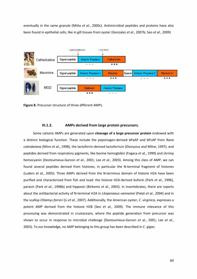
60
eventually in the same granule (Mitta et al., 2000c). Antimicrobial peptides and proteins have also
been found in epithelial cells, like in gill tissues from oyster (Gonzalez et al., 2007b; Seo et al., 2009)
III.1.2. AMPs derived from large protein precursors.
Some cationic AMPs are generated upon cleavage of a large precursor protein endowed with
a distinct biological function. These include the pepsinogen-derived bPaAP and bPcAP from Rana
catesbeiana (Minn et al., 1998), the lactoferrin-derived lactoferricin (Dionysius and Milne, 1997), and
peptides derived from respiratory pigments, like bovine hemoglobin (Fogaca et al., 1999) and shrimp
hemocyanin (Destoumieux-Garzon et al., 2001; Lee et al., 2003). Among this class of AMP, we can
found several peptides derived from histones, in particular the N-terminal fragment of histones
(Luders et al., 2005). Three AMPs derived from the N-terminus domain of histone H2A have been
purified and characterized from fish and toad: the histone H2A-derived buforin (Park et al., 1996),
parasin (Park et al., 1998b) and hipposin (Birkemo et al., 2003). In invertebrates, there are reports
about the antibacterial activity of N-terminal H2A in Litopenaeus vannamei (Patat et al., 2004) and in
the scallop Chlamys farreri (Li et al., 2007). Additionally, the American oyster, C. virginica, expresses a
potent AMP derived from the histone H2B (Seo et al., 2009). The immune relevance of this
processing was demonstrated in crustaceans, where the peptide generation from precursor was
shown to occur in response to microbial challenge (Destoumieux-Garzon et al., 2001; Lee et al.,
2003). To our knowledge, no AMP belonging to this group has been described in C. gigas.
Figure 8. Precursor structure of three different AMPs.

61
III.2. Classification of AMPs
More than 1200 different AMPs have been identified to date and their sequences are
available in the AMSDb database (http://www.bbcm.univ.trieste.it/). AMPs are a diverse group of
molecules divided into different classes on the basis of their amino acid composition and structure
(Boman, 1995; Vizioli and Salzet, 2002). Most AMPs are cationic, meaning that they have a positive
net charge at physiological pH, due to the presence of a higher content in Arginine and Lysine amino
acids than in Aspartic acid and Glutamic acid. This cationic character is sometimes reinforced by a C-
terminal amidation. In addition to their cationic property, AMPs can adopt an amphiphilic structure
with opposite hydrophobic and hydrophilic faces (Bulet et al., 2004). Both the cationic and
amphiphilic character of the AMPs facilitates their interaction with the anionic phospholipid
membranes of microorganisms (Oren and Shai, 1998). AMPs present broad antimicrobial spectra
against Gram-negative and Gram-positive bacteria, fungi, protozoa and enveloped viruses. In
addition, some AMPs can be highly hemolytic. These characteristics are believed to be essential for
their mechanisms of action, like the widely accepted model of membrane disruption (see below).
Cationic AMPs can be classified into three major groups according to their physicochemical
properties and origin. These include (i) linear peptides that can form amphipathic α–helices, (ii)
peptides that contain cysteine residues and form disulphide bonds, and (iii) peptides with one or
two over-represented amino acids (Brogden, 2005) (Fig. 9). Although not as common, an increasing
number of anionic AMPs have been described, but their functional characterization is still poorly
documented.
III.2.1. Amphipatic α-helical peptides
Found in a large variety of organisms quite evolutionarily distant (e.g. arthropods,
amphibians, fishes, rabbits and humans), α-helical AMPs vary remarkably in their sequence.
However, it is possible to fit them into distinct groups on the basis of sequence homologies, which
clearly follow taxonomic lines (Tossi et al., 2000). Most of these peptides are below 30–40 residues in
length, rich in lysine and arginine residues, and C-terminally amidated. Many are unstructured in
solution, and adopt an α-helical conformation upon contact with biological membranes. Some
examples of α-helical peptides include cecropins, ceratotoxin and melittin from insects; magainin,
dermaseptin, bombinin, and buforin II from amphibians, and LL37 from humans.

62
linear peptidesα–helicescysteine-rich peptides
cyclic
β-hairpin
α-helix/ β-sheet
magainin 2 indolicidin
tachyplesin I
Theta defensin
Cg-DefmPlectasin Drosomycin MGD1Cg-DefmPlectasin Drosomycin MGD1
III.2.2. Cationic peptides that contain cysteines and form disulphide
bonds
This is a large group of AMPs that contains pairs of cysteine residues oxidized to form
internal disulfide bridges. These cysteine-containing peptides are often open-ended, but there also
exists a cyclic cysteine-containing peptide from the rhesus monkey named θ-defensin (Tang et al.,
1999). This group gathers peptides (i) with 1 disulphide bond, such as brevinins from amphibians
(Basir et al., 2000); (ii) with 2 disulphide bonds, such as protegrins from pigs and tachyplesins from
horseshoe crabs (Kokryakov et al., 1993), (iii) with 3 disulphide bonds, including α-defensins and β-
defensins from humans (Lehrer, 2004), θ-defensin from the rhesus monkey (RTD-1) (Tang et al.,
1999), big-defensins from horseshoe crab (Kawabata et al., 1997), plectasin from fungus (Mygind et
al., 2005), and insect defensins like defensin A (Cociancich et al., 1993); and finally, (iv) with 4
Figure 9. Structural classes of antimicrobial peptides. Linear, α-helical and cysteine rich peptides are indicated with representative peptides for each class. Cysteine-rich peptides are divided in 3 subgroups depending on the number of cysteine residues and their pairing. Sheet structures are indicated by flat ribbons and arrows. Protein Data Bank ID: Theta defensin [1HVZ], Tachyplesin [1WO0], plectasin [1ZFU], Cg-Defm [2B68], Drosomycin [1MYN], Magainin 2 [PDB 2MAG]

63
disulphide bonds, like drosomycin from Drosophila, plant antifungal defensins (Fehlbaum et al.,
1994) and mollusk defensins (Yang et al., 2000 . Gueguen, 2006 #23). Depending on the number of
cysteine residues and their pairing, they may adopt a β-sheet conformation with triple strands (most
of the vertebrate defensins), a β-hairpin-like structure (like tachyplesin from horseshoe crab or
brevinins and esculentins from amphibians) or a mixed α-helix/β-sheet conformation, as observed
for arthropod and mollusk defensins, for plant defensins and for some mammalian defensins. Despite
structural variations, the cysteine-containing AMPs have been shown to all display one conserved
three-dimensional feature referred to as the γ-core. It was proposed that this motif, which
approximately corresponds to the tachyplesin 3D-structure (β-hairpin), is a key determinant of the
cysteine-containing AMPs conserved throughout evolution (Yount and Yeaman, 2004).
III.2.3. Peptides with one or two over-represented amino acids
They are peptides rich in certain amino acids, lacking cysteine residues, and linear, although
some can form extended coils. These peptides normally show an overrepresentation of one or two
amino acids, such as proline/arginine, proline/histidine or proline/lysine-containing peptides. Among
these, proline-rich antimicrobial peptides are a group of linear peptides, isolated from mammals and
insects, which are characterized by their high content (up to 50%) of proline residues (Gennaro et al.,
2002). Proline-rich antimicrobial peptides were first isolated from honeybees (Casteels et al., 1989)
and cattle (Gennaro et al., 1989) and subsequently found in other arthropod and mammalian species
(Schnapp et al., 1996; Bulet et al., 1999; Otvos, 2000). They are generally cationic, due to a high
content in arginine or, (less frequently) lysine residues. Members of this group are predominantly
active against Gram-negative bacteria, which they kill without bacterial membrane lysis, in contrast
with the majority of known AMPs.
III.2.4. Anionic AMPs
This is one exceptional class of AMPs. Structural characterization shows that they generally
range from - 1 to - 7 in net charge, and from 5 to 70 residues in length. For a number of these
peptides, post-translational modifications are essential for antimicrobial activity. In some cases,
antimicrobial activity appears to be a secondary role for these peptides, other biological activities
constituting their primary role (Harris et al., 2009).
In vertebrates, anionic AMPs are small peptides (721.6–823.8 Da), rich in glutamic and
aspartic acids and found in amphibians, sheep, cattle and humans. In humans, they are present in
surfactant extracts, bronchoalveolar fluid and airway epithelial cells. They are produced in millimolar
concentrations, require zinc as a cofactor for antimicrobial activity and are active against both Gram-
positive and Gram-negative bacteria (Lai et al., 2002; Brogden, 2005).
64
In invertebrates, larger cysteine-rich anionic AMPs (8.5 to 10 kDa) have been characterized
from the leech Hirudo medicinalis (theromacin and theromyzin) (Tasiemski et al., 2004) and the
shrimp Litopeaneus stylirostris (stylicins) (Rolland et al., 2010). Stylicins are expressed by shrimp
hemocytes and are very active (MIC = 2.5 µM) against the shrimp fungal pathogen Fusarium
oxysporum, while theromacin and theromyzin are expressed in large fat cells and epithelia, and are
active against gram-positive bacteria. Two additional anionic AMPs (4.8 and 6.9 kDa) were also
recently described in the insect Galleria mellonella. However, their activity against gram-positive and
fungal strains remains rather poor (MIC = 90 µM) (Cytrynska et al., 2007).
III.3. Mechanisms of action of AMPs
Cationic AMPs have the basic capability to interact with membranes. Some are membrane
active, but others are not (Brogden, 2005). New evidence points to intracellular targets as being
fundamental in the antimicrobial mechanisms of these peptides (Yeaman and Yount, 2003).
III.3.1. Interaction with biological membranes
The initial mechanism by which AMPs target microbes occurs via electrostatic interactions.
Cationic AMPs and negatively charged lipid membranes of bacteria are mutually and vigorously
attracted. The mechanism by which electrostatic attractions drive peptide-membrane interactions
have been examined in numerous studies (Vaz Gomes et al., 1993; Hancock, 1997; Matsuzaki et al.,
1997; Dathe et al., 2001). In the case of Gram-negative organisms, Hancock suggested a mechanism
of peptide interaction with membranes, termed self-promoted uptake (Hancock, 1997). This
mechanism proposes that the initial action of the peptide involves a competitive displacement of the
LPS-associated divalent cations that stabilize the outer membrane of Gram-negative bacteria. In
comparison, Gram-positive organisms lack an outer membrane or LPS, but their cell envelopes are
enriched in negatively charged teichoic and teichuronic acids (Yeaman and Yount, 2003). After the
initial peptide-membrane electrostatic interaction, the following events differ depending on the
specific mode of action.
III.3.2. Membrane-disruptive mechanisms
The mechanisms by which peptides may permeabilize and traverse microbial membranes are
not entirely clear and probably vary between peptides. A number of models for antimicrobial peptide
membrane permeabilization have been proposed but, given the variability in microbial membrane
ultrastructure, a peptide may act via different mechanisms in distinct membrane environments.
Three models are generally accepted, although these were established from results obtained with
individual peptides against artificial membrane systems and there is still no consensus among

65
investigators. These models are the toroidal pore, the barrel-stave and the carpet-like mechanism
(Fig. 10).
III.3.2.1. The Toroidal Pore model
In this model, peptides in the extracellular environment adopt an α-helical structure as they
interact with the charged and hydrophobic bacterial membrane (Fig. 10a). Helices are initially
oriented parallel to the membrane surface and the hydrophobic residues of peptides shift the polar
head groups, creating a break in the hydrophobic region and inducing positive curvature strain in the
membrane. When peptides reach a threshold concentration, they become perpendicularly
orientated to the membrane and helices may begin to self-associate because their polar residues are
no longer exposed to the membrane hydrocarbon chains. Thus, lipids are intercalated with the
peptides in the transmembrane channel. Upon disintegration of the pore, some peptide becomes
translocated to the cytoplasmic leaflet of the membrane (Uematsu and Matsuzaki, 2000). Magainins,
melittin and protegrins are examples that appear to induce transmembrane pores matching the
toroidal model (Yang et al., 2001).
Figure 10. Models for pore formation by AMPs. (1).Outer membrane in Gram-positive bacteria, thick peptidoglycan in Gram-negative bacteria; (2) electrostatic attraction to anionic bacterial membrane surface; (3) formation of an amphiphilic structure capable of membrane insertion; (4) possible permeabilization mechanisms, which depend on peptide sequence/concentration and membrane characteristics. Adapted from Sahl et al., 2005.

66
III.3.2.2. The Barrel-Stave model
In this model, a variable number of channel forming peptides are positioned in a “barrel-like”
ring around an aqueous pore. In this mechanism, the hydrophobic surfaces of an AMP face external
toward the acyl chains of the membrane, whereas the hydrophilic surfaces form the pore lining
(Breukink et al., 1999) (Fig. 10b).The initial step in barrel-stave pore formation involves peptide
binding at the membrane surface, most likely as monomers. Upon binding, the peptide may undergo
a conformational phase transition and the hydrophobic portion of the peptide is inserted into the
membrane. Positioning of the positively charged amino acids near the phospholipid head groups
facilitates this process.
When the bound peptide reaches a threshold concentration, peptide monomers self-
aggregate and insert deeper into the hydrophobic membrane core. Continued accumulation of
peptide monomers results in further expansion of the membrane pore (Yeaman and Yount, 2003).
This model has been proposed, for example, for alamethicin, a fungal AMP (Yang et al., 2001).
III.3.2.3. The Carpet-like Mechanism
Models of nonspecific membrane permeabilization by AMPs traditionally include diffuse
effects that have been compared to detergents. In this sense, some peptides may act against
microorganisms in a relatively diffuse manner, termed the carpet mechanism (Fig. 10c). In the carpet
model, a high density of peptides accumulates on the target membrane surface. Phospholipid
dislocation changes membrane fluidity and/or reduces the barrier properties of membrane, and
therefore leads to membrane disruption. As in other models, peptides initially bind to the membrane
mainly via electrostatic interactions, carpeting the phospholipid bilayer (Shai, 2002). Cecropin P1, an
AMP from Cecropia moths, appears to target microorganisms in this manner (Sitaram and Nagaraj,
1999).
III.3.3. Non membrane-disruptive mechanisms
There is widespread acceptance that cationic AMPs, apart from their membrane-disrupting
properties, also operate through interactions with intracellular targets, and subsequent impairment
of key cellular processes. Although most cationic AMPs act at high concentrations by disrupting the
microbial membrane, recent studies have reported several peptides that affect microbial viability at
low to moderate concentrations through interaction with one or more intracellular targets. Examples
of intracellular activity include inhibition of DNA and protein synthesis, inhibition of chaperone-
assisted protein folding and inhibition of cell wall synthesis (Brogden, 2005; Nicolas, 2009) (Fig. 11).
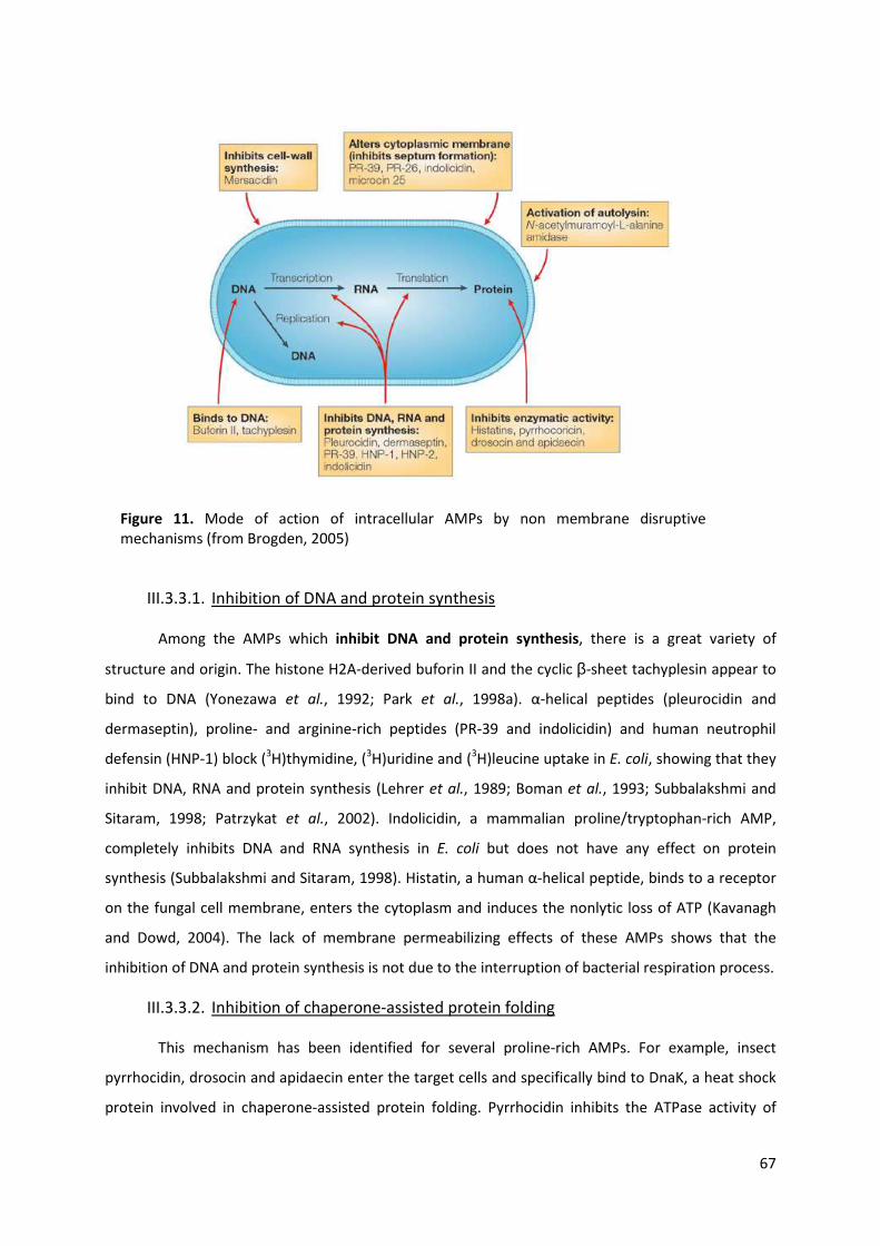
67
III.3.3.1. Inhibition of DNA and protein synthesis
Among the AMPs which inhibit DNA and protein synthesis, there is a great variety of
structure and origin. The histone H2A-derived buforin II and the cyclic β-sheet tachyplesin appear to
bind to DNA (Yonezawa et al., 1992; Park et al., 1998a). α-helical peptides (pleurocidin and
dermaseptin), proline- and arginine-rich peptides (PR-39 and indolicidin) and human neutrophil
defensin (HNP-1) block (3H)thymidine, (3H)uridine and (3H)leucine uptake in E. coli, showing that they
inhibit DNA, RNA and protein synthesis (Lehrer et al., 1989; Boman et al., 1993; Subbalakshmi and
Sitaram, 1998; Patrzykat et al., 2002). Indolicidin, a mammalian proline/tryptophan-rich AMP,
completely inhibits DNA and RNA synthesis in E. coli but does not have any effect on protein
synthesis (Subbalakshmi and Sitaram, 1998). Histatin, a human α-helical peptide, binds to a receptor
on the fungal cell membrane, enters the cytoplasm and induces the nonlytic loss of ATP (Kavanagh
and Dowd, 2004). The lack of membrane permeabilizing effects of these AMPs shows that the
inhibition of DNA and protein synthesis is not due to the interruption of bacterial respiration process.
III.3.3.2. Inhibition of chaperone-assisted protein folding
This mechanism has been identified for several proline-rich AMPs. For example, insect
pyrrhocidin, drosocin and apidaecin enter the target cells and specifically bind to DnaK, a heat shock
protein involved in chaperone-assisted protein folding. Pyrrhocidin inhibits the ATPase activity of
Figure 11. Mode of action of intracellular AMPs by non membrane disruptive mechanisms (from Brogden, 2005)

68
DnaK, which results in the accumulation of misfolded proteins and subsequent cell death (Kragol et
al., 2001).
III.3.3.3. Inhibition of cell wall biosynthesis
In contrast to the great variety of AMPs implicated in the inhibition of DNA, RNA and protein
synthesis and folding, inhibition of cell wall biosynthesis is mostly described for peptides produced
by Gram-positive bacteria, the lanthionine-containing peptide antibiotics (lantibiotics) (Bauer and
Dicks, 2005).
The peptidoglycan biosynthesis cycle consists of two lipid-bound intermediates, lipid I and
lipid II, synthesized at the cytosolic side of the plasmatic membrane. Lipid I is formed by the transfer
of the phospho-N-acetyl muramic acid (MurNAc)-pentapeptide group from the uridine 5’-
diphosphate (UDP)-activated amino sugar to the membrane-bound lipid carrier undecaprenyl-
pyrophosphate (C55P). Lipid II results from coupling of a second UDP-activated amino sugar N-acetyl-
d-glucosamine (GlcNAc) to the MurNAc group of lipid I. Lipid II is subsequently transported to the
exterior side of the membrane by a flippase. The two amino sugars and the pentapeptide are then
coupled to the cell wall and converted into peptidoglycan. The cycle is completed once the remaining
undecaprenyl-pyrophosphate is transported back to the cytosolic side of the membrane, where it is
dephosphorylated to undecaprenylphosphate (Breukink and de Kruijff, 1999; Bauer and Dicks, 2005)
(Fig. 12).
Figure 12. Schematic representation of cell wall biosynthesis in S. aureus (Schneider and Sahl, 2010)

69
The first identified lantibiotic showing inhibition of cell wall biosynthesis was nisin Z, which
uses the membrane-anchored cell wall precursor lipid II as a receptor (Breukink and de Kruijff, 1999).
Nisin is the oldest known and most extensively studied lantibiotic. It is active in nanomolar
concentrations against Gram-positive bacteria, a quality which results from its ability to use lipid II as
a docking molecule and subsequent pore formation. The lantibiotics gallidermin, epidermin,
mersacidin and plantaricin C also target lipid II, inhibiting peptidoglycan biosynthesis by interfering
with membrane-associated transglycosylation and in some cases, producing pore formation (Brotz et
al., 1998; Bonelli et al., 2006). The cyclic lipopeptide friulimicin has a different target and a unique
mode of action. It interrupts the cell wall precursor cycle through the formation of a Ca2+-dependent
complex with the undecaprenyl-phosphate carrier C55P. Since C55P also serves as a carrier in
teichoic acid biosynthesis and capsule formation, it is likely that friulimicin blocks multiple pathways
that are essential for a functional Gram-positive cell envelope (Schneider et al., 2009). Because
activities of lantibiotics are based on combined killing mechanisms, it has become evident that the in
vivo activity of lantibiotics cannot be assigned to a single factor. It appears rather that cell wall
synthesis inhibition and pore formation may contribute differently to the antimicrobial activity
depending on the target strain (Wiedemann et al., 2006).

70
IV. Molecular diversity in immune systems
IV.1. Host-pathogen interactions and the co-evolution theory
The continuous battle between hosts and pathogens has been a major subject of study for
evolutionary biologists. Because hosts and pathogens live in strong relationship with each other,
their interconnected fight for existence is both continual and rapid. At the molecular level, the
evolutionary response is reflected in mutations being under continual selection pressures (Holmes,
2004). Natural selection (see below) may act strongly on immune systems since hosts adapt to novel,
diverse and co-evolving pathogens. Any effective host defense system must have the capacity to
recognize a potential pathogen, activate the immune response and kill the infectious agent. In
parallel, from the perspective of the pathogen, it is essential to avoid the host immune defense. This
places the pathogen under strong evolutionary pressure to generate mechanisms to evade, resist or
suppress host immunity. Every successful advance by the pathogen, however, creates selective
pressure on the host to evolve so as to re-establish immunity (Lazzaro, 2008). Therefore, the general
hypothesis of co-evolution or evolutionary “arms race” proposes that pathogens/parasites evolve
continuously to escape from the immune response of host and, consequently, the immune system
of the host evolves to improve barriers against pathogens/parasites. Consequently, such
interactions might provide rapid evolution in genes involved in host-pathogen interactions. This
adaptation of the Red Queen hypothesis (Van Valen, 1974) predicts that in an “arms race”, both host
and pathogen/parasite develop mechanisms that generate diversity and polymorphism of molecules
that play key roles in their interplay. Thus, it is not surprising that genes involved in immunity
frequently show high levels of genetic diversity and present some of the best examples of positive
selection reported to date (Yang and Bielawski, 2000).
IV.2. Natural selection and molecular adaptation
Natural selection is an important process by which the evolution of organisms takes place. It
is a key mechanism that constitutes one of the bases of modern biology. The term was introduced by
Darwin in his pioneering book The Origin of Species (1859), and remains the primary explanation for
adaptive evolution. It is defined as the process by which heritable traits that increase the probability
of an organism to survive become more common in a population over successive generations
(Hughes, 1999a). Consequently, at the genomic level, different types of selective forces could shape
evolution. On one hand, new alleles that confer a higher fitness to an individual tend to increase in
frequency over time until they reach fixation, thus replacing the ancestral allele in the population.

71
This evolutionary process is called positive selection or adaptive evolution. Conversely, new
mutations that decrease the fitness tend to disappear from populations through a process known as
negative or purifying selection. Finally, it may happen that a mutation is advantageous only in
heterozygotes but not in homozygotes. Such alleles tend to be maintained at an intermediate
frequency in populations by way of the process known as balancing selection. Positive selection
favors the fixation of beneficial mutations that lead to evolution of new traits. Therefore, a mutation
is said to be adaptive if it performs a function that is in some way advantageous in the population.
Negative selection favors the conservation of existing phenotypes or particular genes functionally
constrained, playing an important role in maintaining the long-term stability of biological gene
function (Kimura, 1983). However, the neutral theory of Kimura maintains that the most frequently
observed molecular polymorphism is due to random fixation of neutral mutations by genetic
drift(Kimura, 1983). Random fixation can be associated to balancing selection, which favor the
existence of multiple alleles, rising polymorphism.
One of the most powerful approaches to detect selection pressures at the genomic level is by
comparing the number of nucleotide non-synonymous substitutions per non-synonymous site (dN)
with the number of synonymous substitutions per synonymous site (dS) (Kimura, 1983; Hughes and
Nei, 1989). Mutation at a codon site that results in amino acid change is known as non-synonymous
substitution. In contrast, a mutation which does not lead an amino acid change was known as
synonymous substitution. If the protein-coding gene is functionally constrained, then the rate of non-
synonymous change will be lower than the neutral rate. There are three different types of selective
pressure that can be detected from dN and dS ratio, which are (i) dN/dS < 1, and the gene is
considered to be subjected to strong purifying selection. Alternatively, if non-synonymous mutations
are beneficial then the average rate of non-synonymous changes is expected to be higher than the
neutral rate, resulting in (ii) dN/dS > 1, and the gene is considered to be subjected to positive
selection. When (iii) dN/dS = 1, it indicates neutral evolution. Several tests of neutrality are used to
reject strict neutrality and they can be applied to complete coding sequences or at codon level, by
using codon-based maximum likelihood methods. These models are useful to identify amino acid
sites under diversifying selection (Yang and Bielawski, 2000).
IV.3. Mechanisms of genetic diversity
One major solution for increasing diversity is to make more final products than the number
of inherited genes that one individual could account for. In general, genetic diversity of immune
receptors and effectors could be generated by different processes: (i) gene duplication, (ii) exon
shuffling, (iii) polymorphism, (iv) recombination, (v) somatic hypermutation or (vi) alternative splicing
(Pasquier, 2006).

72
In all domains of life, genomes probably have been generated by gene duplication. Gene
duplication can result from: unequal crossing over, retroposition or chromosomal (or genome)
duplication. Unequal crossing over usually generates tandem gene duplication. Retroposition occurs
when an mRNA is retrotranscribed to complementary DNA and then inserted into the genome; it is
usually unlinked to original gene, because the insertion of cDNA into the genome is more or less
random. Chromosomal or genome duplication probably occurs by a lack of disjunction among
daughter chromosomes after DNA replication (Zhang, 2003).
The existence of duplicate genes is occasionally beneficial just because extra amounts of
protein or RNA products are provided and two paralogous genes can maintain the same function by
gene conversion. Under gene conversion, two paralogous genes will have very similar sequences and
functions, and this mode of evolution is referred to as concerted evolution (Li et al., 2001).
Alternatively, strong negative selection can also prevent duplicated genes from diverging. In parallel,
two duplicate genes can also be maintained when they differ in their functions (Nowak et al., 1997)
and this can occur by subfunctionalization, in which each gene adopts part of the functions of the
original gene, or by neofunctionalization, which is one of the most important outcomes of gene
duplication and represents the origin of a novel function by adaptive evolution (Hughes, 1994).
Exon shuffling leads to the increase of protein structure diversity. By this mechanism,
individual structural or functional units can be put together in various combinations to create many
different proteins. According to the exon shuffling theory, exons in genes correspond to functional
modules in the encoded proteins. Duplication, permutation and rearrangement of these exons would
result in novel genes that express proteins with diverse functional modules (Gilbert, 1978).
Other way to increase the diversity is to add polymorphism to the multiple gene copy
number. This can be done any time during the history of a gene family. As it changes with time,
polymorphism provides allelic diversity that offers flexibility within populations as a function of
environmental variation and pathogens (Pasquier, 2006 392).
Genetic recombination is a process by which a molecule of DNA is broken and then joined to
another DNA molecule. Recombination can occur between similar DNA molecules, as in homologous
recombination, or dissimilar DNA molecules, as in non-homologous end joining. When this process is
associated with polymorphism, recombination moves the mutation into different genetic
backgrounds, which could be associated with functional variation of selective importance (Spencer et
al., 2006). Furthermore, the observed positive correlation between recombination rate and genetic
diversity suggests that many loci showing recombination have been subject to adaptive evolution
(Nachman, 2001).
Somatic hypermutation consists mainly of a programmed process of single nucleotide
changes with rare insertions and deletions. Such changes would be introduced during error-prone

73
repair of lesions involving single-strand DNA breaks or, more likely, double-strand DNA breaks (Wu et
al., 2003a). Somatic hypermutation diversifies the receptors used by the immune system to
recognize antigens and allows the adaptation of its response to new threats during the organism
lifetime (Janeway et al., 2005).
Alternate splicing is the mechanism by which a single primary mRNA transcript can be
transformed into a number of different mature mRNAs encoding different proteins. This is
accomplished by post-transcriptional control of gene expression. After the removal of the introns,
some exons may also be removed. The result is a family of related proteins, formed by the joining of
different combinations of exons, although the exons always remain in the same order as in the
primary transcript (Jensen et al., 2009).
All these mechanisms can act alone or simultaneously to produce the diversification of
immune receptors and effectors of the immune system. Whatever the mechanism used, these
molecular processes have the same goal: creating a variety of molecules that are adequately diverse
to discriminate between the wide spectra of potential pathogens.
IV.4. The adaptive immune response in vertebrates
In vertebrates, the immune system is composed by the innate immune response and the
adaptive immune response. The adaptive response provides the vertebrate immune system with the
ability to recognize and remember specific pathogens. This system offers two major arms, the
immunoglobulins (Ig) (the humoral response) and T cells (the cellular response), both of which set
down an immunological specificity and memory for future defense (Holmes, 2004). Thus, two major
features characterize the adaptive immune system of vertebrates: specificity and memory
(Zinkernagel et al., 1996; Jiang and Chess, 2009). Specificity is characterized by a great diversity of
immune effectors directed against a variety of antigens. Specificity is based on the receptors of
immune cells, called the T and B lymphocytes, presenting Ig, T cell receptors (TCR) and molecules of
the major histocompatibility complex (MHC) (Cresswell, 1994; Engelhard, 1994; McHeyzer-Williams
and Davis, 1995). Memory relies on the selection of these specific molecules in lymphocytes. Once
the infection is over, these cells will remain in the organism to constitute a cellular memory that they
will allow a more rapid response when a second infection by the same pathogen occurs.
IV.4.1. The paradigm shift between adaptive and innate immunity
Invertebrates do not possess an adaptive immune system and have therefore been assumed
to lack diverse and specific immune defenses. Nevertheless, immunology has recently confronted a
major paradigm shift. Recent evidence has demonstrated that some invertebrates have complex

74
immune responses with high levels of immune diversity and specificity. The old notion that
invertebrates have a simple innate immune system that recognize broad groups of pathogens but
lack the capacity for precise discrimination of individual pathogens is actually out of date. Some
works also suggested the existence of memory in invertebrate immunity (Kurtz and Franz, 2003;
Little et al., 2003; Pham et al., 2007) but it is still controversial. Several authors sustains that
conclusions based only on phenomena such as survival or reproductive capacity of organisms is not
sufficient, and that real evidence can only be upheld if supported by descriptions of the underlying
mechanisms (Hauton and Smith, 2007).
Although invertebrates include more than 95% of all species in the animal kingdom, only a
small number of species, mostly arthropods, have been investigated at the molecular level to
establish the mechanisms of defense and the nature of the molecules produced. This is also true for
the second largest invertebrate phylum, the Mollusca. Nevertheless, we can find important studies
on model species that provide evidence of how invertebrates generate diverse innate immune
molecules.
The following examples in both adaptive and innate immune systems were chosen with two
purposes: (i) to highlight the existing mechanisms of diversification of immune receptors and
effectors in vertebrates and invertebrates and (ii) to show the most representative evidence of
innate immune diversification that has supported this paradigm shift.
IV.5. Diversification of immune receptors
IV.5.1. In the adaptive response of vertebrates
The mechanisms underlying the generation of highly variable receptors in the adaptive
immune system have been widely investigated. The recognition molecules, Immunoglobulins (Ig) and
T cell receptors (TCR) are generated by a mechanism of somatic gene rearrangement and
hypermutation, which randomly selects and assembles segments of genes encoding specific proteins
by Recombination Activating Genes (RAG) enzymes. This site-specific recombination reaction
generates a diverse repertoire of Ig and TCR molecules that are necessary for the recognition of
diverse antigens. The product of this rearrangement is expressed clonally in lymphocytes and the
antigen presentation process. This allows the selection of appropriate receptors depending on a set
of enzymes and presenting molecules encoded in the Major Histocompatibility Complex (MHC) (Du
Pasquier, 2004). It is believed that the Ig repertoire generated by somatic hypermutation can
practically recognize an infinite number of non-self structures (Teng and Papavasiliou, 2007).
The MHC is another example of greatly diversified molecules, but the mutational mechanism
underlying the diversity in MHC genes is still controversial (Reusch and Langefors, 2005). MHC

75
comprises a multigene family coding for transmembrane receptor glycoproteins, which bind antigens
and present them to T lymphocytes that initiate the subsequent immune response by recognizing a
foreign peptide (Brown et al., 1993). The MHC genes contain some of the most polymorphic coding
genes in the vertebrate genome (Bernatchez and Landry, 2003) and gene duplication, allelic diversity,
inter and intralocus recombination and selection pressures have all been suggested as mechanisms
of diversification (Reusch and Langefors, 2005; Schaschl et al., 2006; Axtner and Sommer, 2007).
IV.5.2. In the innate response of invertebrates
The following examples of diversification of immune receptors in invertebrates contradict
the paradigm of Medzhitov and Janeway, which established that receptor diversity of the innate
immune system is limited and germline encoded (Medzhitov and Janeway, 1997). These key studies
provide strong evidence that like vertebrates, invertebrates use not only germline-encoded receptors
but also highly diversified receptors generated by somatic mechanisms to react rapidly to antigenic
changes of pathogens (Kurtz and Franz, 2003).
IV.5.2.1. Germline-encoded diversity
Scavenger receptor cysteine-rich repeats (SRCR) proteins from sea urchin
The purple sea urchin Strongylocentrotus purpuratus expresses a complex set of transcripts
belonging to scavenger receptor cysteine-rich repeats (SRCR) in hemocytes (Pancer, 2000). SRCR
domains form part of a superfamily of proteins. SRCR proteins are implicated in development of the
immune system of vertebrates and in the regulation of immune responses, mediating growth,
differentiation and activation of immune cells as well as other cells of the body, most likely via
protein–protein interactions (Resnick et al., 1994). In some invertebrates, members of the SRCR
superfamily have been shown to function as cell surface receptors (Dangott et al., 1989; Blumbach et
al., 1998). Pancer highlighted a heterogeneous and transcriptionally dynamic set of SRCR-containing
molecules which are expressed specifically in sea urchin hemocytes (Pancer, 2000). The
heterogeneity of SRCR domains was demonstrated at genomic and transcriptional levels, and
features a multigene family (150 genes) that encodes an estimated 1200 SRCR domains with specific
patterns particular to each individual. However, the mechanisms by which SRCR-containing
molecules diversify have not yet been clearly shown. Elucidation of the function of sea urchin SRCR
gene products requires further research, but their importance has been illustrated in terms of gene
numbers, complex genomic organization, and dynamic transcription profiles in this marine
invertebrate.

76
Immunoglobulin (Ig)-type variable (V) region-containing chitin binding proteins
(VCBPs) from amphioxus
The amphioxus Branchiostoma floridae presents a unique immune receptor repertoire of
immunoglobulin (Ig)-type variable (V) region-containing chitin binding proteins (VCBPs). These
proteins are soluble proteins consisting of two Ig-type V regions joined to a carboxy-terminal chitin-
binding domain. Although their ligands are unknown, VCBPs are likely candidates for innate immune
receptors and represent the only example of an innate receptor in which the functional unit is a
hyperdiversified Ig-type V region. VCBPs are distributed across at least five families (VCBP 1-5)
(Cannon et al., 2002) and regionalized peptide sequence hypervariability was noted in the N-terminal
V region of pooled amphioxus VCBP2 cDNAs (Cannon et al., 2004). A very high degree of regionalized
hypervariability was observed in VCBP2 at the level of the individual germline, which produces a
unique repertoire of proteins at the individual level. A total of 43 different peptides were encoded
across the amplified region of VCBP2 by only 13 different animals. Although the particular
mechanism that gives rise to the observed genetic polymorphism in VCBP2 genes is unknown,
Cannon et al. (2004) suggested that in species with large populations, such as amphioxus, extensive
polymorphisms may compensate for the absence of somatic modification in the maintenance of
immune receptor diversity (Cannon et al., 2004).
IV.5.2.2. Somatically-generated diversity
Down syndrome cell adhesion molecules (Dscam) from insects
A member of the immunoglobulin superfamily (IgSF), the Down syndrome cell adhesion
molecule (Dscam), in the fruitfly D. melanogaster, and its ortolog AgDscam in the mosquito A.
gambiae, were found to be associated with pathogen resistance and to diversify, through alternative
splicing, to generate more than 30 000 variants (Watson et al., 2005; Dong et al., 2006a). Dscam is a
transmembrane molecule that contains extracellular domains of the Ig and fibronectin types, as well
as a cytoplasmic domain (Schmucker et al., 2000) and the gene is present as a single gene copy in
both species. The extremely diversified Dscam repertoire in D. melanogaster is expressed in the
hemocytes, and the modification of the expression of Dscam gene by RNA interference alters their
phagocytic activity. These data suggested the implication of Dscam in Drosophila immunity.
However, the latest and more important advance was evidenced in the work done by Dong et al.
(2006). The authors demonstrated that, in A. gambiae, the immune response against Plasmodium
does not increase the AgDscam transcript level, but produces a change in the representation of some
changeable exons by alternative splicing. The different Ig domain-containing splice forms are over- or

77
under-represented depending on which pathogen the host cell is exposed to. Thus, challenge with
different pathogens produces specific AgDscam splice-form repertoires with different adhesive
characteristics.
Fibrinogen Related Proteins (FREPs) from mollusks
In mollusks, the diversification of the Fibrinogen Related Proteins (FREPs) has been reported
in the pulmonate gastropod Biomphalaria glabrata (Zhang et al., 2004). FREPs are soluble proteins
and their genes are expressed in response to trematode parasites in hemocytes (Adema et al., 1997).
They are members of the immunoglobulin superfamily (IgSF) and consist of one or two amino-
terminal IgSF domains and a carboxyl-terminal fibrinogen domain. In B. glabrata, 13 different
subfamilies of FREP genes have been identified and they are coded by a variable number (1 to 8) of
gene copies (Zhang et al., 2004). The complete sequences of FREP 2, 3, 4 and 7 are already identified
(Zhang and Loker, 2003; Zhang et al., 2004) and a study focused on the characterization of the IgSF
and fibrinogen domains of the subfamily FREP 3 evidenced a great diversity in these regions (Zhang
et al., 2004). From one single individual, 45 different sequences were isolated, but this number
disagrees with the number of identified gene copies. As a result of further studies, this high level of
diversification was proposed to be generated from a limited number of germline genes that are
submitted to two types of mechanism: (i) point mutations, which allow the generation of new
variants and (ii) somatic recombination using a limited set of source genes, which would lead to new
mosaic genes. This study was the first proposing somatic recombination as a mechanism of
diversification in organisms other than higher vertebrates.
An interesting example of co-evolution was proposed from the B.glabrata/Schistosoma
mansoni model system. B. glabrata is an intermediate host for the human parasite S. mansoni, and if
the arms race hypothesis is applicable, mechanisms of diversification based on polymorphism that
permit the survival of species also have to exist in host parasites. The discovery of highly polymorphic
mucins in S. mansoni, which are potential key factor proteins in the compatibility process of this
parasite with B. glabrata, supports the hypothesis of an arms race taking place in this model (Roger
et al., 2008b). Mucins are known to play a key role in host–parasite interplay and are involved in
immune evasion, host invasion and parasite protection (Hicks et al., 2000; Theodoropoulos et al.,
2001). S. mansoni polymorphic mucins (SmPoMucs) are differentially expressed in response to a
compatible or an incompatible parasite strains (Roger et al., 2008b) and each individual larva
expresses a unique combination of glycosylated SmPoMucs (Roger et al., 2008a). It was found that
SmPoMucs are encoded by a multigene family, in which genes are transcribed in an individual-
specific manner and create multiple splice variants for each gene. Global diversity is finally produced

78
by exon shuffling, gene conversion and frequent recombination, performed by functional genes or by
pseudogenes. Finally, post-transcriptional regulation events, like alternative and aberrant splicing
were shown to contribute to SmPoMucs polymorphism (Roger et al., 2008a).
Concerning the host/parasite interaction, the function of the FREPs family in B. glabrata is
still not clear, although previous evidence suggested that FREPs, as pattern recognition receptors, are
capable of binding molecules of foreign origin similar to SmPoMucs (Adema et al., 1997). In this
context, both host and parasite could have created a system that generates a high degree of
polymorphism to avoid, from the parasite side, the recognition by the host immune response and,
from the host counterpart, to improve barriers against the parasite infection.
IV.6. Diversification of immune effectors
The mechanisms involved in the molecular diversification of immune effectors have been
broadly described in several vertebrate and invertebrate taxa and a high number of these studies are
related to AMP diversification (Hughes, 1999b; Lee et al., 2005; Tennessen, 2005; Cohuet et al.,
2008). On the one hand, AMP diversification was studied from an evolutionary point of view,
focusing on genetic mechanisms such as gene duplication, recombination, and allelic polymorphisms
in both vertebrates and invertebrates. Several hypotheses have been proposed about the role of this
diversification in the immune response. On the other hand, structure-activity relationship of AMP
variants has been studied with the aim of designing novel antimicrobials (Powers and Hancock, 2003;
Chandrababu et al., 2009; Haney et al., 2009). However, few studies have linked the evolutionary
divergence of AMPs to functional analyses of their biological activity. The next examples of AMP
diversification show those who have been characterized up to the biological significance of their
diversity.
IV.6.1. Molecular evolution of AMPs and functional diversification
IV.6.1.1. Antimicrobial activities
Mammalian defensins provide one of the most striking examples of a diverse and evolving
AMP gene family. As presented above, mammalian defensins are classified into three subfamilies
based on the arrangement of the six cysteine motif: α-, β- and θ-defensins (Lehrer and Ganz, 2002),
and α- and β-defensins have been extensively studied at evolutionary, structural and functional
levels. A number of evolutionary studies showed that α- and β-defensins have a common ancestor,
which has evolved by gene duplication and rapid sequence divergence (Maxwell et al., 2003; Semple
et al., 2006; Hollox and Armour, 2008). It seems that they arose initially by unequal crossing over and
tandem duplication (Hughes, 1999b; Morrison et al., 2003). This mechanism of diversification means

79
that each subfamily presents a large degree of sequence variability and that subfamilies display low
sequence similarity (Semple et al., 2006). Additionally both α- and β-defensins have been subjected
to allelic recombination between distinct genomic locations, which generated copy number diversity
(Aldred et al., 2005; Linzmeier and Ganz, 2005; Abu Bakar et al., 2009).
Role of the anionic propiece. Mammalian α and β-defensins generally consist of three and
two exons, respectively. The first exon encodes the signal and pro-region and the second (and third)
exon encodes the mature peptide. Comparison of rates of synonymous and nonsynonymous
nucleotide substitutions suggested that gene duplication has often been followed by the
diversification of exons encoding the mature peptide by positive selection (Maxwell et al., 2003). In
many cases, it appears that amino acid changes in this region occurred in a balance manner between
the mature peptide region and the propiece region (Hughes and Yeager, 1997). These data support
the hypothesis of the neutralizing action of the propiece, which might be important to prevent
intracellular interaction of peptides with membranes (Valore et al., 1996).
Role of the cysteine scaffold. Despite the sequence variation in the mature peptide, the six
cysteines are retained in all defensins and are under negative selection, implying that they are
essential to the functional structure of the molecule. The importance of the six cysteines in the
antimicrobial function of β-defensins has been challenged by the finding that different synthetic
peptides of the human defensin 3 (hBD3), synthesized with different cysteine connectivities,
exhibited similar antimicrobial activity (Kluver et al., 2005). Similarly, a wide range of chemotactic
activities was retained with these analogs. Interestingly, when the disulphide bridges were absent,
the bactericidal activity remained unaffected but the chemotactic activity of linear hBD3 is altered
(Wu et al., 2003b). These results show that the cysteine pairing is required for the peptide
chemotactic activities rather than antimicrobial activities. However, since the presence of the six
cysteines at conserved positions is required for both the antimicrobial and chemotactic activity of
defensins (Taylor et al., 2008), the conservation of the cysteines during evolution reflects their
involvement in both activities. Additionally, a mammalian defensin has recently been shown to have
unexpected involvement in hair pigmentation, suggesting that mammalian AMPs may play roles
outside immunity, which places them under different selective pressures (Candille et al., 2007).
Role of charge distribution. There is experimental evidence that mammalian defensins are
diverse in their potency against different pathogens. Functional differences have been observed in
vitro between a number of β-defensin variants (Lehrer and Ganz, 2002). The correlation of the
antimicrobial activity with the structural changes in hBD-3 reveals that the potency was determined
by the distribution of positively charged and hydrophobic amino acid residues (Kluver et al., 2005).
Several evolutionary studies show evidence of positive selection in sites coding these residues, which
can be linked to the functional diversification (Pazgier et al., 2007). Concerning α-defensins, the

80
mouse neutrophil peptide NP-1 presents a positively charged arginine residue in the active peptide,
which is under adaptive evolution (Lynn et al., 2004) and it seems to be responsible for its
antimicrobial activity against Gram-negative bacteria from genus Actinobacillus (Ouellette and
Bevins, 2001). In humans, HNP1 and HNP3 differ only by one residue under positive selection at the
N-terminus of the mature peptide and HNP1 exhibits potent activity against Candida albicans
whereas HNP3 has little effect (Raj et al., 2000; Lynn et al., 2004). Therefore, small changes in the
primary structure of these molecules can have significant effect on their activities, and positive
selection can act at certain sites to increase their potency.
IV.6.1.2. Ecology
A remarkable example of adaptive evolution and functional divergence in vertebrate
antimicrobials in relation with ecology is the case of amphibian AMPs. Frog species live in different
ecological niches associated to a large spectra of potential pathogens, and every frog species are
equipped with their own unique set of AMPs, constituting families from 2 to 100 closely related
members (Mangoni, 2006). The dermal glands of frogs produce a great number of AMPs with
remarkable levels of interspecific and intraspecific diversity (Nicolas et al., 2003). The principal
classes of amphibian AMPs include bombinins, magainins, dermaseptins, brevinins, esculentins and
temporins (Mangoni and Shai, 2009). These principal classes of amphibian AMPs are genetically
related, with a remarkable identity in signal and propiece sequences of their preprosequences.
Evolutionary relationship between the conserved proregions has been analyzed, indicating that they
all derived from a single family of precursor polypeptides, designated as the preprodermaseptins,
which have been diverged to create several families of peptides that are structurally and functionally
distinct (Vanhoye et al., 2003) (Fig. 13). This unusual pattern acting on the diversification of
amphibian AMPs supports the hypothesis of an evolutionary strategy developed to confer ecological
niche specialization, which arises by the encounter with new pathogens. Consequently, different
species of frogs are equipped with different sets of peptides belonging to different families. The
impressive divergence between and within frog species is such that no one identical peptide has
been found between species at the amino acid level.
Evolutionary analyses showed that frog skin AMPs have arisen by gene duplication, focal
hypermutation and diversifying selection. Each precursor results from transcription of a single
genetic locus that encodes a single mature peptide. The contrast between the conserved
preproregion and the hyperdivergent mature peptides is one of the most extreme examples
observed to date for homologous gene products within a single order of organisms, with positive
selection operating exclusively within their antimicrobial domain (Nicolas et al., 2003). The
combination of targeted hypermutation to generate great variation and the subsequent action of

81
positive selection might explain the hypervariation and large number of peptides per species (Nicolas
et al., 2003; Nicolas and El Amri, 2009).
Figure 13. Molecular phylogeny of preprodermaseptins. Adapted from (Vanhoye et al., 2003)
IV.6.1.3. In invertebrate AMPs
The molecular evolution of invertebrate AMPs has been mainly studied in model species, but
studies that include their evolutionary history up to their gene function have not been as strongly
developed as in vertebrates. Nonetheless, particular patterns of evolution in invertebrate AMPs have
been evidenced in arthropods.
In the Pacific white shrimp, Litopenaeus vannamei, where three distinct classes of
penaeidins, named PEN2, PEN3, and PEN4, are expressed in the hemocytes (Destoumieux et al.,
1999; Cuthbertson et al., 2002). Multiple isoforms, generated by substitutions and deletions, have

82
been reported at the mRNA level for all three classes of penaeidins. Each penaeidin class is encoded
by a unique gene and isoform diversity is generated by allelic polymorphism within each gene family
(O'Leary and Gross, 2006; Padhi et al., 2007). The multiple copies of penaeidins have evolved by gene
duplication and adaptive evolution is the likely cause of accelerated rate of amino acid substitutions
(Padhi et al., 2007). A feature of the penaeidin family is intra-class diversity, particularly for class 3,
where multiple unique isoforms have been reported between species and within an individual
organism (Destoumieux et al., 1997; Cuthbertson et al., 2002; Gueguen et al., 2006a). The
antimicrobial spectra of the synthetic penaeidins from classes 3 and 4 were compared. Penaeidin
class 3 has a broader range of microbial targets and is more potent against certain bacterial species
than class 4 (Destoumieux et al., 1999; Cuthbertson et al., 2005), but the antimicrobial activity of
class 4 appears to be more specifically directed against certain microbial strains (Cuthbertson et al.,
2008). Thus, it seems that diversification of an AMP family like penaeidins could produce a functional
divergence of isoforms to enlarge the antimicrobial spectra.
At the present, diversification of AMPs in both vertebrates and invertebrates seems to
present various common patterns, like gene duplication, allelic polymorphisms and, in some cases,
evidence of adaptive evolution at certain codons. Furthermore, the biological significance of this
diversification has been evidenced at different levels, in the acquisition of synergistic activities,
microbial target specificity or extending the antimicrobial spectrum. However, there are still a lot of
unresolved issues concerning AMP evolution, such as intraspecies AMP diversity. More research at
the genetic level may answer many of these questions, including specific selective pressures,
strength of selection and causes of divergence or conservation. Those who study the genetic
selection to improve resistance to pathogens or find therapeutic uses of AMPs should attempt to
identify the AMP families in which selection has produced high variation in antimicrobial properties.
Or, alternatively, they should attempt to identify AMP families that do not appear to be under
constant diversifying selection, perhaps because pathogens cannot easily evolve resistance to them.
We therefore need to do further research to identify the underlying mechanisms of diversification of
these molecules in order to better understand their function in the antimicrobial response and to be
able to use them for specific purposes.

Chapter 2
Results
83
I. Section 1. Molecular diversity of antimicrobials from
Crassostrea gigas.
Publication N°1. Molecular diversity of antimicrobial effectors in the oyster Crassostrea
gigas. BMC Evolutionary Biology 2010, 10: 23
In recent years, high levels of sequence diversity were reported to be characteristic of several
immune genes from invertebrates (for review see (Schulenburg et al., 2007; Du Pasquier, 2009)).
Thus, the paradigm that diversification of immune genes is an exclusive property of the vertebrate
adaptive immunity is now obsolete. Nowadays, studies in invertebrate model organisms have given
insights into the origin and evolution of innate immune genes, evidencing numerous mechanisms of
diversification and directional selection pressures, related to adaptive molecular evolution (Flajnik
and Du Pasquier, 2004; Lee et al., 2005; Litman et al., 2005; Pujol et al., 2008). Therefore, new
evidences demonstrate that the innate immunity system of invertebrates is more complex that we
previously thought.
In this context, the rapid evolution of AMPs is well documented (Patil et al., 2004; Jiggins and
Kim, 2005; Semple et al., 2006) and suggests that hosts exposed to diverse pathogens may evolve a
broader repertoire of antimicrobials that enhance their defensive potential (Pujol et al., 2008). The
rapid evolution of AMPs has been related with the general hypothesis of co-evolution or "arms race".
In this theory, pathogens evolve continuously to escape from the immune response of hosts and,
consequently, the immune system of hosts evolves to improve new barriers against pathogens
(Dawkins and Krebs, 1979).
In the Pacific oyster, Crassostrea gigas, several antimicrobials have been characterized. First,
an antimicrobial peptide (AMP), member of the defensin family (Cg-Defm) was identified from the
oyster mantle (Gueguen et al., 2006b) and then, two additional defensins named Cg-Defhs (Cg-Defh1
and Cg-Defh2) were characterized from hemocytes (Gonzalez et al., 2007a). Another peptide named
Cg-Prp, which belongs to the Proline-rich AMP family, was also identified from hemocytes (Gueguen
et al., 2009). Additionally, a member of the Bactericidal Permeability Increasing protein (BPI) family
was characterized in hemocytes and oyster tissues (Gonzalez et al., 2007b). From those works, the
existence of a diversity of sequences at amino acid level began to be evidenced, which motivated
further investigation. Hence, the first part of my thesis work deals with the characterization of the
molecular diversity of the oyster antimicrobials by genomic approaches.
84
Nucleotide sequences for the antimicrobials were obtained by specific PCR amplification
from oyster cDNA and genomic DNA, and by in silico search of homologous sequences among the
29,745 unique sequences available at the Sigenae C. gigas EST database (http://public-
contigbrowser.sigenae.org:9090/Crassostrea_gigas/index.html). In the light of the high diversity
observed among the AMP sequences and the discovery of novel variants, we checked the
mechanisms by which the diversity could be generated. We evidenced that the AMPs, namely Cg-
Defs and Cg-Prp are all the product of multigenic families displaying a variety of gene structures and
copy number variation. In contrast, Cg-bpi is encoded by a single copy gene. Moreover, we identified
for both AMPs several genetic mechanisms of diversification such as recombination, parallel
mutations leading to phylogenetic homoplasy and indel events. In addition, the non synonymous to
synonymous substitutions ratio by codon (dN/dS) revealed several negatively and positively selected
sites for both AMPs, suggesting that directional selection pressures have shaped their sequence
variations. Besides, the diversity of the three antimicrobials has been compared with the diversity
present in a non-immune related gene (Cg-actin), showing that variability is not a common
characteristic for all oyster genes.
This is the first evidence a high level of variability in oyster AMP sequences. The results
accumulated reveal that AMPs from C. gigas have been subject to distinct patterns of diversification
and we evidence the existence of different evolutionary routes leading to such sequence variability.
The results are detailed and discussed in publication N°1.

RESEARCH ARTICLE Open Access
Molecular diversity of antimicrobial effectorsin the oyster Crassostrea gigasPaulina Schmitt1, Yannick Gueguen1,3, Erick Desmarais2, Evelyne Bachère1, Julien de Lorgeril1*
Abstract
Background: To gain insight into the molecular diversity of antimicrobial peptides and proteins in the oysterCrassostrea gigas, we characterized and compared the sequence polymorphism of the antimicrobial peptides(AMPs), Cg-Defensins (Cg-Defs) and Cg-Proline Rich peptide (Cg-Prp), and of the bactericidal permeability increasingprotein, Cg-BPI. For that, we analyzed genomic and transcript sequences obtained by specific PCR amplificationand in silico searches.
Results: High diversification among the three antimicrobial effectors was evidenced by this polymorphism survey.On the basis of sequence phylogenies, each AMP aggregates into clearly defined groups of variants and is theproduct of a multigenic family displaying a variety of gene structures. In contrast, Cg-bpi forms a single group andis encoded by a single gene copy. Moreover, we identified for both AMPs several genetic mechanisms ofdiversification such as recombination, parallel mutations leading to phylogenetic homoplasy and indel events. Inaddition, the non synonymous to synonymous substitutions ratio by codon (dN/dS) revealed several negativelyand positively selected sites for both AMPs, suggesting that directional selection pressures have shaped theirsequence variations.
Conclusions: This study shows for the first time in a mollusc that antimicrobial peptides and proteins have beensubject to distinct patterns of diversification and we evidence the existence of different evolutionary routes leadingto such sequence variability.
BackgroundThe Pacific oyster, Crassostrea gigas, is a filter feederbivalve mollusc which is continually exposed to micro-organisms naturally present in the marine environment.Thus, the capacity to control the microflora and over-come infections is essential for the oyster survival. Thehemocytes, immunocompetent cells, play a central rolein innate antimicrobial response. Indeed, in molluscs,hemocytes infiltrate injured tissues, phagocyte microor-ganisms, produce antimicrobial peptides (AMPs) andrelease factors such as lectins and reactive oxygen spe-cies (ROS) [1-3].In C. gigas, several antimicrobial effectors have been
recently characterized. An AMP, member of the defen-sin family named Cg-Defm, was identified from theoyster mantle [4]. Then, two additional defensins named
Cg-Defhs (Cg-Defh1 and Cg-Defh2)have been character-ized from hemocytes [5]. Both Cg-defm and Cg-defhsappear to be continuously expressed in different tissues.More recently, a new peptide, Cg-Prp, which belongs tothe proline-rich AMP family, has been found in hemo-cytes [6]. Whereas Cg-Prp displays alone a weak in vitroantimicrobial activity, it revealed synergistic activity withCg-Defm against both Gram positive and negative bac-teria [6]. Additionally, a member of the bactericidal/per-meability-increasing protein (BPI) family has beencharacterized [7]. Cg-bpi expression is induced in hemo-cytes after oyster bacterial challenge and constitutive invarious tissue epithelia of unchallenged oysters. Similarto vertebrate BPIs, the oyster BPI binds LPS, displaysbactericidal activity against Gram negative bacteria andincreases the permeability of the bacterial cytoplasmicmembrane [7].High levels of sequence diversity were reported to be a
characteristic of several antimicrobial effectors pertain-ing to the innate immunity system of invertebrates [8].
* Correspondence: [email protected], CNRS, Université de Montpellier II, IRD, UMR 5119 « EcosystèmesLagunaires », Place Eugène Bataillon, CC80, 34095 Montpellier Cedex 5(France
Schmitt et al. BMC Evolutionary Biology 2010, 10:23http://www.biomedcentral.com/1471-2148/10/23
© 2010 Schmitt et al; licensee BioMed Central Ltd. This is an Open Access article distributed under the terms of the Creative CommonsAttribution License (http://creativecommons.org/licenses/by/2.0), which permits unrestricted use, distribution, and reproduction inany medium, provided the original work is properly cited.

The functional significance of this diversity has beenaddressed in some cases. Multiple variants of lectinsfrom the horseshoe crab Carcinoscorpius rotundicaudaare able to recognize and differentiate bacteria and fungi[9]. The AMP drosomycin from Drosophila melanoga-ster presents six isoforms with different antifungal activ-ities [10]. Thus, diversification of antimicrobial effectorsby accumulation of multiple variations around an origin-ally unique form may provide significant means ofacquiring microbial target specificity [11], concerned inthe evolutionary arms race between pathogens and theirhosts.In this work, we have investigated the diversity of
three oyster antimicrobial effectors and shown the exis-tence of a high diversity in the transcript and genomicsequences of three antimicrobials, two peptides (Cg-prpand Cg-defs) and one protein (Cg-bpi). We also evidencedistinct phylogenies between them and propose the exis-tence of a combination of different genetic mechanismsfrom which sequence variability arises.
ResultsC. gigas antimicrobial peptides and BPI protein showdistinct phylogeny conformationsTo characterize the diversity of all the genes, i.e. the twoantimicrobial peptides Cg-defs and Cg-prp, the antimi-crobial protein Cg-bpi and the non immune gene Cg-actin, we performed an exhaustive sequencing of clonedPCR products and collected sequences in the Sigenaedatabase http://www.sigenae.org/. Finally, we got a totalof 256 cDNA sequences and 60 gDNA sequences thatwe aligned and used to build phylogenies (Table 1).Alignments are available as Popset in GenBank; cDNAsequences of Cg-prp, Cg-defs, Cg-bpi and Cg-actin:FJ669353-FJ669402, FJ669323-FJ669352, FJ669296-FJ669322, FJ669287-FJ669295 respectively and gDNAsequences of Cg-prp and Cg-defs: FJ669252-FJ669286and FJ669403-FJ669423 respectively. We obtained forboth AMPs, Cg-prp and Cg-defs, clearly structured phy-logenies where sequences clustered in distinct groups,whereas Cg-bpi and Cg-actin sequences formed star-likephylogenies instead (Figure 1). On this basis, we identi-fied three distinct and more or less distant groups ofCg-defs (Figure 1a, group I, II and III), each one com-posed by multiple variants of Cg-defh1, Cg-defh2 andCg-defm, respectively.Furthermore, we uncovered two lengths of transcripts
for Cg-defm, one corresponding to the coding sequence(CDS) of 195 bp previously described [4] and a longerone identified here for the first time with a CDS of 219bp. Similarly, we identified two lengths of transcripts forCg-prp, the one already described with a CDS of 183 bp[6] and a novel shorter one with an indel of 6 nucleo-tides in the C-terminal region. Likewise to Cg-defs, Cg-
Table 1 Number of nucleotide sequences and generalpolymorphism values of antimicrobial effectors and nonimmune gene
Cg-prp Cg-defm Cg-defhs Cg-bpi Cg-actin
PCR gDNA 35 11 14 0 0
PCR cDNA 57 40 43 36 40
in silico 12 2 15 1 10
total 104 53 72 37 50
different* 50 18 12 28 9
Length (bp) 177/183 195/219 195 1431 1128
SNP ratio 3 4.06 12.9 5.2 16.8 66.3
Hd 0.983 0.857 0.454 0.987 0.093
Pi 0.044 0.009 0.022 0.009 0.002
*N° of sequences with a minimum of 1 nucleotide difference founded at leasttwo times (PCR and/or in silico searches)SNP ratio: ratio of the number of polymorphic sites to the length of CDS i.e.reflect the density of polymorphic sitesHd: Proportion of different sequences
Figure 1 Phylogenies from CDS of three antimicrobial effectorsand non immune gene from C. gigas. a. Cg-defs: I. Cg-defh1 (blacktriangles); II. Cg-defh2 (white triangles) and III. Cg-defm (dark graytriangles). Recombinant sequences between Cg-defh1 and Cg-defh2are indicated (light gray triangles). b. Cg-prp: I. Cg-prp short forms(black circles), II. Cg-prp short and long forms, and III. Cg-prp longforms (white circles). Cg-prp recombinant sequences are indicated(gray circles). c. Cg-bpi (white rhombus) and d. Cg-actin (whitesquares). Construction of cladograms were performed with PHYMLv2.4.4 using the models GTR+Γ for Cg-defs, HKY85+Γ for Cg-prp,TN93+I for Cg-bpi and F84+Γ for Cg-actin determined by the Akaikeinformation criterion. For accurate comparison between the fourgenes, cladograms were drawn in the same scale.
Schmitt et al. BMC Evolutionary Biology 2010, 10:23http://www.biomedcentral.com/1471-2148/10/23
Page 2 of 12

97
Section 2. Mechanism of action of oyster defensins.
Publication N°2. Insight into invertebrate defensin mechanism of action: oyster defensins inhibit peptidoglycan biosynthesis by binding to lipid II. The Journal of Biological Chemistry.
2010 in press.
In the past years, antimicrobial peptides were generally assumed to kill microbes by non-
specific pore formation and disruption of the membrane bilayer. Currently, the assumption that
these peptides are uniform and indiscriminant membrane detergents is over. The recognition of
sophisticated and thematic structure activity relationships causing distinct mechanisms of action is
now entirely recognized (Yeaman and Yount, 2003). It is well known that AMPs must display an
appropriate balance of hydrophobicity and net positive charge, since they are involved in their
amphiphilic character and in their capacity to interact with bacterial membranes (Shai, 2002; Bulet et
al., 2004). Nevertheless, after this first contact, they could display a variety of antimicrobial
mechanisms. AMPs can kill microbes by pore formation or by altering metabolic processes without
membrane disruption, like inhibition of cell-wall biosynthesis, nucleic acid and protein synthesis or
enzymatic activity (Brogden, 2005 89).
In the first part of this thesis, we characterized the molecular diversity of three oyster
antimicrobials. Considering oyster defensins, results revealed that they have arisen through gene
duplication, subjected to directional selection pressures and diverged in three distinct groups (Cg-
Defh1, Cg-Defh2 and Cg-Defm), displaying a highly conserved mature peptide within each group. The
evolution and divergence of AMP multigene families have been well described in vertebrates and
invertebrates, (Cohuet et al., 2008; Hollox and Armour, 2008; Lazzaro, 2008; Nicolas and El Amri,
2008; Padhi and Verghese, 2008), but these approaches lack experimental support to understand the
biological consequence of divergence. Interestingly, the emergence of new AMPs variants by gene
duplication, as well as the evidence of strong selective pressures driving evolution has been related
to the functional diversification of these molecules (Lynch and Conery, 2000; Tennessen, 2005).
For these reasons, the diversity of oyster defensins led us to speculate on a functional
divergence of variants, raising the question about the biological meaning of the existence of different
isoforms. Therefore, three major variants representative of oyster defensin diversity (Cg-Defh1, Cg-
Defh2 and Cg-Defm) were produced as recombinant peptides and their antimicrobial activities and
mechanism of action were studied.
We found that oyster defensins display strong but different potency against Gram-positive
bacteria and little to no activity against Gram-negative bacteria. Consequently, their mechanism of
action was studied against the Gram-positive bacteria Staphylococcus aureus. We evidenced that

98
oyster defensins are a non forming pore AMP, which kill S. aureus through binding to the cell wall
precursor lipid II, resulting in the inhibition of peptidoglycan biosynthesis. Interestingly, we identified
a differential binding of the three defensin variants to lipid II, which correlated with their differential
antibacterial activities. Altogether, these results support the hypothesis that selective pressures have
directed oyster defensins towards the design of new variants displaying higher potency. Moreover,
the lipid II-trapping, an essential step of oyster defensin antibacterial activity, would be under strong
functional constraints.
The results are detailed and discussed in publication N°2.

1
INSIGHT INTO INVERTEBRATE DEFENSIN MECHANISM OF ACTION: OYSTER DEFENSINS INHIBIT PEPTIDOGLYCAN BIOSYNTHESIS
BY BINDING TO LIPID II
Paulina Schmitt1,2, Miriam Wilmes3, Martine Pugnière5, André Aumelas4, Evelyne Bachère1,2, Hans-Georg Sahl3, Tanja Schneider3, and Delphine Destoumieux-Garzón1,2
From CNRS1, Ifremer2, IRD, and Université Montpellier 2, UMR 5119, laboratoire « Ecosystèmes Lagunaires », Place Eugène Bataillon, CC80, 34095 Montpellier, France; Institute of Medical Microbiology, Immunology and Parasitology3, Pharmaceutical Microbiology Section, University of Bonn, 53105 Bonn, Germany; CNRS, UMR 5048, INSERM U554, and Université Montpellier 1 & 2, Centre de Biochimie Structurale4, 34090 Montpellier, France; and CNRS, UMR 5236, « Centre d’études d’agents Pathogènes et Biotechnologie pour la Santé »5, Institut de Biologie, F-34965 Montpellier, France.
Running head: Lipid II is a target for invertebrate defensins Address correspondence to: Delphine Destoumieux-Garzón. UMR 5119 « Ecosystèmes Lagunaires » Ifremer, CNRS, Université de Montpellier II, IRD. Place Eugène Bataillon, CC80, 34095 Montpellier Cedex 5 (France). Tel. +33 467 14 46 25; Fax. +33 467 14 46 22; Email : [email protected] Three oyster defensin variants (Cg-Defh1, Cg-Defh2 and Cg-Defm) were produced as recombinant peptides and characterized in terms of activities and mechanism of action. In agreement with their spectrum of activity almost specifically directed against Gram-positive bacteria, oyster defensins were shown here to be specific inhibitors of a bacterial biosynthesis pathway rather than mere membrane active agents. Indeed, at lethal concentrations, the three defensins did not compromise Staphylococcus aureus membrane integrity but inhibited the cell-wall biosynthesis as indicated by the accumulation of the UDP-N-acetylmuramyl-pentapeptide cell wall precursor. In addition, a combination of antagonization assays, thin layer chromatography and surface plasmon resonance measurements showed that oyster defensins bind almost irreversibly to the lipid II peptidoglycan precursor, thereby inhibiting the cell-wall biosynthesis. To our knowledge, this is the first detailed analysis of the mechanism of action of antibacterial defensins produced by invertebrates. Interestingly, the three defensins, which were chosen as representative of the oyster defensin molecular diversity, bound differentially to lipid II. This correlated with their differential antibacterial activities. From
our experimental data and the analysis of oyster defensin sequence diversity, we propose that oyster defensin activity results from selective forces that have (i) conserved residues involved in lipid II-binding, and (ii) diversified residues at the surface of oyster defensins that could improve electrostatic interactions with the bacterial membranes.
INTRODUCTION
Antimicrobial peptides (AMPs) are effector molecules of the innate immune system. Present in virtually all living organisms, they provide successful barriers against invading pathogens. Over the past decades, a wide variety of AMPs have been identified, giving evidence of a great diversity in terms of structure, size, and mode of action. Nevertheless, most AMPs are characterized by the prevalence of cationic and hydrophobic amino acids. It is well known that these peptides must display an appropriate balance of hydrophobicity and net positive charge, since their amphiphilic character is essential for their initial interaction with bacterial membranes (for review see (1,2)). Then, AMPs could display a variety of mechanisms of action, killing microbes by membrane disruption (e.g. pore formation) or by altering metabolic processes such as the septum
http://www.jbc.org/cgi/doi/10.1074/jbc.M110.143388The latest version is at JBC Papers in Press. Published on July 6, 2010 as Manuscript M110.143388
Copyright 2010 by The American Society for Biochemistry and Molecular Biology, Inc.

2
formation, or the cell-wall, nucleic-acid and protein syntheses (for review see (3 89).
Defensins are 3–5 kDa AMPs that contain three to four disulfide bridges (4). They are broadly distributed in the animal and plant kingdom (2). Mammalian defensins (- and -defensins) adopt a three stranded antiparallel -sheet structure while the group of arthropod and plant defensins are composed of an -helix linked to an antiparallel two-stranded -sheet by disulfide bridges, making the so-called cysteine-stabilized -helix/-sheet motif (CS). First evidenced in scorpion toxins, this motif is widespread in invertebrate defensins like arthropod and mollusk defensins (5-7), but is also found in plectasin, a defensin from the saprophytic ascomycete Pseudoplectania nigrella (8). CS-containing defensins are mostly active against Gram-positive bacteria (7-9). Conversely, mammalian - and -defensins are usually both active against Gram-positive and Gram-negative bacteria (10).
The diverse spectra of activity of AMPs are believed to be indicative of different modes of action (5). However, the mechanisms of how defensins kill microorganisms are still incompletely understood. It is well established that the amphiphilic structure they adopt is crucial for the first interaction with the microbial surface (11). In addition, several defensins have been reported to damage bacterial and artificial membranes, including mammalian - and -defensins (12,13), as well as arthropod defensins (14,15). However, non membrane-disruptive mechanisms of action have also been proposed, as for the -defensin HNP-1, which appears to transit across the cytoplasmic membrane with minimal disruption (13). Thus, over the past years, the debate has increased on how far membrane disruption accounts for the antimicrobial activity of defensins and other AMPs (16-18). Strictly antifungal defensins, which include defensins from plants and from lepidopteran insects, are not only membrane-disrupting agents but also interact with fungal glucosylceramides (19). Similarly, antibacterial defensins, which include mammalian, invertebrate (non lepidopteran) and fungal defensins, can be specific inhibitors of a bacterial biosynthesis pathway. For instance, the antibacterial activity of two mammalian and one
fungal defensins has been recently shown to result from an inhibition of peptidoglycan biosynthesis (20-22).
We have performed here a comparative study of the mechanism of action of antibacterial invertebrate defensins, the cellular targets of which are still unknown. For that, we used as a model three defensin variants characterized in the oyster Crassostrea gigas. One was identified from the oyster mantle (Cg-Defm) (7), and two others from the immune cells, the hemocytes (Cg-Defh1 and Cg-Defh2) (23). The best studied is by far Cg-Defm. Like all oyster defensins, and mussel defensin (6), Cg-Defm exhibits four disulfide bonds. Nonetheless, its 3D-structure (7) is very similar to that of plectasin, which contains only three disulfide bonds (8). In a recent study, we showed that the diversity of oyster defensins has arisen through gene duplication and directional selection pressures, and that they cluster in three distinct groups of which Cg-Defh1, Cg-Defh2 and Cg-Defm are the representatives (24). As for other antibacterial invertebrate defensins, almost nothing is known at present about oyster defensin mechanism of action.
Here, we asked whether the oyster defensin diversity had a functional relevance in terms of spectrum of activity and mechanism of action. Three representatives of the oyster defensin diversity were therefore expressed as recombinant peptides and studied for their antimicrobial activities and mechanism of action. We found that oyster defensins are mainly active against Gram-positive bacteria, with Cg-Defh2 and Cg-Defm being noticeably more potent than Cg-Defh1 against several Gram-positive strains. By a combination of in vivo and in vitro assays, including UDP-MurNAc-pp accumulation assays, thin layer chromatography, surface plasmon resonance, and NMR, we showed that all oyster defensins inhibit peptidoglycan biosynthesis by binding to lipid II. We propose that the residues involved in lipid II-binding have been conserved through evolution, and we show that residues conferring improved antibacterial activity to oyster defensins by modifying their charge distribution are under diversifying selection.
116
Section 3. Role of antimicrobials in Crassostrea gigas immune response.
Transcript expression and localization of Cg-BPI, Cg-Prp and Cg-Defs in response to a Vibrio
challenge and biological properties in vitro.
Publication N°3. Role of antimicrobial peptides and proteins in the immune response of the oyster Crassostrea gigas. To be submitted.
Increasing knowledge on the antimicrobial response in Crassostrea gigas has been a major
goal in the characterization of molecular processes involved in the defense reactions of this species.
As already presented in this thesis, several families of antimicrobials, namely Cg-Defs, Cg-Prp and Cg-
BPI, have been characterized from oyster hemocytes and various tissue epithelia. In a first time, we
have shown that these antimicrobials display a high molecular diversity. In a second time, we
determined the mechanism of action of Cg-Defs. Here, we aim to understand how these
antimicrobials are involved in the oyster defense against pathogenic Vibrios. During the summer
mortality outbreaks in France, several Vibrio splendidus strains have been isolated from oysters, and
two strains (LGP31 and LGP32) were shown to cause mortalities when injected to oysters (Gay et al.,
2004a; Gay et al., 2004b). We therefore monitored the expression and colocalization of Cg-Defs, Cg-
Prp and Cg-BPI after a Vibrio splendidus LGP32 bacterial challenge.
For this, we analyzed the expression of genes encoding Cg-Defhs, Cg-Defm, Cg-Prp and Cg-
Bpi by quantitative PCR (qPCR) in circulating hemocytes, gill, mantle and adductor muscle tissues (as
the injection site) collected from oysters 12h after a V. splendidus challenge. The selection of the
tissues was motivated by (i) the potential migration and infiltration of hemocyte populations from
the hemolymph to exposed or infected tissues, and (ii) the previous evidence of epithelial expression
of Cg-Defm (Gueguen et al., 2006b) and Cg-BPI (Gonzalez et al., 2007b). Besides, attention was paid
to the colocalization of the antimicrobials in oyster circulating hemocytes by immunohistochemistry.
To complete this study, the antimicrobial properties of these different immune effectors were
analyzed together with potential synergic activities. For that, three recombinant variants of Cg-Defs,
four synthetic variants of Cg-Prp and one recombinant Cg-BPI were produced.
We evidenced that oyster antimicrobials display a wide variety of expression profiles in
hemocyte populations and tissues, some being constitutively expressed in specific tissues such as
mantle (Cg-Defm) or hemocytes (Cg-Defhs), others being inducible (Cg-BPI) or apparently down-
regulated (Cg-Prp) in hemocytes upon infection. In addition, Cg-Defhs expressing hemocytes
probably migrate towards the infection site and it was highly expressed in gills tissues. As already
shown for Cg-Defhs and Cg-Prp (Gueguen et al., 2009), which are present in different hemocytes but

117
also inside same hemocytes, Cg-Defhs colocalizes also with Cg-BPI in some hemocyte populations.
Finally we showed that oyster antimicrobials exhibit distinct spectra of activity that can be greatly
extended through synergistic effects, between antimicrobials and between variants of the same AMP
family. Altogether, these results suggest that the migratory properties of oyster hemocytes and the
AMP basal expression in tissues could permit the encounter of these antimicrobials during a
pathogenic infection, which might result in synergistic activities of antimicrobials to control diseases.
We propose that such a synergy, which was evidenced both within and between families of
antimicrobials, is essential during the oyster immune response, and that the high degree of sequence
diversity in oyster antimicrobials compensates through synergic activities their low concentration in
oyster tissues. The results are detailed and discussed in publication N°3.
Finally, we provide, as complementary data in this section, preliminary results obtained on
the presence of cationic antimicrobials in C. gigas gills. Using a biochemical method that selectively
enriches gill extracts in cationic molecules, we recovered a mixture of peptides that displayed
antimicrobial activity against both Gram-positive and Gram-negative bacteria. By partial N-terminal
sequencing and LC MS/MS, we identified several fragments of histones and ribosomal proteins.
These new data evidence that the complete repertoire of antimicrobials present in oyster epithelial
tissues remains to be further characterized in order to better understand their role in protecting
oyster against diseases.

Role of antimicrobial peptides and proteins in the immune response of
the oyster Crassostrea gigas
Paulina Schmitt1,2
, Julien de Lorgeril1,2
, Yannick Gueguen3, Evelyne Bachère
1,2 and Delphine
Destoumieux-Garzón1,2
From CNRS
1, Ifremer
2, Université Montpellier 2, IRD. UMR 5119, laboratoire «Ecosystèmes
Lagunaires», Place Eugène Bataillon, CC80, 34095 Montpellier, France. 3From Ifremer, Centre Océanologique du Pacifique. BP 7004-Taravao, 98719 Tahiti-French Polynesia.
To be submitted
Abstract
We have investigated here the implication of antimicrobial peptides and proteins in the
oyster immune response against a pathogenic infection by Vibrio splendidus. Through an analysis of
the transcript expression and immunolocalization of Cg-Defs, Cg-Prp and Cg-BPI, we found that
oyster antimicrobials display a wide variety of expression profiles in hemocyte populations and
tissues, some being constitutively expressed in specific tissues such as mantle (Cg-Defm) or
hemocytes (Cg-Defhs), others being inducible (Cg-BPI) or apparently down-regulated (Cg-Prp) in
hemocytes upon infection. Interestingly, hemocytes were found here to serve as vehicle for given
peptides like Cg-Defhs, driving them to tissues exposed to microorganisms where other peptides like
Cg-Defm or Cg-BPI are constitutively expressed. The migratory behavior of hemocytes that express
the different antimicrobials was found to be of prime importance in response of oysters to a
pathogenic Vibrio infection, contributing to colocalize antimicrobials that were shown here to have
synergistic activities in vitro. We propose that such a synergy, which was evidenced both within and
between families of antimicrobials, is essential during the oyster immune response, and that the high
degree of sequence diversity in oyster antimicrobials compensates through synergic activities their
low concentration in oyster tissues.
Introduction
Great efforts were made in the last years to characterize the molecular processes involved in
the defense reactions of the oyster Crassostrea gigas. A particular attention has been paid to oyster
antimicrobials, leading to the identification of various antimicrobial peptides (AMPs) and proteins by
genomic approaches. First, a member of the defensin family (Cg-Defm) was identified from the
oyster mantle [1], then two additional defensins named Cg-Defhs (Cg-Defh1 and Cg-Defh2) were
characterized from hemocytes [2]. Both Cg-Defm and Cg-Defhs appeared to be constitutively
expressed in different tissues. Another peptide named Cg-Prp, which belongs to the proline-rich AMP

family, was also identified from hemocytes [3]. Additionally, a bactericidal/permeability-increasing
protein (BPI) was characterized, whose gene expression was shown to be induced in hemocytes from
immune challenge oysters and constitutive in various tissue epithelia of challenged and unchallenged
oysters [4].
In a recent study, the molecular diversity of these antimicrobials was investigated [5].
Phylogenetic analyses gave evidence of highly variable sequence diversity among C. gigas
antimicrobials. Cg-Defs cluster in three distinct groups of which Cg-Defh1, Cg-Defh2 and Cg-Defm are
the most representatives. In parallel, Cg-Prp presents a high number of variants with two lengths; the
shorter peptide derived from that previously published [3] by a deletion of a conserved Proline-
Arginine motif in the C-terminal region which corresponds to the putative mature peptide. In
contrast to both AMPs, Cg-BPI is mostly represented by the sequence originally identified [5]. Thus,
C. gigas possesses a variety of diversified antimicrobials.
Over the last 40 years in France, oyster culture has been regularly confronted to high
summer mortality outbreaks. Summer mortalities are a phenomenon known from decades, observed
worldwide with more or less intensity according to the years. During these mortality episodes in
France, several Vibrio splendidus strains have been isolated from oysters, and two strains (LGP31 and
LGP32) were shown to cause mortalities when injected to oysters [6 , 7].
The aim of this study was to understand how antimicrobials are involved in the defense
against pathogenic Vibrios. We therefore monitored the expression and tissue colocalization of Cg-
Defs, Cg-Prp and Cg-BPI protein after a Vibrio splendidus LGP32 bacterial challenge. We proposed
here a global study based on analyses of the antimicrobial modulation of expression and their tissue
localization in response to infection, at the level of transcripts and protein. For this, we considered
the circulating hemocytes, gill, mantle and adductor muscle tissues (as the injection site). The
selection of the tissues was based on the following two criteria: (i) the migration and infiltration of
hemocyte populations from the hemolymph to exposed or infected tissues, including the site of
injection and (ii) the previous evidence of epithelial expression of Cg-Defm [1] and Cg-BPI [4]. To
complete this study, synergic antimicrobial activities of the effectors were investigated.
Results
Expression of oyster antimicrobials after a Vibrio challenge
The expression of genes encoding Cg-Defhs, Cg-Defm, Cg-Prp and Cg-BPI was analyzed by
quantitative PCR (qPCR) in hemocytes, gills and mantle tissues collected from oysters 12h after
challenge (Fig. 1A). In our experiments, oysters received an intramuscular injection of V. splendidus,
which is still considered as the only way to obtain reproducible and standardized Vibrio infections [8 ,

135
Complementary results: Evidence of cationic antimicrobials in C. gigas gills
Introduction
The innate defense at epithelial linings is both a physical and a chemical barrier to infections.
As such, it is fundamental for organisms living in aquatic environments rich in microorganisms.
Epithelia participate in immune regulation and generation of biologically active molecules, because
these surfaces are in constant contact with a variety of potential pathogens (Ganz, 2002). To avoid
infection, unbroken epithelia are a requirement. If a pathogen breaches these barriers, the innate
immune system provides an immediate response through the epithelia, and then the immune cells
and soluble factors operate in a coordinated way to provide protection (Pruzzo et al., 2005).
Therefore, many antimicrobials have been isolated from body surfaces and mucosal linings from
aquatic organisms (Park et al., 1996; Birkemo et al., 2003; Bergsson et al., 2005).
In the oyster C. gigas, knowledge is increasing on the innate defenses at epithelial linings.
One AMP belonging to the defensin family, namely Cg-Defm, was found to be continually and
specifically expressed in mantle tissues (Gueguen et al., 2006b). Besides, one protein member of the
Bactericidal Permeability Increasing protein family, Cg-BPI, was evidenced to be continually
expressed in the epithelia of various organs of the oysters, including gills, mantle, labial palps or
digestive system (Gonzalez et al., 2007b). In addition, a high transcript abundance of the hemocyte
defensins Cg-Defhs was evidenced here in oyster gill tissues (cf. Chapter II. Section 3. Role of
antimicrobials in C. gigas immune response).
This prompted us to isolate and identify by biochemical approaches the antimicrobial
components present in gills. Most AMPs are cationic, a property that promotes the interaction with
the negatively charged microbial membranes (Bulet et al., 2004). Therefore, the isolation method
used in this study was designed to selectively enrich gill extracts in cationic molecules. We recovered
a mixture of peptides that displayed antimicrobial activity against both Gram-positive and Gram-
negative bacteria. By partial N-terminal sequencing and LC MS/MS, we identified several fragments
of histones and ribosomal proteins. These findings suggest that a variety of antimicrobial peptides
can be present in oyster gill tissues and provide evidence that other antimicrobials like histone-
derived non conventional AMPs are present in these tissues.
Experimental procedure (cf. Chapter IV. Materials and Methods III.7.).

136
nc Vs
Overlay assaync Vs
AU gel
A B
1
2
3
4
5
0
Results
Cationic peptide extracts from C. gigas gills display antimicrobial activity
In an attempt to purify cationic antimicrobial peptides from C. gigas epithelial tissues, frozen
gills were pulverized in liquid nitrogen and the resulting powder was subjected to an acid extraction
followed by cation-exchange chromatography. The resulting extract was enriched in cationic
peptides and proteins, as evidenced by electrophoresis in non-denaturant Acid Urea-gels (AU-PAGE).
Indeed, several bands with distinct electrophoretic behavior were observed after commassie blue
staining (Fig. 14A). By comparing the electrophoretic profiles of extracts from oysters subjected or
not to a bacterial challenge, we evidenced a very similar pattern in both extracts. However, they
differed in the intensity of three bands, referred to as bands 0, 1 and 3, which suggested the
generation of cationic peptides upon the bacterial challenge.
To detect antimicrobial activity in the extracts, we used a gel overlay technique that imprints
peptides electrophoresed by AU-PAGE onto a layer of bacteria (Lehrer et al., 1991). After overnight
incubation, inhibition halos were observed, corresponding to different bands in the extracts from
both non-challenged and challenged oysters. A total of 5 different bands showed antimicrobial
activities against both the Gram-positive S. aureus SG511 (Fig. 14B) and the Gram-negative E. coli
SBS363 bacteria. Interestingly, the band 1 which is more intense in challenged oysters, showed
antibacterial activity in the extract of challenged oysters only.
Figure 14. A. Acid-Urea PAGE of cationic extract from gills of non challenged (nc) and challenged (Vs) oysters with V. splendidus LGP32. B. Overlay antibacterial assay against S. aureus.

137
Identification of cationic peptides from active bands
Every active band from the extract of challenged oysters was excised from AU-PAGE and
subjected to Edman degradation. We obtained one single sequence from bands 4 and 5. The 11–
residue N-terminal sequence obtained from band 4 (NH2-AGGKAGKDSGK-COOH) corresponds to a
member of the histone family. Indeed, this fragment is highly similar to the histone H2A sequence
from Crassostrea virginica [Genbank CD649294]. In addition, by BLAST searches in the C. gigas EST
nucleotide database (http://public-contigbrowser.sigenae.org:9090/Crassostrea_gigas/index.html),
several sequences which unknown functions perfectly matched with the corresponding peptide
sequence. From the Edman degradation sequence, it was concluded that the isolated peptide derives
from the translated sequence by elimination of N-terminal Met (Fig. 15A). The 6–residue N-terminal
sequence obtained from band 5 (NH2-AKSKNH-COOH) was identified as the 60S ribosomal protein L29
(RPL29) by exact match to the highly conserved 60S ribosomal protein L29 from many vertebrates
and invertebrates (Fig. 15B). The full-length RPL29 sequence was absent from the C. gigas EST
database. However, from the alignment with L29 proteins from other species (mammals and insects),
it was inferred that similar to Histone 2A, the antimicrobial RPL29 derived from the translated
sequence by elimination of the N-terminal Met (Fig. 15B). Bands 2 and 3 contained a mixture of 2-4
peptides as evidenced by Edman sequencing. The band 1, which is more intense in challenged
oysters, did not give any signal by Edman degradation. Based on the intensity of the band, it was
hypothesized to contain a terminally blocked peptide/protein.
In order to better identify the peptides/proteins present in the active bands, they were
excised from AU-gel, subjected to in-gel trypsin digestion and analyzed by LC MS/MS. Peptide
identification was obtained by matching MS/MS sequenced fragments against the C. gigas EST
sequences. This resulted in the detection of several ribosomal and histone proteins (Table 1). Histone
H2A was confirmed to be present in band 4 (one fragment from the N-terminal region), together
with additional ribosomal proteins. In addition, a fragment similar to H2B from S. mansoni [Genbank
CAZ35189] was identified, suggesting the presence of different histone fragments in this band. We
speculate that the high sensitivity of the MS/MS sequencing revealed the presence of minor products
co-migrating with histone 2A, which appeared as pure by Edman degradation.
Surprisingly, MS/MS sequencing applied to band 5 did not reveal any trypsic fragment from
RPL29. Instead, we found other ribosomal proteins and the same H2A fragment as in band 4 (Fig.
15B). Due to the absence of the RPL29 sequence in the C. gigas database, we could not determine by
in silico analysis whether the RPL29 trypsic fragments are susceptible to be lost during the LC MS/MS
due to poor hydrophobicity.
Noteworthy, in all analyzed bands we identified fragments of ribosomal proteins. Strikingly,
fragments from RPL35 were found in active bands 2 to 5 (Fig. 15C). Although we cannot rule out the

138
possibility of cross contamination at one of the analytical steps, this result strongly suggest that
active fragment of various length are generated from RPL35, giving rise to a series of cationic AMPs
in gill extracts.
Band 1 was the only active band lacking RPL35 fragment in its trypsic digest. Instead, five
other ribosomal proteins were identified, two of which have a Ser and a Pro as a N-terminal residue
(RPL28 and RPL8 respectively). Acetylation/glycosylation of Ser confers resistance to Edman
degradation (Wellner et al., 1990). Besides, examples of N-terminal Pro of proteins forming DNA-
protein complexes have been reported to resist to Edman degradation (Zharkov et al., 1997). Such a
resistance would be consistent with the lack of signal by Edman degradation. Unfortunately, the full-
length sequences of the remaining ribosomal proteins from band 1 are not present in the C. gigas
database. We therefore cannot speculate on their N-terminal blocking.
Discussion
In this study, we aimed to identify antimicrobial components from epithelial linings that may
contribute to the immune defense of C. gigas. By using AU-PAGE and overlay antimicrobial assays,
we evidenced several peptide bands displaying antimicrobial activity against both Gram-positive and
Gram-negative bacteria. The expected antimicrobials, Cg-BPI or Cg-Defhs failed to be isolated by this
method. We assume that their concentrations were below the detection limits. Active peptides were
identified by Edman and LC MS/MS sequencing as corresponding to fragments of histones and
ribosomal proteins.
There are growing evidences that histones are involved in a multitude of biological functions
including DNA stabilization, cellular signaling and innate immunity (Parseghian and Luhrs, 2006).
Histone fragments with antimicrobial properties have been identified and isolated from tissues of
mammals, fish and toads, as well as in invertebrates like shrimp and mollusks. Three AMPs derived
from the N-terminus domain of histone H2A have been purified and characterized from fish and
toad, the buforin (Park et al., 1996), the parasin (Park et al., 1998b) and the hipposin (Birkemo et al.,
2003). In marine invertebrates, N-terminal H2A fragments with antibacterial activity were also
isolated from hemocytes of the shrimp (Patat et al., 2004), scallop (Li et al., 2007) and from the
digestive gland of the abalone Haliotis discus (De Zoysa et al., 2009). Moreover, the complete H2A
protein with antimicrobial activity was found in skin mucus of the rainbow trout (Fernandes et al.,
2002). At this stage, we do not know if full length H2A or fragments are the active antimicrobials
isolated from C. gigas. However, we only obtained sequences from the N-terminal region, which

139
A. Histone 2A C. gigas H2A MAGGKAGKDSGKAKAKAVSRSARAGLQFPVGRIHRHLKNRTTSHGRVGATAAVYSAAILE Band 4 -----------------------AGLQFPVGR---------------------------- Band 5 -----------------------AGLQFPVGR---------------------------- N-ter-4 -AGGKAGKDSGK------------------------------------------------ C. gigas H2A YLTAEVLELAGNASKDLKVKRITPRHLQLAIRGDEELDSLIKATIAGGGVIPHIHKSLIG Band 4 ------------------------------------------------------------ Band 5 ------------------------------------------------------------ N-ter-4 ------------------------------------------------------------ C. gigas H2A KKGTQKTA Band 4 -------- Band 5 -------- N-ter-4 --------
B. Ribosomal protein L29 N-ter Band5 -AKSKNH----------------------------------------------------- L29-M.-mula MAKSKNHTTHNQSRKWHRNGIKKPRSQRYESLKGVDPKFLRNMRFAKKHNKKGLKKMQAN L29 M. sexta MAKSKNHTNHNQNRKAHRNGIKKPTKVRHESTLGMDPKFLRNQRFSKKGNLKPAKQL--- N-ter Band5 ------------------------------------------------------------ L29-M.-mula NAKAMSARAEAVKALVKPKEVKPKIPKGVSGKLDRLAYIAHPKLGKRARARIAKGLRLCR L29 M. sexta ------ARAAERKAVREAKAKK-------------------------------------- N-ter Band5 ----------------------------------- L29-M.-mula PKAKAKDQTKAQAAAPASIPAQAPKGAQATTKATE L29 M. sexta -----------------------------------
C. Ribosomal protein L35 C. gigas RPL35 XXXDLKQELGTLRVAKVTGGAASKLSKIRVVRKSIARVLTVMHQTQKENLRKYYKSKRLK Band 2 ------QELGTLR----------------------------------------------- Band 2 QLDDLKQELGTLR----------------------------------------------- Band 3 -------------------------------------VLTVMHQTQK------------- Band 3 ------------------------------------------------------------ Band 4 ------------------------------------------------------------ Band 5 ------------------------------------------------------------ Band 5 ------------------------------------------------------------ C. gigas RPL35 PTDLRKKKTRAMRRALTPFEKSIKSRKQQRRERLYPMRKFAVKQ Band 2 -------------------------------------------- Band 2 -------------------------------------------- Band 3 -------------------------------------------- Band 3 -------------RALTPFEK----------------------- Band 4 -------------RALTPFEK----------------------- Band 5 -------------------------------ERLYPMR------ Band 5 -------------RALTPFEK-----------------------
Figure 15. Alignment of aminoacid sequences of fragments obtained by LC MS/MS and Edman degradation from active bands from gills extract. A. C. gigas histone 2A [Genbank AM856086]. B. N-terminal sequence from band 5 obtained by Edman degradation with RPL29 aminoacid sequences from M. mulatta [Genbank XP_001091393] and M. Sexta [Genbank GU084325]. C. C. gigas ribosomal protein L35 [Genbank ABY27350].

140
indicates that the active H2A sequence contains the N-terminal region (Fig. 15). MS analysis of
uncleaved band 4 should help answer this question. Besides, the American oyster, C. virginica,
produces an AMP derived from the histone H2B which displays antimicrobial activity in gill tissues
(Seo et al., 2009). The epithelial surface is under constant risk of abrasion, so damage to the cells
may allow histones to become exposed to potential pathogens. Enzymes, such as cathepsin D, also
expressed in fish epidermal mucosa, are known to promote the release of histone fragments (Cho et
al., 2002). The presence of H2A and H2B fragments with antibacterial activity in gills of different
oyster species strengthen the hypothesis of their implication in oyster epithelial defense.
Unlike histones, many fewer reports describe antimicrobial properties of ribosomal proteins
or derived fragments. Ribosomal proteins have been identified as antimicrobials in the skin mucus of
fish (Bergsson et al., 2005) and in human colon epithelium (Howell et al., 2003; Tollin et al., 2003). In
mouse, an heparin/heparan sulfate interacting protein (HIP/RPL29), which is identical to the
ribosomal protein L29 and exhibits antimicrobial activity, was identified from lung and small intestine
(Meyer-Hoffert et al., 2008). Again, further MS analysis is required to identify the length(s) of the
active RPL35-derived fragment(s) found in oyster gill extracts. That those peptide fragments are
actual effectors of oyster immunity is supported by a recent study in which the cellular mechanism of
autophagy was shown to generate antimicrobial peptides from the processing of a ribosomal protein,
RPS30 (Ponpuak et al., 2010). This study proposes that cytoplasmic proteins with no evident
immunological function have an “afterlife” following their autophagic degradation. Thus, autophagy
produces antimicrobial peptides from cytosolic proteins, that are normally engaged in anabolic
processes (protein synthesis on ribosomes), and available in principle in all cells. These proteins have
an affinity for negatively charged molecules and as such, they are a potential source of cationic
antimicrobials.
Although further work will certainly be required to isolate and definitely characterize the
active peptides from gill extracts, conventional AMPs (Cg-Defs, Cg-BPI, Cg-Prp) were not found at
concentrations sufficient for purification in oyster gills. They could not be detected in paraffin
embedded gill sections neither (cf. Chapter II. Section 3. Role of antimicrobials in C. gigas immune
response). Until now, the only method that evidenced the presence of Cg-Defs and Cg-Prp in gills was
by competitive ELISA (B. Romestand, unpublished data). Altogether, this indicates their poor
abundance in oyster gills. On the contrary, histone- and ribosomal protein-derived AMPs appeared
both abundant and active against Gram-positive and Gram-negative bacteria. This may endow
unconventional AMPs with a protective role in the oyster surface tissues rich in potential invaders
from the marine environment.
Although supported by an increasing literature, results from the present study must be
interpreted with care. Indeed, the cation-exchange chromatography step used in this approach

141
favors the isolation of cationic peptides/proteins such as histones and ribosomal proteins. Studies on
the antimicrobial activities of purified or synthetic H2A and RPL35 will help validating their role in the
antimicrobial activity of cationic extracts and their interest in the control of pathogens. Finally, it will
be interest to determine if these antimicrobials derived from histones and ribosomal proteins are
present in organs/tissues other than gills and if their generation is continuous/inducible.
Table 1. Peptides identified by Edman sequencing and LC MS/MS from the 5 AU-PAGE bands of the cationic extract from oyster gill tissues presenting antimicrobial properties. The numbers of peptide fragments found for each sequence are indicated at right.
N-terminal sequencing MALDI TOF
LC MS/MS Genbank N°
N° peptide
fragments (between 7 and
13 residues)
Band 1 No result. Probably bloqued N-ter.
RP L26 A. irradians RP L8 C. gigas RP L4 C. gigas RP L24 C. gigas RP L28 C. gigas
DV736295 AJ563478 BQ426269 CAD91424 CX069200
3 3 2 2 2
Band 2 _*A*_-(_I_KL)-(DV_K_)-(AGN_K_)-L_A_-K-(Q_E_)-(T_R_)-K-G-S (3-4 peptides mixture)
RP L35 C. gigas RP L4 C. gigas RP L24 C. gigas RP L28 C. gigas
CB617368 BQ426269 CAD91424 CX069200
4 3 2 2
Band 3 Peptide mixture Weak signal
RP L35 C. gigas RP S18 C. gigas
CB61736 AB199895
1 1
Band 4 A-G-G-K-A-G-K-D-S-G-K- Histone H2A
RP L35 C. gigas RP L24 C. gigas RP L32 C. gigas H2A C. gigas H2B S. mansoni
RP L31 C. gigas
CB617368 CAD91424 CAD79337 AM856086 CAZ35189 CAD91431
2 2 2 1 1 1
Band 5 AKSKNH 60S RP L29
RP L36 C. gigas RP L35 C. gigas H2A C. gigas
AJ565680 CB617368 AM856086
1 1 1

Chapter 3
General Discussion and Future Prospects

142
The overall aims of this thesis were to identify and characterize the molecular diversity of the
C. gigas antimicrobials and to understand the importance of this diversity in oyster immunity. More
specifically, I examined i) the molecular diversity of Cg-Defensins (Cg-Defs) and Cg-Proline rich
peptide (Cg-Prp) AMPs and the Bactericidal Permeability Increasing protein (Cg-BPI), as well as the
underlying mechanisms that generate their molecular diversity; ii) the biological activities and
mechanism of action of three variants of oyster defensins against the Gram-positive bacteria
Staphylococcus aureus and iii) the role of Cg-Defs, Cg-Prp and Cg-BPI in protecting oysters against
pathogenic infection, by the analysis of their transcript expression and localization after a Vibrio
splendidus challenge and by the in vitro analysis of their biological properties. Additionally, a brief
survey of the presence of antimicrobials in oyster gill tissues was performed.
I. Diversification of AMPs and the coevolutionary “arm
race” between host and pathogens
Over the past years, knowledge has increased on the molecular bases of oyster immunity
through the characterization of several antimicrobials from Crassostrea gigas (Gueguen et al., 2006b;
Gonzalez et al., 2007a; Gonzalez et al., 2007b; Itoh and Takahashi, 2007; Gueguen et al., 2009). In a
previous work, Gonzalez et al. amplified several variants of Cg-Defhs suggesting polymorphism
(unpublished data). We speculated that oyster antimicrobials might present a high level of diversity
produced by the coevolutionary pressures between host and pathogens. Indeed, the diversification
and polymorphism of immune genes has been related with theoretical models of coevolution, like
the "Red Queen hypothesis" (Van Valen, 1974) applied on the host/parasite interactions (Combes,
2000). It predicts an “arm race” between hosts and parasites, with a reciprocal evolution and
antagonism between the resistance mechanisms of the host and the infectivity/virulence of the
parasite. Therefore, molecules that play an essential role in the interaction between host and
pathogens should present high levels of diversity and polymorphism, in order to assure the
coevolutionary dynamic (cf.IV.1 Host pathogen interaction and coevolutionary theory).
There is increasing evidence that all species present a particular arsenal of diversified
antimicrobial families (Vanhoye et al., 2003; Conlon et al., 2009; Nicolas and El Amri, 2009) and it is
proposed that the diversity of AMPs in a host may play a role in determining the pathogenicity of a
microbe in that species (Ganz, 2003). Therefore, great efforts are currently done to identify the
exclusive arsenal that each species can develop and to further recognize the bases of the diversity
(Clark and Wang, 1997; Lee et al., 2005; Tennessen and Blouin, 2007; Cohuet et al., 2008; Hollox and
Armour, 2008).
143
We evidenced high and variable diversity of C. gigas AMPs, generated by a combination of
different genetic mechanisms previously described in the diversification of several immune genes
(Holmes, 2004; Lazzaro, 2008). As observed for oysters, penaeid shrimp have a diverse AMP family
referred as the penaeidins (Gueguen et al., 2006a; Kang et al., 2007). Each penaeidin class (from 2 to
5) is composed by several isoforms and is encoded by unique genes. As found here for oyster AMPs,
diversity of penaeidins is generated by gene duplication and allelic polymorphism within each gene
family, and isoforms arise from multiple length and single nucleotide polymorphisms (O'Leary and
Gross, 2006; Padhi et al., 2007). Evolutionary analyses in penaeidins also indicated that not all codon
sites are positively selected; hence selection appears to act differently on different codons and could
be implicated in the involvement of these sites in the AMP function (O'Leary and Gross, 2006). Thus,
like penaeidins, oyster AMPs appear to be under strong evolutionary pressures to diversify and
create new variants by several genetic mechanisms and selection pressures.
In contrast to the evidence of adaptive evolution in shrimp and oyster AMPs, in Drosophila,
several studies have suggested that, despite remarkable dynamics in genome duplication and
deletion of genes encoding AMPs, their molecular evolution is not driven by natural selection (Clark
and Wang, 1997; Lazzaro and Clark, 2003; Schlenke and Begun, 2003). Despite the rejection of
diversifying selection by evolutionary analyses, the dynamic state of gene duplication in Drosophila
AMPs suggests the potential generation of new functional genes.
In the insect class, a great number of different AMP families have been described, but most
of them are restricted to a few closely related species. For example, Drosophila produces eight
classes of AMPs, but only three are found in the genomes of bees, mosquitoes and beetles. On the
other hand, these insects also produce their own unique set of AMPs (Christophides et al., 2002;
Evans et al., 2006; Waterhouse et al., 2007; Zou et al., 2007). Thus, it appears that each insect lineage
has developed its own repertoire of antimicrobials, probably due to evolutionary strategies directed
to confer ecological niche specialization (Lazzaro, 2008). Niche specialization and, consequently, the
adaptation to new pathogens might produce high levels of inter- and intraspecific diversity in insects,
as already proposed for amphibians (cf. IV 5.1.2. Ecology).
Altogether, these evidences support our hypothesis that the generation of several variants in
AMP families may be indicative of neo- or sub-functionalization processes, due to (i) the high
exposure of hosts to potential pathogens, as observed for aquatic organisms, and/or (ii) a niche
specialization, both phenomena often invoked in the course of evolution of immune genes and in the
coevolutionary arm race between hosts and pathogens.

144
II. Biological meaning of the diversity in oyster
antimicrobials
The diversity of oyster AMPs as a result of selective pressures driving evolution motivated us
to investigate (i) the antimicrobial activities and mechanism of action of three defensin variants
representative of their diversity (Cg-Defh1, Cg-Defh2 and Cg-Defm) and (ii) the potential interplay
between the different AMP families. From the results obtained in these studies we propose that the
significance of their diversity could be explained at least by their different antimicrobial spectra and
the variation in potency between variants, and by the existence of synergistic effects.
II.1. Antimicrobial spectra
We confirmed here the spectra of antimicrobial activities of oyster antimicrobials: oyster
defensins were mainly active against Gram-positive bacteria, Cg-Prp was barely active alone, and Cg-
BPI was only active against Gram-negative bacteria. In addition, we showed that the different
variants of each AMP family could display different antimicrobial potency. Cg-Defh2 was the most
potent among oyster defensins, being from 2 to 40-fold more potent than Cg-Defh1 and Cg-Defm
against Gram-positive strains. Regarding Cg-Prp variants, although they showed reduced activity
against all tested bacteria, the long variants, which displays an extra Arg-Pro motif in the putative
active region, was more potent that the short ones.
Such a difference in the antimicrobial spectra of AMP variants was previously reported for
other invertebrate AMPs. Like in oyster, the four variants of the mytilin AMP family (mytilin B, C, D,
and G1) from the mussel Mytilus galloprovincialis, were reported to display different antimicrobial
activities (Mitta et al., 1999a). Indeed, whereas mytilins B, C and D isoforms are active against both
Gram-positive and Gram-negative bacteria, G1 displays only anti Gram-positive antimicrobial activity.
Additionally, mytilins B and C possess differential activities against the fungus Fusarium oxysporum
and Gram-negative bacteria. In shrimp, the antimicrobial spectra of the synthetic penaeidins from
Litopenaeus setiferus Litset Pen4-1 and Litset Pen3-4 were compared to the spectrum of the
recombinant Litvan Pen3-1 from L. vannamei. Penaeidin class 3 has a broader range of microbial
targets and is more potent against certain bacterial species than class 4 (Destoumieux et al., 1999;
Cuthbertson et al., 2005), but the antimicrobial activity of class 4 appears to be more specifically
directed against certain microbial strains (Cuthbertson et al., 2008). In Drosophila, the 6 variants of
the drosomycin AMP family display different antifungal activities, which has been correlated with the
presence of variable residues among variants (Yang et al., 2006; Tian et al., 2008). Taken together,

145
these evidences are consistent with the theory that different variants in an AMP family could be the
result of the functional divergence of isoforms to extend the antimicrobial spectra.
The variation in oyster defensin potency and the evidence of diversifying selection at certain
sites suggest that selection pressures on the evolution of AMPs may have occurred to preserve an
adaptive phenotype, increase functional divergence, and enhance microbe killing efficiency. Indeed,
the most active oyster defensins, Cg-Defh2 and Cg-Defm, display a charged residue under
diversifying selection (Lys16 and Arg16, respectively), instead of Gly16 in Cg-Defh1, which is a less
active variant (Schmitt et al., 2010a; Schmitt et al., 2010b). Those residues are highly exposed at the
surface of oyster defensins. Since the initial mechanism by which AMPs target microbes relies on
electrostatic interactions (Yeaman and Yount, 2003; Brogden, 2005), we can hypothesize that such a
mutation improves the initial interaction with bacterial membranes. This has also been proposed for
a variant of the fungal AMP plectasin, in which an additional lysine on the surface improved its
antibacterial activity, probably by promoting a better binding to the membrane of target bacteria
(Schneider et al., 2010). In addition, it was demonstrated that amino acid substitutions at sites
subjected to diversifying selection increase the antimicrobial activity of β-defensins against bacterial
pathogens (Higgs et al., 2007). Besides, the increase in antimicrobial activity of two homologous of β-
defensin (BD2) from human and macaque is also caused by amino acid residues subjected to
diversifying selection (Antcheva et al., 2004). Altogether, these evidences allow us to propose that
the variation in oyster defensin potency likely depends, at least in part, on charge distribution
driven by sites under diversifying selection.
II.2. Oyster defensins: inhibitors of peptidoglycan biosynthesis
As earlier mentioned in this manuscript, AMPs have long been considered to kill microbes by
non-specific pore formation and disruption of the membrane bilayer (cf. III.3. Mechanisms of action
of AMPs). Nevertheless, we and others have shown that AMPs can also inhibit a broad diversity of
cellular processes. While oyster defensins can be membrane-active (Duperthuy et al., 2010), they can
also not be membrane-active but be bactericidal by inhibiting the peptidoglycan biosynthesis of S.
aureus in the absence of membrane damages. This relies on binding to the cell wall precursor lipid II,
the membrane bound component involved in peptidoglycan biosynthesis.
The trapping of peptidoglycan precursors is common to many antimicrobials including
glycopeptides, lipopeptides, lipodepsipeptides, lantibiotics, and other AMPs (Schneider and Sahl,
2010). However, such a mechanism of action was only very recently identified for one fungal
defensin named plectasin (Schneider et al., 2010) and two mammalian defensins representative of
the α- defensins, namely HNP-1 (Leeuw, 2010), and β-defensins, namely HBD-3 (Sass et al., 2010).

146
II.2.1. Trapping of Lipid II: an ancient mechanism kept through evolution?
Oyster defensins show a high degree of structural and sequence similarity with plectasin
[GenBank: CAI83768] and with invertebrate defensins from tick [GenBank: BAB41028], mussel
[GenBank: AAD45118], dragonfly [GenBank: AAB24032], spider [GenBank: AAW01792] and scorpion
[GenBank: AAB27538]. Remarkably, the most similar AMPs to oyster and mussel defensins come
from representative species of ancient arthropod taxa (Bulet et al., 1992; Volkoff et al., 2003; Froy
and Gurevitz, 2004) and from saprophytic fungi (Mygind et al., 2005). While other possibilities such
as convergent evolution can also account for the similarities observed between these AMPs, a
common genetic origin for the defensins of fungi, mollusks and ancient arthropods seems highly
probable. This theory is supported by the conservation among these AMPs of the Csαβ motif and of
four residues at the N-terminus, proposed to be involved in the plectasin/lipid II interaction
(Schneider et al., 2010). Furthermore, we evidenced that these sites are under negative selection in
oyster defensins, and consequently under strong functional constraints (Schmitt et al., 2010a), which
could be the result of their potential implication in lipid II-trapping. Nevertheless, the three defensin
variants reveal a differential binding for lipid II that correlates with the variation in their antimicrobial
potency. Consequently, we cannot rule out the possibility that other unidentified residues could be
also implicated in the lipid II-trapping.
Aside from Csαβ-containing defensins, two other defensin families characterized by distinct
cystein arrays have been shown to inhibit peptidoglycan biosynthesis. Indeed, mammalian defensins
members of the α- and the β-defensins (HNP-1 and NBD3 respectively) were recently shown to use
lipid II-trapping as a mode of action (Leeuw, 2010; Sass et al., 2010). Thus, throughtout evolution,
both invertebrate and mammalian defensins have probably evolved by convergent evolution, to
present the lipid II-trapping abilities and acquiring membrane-disrupting properties. Not only gene-
encoded AMPs like defensins but also AMPs synthesized by multi-enzimatic pathways have
developed the ability to both entrap lipid II and disrupt membranes. This has been intensively
demonstrated for bacteriocins (AMPs from procaryotes) in the group of H.G. Sahl. The best described
example is certainly Nisin, the structure of which is completely distinct from defensins. This renforces
our hypothesis of lipid II-trapping and membrane disruption as essential antimicrobial mechanisms
conserved throughout evolution among families of AMPs of the entire living kindom.
No doubt that the combination of those properties is of major advantage in the fight against
bacteria, since membrane permeabilization (i) enables lipid II-trapping in Gram-negative bacteria,
and (ii) gives access to the intracellular pool of lipid II present at the inner leaflet of the cytoplasmic
membrane in both Gram-negative and Gram-positive strains (see Fig. 2, synergistic activities).

147
II.3. Synergistic activities between oyster antimicrobials
Another important aspect that sustains our hypothesis of the functional diversity of oyster
antimicrobials is that their combination produces strong synergistic antimicrobial activities that
enlarge their spectra of antimicrobial activities to both Gram-positive and Gram-negative bacteria.
Synergistic activities between oyster antimicrobials were first evidenced between Cg-Defm and Cg-
Prp22-36 (Gueguen et al., 2009). Here, we showed that strong synergism can be also found between
other antimicrobial families, like Cg-Defhs and Cg-BPI, and between variants of the same AMP family.
Additionally, we also observed different potencies of synergism (as measured through FIC indexes)
according to the combination of variants. This reinforces our theory on the functional diversity of
oyster AMPs. In consequence, it is tempting to speculate that the production of a large number of
structurally similar AMPs is a strategy used by evolution to increase the spectrum of antimicrobial
activities through combinations of peptide isoforms.
For a long time, the biological activities of AMPs have typically been measured for single
peptides, but synergistic relationships between AMPs are now well recognized. It is increasingly
evidenced that AMPs may interact simultaneously against microbial pathogens as a result of their
colocalization (Yeaman and Yount, 2003). Indeed, synergisms occurring between co-expressed AMPs
are now documented. In amphibians, several AMP families display synergism. The S dermaseptins
variants exhibit synergy when combined, resulting in some cases in a 100-fold increase in antibiotic
activity of the mixture over the activity of the peptides when tested alone (Mor et al., 1994). As
observed for oyster defensins or Cg-Prp when mixed with Cg-BPI, temporins 1Ta and 1Tb are weakly
active toward Gram-negative bacteria, but they show a marked synergism when each of them is
mixed with temporin 1Tl (Mangoni and Shai, 2009). Interestingly, the synergistic activity between the
amphibian AMPs, magainin-2 and PGLa, takes place by the formation of a complex between both
peptides. Additionally, a single amino acid mutation in magainin-2 significantly alters the synergistic
activity, suggesting that a precise molecular recognition is involved in the complex formation
(Matsuzaki et al., 1998; Hara et al., 2001).
The different antimicrobial spectra and the synergistic activities between oyster
antimicrobials tempt us to propose a model that could explain in part, the underlying mechanisms of
their interplay. The mechanisms of action of oyster antimicrobials differ between families. Cg-BPI
presents membrane-disrupting properties against Gram-negative bacteria while Cg-Defs are
inhibitors of peptidoglycan biosynthesis. Regarding Cg-Prp, although its mechanism of action is still
not characterized, it may exert its activity by the interaction with intracellular targets, as known for
several proline-rich AMPs (Kragol et al., 2001; Otvos, 2002). Thus, we propose that the combination
of Cg-BPI with Cg-Defs or Cg-Prp against Gram-negative bacteria, allows a better access to lipid II for

148
Cg-Defs
Peptidoglycan access Intracellular target access ?
Cg-Bpi
RPP
PRR
RPP
PRR
Cg-Prp
+ +
OuterMembrane
Gram-negative bacteria
Peptidoglycan
InnerMembrane
RPP
PRR
RPP
PRR
OuterMembrane
Peptidoglycan
RPP
PRR
RPP
PRR
InnerMembrane
A B
Cg-Defs (both in the periplasmic space and at the inner leaflet of the cytoplasmic membrane), or to
the intracellular target for Cg-Prp, resulting in killing bacteria (Fig. 17). Consequently, the combined
action contributes to reduce the peptide concentrations required to kill bacteria when peptides are
acting alone. Still, the molecular mechanisms underlying the synergistic activities between variants of
the same AMP family, or between Cg-Defs and Cg-Prp against Gram-negative bacteria remain to be
studied.
Figure 16. Model of synergism between oyster antimicrobials against Gram-negative bacteria.
Proposed mode of action between A. Cg-BPI and Cg-Defs. and B. Cg-BPI and Cg-Prp.

149
III. Role of antimicrobials in C. gigas immune response
Through the study of the transcript expression, immunolocalization and biological activities
of Cg-Defs, Cg-Prp and Cg-BPI, the aim of the third part of my thesis was to approach the implication
of antimicrobials in the oyster immune response against a pathogenic infection. Results revealed a
great diversity of expression profiles between AMPs in hemocyte populations and oyster tissues
following challenges. We also evidenced a potential interplay between antimicrobials as a result of
their colocalization in hemocytes and of the migration of AMP-expressing hemocytes to AMP-
expressing tissues.
III.1. Chemotactic properties of hemocytes: a major element in the
oyster antimicrobial response?
Our Cg-BPI and Cg-Defs gene expression studies confirmed previous results from our group
(Gueguen et al., 2006b; Gonzalez et al., 2007a; Gonzalez et al., 2007b). The Cg-BPI protein was the
only antimicrobial found to be up-regulated in circulating hemocytes following stimulation by heat-
killed bacteria and continually expressed in epithelia of several tissues. Cg-Defhs and Cg-Defm were
confirmed to be continually expressed in hemocytes and mantle, respectively, and their expression
appeared not to be modulated. However, we also observed here an increase in Cg-Defhs, but not in
Cg-BPI transcript abundance at the site of injection, suggesting differential chemotactic properties
among hemocytes populations expressing these effectors.
Strikingly, in our experimental infections (injection of live pathogenic bacteria), the
expression profiles obtained for Cg-Prp was different from that earlier observed by Gueguen et al.
using In Situ Hybridization on tissue sections of oysters bathed in sea water containing heat-killed
bacteria (Gueguen et al., 2009). While the latter challenge appeared to promote Cg-Prp expression,
in our study, the Vibrio splendidus injection challenge resulted in a decrease of Cg-Prp transcript
abundance in circulating hemocytes, with no increase in gills nor at the site of injection. Cg-Prp
decrease could be due to the down-regulation of this AMP gene expression by the pathogen, as also
shown in other species (Islam et al., 2001; Salzman et al., 2003a), or to the migration of hemocytes
expressing Cg-Prp toward other tissues not analyzed in our study. Further analyses including in situ
hybridization will help elucidate this question. Indeed, we failed in detecting Cg-Prp on whole oyster
body by immunochemical methods, probably due to the low concentration of this peptide. Today, we
do not know whether such divergent results could be related to the challenge methods, bath versus
injection, and/or to a distinct response to killed versus live virulent bacteria.
In this study, we highlighted the importance of chemotactic properties of hemocytes in the
oyster immune response. As in oysters, continual expression of certain AMPs in hemocytes

150
presenting different chemotactic properties was also described in the mussel Mytilus
galloprovincialis and in the shrimp Litopenaeus vannamei (Mitta et al., 2000b; Munoz et al., 2002). In
mussels, hemocytes are the main site for the production of several AMPs, including defensins,
mytilins and myticins. Similarly, subjecting mussels to bacterial challenge did not result in an increase
of transcript abundance of defensins or mytilin B but numerous hemocytes expressing mytilins
migrated and accumulated around the injection site (Mitta et al., 2000a; Mitta et al., 2000b). Besides,
a differential distribution of hemocytes expressing mytilins B and defensins was established (Mitta et
al., 1999b; Mitta et al., 2000c; Mitta et al., 2000d). Interestingly, whereas numerous hemocytes
expressing defensins infiltrate the digestive tubule epithelia, hemocytes expressing mytilins and
myticins are much less or not represented in these epithelia, but they are well represented in gills,
where no defensin-expressing hemocytes were observed. In addition, the colocalization of mytilins
and defensins in some granular hemocytes was evidenced. In shrimp, penaeidins are expressed in
granular hemocytes and their expression and distribution are regulated through dramatic changes in
hemocyte populations, both circulating and infiltrating shrimp tissues after a bacterial challenge. Two
distinct phases in the immune reactions were evidenced: a migration of hemocytes towards the
infection site with a local and massive release of peptides and the appearance into the blood
circulation and tissues of a hemocyte population displaying increased penaeidin-transcriptional
activity. Therefore, like in these species, oyster antimicrobials are expressed in different hemocyte
populations that likely transport the different AMPs to different body areas in response to
infection/injury.
III.2. Involvement of antimicrobials in oyster epithelial defense
Additionally to hemocytes, the epithelia of various organs appear to express AMPs. We have
evidence of tissue expression of various antimicrobials in oyster (Gueguen et al., 2006b; Gonzalez et
al., 2007b). Interestingly, in every tissue we considered, oyster antimicrobials with complementary
spectra of activity were detected. It is well known that the first line of defense in bivalves is
represented by protective barriers such as mantle or gills, and by mucus (Glinski and Jarosz, 1997).
Gill and mantle are involved in major physiological functions, namely food acquisition and gaseous
exchange for gills, and shell secretion and sensory role for mantle. Both are exposed to foreign
microorganisms. Therefore, the expression and colocalization of antimicrobials in pathogen-exposed
tissues/organs argue in favor of a local participation of oyster AMPs in the first line of defense. Such a
role of AMPs in the epithelial local responses against infection has been described in both
vertebrates and invertebrates. In mammals, epithelial cells of the skin, the respiratory, alimentary,
and genitourinary tracts synthesize and release AMPs (Ganz, 2002; Howell et al., 2003; Zasloff, 2006),
either synthesized by epithelial cells or released from storage granules in neutrophils, localized in the

151
vicinity of the epithelium. In insects, AMPs are expressed at basal levels in barrier tissues and are up-
regulated systemically in response to infection (Uvell and Engstrom, 2007). In Drosophila, different
AMPs can be induced in surface epithelia in a tissue-specific manner, and all barrier epithelia express
at least one of these AMPs (Tzou et al., 2000). Furthermore, in Drosophila and mammals, multiple
peptides with overlapping spectra of antimicrobial activity have been described from epithelial
tissues (Huttner and Bevins, 1999; Ouellette and Bevins, 2001).
In the frame of the thesis, we evidenced for the first time a significant level of Cg-Defh
transcripts in gill tissues, which could be a new form of defensin. This prompted us to focus on the
purification of cationic AMPs from gills. In many invertebrate species numerous AMPs have been
isolated by biochemical approaches (Bulet et al., 1992; Destoumieux et al., 1997; Mitta et al., 1999a;
Boulanger et al., 2004; Cytrynska et al., 2007). We identified here a mixture of the non-conventional
antimicrobials (histone 2A and ribosomal protein fragments) but could not detect oyster defensins as
expected. Therefore, from this first approach of identification of antimicrobials from gill tissues, it
appears that oyster can present a wide array of proteins and protein fragments that are derived from
compounds with other primary biological functions that might contribute to the epithelial immune
network. Epithelial immune responses are tightly linked to injury and subsequent wound repair. The
epithelial surface is constantly at risk of abrasion and sloughing, so damage to the cells from such
minor injuries might permit histones and ribosomal protein fragments to become exposed to
potential pathogens from the surrounding water. However, this does not tell how histones or other
intracellular antibacterial factors might interact with infectious agents within the oyster tissues, and
this question remains to be elucidated.
III.3. Antimicrobials and oyster endobiont microflora
Oysters live in permanent contact with the marine environment, and are consequently
exposed to a high variety of microorganisms. A great number of bacteria found in this milieu belong
to the oyster endobiont microflora. With evolution, oysters may have developed an effective
immune system that permits to detect and discriminate beneficial microorganisms from pathogenic
ones to keep infections under control. The continual expression of AMPs in several barrier epithelia
probably contributes to control both the alochtone and endobiont microflora (Zasloff, 2006). In
mammals, the constitutive expression of AMPs in exposed tissues regulates the numbers of
colonizing microbes by creating an antimicrobial environment that prevents heavy colonization and
helps to shape the composition of the colonizing microflora (Salzman et al., 2007; Duerkop et al.,
2009). Besides, the endobiont microflora contributes to host homeostasis and defense against
pathogens, by competing with pathogens for attachment sites and nutrients, and through
elaboration of bacteriocins, a bacterial AMP class (Bevins, 2006). Indeed, in Anopheles mosquitoes,

152
the gut microflora stimulates the epithelium to baseline activity and is thereby primed to respond
vigorously when the malaria parasite Plasmodium faciparum tries to cross it. If the natural flora is
removed, mosquitoes are less competent to deal with Plasmodia (Dong et al., 2006b). Therefore,
through their constitutive expression of antimicrobials in contact epithelia, oysters probably control
their endobiont microbial flora at basal levels beneficial for outcompeting potential pathogens from
the environment.
III.4. Hypothesis on the role of AMPs in oyster antimicrobial defense
From the previous and the present studies, oysters respond to infections by an antimicrobial
response that differs from those described in other invertebrate species. We have earlier discussed
that similar migrations of hemocytes are observed in infected oysters and mussels. However, in the
mussel Mytilus galloprovincialis, all the AMPs characterized to date are continuously expressed in
hemocytes where they are stored. This is in contrast with Cg-Prp and Cg-BPI (Gonzalez et al., 2007b;
Gueguen et al., 2009). Hemocytes containing the AMP mytilin could migrate rapidly toward site of
bacterial injection and could exert its microbicidal activity on engulfed bacteria within hemocytes
after phagocytosis, but also could massively release their AMP content in the immediate hemocyte
environment by exocytosis (Mitta et al., 2000b). Indeed, experimental infections induced a plasma
increase of mytilins and defensins concentrations. We have no evidence of such a release of
antimicrobials in oyster plasma. However, this can be due to their low concentrations. Whether
oyster AMPs are involved in intracellular killing of phagocytosed bacteria remains to be studied. From
their results, Mitta et al. proposed that in mussels, the AMP-expressing granulocytes could be
involved in an early phase response by migrating towards infectious sites and by phagocytizing
microorganisms, and at a later stage in the release of AMPs resulting in systemic response (Mitta et
al., 2000c).
Systemic responses have been observed in several other species. In Drosophila, the
expression of genes encoding AMPs is induced after bacterial challenge and AMPs are rapidly
synthesized by fat body and immediately secreted into the hemolymph, leading to a systemic
response (Hoffmann, 2003). In the chelicerate Tachypleus tridentatus, AMPs are produced and
stored in hemocytes, and after bacterial challenge, hemocytes degranulate and release AMPs into
the plasma (Iwanaga, 2002). As another example, in the Litopenaeus shrimp species, penaeidins are
produced and stored in granular hemocytes and released into the plasma and tissues in response to
Vibrio infection by a phenomenon of intracellular degranulation followed by hemocyte lysis
(Destoumieux et al., 2000; Munoz et al., 2002; Munoz et al., 2004). Besides, the process of
hematopoiesis would be triggered, contributing to a massive hemocyte invasion of the shrimp
tissues, thus providing a systemic response to infection.

153
From our present data, we have no evidence of an AMP systemic response in oysters.
Indeed, after a bacterial challenge, neither plasma increase of oyster AMPs nor hemocyte
proliferation has ever been observed. Oyster antimicrobial response seems to be highly based on
phagocytic processes (Cheng, 1996; Hine, 1999; Takahashi and Mori, 2000). Therefore, AMPs could
have an intracellular role in the oyster antimicrobial response after the recognition and phagocytosis
of microorganisms by hemocytes, where diverse AMPs might interplay to increase their antimicrobial
spectra by synergistic activities. In addition, they could display antimicrobial activities in a coordinate
manner with additional microbicidal reactions that involve lysosomal enzymes and reactive oxygen
species (ROS). The expression and colocalization of oyster AMPs like Cg-Defm and Cg-BPI in exposed
epithelial tissues could argue also in favor of a local participation in the antimicrobial response.
Our hypothesis is reinforced by works performed by pathologists. The cellular hemocyte
reactions in oyster appear to be the target of V. splendidus and V. aestuarianus pathogenicity (Schott
et al., 2003; Labreuche et al., 2006)(Duperthuy et al., cf. Annexes). Both pathogens, which have been
associated to the summer mortalities of oysters, evade hemocyte reactions in two different manners.
On the one hand, V. splendidus invades the oyster hemocytes by the adhesive properties of its major
outer membrane protein OmpU. Then, it retains intracellular viability within hemocytes by the
inhibition of the phagolysosome formation and an attenuated ROS production, two mechanisms
involved in the elimination of intracellular pathogens (Duperthuy et al., cf. Annexes). On the other
hand, the evasion of V. aestuarianus from oyster immune response relies on the avoidance of
phagocytosis by the secretion of inhibitory extracellular products (Labreuche et al., 2006). Thus,
these two pathogens have developed different strategies that would reduce the probability to
encounter oyster AMPs within the hemocytes. In addition, a variety of resistance mechanisms to
AMPs are known in several microorganisms (Yeaman and Yount, 2003; Kraus and Peschel, 2006;
Peschel and Sahl, 2006) and we have evidence that Vibrio spp. display high in vitro resistance to
oyster AMPs (Duperthuy et al., 2010; Schmitt et al., 2010b) that could also be related to the
strategies developed by Vibrio spp. to inhibit the antimicrobial response. However, oyster AMPs are
active against a large spectra of bacteria that could belong to oyster microflora, suggesting their
potential implication in the maintenance of oyster homeostasis through the regulation of its
microflora.
154
IV. Future Prospects
By the characterization of oyster antimicrobial diversity and its potential role in the
antimicrobial response, this thesis is a contribution to the knowledge of the molecular bases of
oyster immunity.
Pending questions on the immune function of oyster antimicrobials
One major difficulty encountered in this study to better characterize the oyster antimicrobial
response was our impossibility to detect and localize antimicrobial peptides/proteins in oyster
tissues. Attention must be paid to the immunological detection and ultrastructural localization of the
antimicrobials in the whole oyster body and hemocyte populations in response to both stimuli and
pathogenic infections. Such data would help clarifying the different migratory behaviors of hemocyte
populations. Our results evidenced once again that the characterization of the hemocyte populations
or lineages, never elucidated until now in this mollusk species, is of prime importance. Indeed,
besides potential differences in hemocyte migratory behavior, we also showed that different
hemocyte populations can express different antimicrobials. Such ultrastructural data should help
fully elucidate the colocalisation of AMPs in hemocytes and investigate their ability to encounter
pathogens intracellularly.
Progress has been made in the antimicrobial response of the oyster by the characterization
of various conventional AMP families. Additionally, we evidenced here that non conventional AMPs,
such as peptides derived from histones and ribosomal proteins are present in gills, which may
participate in the oyster immune protection. Such new results motivate further investigation. Are
they produced in other tissues and do they interact with conventional AMPs to eliminate pathogens?
Interplay between antimicrobials and oyster microflora
In numerous species, it is now established that AMPs interact with the endobiont microflora
(Dong et al., 2006b; Duerkop et al., 2009). Nevertheless, the organization of the microbial
communities that constitute the oyster microflora and the host factors involved in the regulation of
this microflora remain to be documented. In this species, which lives in permanent contact with a
great variety of microorganisms, it is of prime importance to understand the interaction of AMPs
with the microflora and the mechanisms developed for maintaining the oyster homeostasis and for
discriminating commensal and pathogenic bacteria.

155
Role of AMPs in fighting pathogenic Vibrios
Several questions remain to be elucidated, in particular the real implication of AMPs in the
oyster capacity to fight pathogenic bacteria. This could be clarified by developing functional
genomics approach such as the RNA interference method. This technique has been successfully used
in oysters for demonstrating the implication of the vasa-like gene (Oyvlg) in the germ cell
development (Fabioux et al., 2009). Regarding AMPs, the role of crustins and ALF in protecting
shrimp against V. penaeicida and V. harveyi infection respectively, was evidenced by RNAi (Tharntada
et al., 2008; Shockey et al., 2009). RNAi could also help in understanding the role of AMPs in
maintaining oyster homeostasis and its dialogue with the endobiont microflora.
Potential role of AMPs in oyster resistance to summer mortalities
Through this thesis we have now evidence that Cg-Defs and Cg-Prp are composed by
multigenic families presenting a great gene copy number polymorphism, a subject worthy to be
further investigated. Copy number variations in immune genes could possibly contribute to immune
differences observed between individuals (Ballana et al., 2007). Contrary to what one might guess, a
higher number of gene copies is not necessarily related with an enhanced immune protection. As an
example, high copy number of human neutrophil peptides 1-3 and β-defensin genes was associated
with propensity to severe sepsis and psoriasis respectively (Hollox et al., 2008; Chen et al., 2010).
Therefore, as genetic variability exists for susceptibility to oyster summer mortalities (Degremont et
al., 2005; Boudry et al., 2007), it could be interesting to determine the copy number variation of AMP
genes between oyster populations with different genotypes associated to survival capacities. In this
context, we can propose a comparison of individuals belonging to the “resistant (R)” and “susceptible
(S)” oyster lines, produced by divergent selection in the MOREST project (Boudry et al., 2007). By
studying the variation of AMP gene copy number between these two selected lines we might provide
new arguments to clarify the role of AMPs in oyster resistance to diseases.
Finally, we characterized the oyster AMP diversity from a pool of a great number of oyster
individuals issued from a large spectrum of origin and challenge conditions, in order to give a good
insight into the overall polymorphism. Present evidence obtained by our group suggests the
existence of a high inter-individual variability in terms of sequence polymorphism, including the
presence and absence of complete groups of AMP variants at the level of transcript and/or genome
sequences (unpublished data). Thus, the analysis of AMP diversity at individual level can give news
clues to understand their role in resistance to infectious diseases. This argument is supported by the
different potency of AMP variants shown here. As a consequence, it is temping to speculate that the
presence or absence of specific variants could have an effect on the overall resistance of oysters.

156
Interestingly, a high variability among individuals is also found in the transcript expression level of
AMPs (unpublished data). Therefore, future work must be oriented toward the comprehension of the
inter-individual variability that may contribute to successful immune response in oyster.

Chapter 4
Materials and Methods

157
I. Biological material
I.1. Bacterial strains and culture media
Table 2. List of bacterial strains used in the antimicrobial assays, oyster challenges and study of
mechanism of action of oyster defensins.
Strains Culture media T(°C) Origin
Gram-positive
Staphylococcus aureus SG511-Berlin
LB or HMB 37° (Sass and Bierbaum, 2009)
Micrococcus lysodeikticus
CIP 5345 LB 30° Institute Pasteur Collection
Staphylococcus aureus
CIP 103428 LB 37° Institute Pasteur Collection
Bacillus megaterium CIP 6620
LB 30° Institute Pasteur Collection
Mycrobacterium maritypicum
CIP 105733T EPS 30° Institute Pasteur Collection
Brevibacterium stationis
CIP 101282 EPS 30° Institute Pasteur Collection
Staphylococcus. haemolyticus
LB 37° IBMC Strasbourg
Gram-negative
V. splendidus
LGP32 CIP 107 715 Zobell 20° Institute Pasteur Collection
Vibrio aestuarianus
CIP 102971 LPi 02/41 Zobell 20° IFREMER Collection
Vibrio tasmaniensis
LMG 20 012 Zobell 20° IFREMER Collection
Escherichia coli SBS 363
LB 30° (short-chain LPS)
Vibrio nigripulchritudo CIP103195 Zobell or EPS 25° Institute Pasteur Collection

158
Table 3. Bacteria and yeast strains used in general cloning and recombinant expression.
Culture Media
Luria-Bertani broth (LB): 10g of tryptone, 5g of yeast extract, and 10g of sodium chloride in 1
liter of distilled water. Adjust the pH to 7.4 using sodium hydroxide. It is a rich broth and is the most
common liquid medium used in the cultivation of bacteria.
Mueller-Hinton broth (MHB) (Oxoid): 2g of beef extract powder; 17.5g of acid digest of
casein and 1.5g of starch in 1 liter of distilled water (final pH 7.3). It is also a general purpose medium
that is used in the cultivation of a wide variety of bacteria.
BMGY broth: 10g of yeast extract and 20g of peptone in 700ml distilled water. After
autoclave, add 100ml of 1M potassium phosphate buffer (pH 6), 100ml of 13.4 yeast nitrogen base
(without amino acids), 2ml of 0.02% biotine, and 100ml of 10% glycerol.
BMMY broth: the same as BMGY but replacing the 100ml of 10% glycerol for 100ml of 5%
methanol.
EPS broth: 15g of bacto peptone and 15g of Sodium chloride in 1 liter of distilled water.
Adjust the pH to 7.2 using sodium hydroxide.
SOC medium: (Invitrogen) 2% of tryptone, 0.5% of Yeast Extract, 10mM of sodium chloride,
2.5mM potassium chloride, 10mM of magnesium chloride, 10mM of magnesium sulfate and 20mM
of glucose.
Poor Broth (PB): 10g of tryptone and 5g of sodium chloride in 1 liter of distilled water. Adjust
the pH 7.5 using sodium hydroxide. This broth is used in the antimicrobial tests by liquid growth
inhibition assay. When needed, it is supplemented with 0.5M of sodium chloride (PB + NaCl).
Strains plasmid resistance Characteristics Culture media
T(°C) Origin
E. coli BL21 Rosetta (DE3)
pLysS Cm DE3 expresses T7 polymerase upon IPTG induction. The pLysS plasmid produces T7 lysozyme to reduce basal level expression of the gene of interest. Thus it is suitable for expression of toxic genes.
LB 37° Novagen
E.coli
TOP10 Chemically competent strain.
Use for general cloning. SOC or LB 37° Invitrogen
Pichia
pastoris
Designed for high-level expression of recombinant protein
BMMY 30° Invitrogen

159
Artificial sea water (ASW): Prepared at 10X. 234g of sodium chloride, 14g of potassium
chloride, 12g of magnesium sulfate, 2g of calcium chloride in 1 liter of distilled water.
Zobell broth: 4g of bactopeptone and 1g of yeast extract in 1 liter of ASW 1X. Adjust the pH
7.5 using sodium hydroxide. This broth is used for the growth of marine bacteria.
Tryptic Soy Broth (Difco): 17g of pancreatic digest of casein, 3g of papaic digest of soybean
meal, 2.5g of dextrose, 5g of sodium chloride and 2.5g of dipotassium phosphate in 1 liter of distilled
water. Final pH 7.3.
Overlay agar: 6% of Tryptic Soy Broth (Difco) and 1% of agarose.
Underlay agar: 10mM of Sodium phosphate buffer p 7.4, 0.03% of Tryptic Soy Broth (Difco)
and 1% of agarose.
To obtain culture media agar, 20g of agar was added to 1 liter of specific broth.
All culture agar and media are autoclaved at 120°C for 15 min to sterilize them. In some
cases, selective broths were produced by the addition of antibiotics after the broth cool down:
Kanamycin (50µg/ml), Chloramphenicol (34µg/ml) or Tetracycline (10μg/ml).
I.2. Oysters
Adult C. gigas oysters (∼1-2 years old) from three geographic origins of France (Atlantic
coast: La Tremblade, Normandie-Bay des Veys and Mediterranean Sea-Thau lagoon) were purchased
from local oyster farms. Oysters were kept in sea water tanks at 15°C until bacterial challenges
(described below) and RNA extraction. RNAs obtained from these oysters were used for the
sequence obtention of the three antimicrobials and the non immune gene Cg-actin for molecular
diversity analyses.
For the transcript expression analyses of antimicrobials, rapid amplification of cDNA ends
(RACE) of Cg-defs and purification of cationic peptides from gills tissue, adult oysters (∼1-2 years old)
were purchased from the local farm “SODIMER” located in the Mediterranean Sea-Thau lagoon,
Mèze, France. Oysters were kept in sea water tanks at 15°C at the IFREMER research Center at
Palavas les Flots, France, until their use.
I.2.1. Oyster bacterial challenges
Two different types of challenges were performed:
For the obtention of sequences coding for the three antimicrobials and the non immune
gene Cg-actin, C. gigas adult oysters were exposed to four kinds of bacterial challenges, performed
by oyster immersion in sea water supplemented with: (i) alive non virulent Micrococcus lysodeikticus
CIP 5345 and Vibrio tasmaniensis LMG 20 012 (2.5 x 10⁸ CFU/ L for each strain), (ii) alive virulent V.
splendidus LGP32 CIP 107715 (5 x 10⁸ CFU/ L), (iii) mix of heat killed virulent V. splendidus LGP32 and

160
V. aestuarianus CIP 102971 LPi 02/41 (2.5 x 10⁸ CFU/ L for each strain) and (iv) unchallenged oysters.
For each condition, hemolymph was collected at 12 and 24 hours after challenge from the pericardial
cavity through the adductor muscle. After hemolymph collection, hemocytes were isolated by
centrifugation to discard plasma (700g for 10 min. at 4°C) and resuspended in TRIzol® Reagent
(Invitrogen) for further RNA extraction.
For transcript expression analyses, rapid amplification of cDNA ends (RACE) and purification
of cationic peptides from gills, a small notch was carved in the dorsal side of the oyster shell,
adjacent to the adductor muscle. After filing the shell in the seawater tanks, animals were acclimated
for 5 days to let oysters recover from the stress induced by the notch and also to naturally clear the
notch from small shell debris. After the acclimatization period, oysters were challenged by the
injection in the adductor muscle of 100µl of (i) alive virulent V. splendidus LGP32 (1.5 x 10⁸
CFU/oyster) or (ii) sea water as a control. Bacteria doses were calculated to stimulate the oyster
immune response but not to produce oyster mortality. In parallel, bacterial strains were grown
separately overnight at 20°C in Zobell broth. The bacterial cells were collected by centrifugation
(1000g, 10min 20°C), washed twice, centrifuged and resuspended in sterile seawater before
injection. Bacterial concentration was calculated from the optical density at 600nm (1 unit OD600
corresponds to 2 X 10⁸ CFU/ml). After injection, oysters were returned to seawater tanks.
Hemolymph from unchallenged or challenged oysters was collected at 12 h and 24 h using a 1ml
plastic syringe fitted with a 25-gauge needle via the notch, from the adductor muscle or directly from
the pericardial cavity, and immediately centrifuged at 700xg for 15min at 4°C. Hemocyte pellets were
resuspended in 1 ml of TRIzol® reagent (Invitrogen) and used for RNA extraction. For RNA extraction
of mantle and gills, tissues were harvested by dissection and a pool from each tissue was
resuspended in 1ml of TRIzol® reagent. For protein extraction of gills, tissues were harvested by
dissection, stored in clean plastic bags and immediately frozen in liquid nitrogen. Bags were kept at -
80°C until use. All experimental infections were performed according to the IFREMER animal care
guideline and policy.
I.2.2. Hemocyte obtention for immunochemistry
For immunofluorescence assays, hemolymph was collected as described above in ice cold 1:1
anti-aggregant Modified Alsever Solution MAS (9mM EDTA, 27mM Na-citrate, 336mM NaCl, 115mM
Glucose, pH 7). Hemocytes were collected by centrifugation (700g, 10min, 4°C) and then fixed in MAS
4% paraformaldehyde for 10min at room temperature. Then, the number of hemocytes was
determined using a Malassez counting chamber (0.2mm cell depth). Concentration was adjusted to
obtain 2 x 10⁵ hemocytes/slide and 200µl was centrifuged on L-polyLysine slides at 1000rpm for 5min
at room temperature in a Cyto-TEK® centrifuge.

161
II. Methods in Molecular Biology
II.1. Primers and plasmids
Table 3. Primers used for 5‘ and 3’ rapid amplification cDNA ends (RACE), PCR amplification from cDNA and gDNA, plasmid sequencing and construction of recombinants Cg-Defhs
5'-3'
RACE amplification
race(dT) anchor GACCACGCGTATCGATGTCGAC
oligo(dT) anchor GACTCGAGTCGACATCGATTTTTTTTTTTTTTTTV
Cg-Defh5’ sp1 CACAGTAGCCCGCTCTACA
Cg-Defh5’ sp2 CGGACACCCAAATCCAGC
Cg-Defh5’ sp3 CCATYTCTGCAGAAACCATC
Cg-Defm5’ sp1 CATCTTAACCAGAGCGTGGC
Cg-Defm5’ sp2 GGACTTGCAGTGATTGTTGC
Cg-Defm5’ sp3 CATCAGAAGGACAGCTAGTG
Cg-Defh3’ sp1 GCTGGATTTGGGTGTCCG
PCR amplification from cDNA and gDNA
Cg-Prp F CACCATGTTCTCTCGGAGGA
Cg-Prp R TCTCTTCATCAAAACAAAGTCG
Cg-Defm F CCACTTTCTGGTTTGCTGAG
Cg-Defm R TCTTGGTCAGATTCAGWCTGG
Cg-Defh F CTACCAGTTGTTCATACAGAG
Cg-Defh R TCTTGGTCAGATTCAGWCTGG
Cg-BPI F CTACCAGTTGTTCATACAGAG
Cg-BPI R GGATTTAATATATCCGTCTTCTG
Cg-Actin F CTTCACAATGGGAGATGAAGA
Cg-Actin R GTAAACTCCTATCACAGCCAC
Plasmid amplification
M13 F GTAAAACGACGGCCAG
M13 R CAGGAAACAGCTATGAC
T7 TAATACGACTCACTATAGGG
T3 ATTAACCCTCACTAAAGGGA
rCg-Defhs construction
Cg-Defh F GCGCGAATTCATGGGATTTGGGTGTCCG
Cg-Defh R ATATATGTCGACCTTGAAAGATCTTTACTTC

162
Table 4. Primers used for gene copy number estimation and expression analysis
Table 5. List of plasmids used in cloning and expression.
5'-3'
Gene copy number estimation
Cg-prp F CCACCATGTTCTCTCGGAGG
Cg-prp R CTTGTAGAACGGGCTAGCAC
Cg-defs F CTGCAGACATGGCTTTTGCTG
Cg-defs R GTACATCTTGACCAGAGCGTG
Cg-bpi F AGATAGAAATAGGAATGGACGG
Cg-bpi R GTTATAGATCCACGCTGCTCC
Cg-actin F CAGCTATGTAGGAGACGAG
Cg-actin R CACGGAGTTCATTGTAGAAGG
qPCR analysis of transcript expression
Cg-Prp F CCACCATGTTCTCTCGGAGG
Cg-Prp R CTTGTAGAACGGGCTAGCAC
Cg-Defs F GCTGGATTTGGGTGTCC
Cg-Defs R ACAGTAGCCCGCTCTACAAC
Cg-Defm F CGGGTAACCAGTTAAAGTGC
Cg-Defm R ACATCTTAACCAGAGCGTGG
Cg-Defh F GTTGTAGAGCGGGCTACTGTG
Cg-Defh R CTTGGTCAGATTCAGACTGG
Cg-BPI F GATAGAAATAGGAATGGACGG
Cg-BPI R GTTATAGATCCACGCTGCTCC
Cg-Elf-1 F GAGCGTGAACGTGGTATCAC
Cg-Elf-1 R ACAGCACAGTCAGCCTGTGA
plasmids resistance Characteristics Origin
pET28a+ Kanamycin
Expression vector which carry an N-terminal His-Tag configuration. Allow the expression of His-Tag fusion proteins in presence of IPTG.
Novagen
pCR®2.1-TOPO
Kanamycin Ampicillin
Cloning vector for the direct insertion of Taq polymerase-amplified PCR products.
Invitrogen
pPICK9 Geneticin ® (G418 sulfate) Ampicillin
Invitrogen

163
II.2. Extraction and quantification of total RNA
RNA extraction was performed following the TRIzol® Reagent manual (Invitrogen). Samples
were homogenized by vigorous vortexing between 10-18h prior to extraction. Following
homogenization, insoluble material from the homogenate was removed by centrifugation at 12000xg
for 10min at 4°C. The resulting pellet contained extracellular membranes, polysaccharides, and high
molecular weight DNA, while the supernatant contained RNA. The supernatant was transfer to a
fresh tube and incubated for 5min at room temperature to permit the complete dissociation of
nucleoprotein complexes. Then, 0.2ml of chloroform per 1ml of TRIzol® was added. Tubes were
shacked by hand for 15s and then incubated at room temperature for 3min. Tubes were centrifuged
at 12000xg for 15min at 4°C. Following centrifugation, the mixture separates into a lower pink,
phenol-chloroform phase, an interphase, and a colorless upper aqueous phase. RNA remains
exclusively in the aqueous phase. Thus, the aqueous phase was transfer to a fresh tube and RNA was
precipitated from the aqueous phase by mixing with 0.5ml of isopropyl alcohol. Samples were
incubated at room temperature for 10min and centrifuged 12000xg for 10min at 4°C. Then, the
supernatant was removed and the RNA pellet was washed once with 1ml of 75% ethanol, and
centrifuged at 7500g for 5min at 4°C. Ethanol was discarded and the RNA pellet was vacuum-dried
for 5min at room temperature. It was important not to let the RNA pellet dry completely as this
greatly decreases its solubility. RNA was dissolved in RNase-free water by passing the solution a few
times through a pipette tip, and incubating for 10min at 55°C.
Next, RNA samples were treated by Deoxyribonuclease I amplification grade (DNase I)
(Invitrogen). DNAI treatment is suitable for eliminating DNA during critical RNA purification
procedures. DNase I digests single- and double-stranded DNA to oligodeoxyribonucleotides
containing a 5' phosphate. 1X DNase I Buffer (200mM Tris-HCl (pH 8.4), 20mM MgCl2, 500mM KCl)
and DNAse I (1U/μl) were added to RNA samples and treated 15min at room temperature. DNAse I
was inactivated by heating 10min at 65°C. It is important not to exceed the 15min incubation time.
Higher temperatures and longer time could lead to Mg++-dependent hydrolysis of the RNA.
A second precipitation was performed to purify RNA. For RNA precipitation from solution, it
needs to have a high salt concentration. 1:10 volume of 3M Na-acetate was added to RNA sample
and mix briefly. Then 1:1 volume of 100% isopropyl alcohol was added, mixed briefly, incubated at
room temperature for 10min and centrifuged at 10000xg for 10min at 4°C. The supernatant was
removed carefully and the pellet was washed by adding 5 volumes of 75% cold ethanol. Samples
were centrifuged at 10000g for 5min at 4°C, and ethanol was discarded. The RNA pellet was vacuum-
dried for 5min at room temperature and then resuspended and dissolved in RNase-free water as
described above. RNA samples were kept at -80°C until use.

164
Quantification of total RNA were determined using a NanoDrop spectrophotometer
(NanoDrop Thermo Fisher Scientific) assuming that an OD260=1 the solution has 40µg/ml of RNA.
Quality of RNA was verified by 1% agarose gel electrophoresis.
II.3. Extraction and quantification of genomic DNA
For the genomic DNA extractions (gDNA), hemocytes were recovered as described above.
Genomic DNA (gDNA) was isolated from oyster hemocytes individually, by incubation in 500µl of
buffer (100mM NaCl, 10mM Tris-HCl pH 8, 25mM EDTA pH 8, 0.5% SDS and 0.1mg/ml proteinase K),
4h at 50°C, followed by phenol/chloroform extraction. 500µl of phenol chloroform isoamyl alcohol
25:24:1 was added. Sample was mixed gently for 15s and centrifuged at 12000xg for 30min at 4°C.
The aqueous upper phase which contains the DNA was removed, transfer to a fresh tube and an
equal amount of chloroform was added to removes phenol. Sample was mixed gently for 15s and
centrifuged at 12000xg for 15min at 4°C. The aqueous upper phase (500µl approximately) was
recovered and DNA was precipitated with 1.5ml of 100% cold ethanol for 2h at -80°C. Ethanol pulls
water from the DNA molecule so that it precipitates and becomes visible as white mucous strands.
DNA was spooled with a pipette tip and transfer to tubes containing 75% ethanol. DNA was
centrifuged at 4000xg for 3min at 4°C, ethanol was discarded and the DNA pellet was vacuum-dried
for 5min at room temperature. The pellet was then resuspended in DNase-free water and treated
with RNAse (Invitrogen) 50µg/ml, 30min at 37°C. A second precipitation with Na-acetate was used to
purify DNA as describe above. DNA samples were kept at -20°C until use. Quantification of gDNA
were determined using a NanoDrop spectrophotometer (NanoDrop Thermo Fisher Scientific)
assuming that an OD260=1 the solution has 50µg/ml of gDNA. Quality of gDNA was verified by 0.8%
agarose gel electrophoresis.
II.4. Complementary DNA (cDNA) synthesis
For cDNA synthesis we used the Moloney Murine Leukemia Virus Reverse Transcriptase (M-
MLV RT) (Invitrogen). In a 20μl reaction volume it was added 1μl oligo (dT)12-18 (500μg/ml,
Invitrogen), 1μg of total RNA, 1μl 10mM dNTP mix (2.5mM of each dATP, dGTP, dCTP and dTTP) and
RNase-free water up to 12μl. The mixture was heated 65°C for 5min for denaturation and after
brought to 4°C. Then, a mix containing 4μl 5X First-Strand Buffer (250mM Tris-HCl pH 8.3, 375mM
KCl, 15mM MgCl2), 2μl 0.1 M DTT and 1μl at 40U/μl of Recombinant Ribonuclease Inhibitor
RNaseOUT® (Invitrogen) was added. Mix was incubated at 37°C for 2min. First strand synthesis was
carried out with 1μl (200U) of M-MLV RT and samples were incubated 50min at 37°C. Reaction was
finally inactivated by heating at 70°C for 15min. cDNAs were kept at -20°C until use.

165
II.5. Polymerase chain reaction (PCR) amplification
cDNA and gDNA were amplified by PCR. They were all performed by the Taq DNA polymerase
(Promega®) following manufacturer protocol. In a 25μl reaction volume was added 1X PCR buffer,
2µM MgCL2 and 0.25U of Taq polymerase supplied by manufacturers, 0.4µM of each primer, 100µM
dNTP (25µM each), and 1µl of cDNA or 100ng of gDNA. For cDNA amplification, they were all carried
out under the following conditions: 10min at 95°C, followed by 35 cycles of 95°C for 30s, 57°C for
30s, 72°C for 1min and a final elongation step of 72°C for 7min. For gDNA amplification, they were all
carried out under the following conditions: 10min at 95°C, followed by 35 cycles of 95°C for 1min,
57°C for 1min, 72°C for 2min and a final elongation step of 72°C for 7min.
All PCR products were analyzed by agarose electrophoresis 1-2%. When it was necessary, PCR
products were gel purified using a QIAQuick® gel extraction kit (Qiagen) DNA fragment was excised
the from the agarose gel with a clean, sharp scalpel and then it was purified following the
manufacturer protocol.
II.6. PCR product cloning
For the direct insertion of Taq polymerase-amplified PCR products into a plasmid vector,
fresh or purified PCR products were cloned using TOPO TA cloning kit with the pCR 2.1 TOPO® vector
(Invitrogen), following the manufacturer protocol. Taq polymerase has a nontemplate-dependent
terminal transferase activity that adds a single deoxy-adenosine (A) to the 3´ ends of PCR products.
The linearized vector supplied in this kit has single, overhanging 3´ deoxy-thymidine (T) residues,
allowing PCR inserts to ligate efficiently with the vector. TOPO® cloning reaction was performed in
6μl reaction volume in which 0.5 to 4μl PCR product, 2µl of salt solution (1.2M NaCl; 0.06M MgCl2),
water to a total volume of 5μl and 1μl TOPO® vector were added and incubated for 25min at 23°C.
Reaction was placed on ice for 5min and then 2µl was added into a vial of One Shot® chemically
competent E. coli (Invitrogen), mixed gently and incubated on ice for 5min. Cells were heat-shocked
for 30s at 42°C without shaking and immediately transferred to ice. 250μl of SOC medium at room
temperature was added and the tube was shacked horizontally (200rpm) at 37°C for 1h. 50 and 100μl
from each transformation were spread on a pre-warmed selective plate LB agar supplemented with
Kanamycin (50µg/ml) and incubated overnight at 37°C. Positive clones were confirmed by PCR as
described above, but replacing the cDNA by colonies and amplifying the specific fragment with M13
forward and reverse primers.

166
II.7. Plasmid purification
Plasmid DNA from positive clones was purified using a silica membrane-based system from
the Wizard® SV miniprep DNA purification kit (Promega). 10ml of overnight E. coli culture in LB broth
supplemented with Kanamycin (50µg/ml) were centrifuged and cell pellet was then treated following
the manufacturer protocol. Plasmids were eluted with DNase-free water and kept at -20°C until use.
II.8. Plasmid sequencing
Plasmid sequencing was performed using Big Dye® Terminator sequencing kit on a DNA
sequencer model ABI Prism 3130XL (Applied Biosystems). In a 96-well reaction plate, for each
reaction was added 400ng of plasmid, 1µl of Big Dye®, 4pmol of M13 primer and water up to 10μl. To
incorporate the fluorescent dyes, cycle sequencing were performed under the following conditions:
10min at 95°C, followed by 60 cycles of 95°C for 10s, 50°C for 5s and 60°C for 4min. Reactions were
kept at 4°C until purification. The best results are obtained when unincorporated dye terminators are
completely removed by an ethanol/EDTA precipitation. For that, 5μl of 125mM EDTA were added to
each well follow by adding 60μl of 100% cold ethanol. The plate was sealed with aluminum foil,
mixed by vortexing and incubated at room temperature for 5min. Plate was centrifuged at 4200xg for
30min at 4°C. The plate was inverted and centrifuged at 700xg for 30s at 4°C. Then, 60μl of 70% cold
ethanol was added to each well and plate was centrifuged at 4200xg for 15min at 4 °C. The plate was
inverted and centrifuged at 700xg for 1min at 4°C. After verified the wells were dry, samples were
resuspended in 15µl of water, the plate was covered with aluminum foil, protected from light and
store at 4°C prior sequencing.
II.9. 5‘ and 3‘ Rapid amplification of cDNA ends (RACE)
5‘ and 3‘ Rapid amplification cDNA ends (RACE) was performed from C. gigas hemocytes and
mantle tissue RNA to generate the full-length coding sequences (CDS) of Cg-Defs. The procedure was
performed according to Roche 5’/3‘ Race protocol (Roche). First, we designed three specific reverse
primers and one specific forward primer from partial cDNA sequences from hemocytes defensins Cg-
defh1 [GeneBank DQ400101] and Cg-defh2 [GeneBank DQ400102], and three reverse primers from
mantle defensin Cg-Defm [GenBank AM050547](Table 3).
5‘ RACE for hemocytes defensins was carried out using for first strand synthesis 1μg of total
RNA, 1.6µM of Cgdefh5’ SP1 antisense primer, 1mM dNTPs, 40U RnaseOUT® (Invitrogen) and
200units of M-MLV reverse transcriptase in reverse transcriptase buffer (Invitrogen). The cDNA was
then treated with 2units of RNaseH (Invitrogen) for 20min at 37°C. ssDNA product was purified with
High Pure PCR Product purification kit (Roche), and a poly A end was added with 80U of terminal

167
transferase (Promega). The 5’UTR was obtained after two PCR reactions, the first reaction with the
antisense primer Cgdefh5’SP2 and the oligo d(T)-anchor primer (Roche), and the second one with the
antisense primer Cg-defh5’SP3 and the racedT anchor primer (Roche). Both PCR reactions were
carried out in a total volume of 25µl under the following conditions: 10min at 95°C, followed by 35
cycles of 95°C for 30s, 53°C for 30s, 72°C for 2min and a final elongation step of 72°C for 5min.
5’RACE for mantle defensin was performed with the three antisense primers CgDefm5’ SP1,
CgDefm5’ SP2 and CgDefm5’ SP3 and procedure was performed as described above.
3‘ RACE for hemocytes defensins was performed in the same conditions as 5’ RACE but using
the oligo d(T)-anchor primer (Roche) for first strand synthesis. Only one round of PCR was performed
using the specific sense primer Cgdefh3’SP1 (Table 1) and racedT anchor primer (Roche). PCR
reactions, gel purification of PCR products, cloning using TOPO TA with the pCR 2.1 TOPO®
vector(Invitrogen), plasmid purification and plasmid sequencing were performed as describe above.
II.10. Gene copy number estimation
For each gene, specific pairs of primers were designed from a conserved region on one single
exon. As standard of gene quantification, we used equimolar amounts of pCR 2.1 TOPO® vector
plasmid DNA (pDNA) containing the inserts of interest that were pooled and diluted in 40ng of
herring sperm DNA. PCR efficiency was calculated by serial dilutions ranging from 103 to 109 copies
per reaction which were tested in duplicate with each primer pair. Primer pair efficiencies were
calculated from the given slopes in LightCycler software. The corresponding efficiency (E) of one cycle
in the exponential phase was calculated according to the equation: E = 10[–1/slope]. Quantitative PCR
(qPCR) assays were then performed in duplicate with four serial dilutions of gDNA (between 5 to
40ng per reaction). qPCR was carried out on the LightCycler 480 System (Roche). The reaction
mixture consisted in LightCycler 480 master mix (1X), gDNA and 0.5μM of each primer, which was
submitted to 900s of denaturation at 95°C, 40 cycles of 10s of denaturation at 95°C, 20s of annealing
at 60°C and 20s of extension at 72°C, and fluorescence detection. After an initial 10s denaturation
step at 95°C, a melting curve was obtained from a start temperature of 65°C to a final temperature of
95°C, with an increase of 0.06°C/s. The data were analyzed using Light Cycler 480 software version
1.5.0.39 and the 2nd derivative max algorithm.
Gene copy number was calculated by absolute quantification. Standard curves for gDNA and
pDNA of each gene were constructed from the mean Cq values using linear regression, from which
slope and correlation coefficients were calculated. Using an estimation of the C. gigas genome size of
about 823 Mb, the number of molecules represented in 20ng of gDNA was then calculated.
Quantification was finally achieved by plotting the measured threshold cycle (Ct) on the standard
curve obtained with the serial dilutions of pDNA.

168
II.11. Quantitative PCR (qPCR) analysis of gene expression
Gene expression of antimicrobials after bacterial challenges was determined by qPCR relative
quantification, which determines the changes in steady-state mRNA levels of a target gene and
expresses it relative to the levels of mRNA levels of a reference gene. Therefore, relative
quantification does not require standards with known concentrations and the reference can be any
transcript, as long as it is a reference gene. qPCR assays were carried out on the Light-Cycler 480
System (Roche). The 5µl-volume reaction consisted in 1X Light-Cycler 480 master mix, 0.5μM of each
primer and 1µl of cDNA diluted at 1/8. qPCR assays were performed in triplicate, and primer pair
efficiencies (E) were calculated as described above. qPCR assays were submitted to an initial
denaturation step of 900s at 95°C followed by an amplification of the target cDNA (35 cycles of
denaturation at 95 C for 10s, annealing at 57°C for 20s and extension time at 72 C for 25s) and
fluorescence detection. The melting curves were obtained as described above
To calculate the expression of target genes, we used the method described by Pfaffl (Pfaffl,
2001), using the C. gigas Elongation Factor-1 α [GenBank AB122066] as reference gene. The relative
expression ratio (R) of the target gene was calculated based on the efficiency (E) and the
quantification cycle (Cq) of the target gene expressed in comparison to the reference gene. Cq is
defined as the point at which the fluorescence rises appreciably above the background fluorescence.
Thus, changes in relative expression normalized to reference gene were determined using
the equation (R) = (Etarget)ΔCqtarget (control – sample)/(Eref)
ΔCqref (control – sample), were Etarget is the amplification
efficiency of target gene transcript; Eref is the amplification efficiency of a reference gene transcript
and Cq is the quantification cycle of a designated threshold level. ΔCqtarget is the Cq deviation of
control – sample of the target gene transcript; ΔCqref = Cq deviation of control – sample of reference
gene transcript. Efficiencies of tested primers in this study varied between 1.98 and 2.07. Statistical
significance was determined using the Student’s t-test between conditions and differences were
considered when p < 0.05.
II.12. Agarose gels
To evaluate the presence and quality of total RNA and gDNA extraction, and PCR products,
agarose gel electrophoresis were used. Between 0,8% and 2% (w/v) of agarose (depending on
analyzed fragment sizes) was dissolved by heat in TAE buffer (40mM Tris, 0.001% acetic acid (v/v),
1mM EDTA, pH 8) and cooled down to about 60°C. Then, agarose solution was put into a gel rack
until gel became solid. Samples were mixed with 1X sample buffer (bromophenol blue 0.04% (w/v),
xylene cyanol 0.04% (w/v), saccharose 4% (w/v), EDTA 17mM, pH 8) and loaded in the gel. Migration
was performed for 30 min-1h at 100V in TAE buffer and DNA ladders of 1kB and 10kB (Eurogentec)

169
were used as molecular weight markers. Gel was stained by incubation in ethidium bromide solution
at 0.01% (v/v) in distilled water for 10min, washed 5min in distilled water and then observed under
ultraviolet light.
II.13. Bioinformatics analysis
II.13.1. In silico searches
Sequences previously identified for Cg-prp, Cg-defm, Cg-defh1, Cg-defh2, Cg-bpi and Cg-actin
were used for the search of homologous sequences among the 29,745 unique sequences contained
in the Sigenae C.gigas EST database (http://public-
contigbrowser.sigenae.org:9090/Crassostrea_gigas/index.html). From all the identified sequences,
we kept only sequences with the complete CDS. In addition, to avoid scoring PCR or sequencing
artifacts, we have only considered sequences shared between PCR and in silico approaches or those
observed at least twice in the same approach for global analyses.
II.13.2. Sequence data analysis.
The multiple alignments were generated using the MAFFT alignment program
(http://align.bmr.kyushu-u.ac.jp/mafft/online/server/, version 6). Prediction of signal peptide was
performed with the SignalP program (http://www.cbs.dtu.dk/services/SignalP/, version 3.0).
Construction of phylogenetic trees was performed with PHYML v2.4.4 using the models with the best
Akaike information criterion (Phymltest function from the APE package). Measure of sequence
diversity as the mean nucleotide diversity (Pi) (intra-group) and the mean nucleotide divergence
(inter-groups) values were calculated using the Maximum Composite Likelihood method included in
the Molecular Evolutionary Genetics Analysis program (http://www.megasoftware.net/ version 4.0).
Polymorphism values and minimum numbers of recombination events (Rm) were calculated with
DNAsp program (www.ub.es/dnasp/ version 4.20.2). The ratio of the rate of non-synonymous
substitutions (dN) to the rate of synonymous substitutions (dS) for each codon was calculated with
Selecton web server (http://selecton.tau.ac.il/index.html), based on M8 evolutionary model which
allows for positive selection. Statistical significance of results (p<0.05) was assessed using the
likelihood ratio test (LRT) which compares the log likelihood of M8 model to the log likelihood of M8a
alternative model, that allows for negative and neutral selection.

170
III. Methods in Biochemistry
III.1. Characterization of peptides and proteins
III.1.1. Protein quantification
III.1.1.1. Micro BCA® Protein Assay Kit (Pierce)
This colorimetric method utilizes bicinchoninic acid (BCA) as the detection reagent for Cu+1,
which is formed when Cu+2 is reduced by protein in an alkaline environment. A purple-colored
reaction product is formed by the chelation of two molecules of BCA with one cuprous ion (Cu+1).
This water-soluble complex exhibits a strong absorbance at 562 nm that is linear with increasing
protein concentrations.
A standard curve of seven serial dilutions between 0 and 100µg/ml was constructed with
bovine serum albumin (BSA). 1 volume of each standard or diluted target sample was mixed with an
equal volume of BCA reaction mix and placed in a microplate. After the incubation for 60min at 60°C,
the microplate was read at A562 on a Multiscan microplate reader (Labsystems).
III.1.1.2. Protein quantification by spectrometry
NanoDrop spectrophotometer (NanoDrop Thermo Fisher Scientific) is used to measure the
concentration of proteins, based on the molecular weight and extinction coefficients of the
molecules and reading at A225 or A280. Nanodrop spectrophotometer employs a sample retention
system that micro volumes as small as 2µl can be quantified.
III.2. Electrophoresis
III.2.1. Sodium Dodecyl Sulfate PolyAcrylamide Gel Electrophoresis (SDS PAGE)
Tris-tricine SDS PAGE and Tris-glycine SDS PAGE, based on Tris-glycine and Tris-tricine buffer
systems, respectively, are the most commonly used SDS electrophoretic techniques for separating
proteins. Tricine–SDS-PAGE are preferentially used for the optimal separation of proteins <30kDa,
whereas Tris-glycine SDS PAGE are used for separate proteins >30kDa. Tricine is an organic
compound which presents a lower negative charge than glycine. This high ionic strength causes more
ion movement and less protein movement, allowing for low molecular weight proteins to be
separated in lower percent acrylamide gels. Polyacrylamide gels are characterized by the total
percentage concentration (%T) of two monomers (acrylamide and the crosslinker bis-acrylamide) and
the percentage concentration of the crosslinker (% C) relative to the total concentration.

171
In an Acetic Acid-Urea polyacrylamide gel electrophoresis (AU-PAGE), both the molecular size
and charge act as bases for protein separation. The pH in a system is commonly about 3.0. Thus all
proteins are likely to be positively charged (+) and to travel towards the cathode (-) in an electric
field. In such an AU-gel electrophoresis system, two proteins of similar size but different charge may
be separated from each other. Since SDS gels may be unable to achieve this end, these two
electrophoresis systems usefully complement each other for analysis of small amounts of proteins. In
addition, AU-PAGE is a non-denaturant gel electrophoresis, because it runs in the absence of SDS and
thus, proteins keep their biological activities.
III.2.2. Tris-tricine SDS PAGE
SDS Tris-tricine gels (100mm wide, 75mm long, 0.75mm thick) were prepared having a
resolving and stacking gels. Resolving gel was prepared with 16.5% acrylamide; 38:1.17 (w/w)
acrylamide/bis solution, 15.4% glycerol, 1.15 M Tris pH 8.45, 0.12% SDS, 0.06% (v/v) N,N,N’,N’-
Tetramethyl-ethylenediamine (TEMED), and 0.06% (w/v) ammonium persulfate (APS). Gels were
poured and 100µl of isopropanol was added to each gel by gently drooping onto the midth of the gel.
Gels were polymerized at room temperature, isopropyl alcohol was removed and gels were washed
with distilled water. The stacking gel was prepared with 10% acrylamide; 38/1.17 (w/w)
acrylamide/bis solution, 0.75M Tris pH 8.45, 0.075% SDS, 0.08% (v/v) TEMED and 0.08% (w/v) APS
and poured above the resolving gel. Combs were put avoiding air bubbles. After complete
polymerization, gels were mounted in a Mini PROTEAN® Tetra Electrophoresis Cell (Bio-Rad). Upper
chamber was filled with cathode buffer (0.1M Tricine, 0.1M Tris, 0.1%SDS pH 8.25) and lower
chamber with anode buffer (0.2 M Tris-base, pH 8.9). Samples were prepared in 1x Tris-tricine
sample loading buffer (Biorad), boiled for 3-5min and then loaded. Samples were running at 10mA,
75V for stacking gel and at 20mA, 120V for separating gel. When dye front reached the bottom, the
chamber was disconnected, gels were took out and stained with staining buffer containing 40%
ethanol (v/v), 7% acetic acid (v/v) and 0.025% Coomassie blue R-250 (p/v). Once gels were colored,
they were placed in distaining buffer (40% ethanol (v/v), 7% acetic acid (v/v)) until bands were
visible.
III.2.3. Tris-glycine SDS PAGE
SDS Tris-glycine gels (100mm wide, 75mm long, 0.75mm thick) were prepared as described
above. Resolving gel was prepared with 12% acrylamide; 30:0.8 (w/w) acrylamide/bis solution,
0.24M Tris HCl pH 8.8, 0.1% SDS, 0.04% (v/v) TEMED, and 0.04% (w/v) APS. The stacking gel was
prepared with 5% acrylamide; 30:0.8 (w/w) (w/w) acrylamide/bis solution, 0.12M Tris HCl pH 6.8,
0.1% SDS, 0.08% (v/v) TEMED and 0.06% (w/v) APS and poured above the resolving gel. Both
chambers were filled with running buffer (0.1M Glycine, 0.1M Tris, 0.1%SDS). Samples were prepared

172
in 1x Laemmli loading dye (2% SDS, 10% glycerol, 5% β-mercaptoethanol, 0.002% bromphenol blue,
0.06M Tris HCl, pH 6.8), boiled for 3-5min and then loaded. Running and coloration of gels are
performed as described above.
III.2.4. Acid–Urea PAGE
Gels (100mm wide, 75mm long, 0.75mm thick) were prepared having 12.5% acrylamide;
37.5/1 (w/w) acrylamide/bis solution, 4.8M urea, 5% acetic acid (HAc), 0.48% (v/v) N,N,N’,N'-
Tetramethyl-ethylenediamine (TEMED), and 0.22% (w/v) ammonium persulfate (APS). The gel was
polymerized at room temperature. No stacking gel was used. The gel was pre-running with sample
solution (3M urea in 5% HAc containing methyl green as a tracking dye)with 5% HAc for 90min at 100
V/20mA with reversed polarity using a Mini PROTEAN® Tetra Electrophoresis Cell (Bio-Rad) until the
dye reached the bottom of the gel. Then, the HAc was discarded and gel assembly was rinsed with
distilled water. Wells were washed out just before loading samples with 5% HAc using a gauge
needle and a small syringe. Samples in 5% HAc were mixed (1:1) with sample solution and
electrophoresed with 5% HAc at 100 V/20mA for 90min at room temperature with reversed polarity.
After electrophoresis, the gel was stained with staining buffer and appropriately distained in
distaining buffer.
III.3. Edman Degradation Sequence analysis
III.3.1. Transfer PVDF membrane
AU gel was performed as described above. In parallel, PSQ-PVDF 0.2µm membrane (Biorad)
was cut and activated in 100% methanol for 5min and then immersed in transfer buffer (25mM Tris
pH 8.3, 192mM glycine, 20% ethanol, 0.05% SDS) for 10min. After running, AU gel, Whatman papers
and filter pads from the minitransblot MTB® module transfer system (Biorad) were also immersed in
transfer buffer for 10min. The membrane sandwich was assembled according to the transfer sense
(filter pad, Whatman paper, gel, membrane, whatman paper and filter pad) and placed into the
transfer assembly. The transfer tank was filled with transfer buffer and transfer migration was
reversed by inverting the electrodes. Transfer was performed at 4°C, 160mA, 60V for 25min. After
the transfer, membrane was stained 30s with 0.1% (p/v) Comaassie R-250, 1% (v/v) acetic acid and
40% (v/v) methanol and distained with 3 washes of 50% methanol.
III.3.2. Edman degradation
After transfer in PDVF membrane, bands of interest were cut and analyzed by Edman
degradation. Edman degradation is a method of sequencing amino acids in a peptide. In this method,
the amino-terminal residue is labelled and cleaved from the peptide without disrupting the peptide

173
bonds between other amino acid residues. Phenyl-isothiocyanate (PTC) is reacted with an uncharged
terminal amino group, under mildly alkaline conditions, to form a cyclical phenyl-thiocarbamoyl
derivative. Then, under acidic conditions, this derivative of the terminal amino acid is cleaved as a
thiazolinone derivative. The thiazolinone amino acid is then selectively extracted into an organic
solvent and treated with acid to form the more stable Phenyl-Thio-Hydantoin (PTH)- amino acid
derivative that can be identified by using HPLC. This procedure can then be repeated again to identify
the next amino acid. Because the Edman degradation proceeds from the N-terminus of the protein, it
will not work if the N-terminal amino acid has been chemically modified or if it is concealed within
the body of the protein. It also requires the use of either guesswork or a separate procedure to
determine the positions of disulfide bridges.
Edman degradation analyses were performed by Marie-Hélène Metz-Boutigue, INSERM
Z575, University of Strasbourg, France.
III.4. Matrix-assisted laser desorption ionization mass
spectrometry
Mass spectrometry (MS) is an analytical technique for the determination of the elemental
composition or the chemical structures of a sample or molecule. The MS principle consists of ionizing
chemical compounds to generate charged molecules or molecule fragments and measurement of
their mass-to-charge ratios. In a typical MS procedure, a sample is loaded onto the MS instrument,
and undergoes vaporization. The components of the sample are ionized by one of a variety of
methods (e.g., by impacting them with an electron beam), which results in the formation of charged
particles. The positive ions are then accelerated by an electric field and to obtain the mass-to-charge
ratio (m/z) of the particles, based on the details of motion of the ions as they transit through
electromagnetic fields.
In matrix-assisted laser desorption/ionization (MALDI) analysis, the sample or molecule is
first co-crystallized with a large molar excess of a matrix compound, usually a UV-absorbing weak
organic acid, after which laser radiation of this sample–matrix mixture results in the vaporization of
the matrix which carries the molecules with it. One MALDI mass analyzer is the linear time of flight
(TOF). TOF analysis is based on accelerating a set of ions to a detector where all of the ions are given
the same amount of energy. Because the ions have the same energy, yet a different mass, the ions
reach the detector at different times. The smaller ions reach the detector first because of their
greater velocity while the larger ions take longer owing to their larger mass (Ragoussis et al., 2006).
Molecular masses of the purified recombinant defensins were verified by MALDI-TOF MS,
performed by the Laboratory of Physical Measurements, University of Montpellier 2, France.

174
III.5. MALDI-TOF Liquid chromatography coupled to tandem MS
Liquid chromatography-mass spectrometry (LC-MS) is an analytical chemistry technique that
combines the physical separation capabilities of liquid chromatography (LC) with the mass analysis
capabilities of MS. LC-MS is a powerful technique used for many applications which has very high
sensitivity and specificity, generally oriented towards the specific detection and potential
identification of chemicals in complex mixtures. LC-MS approach to proteomics generally involves
protease digestion and denaturation (usually trypsin as a protease, urea to denature tertiary
structure and iodoacetamide to cap cysteine residues) followed by LC-MS/MS (tandem MS) to derive
sequence of individual peptides (Wysocki et al., 2005). This “shotgun” technology does not require
the initial separation of individual proteins and therefore can be applied to complex mixtures.
Typically, after the enzymatic digestion of the peptide mixture, is then separated by LC, ionized and
sent to a mass spectrometer to measure the mass/charge ratio of each peptide. Peptides of interest
are selected for further fragmentation in a collision cell to produce tandem (MS/MS) mass spectra. A
MS/MS spectrum consists of a sequence of peaks, each characterizing the mass/charge ratio and
intensity of an ion. Computer software is then used to identify the peptide sequence associated with
each MS/MS spectrum.
Protein bands were excised and subjected to in-gel trypsin digestion: 0.15 µg of modified
trypsin (Promega, sequencing grade) in 25 mM NH4HCO3 was added to the dehydrated gel spots for
an overnight incubation at 37°C. Peptides were then extracted from gel pieces in three 15 min
sequential extraction steps with 30 µl of 50% acetonitrile, 30 µl of 5% formic acid and finally 30µl of
100% acetonitrile. The pooled supernatants were then dried under vacuum. These operations were
done automatically on a robot (EVO150, Tecan). Peptides extracts were analyzed by LC-MS/MS on a
Ultimate 3000 HPLC (Dionex) coupled with a LTQ-ORBITRAP discovery (Thermo Fisher Scientific) mass
spectrometer. The MS/MS sequenced peptides were analyzed by the Mascot search engine for
protein identification, against the 29,745 unique EST sequences available for C. gigas (http://public-
contigbrowser.sigenae.org:9090/Crassostrea_gigas/index.html). Results were validated using the le
IRMa (Mascot Results Interpretation) program. Analyses were performed by Ms. Silvie Kieffer-
Jaquinod, from the Platform of proteomic and protein analysis “Etude de la Dynamique des
Protéomes EDyP Service”, INSERM U880, University of Joseph Fourier, Grenoble, France.

175
III.6. Production of recombinant and synthetic antimicrobial
peptides and protein
III.6.1. Production and purification of Cg-BPI
The recombinant construction of the bactericidal permeability increasing protein was
produced in the recombinant system of Pichia pastoris using the pPICK9 vector system (Invitrogen)
following manufacturer protocol (Gonzalez et al., 2007b). Expression and purification from culture
supernatants was optimized as described below. The yeast cells were grown as a pre-culture in
BMGY medium at 220rpm, 30°C for 48h. Pre-culture was used to inoculate 1/100 of BMGY medium
and then incubated at 220rpm, 30°C for 72h. Yeast cells were harvested by centrifugation at
3000rpm, 15min at 4°C. Expression of Cg-BPI was induced by the resuspension of yeast cells in1/10
BMMY medium and incubated at 220rpm, 30°C for 72 h, adding 0.5% methanol every 24 h. After
induction, culture was centrifuged at 3000rpm for 30min at 4°C and supernatant was recovered. Cg-
BPI was purified by a step gradient precipitation with 20% and 50% ammonium sulfate. 50%
precipitate was resuspended in 10mM Potassium phosphate buffer (pH 6) and subjected to dialysis in
Cellu-Sep® cellulose tubular membrane (cut-off 6000-8000)(Interchim) against 25mM Potassium
phosphate buffer (pH 6), 150mM sodium chloride, overnight at 4°C. Then, sample was centrifuged at
12000xg for 30min at 4°C, and subjected to a cation exchange chromatography by carboxymethyl
weak cation exchange resin (Macro-Prep® CM support, Bio-Rad). The cation exchange resin was
washed twice with deionized water and equilibrated with 25mM sodium phosphate buffer pH 7.3.
Sample was diluted four times in 25mM potassium phosphate buffer pH 6 and resin was then added
in a 1:50 ratio. Sample was incubated overnight at 4°C with gentle agitation. The resin was packed in
a 1ml column and washed with 10 resin volumes of 25mM sodium phosphate buffer pH 7.3 at
5ml/min. Elution was performed with 25mM sodium phosphate buffer pH 7.3 supplemented with
200mM NaCl, in 250µl fractions. Protein concentration was determined using the microBCA method
and protein profile was controlled by acid-urea gel electrophoresis. The corresponding fractions
containing Cg-BPI were kept at 4°C until use.
III.6.2. Chemical synthesis of Cg-Prp variants
Chemical synthesis of four variants of Cg-Prp: Cg-Prp22-36 and Cg-Prp22-36 P33Q long
variants; Cg-Prp22-34 and Cg-Prp22-34 K28E short variants was performed by Fmoc chemistry and
purchased from GENEPEP (Montpellier, France).

176
III.6.3. Production and purification of Cg-Defhs
III.6.3.1. Recombinant construction of Cg-Defhs
Recombinant Cg-Defh1 and Cg-Defh2 peptides were obtained according to the method
described for recombinant Cg-Defm production with some modifications (Gueguen 2006).
Recombinant Cg-Defh1 and Cg-Defh1 were expressed in E. coli as an N-terminal His6-tagged fusion
protein using the pET-28a system (Novagen, Madison, WI). By PCR amplification, a Met-coding
trideoxynucleotide was incorporated 5’ of the cDNA of Cg-defh1 and Cg-defh2, and cloned in-frame
with the N-terminal His6 in the EcoRI/SalI sites of pET-28a.
To obtain the construct pET-28a/Cg-Defh1 and pET-28a/Cg-Defh2, their coding sequences
were amplified from two pCR 2.1 TOPO® plasmid vectors, containing the coding sequence of each
hemocyte defensin (primers in Table 3). The underlined codon in the forward primer denotes the
Met codon to incorporate a CNBr cleavage site immediately upstream of the N-terminus of the
designed peptide. In a 25μl reaction volume were added 1X PCR buffer, 2µM MgCL2, 0.25U Taq
polymerase, 0.4µM of each primer, 100µM dNTP and 50ng of plasmid. Amplification was carried out
under the following conditions: 10min at 95°C, followed by 35 cycles of 95°C for 30s, 50°C for 30s,
72°C for 30s and a final elongation step of 72ºC for 10min. PCR fragment was purified by gel
extraction using the QIAEX II Gel Extraction Kit (Qiagen). Ligation was performed with 10ng of
digested pET-28a and 30ng of digested PCR product with EcoRI and SalI, for 4h at room temperature
and inactivated at 65°C for 10min.
III.6.3.2. Obtention of competent cells and transformation
Competent cells are bacterial cells that have been chemically treated to allow incorporation
of foreign DNA/plasmids. Thus, to express recombinant hemocyte defensins, E. coli Rosetta (DE3)
pLysS strain (Novagen) was chosen for this effect. Cells were grown at 37°C to DO600 0.2 in LB
medium, kept on ice for 10min and centrifuged at 3000g for 10min at 4°C. The pellet was gently
resuspended on ice in 1/4 volume of ice cold 0.1M CaCl2, treated for 20min on ice and centrifuged at
3000g for 10min at 4°C. Cells were resuspended in 1/20 volume of 0.1M CaCl2 and suspension was
kept on ice for at least 2h.
Transformation was performed by adding 2µl of ligate vector to the competent E. coli cell
suspension, mixed gently and kept in ice for 10min. After that, the cells were heat-shocked for 30s at
42°C without shaking and tubes were transferred immediately to ice for 2min. 250μl of SOC medium
were added at room temperature and tubes were shacked horizontally (200rpm) at 37°C for 1hour.
50μl from each transformation were spread on a prewarmed selective plate LB supplemented with
50µg/ml kanamycin. Plates were incubated at 37°C overnight. Positive clones were confirmed by

177
PCR, amplifying the specific fragment with T7 and T3 primers and further sequencing as described
above.
III.6.3.3. Expression of recombinant Cg-Defhs
Recombinant Cg-Defh1 and Cg-Defh2 were expressed in E. coli Rosetta (DE3) pLysS cells
(Novagen) transformed with the pET-28a/Cg-Defh construct. The cells were grown at 37°C to DO600
0.9 in LB medium supplemented with 50µg/ml kanamycin. Expression of fusion proteins was induced
with 0.5mM IsoPropyl-β-D-1-ThioGalactopyranoside (IPTG). After growth for 3h at 37 °C, bacterial
cells were harvested by centrifugation at 6000xg for 20min at 4°C, resuspended in 6M guanidine HCl,
50mM Sodium phosphate, 300mM NaCl, 5mM imidazole pH 8.5 and stored at -20 °C. The cells were
lysed by the resuspension of bacteria pellets in this solution followed by sonication at 50%
amplitude, 3 X 45s using a Vibracell (Bioblock Scientific). The lysate was clarified by centrifugation at
12000xg for 30min at 4 °C prior to protein purification.
III.6.3.4. Purification and folding of recombinant Cg-Defhs
Cg-Defhs fusion proteins were purified by affinity chromatography by incubating cell lysates
with TALON® metal affinity resin (Clontech) at a ratio of 25:1 (v/v) in 6M guanidine HCl, 50mM
Sodium phosphate, 300mM NaCl, 5mM imidazole pH 8.5 for 4h at 4 °C with gently agitation. Then,
resin was washed twice by decantation in 6M guanidine HCl, 50mM Sodium phosphate, 300mM NaCl
pH 8.5, and fusion proteins were eluted by decantation with two column volumes of 6M guanidine
HCl, 50mM Sodium phosphate and 1M imidazole pH 6.4. Elution was desalted using a reverse phase
Sep-pak® C-18 cartridge (Waters) which was previously activated with 3 column volumes of methanol
and equilibrated with 5 column volumes of deionized water/TriFluoroAcetic acid (TFA) 0.05%. After
the sample load, it was washed with 10 column volumes of deionized water/TFA 0.05% and then the
peptide mixture was eluted with a step gradient of 5% and 80% of Acetonitrile (ACN)/TFA 0.05%. The
80% ACN/TFA 0.05% fraction containing the peptide mixture was then frozen and lyophilized. The
methionine residue introduced at the peptide N-terminus was then subjected to CNBr cleavage by
dissolving the lyophilized His6 fusion proteins in 50% formic acid, adding CNBr dissolved in 50%
formic acid to a final concentration of 10 mg/ml and incubating the mixture for 8h in the dark at
room temperature. The cleavage reaction was terminated by adding 10 volumes of deionized water,
followed by freezing and lyophilization. Then, the cleaved fusion peptide mixture was directly folded
at pH 8.1 in a refolding solution containing 0.1M NaHCO3, 3mM reduced glutathione and 0.3mM
oxidized glutathione in the presence of 2M urea and 25% N,N-dimethylformamide, at room
temperature for 72h. The peptide mixture was then prepurified by a reverse phase Sep-Pak® C18

178
chromatography and purified by a reverse phase high performance liquid chromatography (RP-HPLC)
as described below.
III.6.3.5. Reverse phase Sep-Pak® C18
Sep-Pak® C18 12 cc cartridges were activated with 3 column volumes of Methanol and
washed with 5 column volumes of deionized water/TFA 0.05%, pH 2. The refolded peptide mixture
was acidified with 3 volumes of deionized water/TFA 0.05%, and then was loaded in a Sep-Pak®
cartridge. Column was washed with 10 volumes of deionized water/TFA 0.05% and the peptide was
eluted with 3 column volumes of a step gradient of 20%, 40% and 70% of ACN/TFA 0.05%. The 40%
elution containing the refolded peptide was frozen, lyophilized and resuspended in sterile deionized
water to further purification by RP-HPLC.
III.6.3.6. Reverse Phase High Performance Liquid Chromatography
40% ACN/TFA 0.05% Sep-pak® fraction was resolved by RP-HPLC with a 0–60% ACN/ TFA
0.05%. gradient developed over 30min at a flow rate of 1ml/min on a UP5ODB-25QS column
(Interchrom modulo-cart uptisphere 5 C18, 250 x 4.6 mm). Peptide purity was controlled by Tris-
tricine PAGE, and the peptide concentration was determined using a NanoDrop spectrophotometer
NanoDrop (Thermo Fisher Scientific) at 280 nm based on the extinction coefficients of the molecules.
Molecular masses of the purified peptides were determined using Matrix-Assisted Laser Desorption
Ionization mode Mass Spectrometry (MALDI-TOF MS).
III.7. Purification of cationic peptides from gills
Frozen gill tissues was pulverized in liquid nitrogen and powder were transferred to
polypropylene tubes and resuspended in 16 volumes of cold 5% acetic acid (w/v) supplemented with
protease inhibitor cocktail (1µM pepstatin A ,25µM leupeptin and 6.1µM aprotinin (Sigma). The
tissue was then homogenized on wet ice by sonication at 50% amplitude, 3 X 45s using a Vibracell
(Bioblock Scientific). After homogenization, the extract was agitated for 3h at 4°C and then
centrifuged at 12000xg for 30min at 4°C. The supernatant was recovered and placed on wet ice, and
pH was increased by adding 10M NaOH gradually to avoid protein precipitation. Then, extract was
centrifuged at 2500xg for 30min at 4°C, and a carboxymethyl weak cation exchange resin (Macro-
Prep® CM support, Bio-Rad) was used to enrich the supernatant in cationic peptides. The cation
exchange resin was washed twice with deionized water and equilibrated with acetate ammonium
buffer (25mM acetate ammonium pH 6.8). Then, resin was added in a ratio 1:100 resin:extract, and
then incubated overnight at 4°C with gentle agitation. The sample was centrifuged at 1000xg,
washed twice with 40 resin volumes of 25mM acetate ammonium pH 6.8 and the cationic peptides

179
bound to the resin were eluted with 2serial elutions using 5 resin volumes of 1%TFA in deionized
water. The elutions were pooled, lyophilized, resuspended in deionized sterile water and kept at -
20°C. Protein concentration was determined using the microBCA method and protein profile was
controlled by acid-urea gel electrophoresis.
1%TFA elution was resolved by RP-HPLC with a 0–60% acetonitrile gradient developed over
60min at a flow rate of 0.3 ml/min on a BEH 130 column (Waters C18, 3.5µm, 100 x 2.1 mm). Peptide
purity was controlled by Acid Urea PAGE.
III.8. Surface Plasmon Resonance
When a beam of light passes from material with a high refractive index (e.g. glass) into
material with a low refractive index (e.g. water) some light is reflected from the interface. When the
angle at which the light strikes the interface (the angle of incidence or θ) is greater than the critical
angle (θc), the light is completely reflected (total internal reflection). If the surface of the glass is
coated with a thin film of a noble metal (e.g. gold), this reflection is not total; some of the light is
“lost” into the metallic film. There then exists a second angle greater than the critical angle at which
this loss is greatest and at which the intensity of reflected light reaches a minimum. This angle is
called the surface plasmon resonance angle (θspr). It is a consequence of the oscillation of mobile
electrons or “plasma” at the surface of the metal film. These oscillating plasma waves are called
surface plasmons. When the wave vector of the incident light matches the wavelength of the surface
plasmons, the electrons “resonate”, hence the term surface plasmon resonance.
Surface plasmon resonance based instruments use an optical method to measure the
refractive index near a sensor surface. In the BIAcore, this surface forms the floor of a small flow cell,
through which an aqueous running buffer passes under continuous flow. In order to detect an
interaction, one molecule (the ligand) is immobilised onto the sensor surface. Its binding partner (the
analyte) is injected in aqueous sample buffer through the flow cell, also under continuous flow. As
the analyte binds to the ligand, the accumulation of protein on the surface results in an increase in
the refractive index, and the BIAcore uses a photo-detector array to measure very small changes in
θspr. This change in refractive index is measured in real time, and the result plotted as response or
resonance units (RU) versus time (a sensorgram). Importantly, a background response will also be
generated if there is a difference in the refractive indices of the running and sample buffers. This
background response must be subtracted from the sensorgram to obtain the actual binding
response. The background response is recorded by injecting the analyte through a control or
reference flow cell, which has no ligand or an irrelevant ligand immobilized to the sensor surface (van
der Merwe and Barclay, 1996).

180
III.8.1. Preparation of liposomes.
Liposomes (lipid vesicles) are formed when thin lipid films or lipid cakes are hydrated and
stacks of liquid crystalline bilayers become fluid and swell. The hydrated lipid sheets detach during
agitation and self-close to form large multilamellar vesicles (LMV) which prevents interaction of
water with the hydrocarbon core of the bilayer at the edges. Once these particles have formed,
reducing the size of the particle requires energy input in the form of sonic energy (sonication) or
mechanical energy (extrusion). The general elements of the procedure involve preparation of the
lipid for hydration, hydration with agitation, and sizing to a homogeneous distribution of vesicles.
10mg of DOPC lipid (18:1 1,2-dioleoyl-sn-glycero-3-phosphocholine, Avanti Polar Lipids ®)
was dissolved in chloroform and mixed with 0.8% of lipid II in chloroform/methanol 1:1 to assure a
homogeneous mixture of lipids. Once the lipids were thoroughly mixed in the organic solvent, the
solvent was removed by 2 hours of rotary evaporation on a vacuum pump to yield a lipid film yielding
a thin lipid film on the sides of a round bottom flask. Hydration of the dry lipid film was accomplished
by adding 2ml of phosphate saline buffer (PBS) to the container of dry lipid and agitating, without
making bubbles. The temperature of the hydrating medium should be above the gel-liquid crystal
transition temperature (Tc) of the lipid with the highest Tc before adding to the dry lipid. After
addition of PBS, the lipid suspension was maintained above the Tc during the hydration period by
spinning the round bottom flask in a warm water bath maintained at 30°C for 1 hour. Then, hydrated
large multilamellar vesicles (LMV) were frozen at -20°C and after freezing completely, air was
removed with argon. Extemporary, LMV were downsized by sonication for 20min in wet ice by a
probe tip sonicator. Disruption of LMV suspensions using sonication produces Small Unilamellar
Vesicles (SUV), with diameters in the range of 15-50nm. By sonication, the lipid suspension should
clarify to yield a slightly hazy transparent solution. After sonication, particles into the lipid suspension
were removed by centrifugation at 12000xg, 10 min at 4°C prior to use.

181
IV. Methods in Microbiology
IV.1. Antimicrobial assays
IV.1.1. Liquid growth inhibition assay
Minimum inhibitory concentrations (MICs) of peptides were determined in triplicate by the
liquid growth inhibition assay based on the procedure described by Hetru & Bulet (Hetru and Bulet,
1997). MIC values are expressed as the lowest concentration tested that causes 100% of growth
inhibition (µM). Poor broth was used for standard bacteria, and Zobell medium at a third strength or
saline poor broth was used for marine bacteria. Tests were performed as follow: exponential phase
bacteria culture (Optical Density at 600 nm (OD600) between 0.2-0.4) was diluted in the
corresponding media to obtain a final OD600 of 0.001 (about 1x10⁶ CFU/ml). 10µl of peptide was
incubated with 100µl of this bacterial suspension in a 96-well plate an incubated at the
corresponding temperature for 18h at 200rpm. Two sterility controls were performed: (i) 10µl of
solution the peptide was resuspended and (ii) culture media used to the bacterial dilution. After
incubation time, growth was monitored spectrophotometrically at 620nm on a Multiscan microplate
reader (Labsystems).
IV.1.2. Checkerboard microtiter assay
Synergy between 2 antibacterial molecules was tested using the checkerboard microtiter
assay (Rabel et al., 2004). This assay uses the same method of the liquid growth inhibition assay
described above. The MIC of each peptide must be well verified, because in this assay the tested
peptide concentration begins at half the MIC in order to detect synergistic effects. Thus, a fixed
concentration of half the MIC of peptide 1 was distributed in several wells and 2-fold serial dilutions
of peptide 2 were tested against the fixed concentration of peptide 1. The same procedure was
tested with a fixed concentration of peptide 2 and a variable concentration of peptide 1. An
additional control was incorporated, corresponding to the MIC of each peptide in each assay.
Volumes, bacterial dilution, incubation and growth monitoring is performed as described above.
Results were expressed as the fractional inhibitory concentration (FIC) index according to the
following formula: FIC = (A)/MICA + (B)/MICB where MICA and MICB are the MICs of peptides A and B
tested alone and (A) and (B) are the MICs of the two peptides tested in combination. An FIC index ≤
0.5 indicates strong synergy (representing the equivalent of a fourfold decrease in the MIC of each
compound tested), between 0.5-1.0 indicates synergy, ≥ 1 indicates that the antimicrobial activity of

182
the two compounds is additive (a twofold decrease in the MIC of each compound tested), =2
indicates no effect and ≥ 2 indicates antagonism.
IV.1.3. Gel overlay antibacterial assay
Gel overlay assay was performed according to the protocol of Lehrer with minor
modifications (Lehrer et al., 1991). Briefly, S. aureus SG511 or E. coli SBS363 cells were grown
overnight in LB broth and centrifuged at 1000xg for 10min at 4°C. The pellet was washed twice using
ice cold 10mM NaPO4 buffer pH 7.2. The bacteria pellet was resuspended in ice cold buffer and
bacterial concentration was calculated from the optical density at 600nm. The underlay agar was
kept molten at 42°C and bacteria were added to the gel solution to have the final concentration of 4 x
106 CFU in 10ml of gel solution. The mixture was immediately poured into a Petri dish to form a
uniform layer of 1mm deep.
Gill extracts were subjected to non-denaturing AU-PAGE in duplicate. After electrophoresis,
the gel was cut into two identical halves. One half was washed 3 times with ice cold 10mM NaPO4
buffer pH 7.2, 5min each to decrease the acetic acid and urea content. Then the PAGE gel was placed
on the top of the underlay gel. The Petri dish was incubated for 3h at 37°C to allow the peptides to
diffuse into the underlay gel from the AU gel. The gel was removed, replaced by 10ml of overlay agar
and incubated overnight at 30°C. Clear bacteria-free regions were seen where the growth of bacteria
was suppressed by protein bands with antibacterial activity. The second half of the gel, an exact
replica of the first half, was stained with R-250 Coomassie blue to verify the protein bands with
antibacterial activity.
IV.1.4. Antagonization assays
Different peptidoglycan precursors, namely undecaprenyl phosphate (C55P), UDP-N-
acetylmuramyl-pentapeptide (UDP-MurNAc-pp), Lipid II, or UDP-N-acetyl glucosamine (UDP-GlcNAc)
were tested for antagonization of the oyster defensin antimicrobial activity. Tests were performed
against S. aureus SG511 strain in half-concentrated MH broth. Serial dilutions of oyster defensins
were performed from 0.25 to 8 X MIC were incubated in a 96-well plate with the peptidoglycan
precursors in a 1:1, 1:2 or 1:5 molar ratio. S.aureus SG511 was then added to the microtiter plate as
for a conventional MIC determination. After the incubation for 18h at 37°C, the lowest
peptide/peptidoglycan precursor molar ratio that antagonized the antimicrobial activity of the
highest defensin concentration was determined.

183
IV.2. Determination of bacterial membrane potential using TPP+
S. aureus SG511 was grown in half-concentrated MH broth to an OD600 of 0.5-0.6. To monitor
the membrane potential, 1 µCi/ml of [3H]TetraPhenylPhosphonium bromide (TPP+; 26 Ci/mMol) was
added (the lipophilic TPP+ diffuses across the bacterial membrane in response to a trans-negative
membrane potential). The culture was then treated with each defensin at 10X MIC (1.2, 2.5 and 5µM
Cg-Defh2, Cg-Defm and Cg-Defh1 respectively), and sample aliquots of 100µl were filtered through
cellulose acetate filters (pore size: 0.2µm) and washed twice with 5ml of 50mM potassium
phosphate buffer. The filters were dried and placed into scintillation fluid, and the radioactivity was
measured with a liquid scintillation counter. Non specific TTP+ binding was determined by measuring
the TPP+ incorporation in cells treated with butanol. The pore-forming lantibiotic nisin (3.6µM
corresponds to 10X the MIC) was used as control. For calculation of the membrane potential (∆ψ),
the TPP+ concentrations were applied in the Nernst equation (∆ψ) = (2.3 X R X T/F) X log(TPP+
inside/TPP+outside), where T is absolute temperature, R is the universal gas constant and F is the
Faraday constant.
IV.3. Purification of cytoplasmic peptidoglycan precursor pool
The final soluble cell wall precursor UDP-MurNAc-pp accumulates if cells were treated with
substances that interact with membrane-bound steps of peptidoglycan biosynthesis, like the
antibiotic vancomycin (Kohlrausch and Holtje, 1991). Therefore, S. aureus SG511 was grown in 5ml
half-concentrated MH broth and incubated overnight at 37°C (160rpm). Next, 50ml of half-
concentrated MH broth were inoculated with 100µl of the overnight pre-culture and grown to and
OD600 of 0.5. Then, 5ml of the culture were filled in a sterile tube (control) and the remaining culture
was supplemented with 130µg/ml of chloramphenicol and incubated for 15min at 37°C.
Cloramphenicol prevents that cellular autolysis occurs and disrupts feedback mechanisms that
normally block accumulation of UDP-MurNAc-pentapeptide (Dai and Ishiguro, 1988). After 15min of
incubation, culture was divided and each defensin and vancomycin as positive control was separately
added at 10X MIC and incubated for another 30min. The cells were centrifuged at 13000xg for 5min
at 4°C, resuspended in 250µl deionized sterile water and the suspensions were added to 500µl
boiling deionized water. After boiling for 15min, the suspensions were cooled down and the cell
extracts were adjusted to pH 2 with H3PO4. Insoluble components were removed by centrifugation at
12000xg for 5min at room temperature and the supernatants were filtered through cellulose acetate
filters (pore size: 0.2µm) and analyzed by RP-HPLC in 50mM sodium phosphate buffer pH 5.2,
developed in isocratic mode over 30min at a flow rate 1ml/min on a Nucleosil 100-C18 column
(Schambeck SFD GmbH, Bad Honnef, Germany). UDP-linked cell wall precursors were analyzed and

184
corresponding fractions were confirmed using Matrix-Assisted Laser Desorption Ionization Mass
Spectrometry (MALDI-TOF MS), in negative mode with 6-aza-2-thiothymine dissolved in 50% (v/v)
ethanol/20 mM ammonium citrate as matrix.
V. Methods in Immunochemistry
V.1 Polyclonal antibodies
Antibodies against Cg-Defs were raised in New-Zeland rabbits by Eurogentec. Antibodies
against Cg-BPI were raised in Balb/C mice. Briefly, mice (4 weeks old) received three subcutaneous
injections (50µg each) of recombinant Cg-BPI at day 0 with complete Freund’s adjuvant, and at days
15 and 30 with incomplete Freund’s adjuvant. Mouse tumor cells (5x10⁶ cells in 500µl RPMI 1640
medium, Gibco) were injected in the mouse peritoneal cavity at day 30. Ten days after cell injection,
ascitic fluid was withdrawn and clarified by centrifugation (800xg, 15 min, 4°C). Immunoglobulins G
(IgG) contained in each immune sera were purified on a Hitrap protein G Sepharose column
(Pharmacia). Reactivity and specificity of purified IgG were tested by ELISA against rCg-BPI.
V.2 Immunofluorescence assays
Immunofluorescence analyses were carried out using polyclonal antibodies specific for Cg-
Prp, Cg-Defs and Cg-BPI. Fixed hemocytes on L-polyLysine slides were permeabilized for 10min at
room temperature with 25mM Tris, 0.2% gelatin, 0.5% Triton X100 and 50mM ammonium chloride,
washed with PBS and blocked 2 hrs at room temperature with PBS/5% nonfat milk. After three
washes for 3min at room temperature, slides were incubated 2h at 37°C with both anti-Cg-Def
(20μg/ml) and anti-Cg-BPI (20μg/ml) antibodies in PBS/0.5% nonfat milk. As control, non relevant
antibodies were used. After three washes in PBS, a second incubation was performed for 1h at room
temperature with anti-mouse Alexa Fluor® 488-conjugated (Invitrogen) and anti-rabbit Cy5-
conjugated IgG (Jackson) diluted at 1:100 in PBS/0.5% nonfat milk. Slides were washed three times in
PBS and mounted in ProLong Gold® Antifade Reagent (Invitrogen). Slides were observed by confocal
microscopy Leica TCS 4D.

References

185
A Abu Bakar S, Hollox EJ, Armour JA (2009) Allelic recombination between distinct genomic locations generates copy number diversity in human beta-defensins. Proc Natl Acad Sci U S A 106(3): 853-858. Adema CM, Hertel LA, Miller RD, Loker ES (1997) A family of fibrinogen-related proteins that precipitates parasite-derived molecules is produced by an invertebrate after infection. Proc Natl
Acad Sci U S A 94(16): 8691-8696. Aldred PM, Hollox EJ, Armour JA (2005) Copy number polymorphism and expression level variation of the human alpha-defensin genes DEFA1 and DEFA3. Hum Mol Genet 14(14): 2045-2052. Andrews JD (1988) Epizootiology of the disease caused by the oyster pathogen Perkinsus marinus and its effects on the oyster industry. Amer Fish Soc Special Publication 18: 47- 63. Antcheva N, Boniotto M, Zelezetsky I, Pacor S, Verga Falzacappa MV, Crovella S, Tossi A (2004) Effects of positively selected sequence variations in human and Macaca fascicularis beta-defensins 2 on antimicrobial activity. Antimicrob Agents Chemother 48(2): 685-688. Armstrong PB, Quigley JP (1999) Alpha2-macroglobulin: an evolutionarily conserved arm of the innate immune system. Dev Comp Immunol 23(4-5): 375-390. Arzul I, Renault T, Lipart C, Davison AJ (2001) Evidence for interspecies transmission of oyster herpesvirus in marine bivalves. J Gen Virol 82(Pt 4): 865-870. Ashok Y (2009) Drosophila toll pathway: the new model. Sci Signal 2(52): jc1. Axtner J, Sommer S (2007) Gene duplication, allelic diversity, selection processes and adaptive value of MHC class II DRB genes of the bank vole, Clethrionomys glareolus. Immunogenetics 59(5): 417-426. Azevedo C, Villalba A (1991) Extracellular Giant Rickettsiae Associated with Bacteria in the Gill of Crassostrea gigas (Mollusca, Bivalvia). Journal of Invertebrate Pathology 58(1): 75-81.
B Bachali S, Jager M, Hassanin A, Schoentgen F, Jolles P, Fiala-Medioni A, Deutsch JS (2002) Phylogenetic analysis of invertebrate lysozymes and the evolution of lysozyme function. J Mol Evol 54(5): 652-664. Bachère E, Chagot D, Grizel H (1988) Separation of Crassostrea gigas hemocytes by density gradient centrifugation and counterflow centrifugal elutriation. Dev Comp Immunol 12(3): 549-559. Bachère E, Gueguen Y, Gonzalez M, de Lorgeril J, Garnier J, Romestand B (2004) Insights into the anti-microbial defense of marine invertebrates: the penaeid shrimps and the oyster Crassostrea gigas. Immunol Rev 198: 149-168. Bachère E, Hervio D, Mialhe E (1991) Luminol-dependent chemiluminescence by hemocytes of two marine bivalves, Ostrea edulis and Crassostrea gigas. Dis Aquat Org 11: 173-180. Ballana E, Gonzalez JR, Bosch N, Estivill X (2007) Inter-population variability of DEFA3 gene absence: correlation with haplotype structure and population variability. BMC Genomics 8: 14.

186
Ballarin L, Scanferla M, Cima F, Sabbadin A (2002) Phagocyte spreading and phagocytosis in the compound ascidian Botryllus schlosseri: evidence for an integrin-like, RGD-dependent recognition mechanism. Developmental and Comparative Immunology 26(4): 345-354. Balouet G, Poder M, Cahour A (1983) Hemocytic Parasitosis - Morphology and Pathology of Lesions in the French Flat Oyster, Ostrea edulis L. Aquaculture 34(1-2): 1-14. Barber BJ, Ford SE, Littlewood DT (1991) A physiological comparison of resistan and susceptible oysters C. virginica exposed to the endoparasite Haplosporidium nelsoni. Journal of Experimental
Marine Biology and Ecology 146: 101-112. Barczyk M, Carracedo S, Gullberg D (2010) Integrins. Cell Tissue Res 339(1): 269-280. Basir YJ, Knoop FC, Dulka J, Conlon JM (2000) Multiple antimicrobial peptides and peptides related to bradykinin and neuromedin N isolated from skin secretions of the pickerel frog, Rana palustris. Biochim Biophys Acta 1543(1): 95-105. Bauer R, Dicks LM (2005) Mode of action of lipid II-targeting lantibiotics. Int J Food Microbiol 101(2): 201-216. Bayne CJ (1990) Phagocytosis and non-self recognition in invertebrates. Phagocytosis appears to be an ancient line of defense. Bioscience 40: 723-731. Bergsson G, Agerberth B, Jornvall H, Gudmundsson GH (2005) Isolation and identification of antimicrobial components from the epidermal mucus of Atlantic cod (Gadus morhua). FEBS J 272(19): 4960-4969. Bernatchez L, Landry C (2003) MHC studies in nonmodel vertebrates: what have we learned about natural selection in 15 years? J Evol Biol 16(3): 363-377. Beutler B (2004) Innate immunity: an overview. Mol Immunol 40(12): 845-859. Bevins CL (2006) Paneth cell defensins: key effector molecules of innate immunity. Biochem Soc
Trans 34(Pt 2): 263-266. Biragyn A, Surenhu M, Yang D, Ruffini PA, Haines BA, Klyushnenkova E, Oppenheim JJ, Kwak LW (2001) Mediators of innate immunity that target immature, but not mature, dendritic cells induce antitumor immunity when genetically fused with nonimmunogenic tumor antigens. J Immunol 167(11): 6644-6653. Birkemo GA, Luders T, Andersen O, Nes IF, Nissen-Meyer J (2003) Hipposin, a histone-derived antimicrobial peptide in Atlantic halibut (Hippoglossus hippoglossus L.). Biochim Biophys Acta 1646(1-2): 207-215. Blandin S, Moita LF, Kocher T, Wilm M, Kafatos FC, Levashina EA (2002) Reverse genetics in the mosquito Anopheles gambiae: targeted disruption of the Defensin gene. Embo Reports 3(9): 852-856. Blumbach B, Pancer Z, Diehl-Seifert B, Steffen R, Munkner J, Muller I, Muller WE (1998) The putative sponge aggregation receptor. Isolation and characterization of a molecule composed of scavenger receptor cysteine-rich domains and short consensus repeats. J Cell Sci 111 ( Pt 17): 2635-2644.

187
Bodoy A, Garnier J, Razet D, Geairon P (1990) Mass mortalities of oysters (Crassostrea gigas) during spring 1988 in the bay of Marennes-Oléron, related to environmental conditions. Note ICES CM 1990
/ K 11: 1-23. Boman HG (1995) Peptide antibiotics and their role in innate immunity. Annu Rev Immunol 13: 61-92. Boman HG, Agerberth B, Boman A (1993) Mechanisms of action on Escherichia coli of cecropin P1 and PR-39, two antibacterial peptides from pig intestine. Infection and immunity 61(7): 2978-2984. Bonelli RR, Schneider T, Sahl HG, Wiedemann I (2006) Insights into in vivo activities of lantibiotics from gallidermin and epidermin mode-of-action studies. Antimicrob Agents Chemother 50(4): 1449-1457. Bouchet JM, Deltreil JP, Manaud P, Maurer D, Trut G (1997) Etude intégrée du bassin d’Arcachon. In Pêche et Ostréiculture, 97.09/Arcachon. IRD (ed), p 128. Boudry P, Chatain B, Naciri-Graven Y, Lemaire C, Gérard A (1996) Genetical improvement of marine fish and shellfish: a French perspective. Proceedings of FOID 5: 141-150. Boudry P, Dégremont L, Haffray P (2007) Etude des bases génétiques de la mortalité estivale du naissain et des possibilités d'amélioration de la survie par sélection. In Mortalités estivales de l'huître
creuse Crassostrea gigas. Défi Morest., Samain JF, McCombie H (eds), pp 141-184. Ifremer/Quae. Boulanger N, Lowenberger C, Volf P, Ursic R, Sigutova L, Sabatier L, Svobodova M, Beverley SM, Spath G, Brun R, Pesson B, Bulet P (2004) Characterization of a defensin from the sand fly Phlebotomus
duboscqi induced by challenge with bacteria or the protozoan parasite Leishmania major. Infection
and immunity 72(12): 7140-7146. Boutet I, Tanguy A, Moraga D (2004) Response of the Pacific oyster Crassostrea gigas to hydrocarbon contamination under experimental conditions. Gene 329: 147-157. Breukink E, de Kruijff B (1999) The lantibiotic nisin, a special case or not? Biochim Biophys Acta 1462(1-2): 223-234. Breukink E, Wiedemann I, van Kraaij C, Kuipers OP, Sahl H, de Kruijff B (1999) Use of the cell wall precursor lipid II by a pore-forming peptide antibiotic. Science (New York, NY 286(5448): 2361-2364. Brogden KA (2005) Antimicrobial peptides: pore formers or metabolic inhibitors in bacteria? Nat Rev
Microbiol 3(3): 238-250. Brotz H, Josten M, Wiedemann I, Schneider U, Gotz F, Bierbaum G, Sahl HG (1998) Role of lipid-bound peptidoglycan precursors in the formation of pores by nisin, epidermin and other lantibiotics. Mol Microbiol 30(2): 317-327. Brown JH, Jardetzky TS, Gorga JC, Stern LJ, Urban RG, Strominger JL, Wiley DC (1993) Three-dimensional structure of the human class II histocompatibility antigen HLA-DR1. Nature 364(6432): 33-39. Buestel D, Ropert M, Prou J, Goulletquer P (2009) History, Status and Future of oyster culture in France. Journal of Shellfish Research 28(4): 813-820.

188
Bulet P, Cociancich S, Reuland M, Sauber F, Bischoff R, Hegy G, Van Dorsselaer A, Hetru C, Hoffmann JA (1992) A novel insect defensin mediates the inducible antibacterial activity in larvae of the dragonfly Aeschna cyanea (Paleoptera, Odonata). Eur J Biochem 209(3): 977-984. Bulet P, Hetru C, Dimarcq JL, Hoffmann D (1999) Antimicrobial peptides in insects; structure and function. Dev Comp Immunol 23(4-5): 329-344. Bulet P, Stocklin R, Menin L (2004) Anti-microbial peptides: from invertebrates to vertebrates. Immunol Rev 198: 169-184. Butt D, Raftos D (2008) Phenoloxidase-associated cellular defence in the Sydney rock oyster, Saccostrea glomerata, provides resistance against QX disease infections. Dev Comp Immunol 32(3): 299-306.
C Candille SI, Kaelin CB, Cattanach BM, Yu B, Thompson DA, Nix MA, Kerns JA, Schmutz SM, Millhauser GL, Barsh GS (2007) A -defensin mutation causes black coat color in domestic dogs. Science (New
York, NY 318(5855): 1418-1423. Canesi L, Gallo G, Gavioli M, Pruzzo C (2002) Bacteria-hemocyte interactions and phagocytosis in marine bivalves. Microsc Res Tech 57(6): 469-476. Cannon JP, Haire RN, Litman GW (2002) Identification of diversified genes that contain immunoglobulin-like variable regions in a protochordate. Nat Immunol 3(12): 1200-1207. Cannon JP, Haire RN, Schnitker N, Mueller MG, Litman GW (2004) Individual protochordates have unique immune-type receptor repertoires. Curr Biol 14(12): R465-466. Caselgrandi E, Kletsas D, Ottaviani E (2000) Neutral endopeptidase-24.11 (NEP) deactivates PDGF- and TGF-beta-induced cell shape changes in invertebrate immunocytes. Cell Biology International 24(2): 85-90. Casteels P, Ampe C, Jacobs F, Vaeck M, Tempst P (1989) Apidaecins: antibacterial peptides from honeybees. Embo J 8(8): 2387-2391. Cesaretti M, Luppi E, Maccari F, Volpi N (2004) Isolation and characterization of a heparin with high anticoagulant activity from the clam Tapes phylippinarum: evidence for the presence of a high content of antithrombin III binding site. Glycobiology 14(12): 1275-1284. Chandrababu KB, Ho B, Yang D (2009) Structure, dynamics, and activity of an all-cysteine mutated human beta defensin-3 peptide analogue. Biochemistry 48(26): 6052-6061. Charlet M, Chernysh S, Philippe H, Hetru C, Hoffmann JA, Bulet P (1996) Innate immunity. Isolation of several cysteine-rich antimicrobial peptides from the blood of a mollusc, Mytilus edulis. J Biol Chem 271(36): 21808-21813. Chen Q, Hakimi M, Wu S, Jin Y, Cheng B, Wang H, Xie G, Ganz T, Linzmeier RM, Fang X (2010) Increased genomic copy number of DEFA1/DEFA3 is associated with susceptibility to severe sepsis in Chinese Han population. Anesthesiology 112(6): 1428-1434.

189
Cheng TC (1981) Invertebrates blood cells. In Bivalves, Ratcliffe NA, Rowley AF (eds), pp 233-301. London: Academic Press. Cheng TC (1996) Hemocytes: Forms and functions. In The eastern oyster Crassotrea virginica, Maryland Sea Grant College UoMS, College Park (ed). Maryland. Cheng TC, Rodrick GE (1975) Lysozyme and other enzymes in the hemolymph of Crassostrea virginica
and Mercenaria mercenaria. Comp Biochem Physiol 52: 443. Cho JH, Park IY, Kim HS, Lee WT, Kim MS, Kim SC (2002) Cathepsin D produces antimicrobial peptide parasin I from histone H2A in the skin mucosa of fish. FASEB J 16(3): 429-431. Choi DL, Lee NS, Choi HJ, Park MA, McGladdery SE, Park MS (2004) Viral gametocytic hypertrophy caused by a papova-like virus infection in the Pacific oyster Crassostrea gigas in Korea. Diseases of
Aquatic Organisms 59(3): 205-209. Christophides GK, Zdobnov E, Barillas-Mury C, Birney E, Blandin S, Blass C, Brey PT, Collins FH, Danielli A, Dimopoulos G, Hetru C, Hoa NT, Hoffmann JA, Kanzok SM, Letunic I, Levashina EA, Loukeris TG, Lycett G, Meister S, Michel K, Moita LF, Muller HM, Osta MA, Paskewitz SM, Reichhart JM, Rzhetsky A, Troxler L, Vernick KD, Vlachou D, Volz J, von Mering C, Xu J, Zheng L, Bork P, Kafatos FC (2002) Immunity-related genes and gene families in Anopheles gambiae. Science (New York, NY 298(5591): 159-165. Clark AG, Wang L (1997) Molecular population genetics of Drosophila immune system genes. Genetics 147(2): 713-724. Cochennec-Laureau N, Auffret M, Renault T, Langlade A (2003) Changes in circulating and tissue-infiltrating hemocyte parameters of European flat oystes, Ostrea edulis, naturally infected with Bonamia ostreae. Journal of Invertebrate Pathology 83: 23-30. Cociancich S, Ghazi A, Hetru C, Hoffmann JA, Letellier L (1993) Insect Defensin, an Inducible Antibacterial Peptide, Forms Voltage-Dependent Channels in Micrococcus-Luteus. Journal of
Biological Chemistry 268(26): 19239-19245. Cohuet A, Krishnakumar S, Simard F, Morlais I, Koutsos A, Fontenille D, Mindrinos M, Kafatos FC (2008) SNP discovery and molecular evolution in Anopheles gambiae, with special emphasis on innate immune system. BMC Genomics 9: 227. Combes C (2000) Selective pressure in host-parasite systems. J Soc Biol 194(1): 19-23. Comps M (1988) Epizootie Diseases of Oysters Associated with Viral Infections. American Fisheries
Society Special Publication 18: 23-37. Comps M, Bonami JR, Vago C, Razet D (1976) La mise en évidence d'une infection virale chez l'huître portugaise à l'occasion de l'épizootie de 1970-1974. Science et Pêche, Bull Inst Pêches marit 256: 13-15. Comps M, Cochennec N (1993) A Herpes-Like Virus from the European Oyster Ostrea edulis L. Journal
of Invertebrate Pathology 62(2): 201-203. Comps M, Duthoit JL (1979) Infections virales chez les huîtres Crassostrea angulata (Lmk) et C. gigas (Th.). Haliotis 8: 301-308.

190
Comps M, Park MS, Desportes I (1986) Etude ultrastructurale de Marteilioides chungmuensis n.g., n. sp. parasite des ovocytes de l’huitre Crassostrea gigas Th Protistologica 22(279–285). Conlon JM, Kolodziejek J, Nowotny N (2009) Antimicrobial peptides from the skins of North American frogs. Biochim Biophys Acta 1788(8): 1556-1563. Cresswell P (1994) Antigen presentation. Getting peptides into MHC class II molecules. Curr Biol 4(6): 541-543. Crovella S, Antcheva N, Zelezetsky I, Boniotto M, Pacor S, Verga Falzacappa MV, Tossi A (2005) Primate beta-defensins--structure, function and evolution. Curr Protein Pept Sci 6(1): 7-21. Culloty SC, Novoa B, Pernas M, Longshaw M, Mulcahy MF, Feist SW, Figueras A (1999) Susceptibility of a number of bivalve species to the protozoan parasite Bonamia ostreae and their ability to act as vectors for this parasite. Diseases of Aquatic Organisms 37(1): 73-80. Cuthbertson BJ, Deterding LJ, Williams JG, Tomer KB, Etienne K, Blackshear PJ, Bullesbach EE, Gross PS (2008) Diversity in penaeidin antimicrobial peptide form and function. Dev Comp Immunol 32(3): 167-181. Cuthbertson BJ, Shepard EF, Chapman RW, Gross PS (2002) Diversity of the penaeidin antimicrobial peptides in two shrimp species. Immunogenetics 54(6): 442-445. Cuthbertson BJ, Yang Y, Bachere E, Bullesbach EE, Gross PS, Aumelas A (2005) Solution structure of synthetic penaeidin-4 with structural and functional comparisons with penaeidin-3. J Biol Chem 280(16): 16009-16018. Cytrynska M, Mak P, Zdybicka-Barabas A, Suder P, Jakubowicz T (2007) Purification and characterization of eight peptides from Galleria mellonella immune hemolymph. Peptides 28(3): 533-546.
D Dai D, Ishiguro EE (1988) murH, a new genetic locus in Escherichia coli involved in cell wall peptidoglycan biosynthesis. J Bacteriol 170(5): 2197-2201. Dangott LJ, Jordan JE, Bellet RA, Garbers DL (1989) Cloning of the mRNA for the protein that crosslinks to the egg peptide speract. Proc Natl Acad Sci U S A 86(7): 2128-2132. Dathe M, Nikolenko H, Meyer J, Beyermann M, Bienert M (2001) Optimization of the antimicrobial activity of magainin peptides by modification of charge. FEBS Lett 501(2-3): 146-150. Davids BJ, Yoshino TP (1998) Integrin-like RGD-dependent binding mechanism involved in the spreading response of circulating molluscan phagocytes. Developmental and Comparative
Immunology 22(1): 39-53. Davison AJ, Trus BL, Cheng N, Steven AC, Watson MS, Cunningham C, Le Deuff RM, Renault T (2005) A novel class of herpesvirus with bivalve hosts. J Gen Virol 86(Pt 1): 41-53.

191
Dawkins R, Krebs JR (1979) Arms races between and within species. Proc R Soc Lond B Biol Sci 205(1161): 489-511. De Zoysa M, Nikapitiya C, Whang I, Lee JS, Lee J (2009) Abhisin: a potential antimicrobial peptide derived from histone H2A of disk abalone (Haliotis discus discus). Fish Shellfish Immunol 27(5): 639-646. Degremont L, Bedier E, Soletchnik P, Ropert M, Huvet A, Moal J, Samain JF, Boudry P (2005) Relative importance of family, site, and field placement timing on survival, growth, and yield of hatchery-produced Pacific oyster spat (Crassostrea gigas). Aquaculture 249(1-4): 213-229. Destoumieux-Garzon D, Saulnier D, Garnier J, Jouffrey C, Bulet P, Bachere E (2001) Crustacean immunity. Antifungal peptides are generated from the C terminus of shrimp hemocyanin in response to microbial challenge. J Biol Chem 276(50): 47070-47077. Destoumieux D, Bulet P, Loew D, Van Dorsselaer A, Rodriguez J, Bachere E (1997) Penaeidins, a new family of antimicrobial peptides isolated from the shrimp Penaeus vannamei (Decapoda). J Biol Chem 272(45): 28398-28406. Destoumieux D, Bulet P, Strub JM, Van Dorsselaer A, Bachere E (1999) Recombinant expression and range of activity of penaeidins, antimicrobial peptides from penaeid shrimp. Eur J Biochem 266(2): 335-346. Destoumieux D, Munoz M, Cosseau C, Rodriguez J, Bulet P, Comps M, Bachere E (2000) Penaeidins, antimicrobial peptides with chitin-binding activity, are produced and stored in shrimp granulocytes and released after microbial challenge. J Cell Sci 113 ( Pt 3): 461-469. Di Salvo L, Blecka J, Zebal R (1978) Vibrio anguillarum and Larval mortality in a California coastal shellfish hatchery. Appl Environ Microbiol 35: 219-221. Dietrich CP, de Paiva JF, Moraes CT, Takahashi HK, Porcionatto MA, Nader HB (1985) Isolation and characterization of a heparin with high anticoagulant activity from Anomalocardia brasiliana. Biochim
Biophys Acta 843(1-2): 1-7. Dionysius DA, Milne JM (1997) Antibacterial peptides of bovine lactoferrin: purification and characterization. J Dairy Sci 80(4): 667-674. Dong Y, Taylor HE, Dimopoulos G (2006a) AgDscam, a hypervariable immunoglobulin domain-containing receptor of the Anopheles gambiae innate immune system. PLoS Biol 4(7): e229. Dong YM, Aguilar R, Xi ZY, Warr E, Mongin E, Dimopoulos G (2006b) Anopheles gambiae immune responses to human and rodent Plasmodium parasite species. Plos Pathogens 2(6): 513-525. Du Pasquier L (2004) Speculations on the origin of the vertebrate immune system. Immunol Lett 92(1-2): 3-9. Du Pasquier L (2009) Metazoa immune receptors diversification during evolution. Med Sci (Paris) 25(3): 273-280. Duerkop BA, Vaishnava S, Hooper LV (2009) Immune responses to the microbiota at the intestinal mucosal surface. Immunity 31(3): 368-376.

192
Dungan CF, Elston RA (1989) Evidence and colonisation and destruction of hinge ligaments in cultured juvenile oysters (Crassostrea gigas) by cytophaga-like bacteria. Appl Environ Microbiol 55: 1128-1135. Duperthuy M, Binesse J, Le Roux F, Romestand B, Caro A, Got P, Givaudan A, Mazel D, Bachere E, Destoumieux-Garzon D (2010) The major outer membrane protein OmpU of Vibrio splendidus contributes to host antimicrobial peptide resistance and is required for virulence in the oyster Crassostrea gigas. Environ Microbiol 12: 951-963.
E Elston RA (1993) Infectious diseases of the pacific oyster Crassotrea gigas. Annual Review of Fish
Diseases 3: 259-276. Elston RA, Frelier P, Cheney D (1999) Extrapallial abscesses associated with chronic bacterial infections in the intensively cultured juvenile Pacific oyster Crassostrea gigas. Diseases of Aquatic
Organisms 37(2): 115-120. Elston RA, Wilkinson MT (1985) Pathology, management and diagnosis of oyster velar virus disease (OVVD). Aquaculture 48: 189-210. Engelhard VH (1994) How cells process antigens. Sci Am 271(2): 54-61. Escoubas JM, Briant L, Montagnani C, Hez S, Devaux C, Roch P (1999) Oyster IKK-like protein shares structural and functional properties with its mammalian homologues. Febs Letters 453(3): 293-298. Evans JD, Aronstein K, Chen YP, Hetru C, Imler JL, Jiang H, Kanost M, Thompson GJ, Zou Z, Hultmark D (2006) Immune pathways and defence mechanisms in honey bees Apis mellifera. Insect Mol Biol 15(5): 645-656.
F Fabioux C, Corporeau C, Quillien V, Favrel P, Huvet A (2009) In vivo RNA interference in oyster -vasa silencing inhibits germ cell development. Febs Journal 276(9): 2566-2573. Faisal M, MacIntyre EA, Adham KG, Tall BD, Kothary MH, La Peyre JF (1998) Evidence for the presence of protease inhibitors in eastern (Crassostrea virginica) and Pacific (Crassostrea gigas) oysters. Comparative Biochemistry and Physiology B-Biochemistry & Molecular Biology 121(2): 161-168. FAO. (2009) The State of World Fisheries and Aquaculture 2008. Food and Agriculture Organization of the United Nations. Fisheries and Aquaculture Department., Rome, p. 196. Farley CA, Banfield WG, Kasnic JRG, Foster WS (1972) Oyster herpes-type virus. Science (New York,
NY 178: 759-760. Farley CA, Wolf PH, Elston RA (1988) A long-term study of "microcell" disease in oysters with a description of new genus, Mikrocytos (g. n.) and two new species, Mikrocytos mackini (sp.N.) and
Mikrocytos roughleyi (sp.N.). US National Marine Fish Service Bulletin 86: 581-593.

193
Fauvel Y. (1985) L'étang de Thau: compé tition dans l’exploitation, une redite. In IFREMER/DRV/SDA. R (ed.). IFREMER, Nantes, p. 48. Fehlbaum P, Bulet P, Michaut L, Lagueux M, Broekaert WF, Hetru C, Hoffmann JA (1994) Insect immunity. Septic injury of Drosophila induces the synthesis of a potent antifungal peptide with sequence homology to plant antifungal peptides. J Biol Chem 269(52): 33159-33163. Fernandes JM, Kemp GD, Molle MG, Smith VJ (2002) Anti-microbial properties of histone H2A from skin secretions of rainbow trout, Oncorhynchus mykiss. Biochem J 368(Pt 2): 611-620. Figueras AJ, Montes J (1988) Aber disease of edible oysters caused by Marteilia refringens. Disease processes in marine bivalve molluscs Amer Fish Soc Spec Publ 38-46. Fisher WS (1986) Structure and function of oyster hemocytes. In Immunity in invertebrates, Brehelin M (ed), pp 25-35. Berlin Springer-Verlag. Flajnik MF, Du Pasquier L (2004) Evolution of innate and adaptive immunity: can we draw a line? Trends Immunol 25(12): 640-644. Fleury E, Huvet A, Lelong C, de Lorgeril J, Boulo V, Gueguen Y, Bachere E, Tanguy A, Moraga D, Fabioux C, Lindeque P, Shaw J, Reinhardt R, Prunet P, Davey G, Lapegue S, Sauvage C, Corporeau C, Moal J, Gavory F, Wincker P, Moreews F, Klopp C, Mathieu M, Boudry P, Favrel P (2009) Generation and analysis of a 29,745 unique Expressed Sequence Tags from the Pacific oyster (Crassostrea gigas) assembled into a publicly accessible database: the GigasDatabase. BMC Genomics 10: 341. Fleury PG, Goyard E, Mazurie J, Claude S, Bouget JF, Langlade A, Le Coguic Y (2001) The assessing of Pacific oyster (Crassostrea gigas) rearing performances by the IFREMER/REMORA network: method and first results (1993-98) in Brittany (France). Hydrobiologia 465(1-3): 195-208. Fogaca AC, da Silva PI, Jr., Miranda MT, Bianchi AG, Miranda A, Ribolla PE, Daffre S (1999) Antimicrobial activity of a bovine hemoglobin fragment in the tick Boophilus microplus. J Biol Chem 274(36): 25330-25334. Ford SE, Kanaley SA, Littlewood DTJ (1993) Cellular responses of oysters infected with Haplosporidium nelsoni: changes in circulating and tissue-infiltrating hemocytes. Journal of
invertebrate pathology 61: 49-57. Ford SE, Tripp MR (1996) Diseases and Defense Mechanisms. In The Eastern Oyster Crassostrea
virginica, Kennedy VS, Newell RIE, Eble AF (eds), pp 581-660. Maryland: Maryland Sea Grant College, University of Maryland System, College Park. Friedman CS, Estes RM, Stokes NA, Burge CA, Hargove JS, Barber BJ, Elston RA, Burreson EM, Reece KS (2005) Herpes virus in juvenile Pacific oysters Crassostrea gigas from Tomales Bay, California, coincides with summer mortality episodes. Diseases of Aquatic Organisms 63(1): 33-41. Friedman CS, Hedrick RP (1991) Pacific oyster nocardiosis: Isolation of the bacterium and induction of laboratory infections Journal of Invertebrate Pathology 57(1): 109-120. Froy O, Gurevitz M (2004) Arthropod defensins illuminate the divergence of scorpion neurotoxins. J Pept Sci 10(12): 714-718.

194
G Gagnaire B, Soletchnik P, Madec P, Gealron P, Le Moine O, Renault T (2006) Diploid and triploid Pacific oysters, Crassostrea gigas (Thunberg), reared at two heights above sediment in Marennes-Oleron Basin, France: Difference in mortality, sexual maturation and hemocyte parameters. Aquaculture 254(1-4): 606-616. Ganz T (2002) Epithelia: not just physical barriers. Proc Natl Acad Sci U S A 99(6): 3357-3358. Ganz T (2003) Microbiology: Gut defence. Nature 422(6931): 478-479. Garland C, Nash G, Summer C, McMeekin T (1983) Bacterial pathogens od oyster larvae (Crassostrea
gigas) in a Tasmanian hatchery. Marine and Freshwater research 34: 483-487. Garnier M, Labreuche Y, Nicolas JL (2008) Molecular and phenotypic characterization of Vibrio
aestuarianus subsp francensis subsp nov., a pathogen of the oyster Crassostrea gigas. Systematic and
Applied Microbiology 31(5): 358-365. Gay M, Berthe FCJ, Le Roux F (2004a) Screening of Vibrio isolates to develop an experimental infection model in the Pacific oyster Crassostrea gigas. Diseases of Aquatic Organisms 59(1): 49-56. Gay M, Renault T, Pons AM, Le Roux F (2004b) Two Vibrio splendidus related strains collaborate to kill Crassostrea gigas: taxonomy and host alterations. Dis Aquat Organ 62(1-2): 65-74. Gennaro R, Skerlavaj B, Romeo D (1989) Purification, composition, and activity of two bactenecins, antibacterial peptides of bovine neutrophils. Infection and immunity 57(10): 3142-3146. Gennaro R, Zanetti M, Benincasa M, Podda E, Miani M (2002) Pro-rich antimicrobial peptides from animals: structure, biological functions and mechanism of action. Curr Pharm Des 8(9): 763-778. Gilbert W (1978) Why genes in pieces? Nature 271(5645): 501. Glinski Z, Jarosz J (1997) Molluscan immune defenses. Archivum Immunologiae et Therapiae
Experimentalis 45: 149-155. Glude JB (1975) A summary report of Pacific coast oyster mortality investigations 1965- 1972. In Proceedings of the Third U.S.-Japan Meeting on Aquaculture, pp 1-28. Tokyo, Japan Gomez DE, Alonso DF, Yoshiji H, Thorgeirsson UP (1997) Tissue inhibitors of metalloproteinases: structure, regulation and biological functions. Eur J Cell Biol 74(2): 111-122. Gonzalez M (2005) Etude de la réponse immunitaire chez l’huître Crassostrea gigas: caractérisation et fonctions de protéines de reconnaissance aux LPS et d’effecteurs antimicrobiens. PhD Thesis, Sciences et Techniques du Languedoc, University of Montpellier 2, Montpellier. Gonzalez M, Gueguen Y, Desserre G, de Lorgeril J, Romestand B, Bachere E (2007a) Molecular characterization of two isoforms of defensin from hemocytes of the oyster Crassostrea gigas. Dev
Comp Immunol 31(4): 332-339. Gonzalez M, Gueguen Y, Destoumieux-Garzon D, Romestand B, Fievet J, Pugniere M, Roquet F, Escoubas JM, Vandenbulcke F, Levy O, Saune L, Bulet P, Bachere E (2007b) Evidence of a bactericidal

195
permeability increasing protein in an invertebrate, the Crassostrea gigas Cg-BPI. Proc Natl Acad Sci U
S A 104(45): 17759-17764. Gonzalez M, Romestand B, Fievet J, Huvet A, Lebart MC, Gueguen Y, Bachere E (2005) Evidence in oyster of a plasma extracellular superoxide dismutase which binds LPS. Biochem Biophys Res
Commun 338(2): 1089-1097. Goulletquer P, Héral M. (1997) Marine molluscan production trends in France: from Fisheries to aquaculture. U.S. Dep. Commerce. NOAA Tech. Rep. NMSF., Vol. 129, pp. 137–164. Green T (1996) Haematopoiesis. Master regulator unmasked. Nature 383(6601): 575, 577. Grizel H, Heral M (1991) Introduction into France of the Japanese Oyster (Crassostrea gigas). Journal
du Conseil: ICES Journal of Marine Science 47(3): 399-403. Gueguen Y, Cadoret JP, Flament D, Barreau-Roumiguiere C, Girardot AL, Garnier J, Hoareau A, Bachere E, Escoubas JM (2003) Immune gene discovery by expressed sequence tags generated from hemocytes of the bacteria-challenged oyster, Crassostrea gigas. Gene 303: 139-145. Gueguen Y, Garnier J, Robert L, Lefranc MP, Mougenot I, de Lorgeril J, Janech M, Gross PS, Warr GW, Cuthbertson B, Barracco MA, Bulet P, Aumelas A, Yang Y, Bo D, Xiang J, Tassanakajon A, Piquemal D, Bachere E (2006a) PenBase, the shrimp antimicrobial peptide penaeidin database: sequence-based classification and recommended nomenclature. Dev Comp Immunol 30(3): 283-288. Gueguen Y, Herpin A, Aumelas A, Garnier J, Fievet J, Escoubas JM, Bulet P, Gonzalez M, Lelong C, Favrel P, Bachere E (2006b) Characterization of a defensin from the oyster Crassostrea gigas. Recombinant production, folding, solution structure, antimicrobial activities, and gene expression. J Biol Chem 281(1): 313-323. Gueguen Y, Romestand B, Fievet J, Schmitt P, Destoumieux-Garzon D, Vandenbulcke F, Bulet P, Bachere E (2009) Oyster hemocytes express a proline-rich peptide displaying synergistic antimicrobial activity with a defensin. Molecular Immunology 46(4): 516-522. Gunther R (1897) The Oyster Culture of the Ancient Romans. Journal of the Marine Biological
Association of the United Kingdom 4: 360-365. Guo XM, Allen SK (1994) Reproductive Potential and Genetics of Triploid Pacific Oysters, Crassostrea
gigas (Thunberg). Biological Bulletin 187(3): 309-318. Guo XM, DeBrosse GA, Allen SK (1996) All-triploid Pacific oysters (Crassostrea gigas Thunberg) produced by mating tetraploids and diploids. Aquaculture 142(3-4): 149-161.
H Habicht GS, Beck G (1996) Evidence for invertebrate inflammatory cytokines. Adv Comp Environ
Physiol 24: 29-47. Hadjicharalambous C, Sheynis T, Jelinek R, Shanahan MT, Ouellette AJ, Gizeli E (2008) Mechanisms of alpha-defensin bactericidal action: comparative membrane disruption by Cryptdin-4 and its disulfide-null analogue. Biochemistry 47(47): 12626-12634.

196
Halliwell B, Gutteridge JM (1984) Oxygen toxicity, oxygen radicals, transition metals and disease. Biochem J 219(1): 1-14. Han-Ching Wang K, Tseng CW, Lin HY, Chen IT, Chen YH, Chen YM, Chen TY, Yang HL (2010) RNAi knock-down of the Litopenaeus vannamei Toll gene (LvToll) significantly increases mortality and reduces bacterial clearance after challenge with Vibrio harveyi. Dev Comp Immunol 34(1): 49-58. Hancock RE (1997) Peptide antibiotics. Lancet 349(9049): 418-422. Haney EF, Hunter HN, Matsuzaki K, Vogel HJ (2009) Solution NMR studies of amphibian antimicrobial peptides: linking structure to function? Biochim Biophys Acta 1788(8): 1639-1655. Hara T, Mitani Y, Tanaka K, Uematsu N, Takakura A, Tachi T, Kodama H, Kondo M, Mori H, Otaka A, Nobutaka F, Matsuzaki K (2001) Heterodimer formation between the antimicrobial peptides magainin 2 and PGLa in lipid bilayers: a cross-linking study. Biochemistry 40(41): 12395-12399. Harris F, Dennison SR, Phoenix DA (2009) Anionic antimicrobial peptides from eukaryotic organisms. Curr Protein Pept Sci 10(6): 585-606. Hauton C, Smith VJ (2007) Adaptive immunity in invertebrates: a straw house without a mechanistic foundation. Bioessays 29(11): 1138-1146. Hayden MS, West AP, Ghosh S (2006) NF-kappa B and the immune response. Oncogene 25(51): 6758-6780. Hellio C, Bado-Nilles A, Gagnaire B, Renault T, Thomas-Guyon H (2007) Demonstration of a true phenoloxidase activity and acticvation of a ProPO cascade in Pacific oyster, Crassostrea gigas (Thunberg) in vitro. Fish Shellfish Immunol 22: 433–440. Héral M (1989) Traditional oyster culture in France. In Barnabe Aquaculture, pp 342-387. Herpin A, Favrel P, Cunningham C (2002) Gene structure and expression of cg-ALR1, a type I activin-like receptor from the bivalve mollusc Crassostrea gigas. Gene 301(1-2): 21-30. Hetru C, Bulet P (1997) Strategies for the isolation and characterization of antimicrobial peptides of invertebrates. Methods Mol Biol 78: 35-49. Hibbetts K, Hines B, Williams D (1999) An overview of proteinase inhibitors. Journal of Veterinary
Internal Medicine 13(4): 302-308. Hicks SJ, Theodoropoulos G, Carrington SD, Corfield AP (2000) The role of mucins in host-parasite interactions. Part I-protozoan parasites. Parasitol Today 16(11): 476-481. Higgs R, Lynn DJ, Cahalane S, Alana I, Hewage CM, James T, Lloyd AT, O'Farrelly C (2007) Modification of chicken avian beta-defensin-8 at positively selected amino acid sites enhances specific antimicrobial activity. Immunogenetics 59(7): 573-580. Hine PM (1999) The inter-relationships of bivalves hemocytes. Fish Shellfish Immunol 9: 367-385. Hine PM, Wesney B, Hay BE (1992) Herpesvirus associated with mortalities among hatchery-reared larval Pacific oysters, Crassostrea gigas. Dis Aquat Org 12: 135-142.

197
Hoffmann JA (2003) The immune response of Drosophila. Nature 426(6962): 33-38. Hoffmann JA, Reichhart JM (2002) Drosophila innate immunity: an evolutionary perspective. Nature
Immunology 3(2): 121-126. Hollox EJ, Armour JA (2008) Directional and balancing selection in human beta-defensins. BMC
evolutionary biology 8: 113. Hollox EJ, Huffmeier U, Zeeuwen PL, Palla R, Lascorz J, Rodijk-Olthuis D, van de Kerkhof PC, Traupe H, de Jongh G, den Heijer M, Reis A, Armour JA, Schalkwijk J (2008) Psoriasis is associated with increased beta-defensin genomic copy number. Nat Genet 40(1): 23-25. Holmes EC (2004) Adaptation and immunity. PLoS Biol 2(9): E307. Howell SJ, Wilk D, Yadav SP, Bevins CL (2003) Antimicrobial polypeptides of the human colonic epithelium. Peptides 24(11): 1763-1770.
Hubert F, Noel T, Roch P (1996) A member of the arthropod defensin family from edible Mediterranean mussels (Mytilus galloprovincialis). Eur J Biochem 240(1): 302-306. Hughes AL (1994) The evolution of functionally novel proteins after gene duplication. Proc Biol Sci 256(1346): 119-124. Hughes AL (1999a) Adaptive evolution from genes and genomes, New York. Hughes AL (1999b) Evolutionary diversification of the mammalian defensins. Cell Mol Life Sci 56(1-2): 94-103. Hughes AL, Nei M (1989) Nucleotide substitution at major histocompatibility complex class II loci: evidence for overdominant selection. Proc Natl Acad Sci U S A 86(3): 958-962. Hughes AL, Yeager M (1997) Coordinated amino acid changes in the evolution of mammalian defensins. J Mol Evol 44(6): 675-682. Huttner KM, Bevins CL (1999) Antimicrobial peptides as mediators of epithelial host defense. Pediatr
Res 45(6): 785-794. Hynes RO (1992) Integrins - Versatility, Modulation, and Signaling in Cell-Adhesion. Cell 69(1): 11-25.
I IFREMER Internal report. (2009) Mortalités anormales d’huîtres creuses en 2009. In IFREMER (ed.). Institut français de recherche pour l'exploitation de la mer Vol. 6. Islam D, Bandholtz L, Nilsson J, Wigzell H, Christensson B, Agerberth B, Gudmundsson G (2001) Downregulation of bactericidal peptides in enteric infections: a novel immune escape mechanism with bacterial DNA as a potential regulator. Nat Med 7(2): 180-185. Ito Y, Yoshikawa A, Hotani T, Fukuda S, Sugimura K, Imoto T (1999) Amino acid sequences of lysozymes newly purified from invertebrates imply wide distribution of a novel class in the lysozyme family. Eur J Biochem 259(1-2): 456-461.

198
Itoh N, Kamitaka R, Takahashi KG, Osada M (2009) Identification and characterization of multiple beta-glucan binding proteins in the Pacific oyster, Crassostrea gigas. Dev Comp Immunol. Itoh N, Komiyama H, Ueki N, Ogawa K (2004) Early developmental stages of a protozoan parasite, Marteilioides chungmuensis (Paramyxea), the causative agent of the ovary enlargement disease in the Pacific oyster, Crassostrea gigas. Int J Parasitol 34(10): 1129-1135. Itoh N, Takahashi KG (2007) cDNA cloning and in situ hybridization of a novel lysozyme in the Pacific oyster, Crassostrea gigas. Comp Biochem Physiol B Biochem Mol Biol 148(2): 160-166. Itoh N, Takahashi KG (2008) Distribution of multiple peptidoglycan recognition proteins in the tissues of Pacific oyster, Crassostrea gigas. Comparative Biochemistry and Physiology B-Biochemistry &
Molecular Biology 150(4): 409-417. Itoh N, Takahashi KG (2009) A novel peptidoglycan recognition protein containing a goose-type lysozyme domain from the Pacific oyster, Crassostrea gigas. Molecular Immunology 46(8-9): 1768-1774. Iwanaga S (2002) The molecular basis of innate immunity in the horseshoe crab. Curr Opin Immunol 14(1): 87-95. Iwanaga S, Muta T, Shigenaga T, Miura Y, Seki N, Saito T, Kawabata S (1994) Role of hemocyte-derived granular components in invertebrate defense. Ann N Y Acad Sci 712: 102-116.
J Janeway CA, Travers P, Walport M, Shlomchik MJ (2005) Immunobiology, the immune system in
health and disease Jayaraj SS, Thiagarajan R, Arumugam M, Mullainadhan P (2008) Isolation, purification and characterization of beta-1,3-glucan binding protein from the plasma of marine mussel Perna viridis. Fish Shellfish Immunol 24(6): 715-725. Jeffries VE (1982) Three vibrio strains pathogenic to larvae of C. gigas and O. edulis. Aquaculture 29: 201-226. Jensen CJ, Oldfield BJ, Rubio JP (2009) Splicing, cis genetic variation and disease. Biochem Soc Trans 37(Pt 6): 1311-1315. Jiang H, Chess L (2009) How the immune system achieves self-nonself discrimination during adaptive immunity. Adv Immunol 102: 95-133. Jiggins FM, Kim KW (2005) The evolution of antifungal peptides in Drosophila. Genetics 171(4): 1847-1859. Johansson MW, Soderhall K (1989) A Peptide Containing the Cell-Adhesion Sequence Rgd Can Mediate De-Granulation and Cell-Adhesion of Crayfish Granular Hemocytes Invitro. Insect
Biochemistry 19(6): 573-579.

199
Jung WK, Jo HY, Qian ZJ, Jeong YJ, Park SG, Choi IW, Kim SK (2007) A novel anticoagulant protein with high affinity to blood coagulation factor Va from Tegillarca granosa. J Biochem Mol Biol 40(5): 832-838.
K Kang CJ, Xue JF, Liu N, Zhao XF, Wang JX (2007) Characterization and expression of a new subfamily member of penaeidin antimicrobial peptides (penaeidin 5) from Fenneropenaeus chinensis. Mol
Immunol 44(7): 1535-1543. Kanost MR (1999) Serine proteinase inhibitors in arthropod immunity. Dev Comp Immunol 23(4-5): 291-301. Kavanagh K, Dowd S (2004) Histatins: antimicrobial peptides with therapeutic potential. J Pharm
Pharmacol 56(3): 285-289. Kawabata S, Saito T, Saeki K, Okino N, Mizutani A, Toh Y, Iwanaga S (1997) cDNA cloning, tissue distribution, and subcellular localization of horseshoe crab big defensin. Biol Chem 378(3-4): 289-292. Kawasaki H, Nose T, Muta T, Iwanaga S, Shimohigashi Y, Kawabata S (2000) Head-to-tail polymerization of coagulin, a clottable protein of the horseshoe crab. J Biol Chem 275(45): 35297-35301. Kennedy V, Newell R, Eble A (1996) The Eastern Oyster Crassostrea virginica, Vol. 1. Kimura M (1983) The Neutral Theory of Molecular Evolution, Cambridge. Kluver E, Schulz-Maronde S, Scheid S, Meyer B, Forssmann WG, Adermann K (2005) Structure-activity relation of human beta-defensin 3: influence of disulfide bonds and cysteine substitution on antimicrobial activity and cytotoxicity. Biochemistry 44(28): 9804-9816. Koganezawa A (1975) status of studies on the mass mortality of cultured oysters in Japan and its prevention. In Proceedings of the Third U.S.-Japan Meeting on Aquaculture, pp 29-34. Tokyo, Japan Kohlrausch U, Holtje JV (1991) One-step purification procedure for UDP-N-acetylmuramyl-peptide murein precursors from Bacillus cereus. FEMS microbiology letters 62(2-3): 253-257. Kokryakov VN, Harwig SS, Panyutich EA, Shevchenko AA, Aleshina GM, Shamova OV, Korneva HA, Lehrer RI (1993) Protegrins: leukocyte antimicrobial peptides that combine features of corticostatic defensins and tachyplesins. FEBS Lett 327(2): 231-236. Kragol G, Lovas S, Varadi G, Condie BA, Hoffmann R, Otvos L, Jr. (2001) The antibacterial peptide pyrrhocoricin inhibits the ATPase actions of DnaK and prevents chaperone-assisted protein folding. Biochemistry 40(10): 3016-3026. Kraus D, Peschel A (2006) Molecular mechanisms of bacterial resistance to antimicrobial peptides. Curr Top Microbiol Immunol 306: 231-250. Kuchel RP, Raftos DA, Birch D, Vella N (2010) Haemocyte morphology and function in the Akoya Pearl Oyster, Pinctada imbricata. J Invertebr Pathol.

200
Kurtz J, Franz K (2003) Innate defence: evidence for memory in invertebrate immunity. Nature 425(6953): 37-38.
L Labreuche Y, Soudant P, Goncalves M, Lambert C, Nicolas JL (2006) Effects of extracellular products from the pathogenic Vibrio aestuarianus strain 01/32 on lethality and cellular immune responses of the oyster Crassostrea gigas. Dev Comp Immunol 30(4): 367-379. Lacoste A, Jalabert F, Malham S, Cueff A, Gelebart F, Cordevant C, Lange M, Poulet SA (2001) A Vibrio
splendidus strain is associated with summer mortality of juvenile oysters Crassostrea gigas in the Bay of Morlaix (North Brittany, France). Diseases of Aquatic Organisms 46(2): 139-145. Lai R, Liu H, Hui Lee W, Zhang Y (2002) An anionic antimicrobial peptide from toad Bombina maxima. Biochem Biophys Res Commun 295: 796–799 Lambert C, Nicolas JL, Bultel V (2001) Toxicity to bivalve hemocytes of pathogenic Vibrio cytoplasmic extract. Journal of Invertebrate Pathology 77(3): 165-172. Lambert C, Soudant P, Choquet G, Paillard C (2003) Measurement of Crassostrea gigas hemocyte oxidative metabolism by flow cytometry and the inhibiting capacity of pathogenic vibrios Fish &
Shellfish Immunology 15(3): 225-240. Lambert C, Soudant P, Jegaden M, Delaporte M, Labreuche Y, Moal J, Boudry P, Jean F, Huvet A, Samain J (2007) In-vitro modulation of reactive oxygen and nitrogen intermediate (ROI/RNI) production in Crassostrea gigas hemocytes. Aquaculture 270: 413-421. Lauckner G (1983) Diseases of Mollusca: Bivalvia. In Diseases of Marine Animals Kinne O (ed), pp 477-963. Hamburg: Biologishe Anstalt Helgoland Lauth X, Babon JJ, Stannard JA, Singh S, Nizet V, Carlberg JM, Ostland VE, Pennington MW, Norton RS, Westerman ME (2005) Bass hepcidin synthesis, solution structure, antimicrobial activities and synergism, and in vivo hepatic response to bacterial infections. J Biol Chem 280(10): 9272-9282. Lazzaro BP (2008) Natural selection on the Drosophila antimicrobial immune system. Curr Opin
Microbiol 11(3): 284-289. Lazzaro BP, Clark AG (2003) Molecular population genetics of inducible antibacterial peptide genes in Drosophila melanogaster. Mol Biol Evol 20(6): 914-923. Le Roux F, Gay M, Lambert C, Waechter M, Poubalanne S, Chollet B, Nicolas JL, Berthe F (2002) Comparative analysis of Vibrio splendidus-related strains isolated during Crassostrea gigas mortality events. Aquatic Living Resources 15(4): 251-258. Le Roux F, Zouine M, Chakroun N, Binesse J, Saulnier D, Bouchier C, Zidane N, Ma L, Rusniok C, Lajus A, Buchrieser C, Medigue C, Polz MF, Mazel D (2009) Genome sequence of Vibrio splendidus: an abundant planctonic marine species with a large genotypic diversity. Environmental Microbiology 11(8): 1959-1970. Lee SY, Lee BL, Soderhall K (2003) Processing of an antibacterial peptide from hemocyanin of the freshwater crayfish Pacifastacus leniusculus. J Biol Chem 278(10): 7927-7933.

201
Lee WH, Li Y, Lai R, Li S, Zhang Y, Wang W (2005) Variety of antimicrobial peptides in the Bombina
maxima toad and evidence of their rapid diversification. Eur J Immunol 35(4): 1220-1229. Leeuw ELC, Zeng P, Li C, Buin MD, Lu WY, Breukink E, Lu W. (2010) Functional interaction of human neutrophil peptide-1 with the cell wall precursor lipid II. FEBS Letters 584: 1543-1548. Lehrer RI (2004) Primate defensins. Nat Rev Microbiol 2(9): 727-738. Lehrer RI, Barton A, Daher KA, Harwig SS, Ganz T, Selsted ME (1989) Interaction of human defensins with Escherichia coli. Mechanism of bactericidal activity. J Clin Invest 84(2): 553-561. Lehrer RI, Ganz T (2002) Defensins of vertebrate animals. Curr Opin Immunol 14(1): 96-102. Lehrer RI, Rosenman M, Harwig SS, Jackson R, Eisenhauer P (1991) Ultrasensitive assays for endogenous antimicrobial polypeptides. J Immunol Methods 137(2): 167-173. Lelong C, Badarlotti F, Le Quere H, Rodet F, Dubos MP, Favrel P (2007) Cg-TGF-beta, a TGF-beta/activin homologue in the Pacific Oyster Crassostrea gigas, is involved in immunity against Gram-negative microbial infection. Dev Comp Immunol 31(1): 30-38. Lepore LS, Roelvink PR, Granados RR (1996) Enhancin, the granulosis virus protein that facilitates nucleopolyhedrovirus (NPV) infections, is a metalloprotease. Journal of Invertebrate Pathology 68(2): 131-140. Li C, Song L, Zhao J, Zhu L, Zou H, Zhang H, Wang H, Cai Z (2007) Preliminary study on a potential antibacterial peptide derived from histone H2A in hemocytes of scallop Chlamys farreri. Fish Shellfish
Immunol 22(6): 663-672. Li WH, Gu Z, Wang H, Nekrutenko A (2001) Evolutionary analyses of the human genome. Nature 409(6822): 847-849. Linzmeier RM, Ganz T (2005) Human defensin gene copy number polymorphisms: comprehensive analysis of independent variation in alpha- and beta-defensin regions at 8p22-p23. Genomics 86(4): 423-430. Litman GW, Cannon JP, Rast JP (2005) New insights into alternative mechanisms of immune receptor diversification. Adv Immunol 87: 209-236. Little TJ, O'Connor B, Colegrave N, Watt K, Read AF (2003) Maternal transfer of strain-specific immunity in an invertebrate. Curr Biol 13(6): 489-492. Liu H, Jiravanichpaisal P, Cerenius L, Lee BL, Soderhall I, Soderhall K (2007) Phenoloxidase is an important component of the defense against Aeromonas hydrophila Infection in a crustacean, Pacifastacus leniusculus. J Biol Chem 282(46): 33593-33598. Luders T, Birkemo GA, Nissen-Meyer J, Andersen O, Nes IF (2005) Proline conformation-dependent antimicrobial activity of a proline-rich histone h1 N-terminal Peptide fragment isolated from the skin mucus of Atlantic salmon. Antimicrob Agents Chemother 49(6): 2399-2406. Lynch M, Conery JS (2000) The evolutionary fate and consequences of duplicate genes. Science (New
York, NY 290(5494): 1151-1155.

202
Lynn DJ, Lloyd AT, Fares MA, O'Farrelly C (2004) Evidence of positively selected sites in mammalian alpha-defensins. Mol Biol Evol 21(5): 819-827.
M Mangoni ML (2006) Temporins, anti-infective peptides with expanding properties. Cell Mol Life Sci 63(9): 1060-1069. Mangoni ML, Epand RF, Rosenfeld Y, Peleg A, Barra D, Epand RM, Shai Y (2008) Lipopolysaccharide, a key molecule involved in the synergism between temporins in inhibiting bacterial growth and in endotoxin neutralization. J Biol Chem 283(34): 22907-22917. Mangoni ML, Shai Y (2009) Temporins and their synergism against Gram-negative bacteria and in lipopolysaccharide detoxification. Biochim Biophys Acta 1788(8): 1610-1619. Matsumoto T, Nakamura AM, Takahashi KG (2006) Cloning of cDNAs and hybridization analysis of lysozymes from two oyster species, Crassostrea gigas and Ostrea edulis. Comp Biochem Physiol B
Biochem Mol Biol 145(3-4): 325-330. Matsuzaki K, Mitani Y, Akada KY, Murase O, Yoneyama S, Zasloff M, Miyajima K (1998) Mechanism of synergism between antimicrobial peptides magainin 2 and PGLa. Biochemistry 37(43): 15144-15153. Matsuzaki K, Nakamura A, Murase O, Sugishita K, Fujii N, Miyajima K (1997) Modulation of magainin 2-lipid bilayer interactions by peptide charge. Biochemistry 36(8): 2104-2111. Maurer D, Comps M (1986) Mortalités estivales de l’huître Crassostrea gigas dans le bassin d’Arcachon : facteurs du milieu, aspects biochimiques et histologiques. In Pathology in Marine
Aquaculture. European Aquaculture Society, Special Publication N°9, Vivarès CP, Bonami JR, Jaspers E (eds), pp 29-41. Bredene. Maxwell AI, Morrison GM, Dorin JR (2003) Rapid sequence divergence in mammalian beta-defensins by adaptive evolution. Mol Immunol 40(7): 413-421. McCombie H, Ledu C, Phelipot P, Lapegue S, Boudry P, Gerard A (2005) A complementary method for production of tetraploid Crassostrea gigas using crosses between diploids and tetraploids with cytochalasin B treatments. Marine Biotechnology 7(4): 318-330. McCormick-Ray MG, Howard T (1991) Morphology and mobility of oyster hemocytes: evidence for seasonal variations. J Invertebr Pathol 58: 219-230. McGladdery SE, Stephenson MF (1994) A viral infection of the gonads of eastern oyster (Crassostrea
virginica) from Atlantic Canada. Bull Aquac Assoc Can 94: 84-86. McHenery JG, Birkbeck TH (1982) Characterization of the lysozyme of Mytilus edulis (L). Comp
Biochem Physiol B 71(4): 583-589. McHeyzer-Williams MG, Davis MM (1995) Antigen-specific development of primary and memory T cells in vivo. Science (New York, NY 268(5207): 106-111.

203
Medzhitov R, Janeway CA, Jr. (1997) Innate immunity: the virtues of a nonclonal system of recognition. Cell 91(3): 295-298. Meyer-Hoffert U, Hornef M, Henriques-Normark B, Normark S, Andersson M, Putsep K (2008) Identification of heparin/heparan sulfate interacting protein as a major broad-spectrum antimicrobial protein in lung and small intestine. FASEB J 22(7): 2427-2434. Meyers TR (1981) Endemic Diseases of Cultured Shellfish of Long-Island, New-York - Adult and Juvenile American Oysters (Crassostrea virginica) and Hard Clams (Mercenaria mercenaria). Aquaculture 22(4): 305-330. Minakhina S, Steward R (2006) Nuclear factor-kappa B pathways in Drosophila. Oncogene 25(51): 6749-6757. Minn I, Kim HS, Kim SC (1998) Antimicrobial peptides derived from pepsinogens in the stomach of the bullfrog, Rana catesbeiana. Biochim Biophys Acta 1407(1): 31-39. Mitta G, Hubert F, Dyrynda EA, Boudry P, Roch P (2000a) Mytilin B and MGD2, two antimicrobial peptides of marine mussels: gene structure and expression analysis. Dev Comp Immunol 24(4): 381-393. Mitta G, Hubert F, Noel T, Roch P (1999a) Myticin, a novel cysteine-rich antimicrobial peptide isolated from haemocytes and plasma of the mussel Mytilus galloprovincialis. Eur J Biochem 265(1): 71-78. Mitta G, Vandenbulcke F, Hubert F, Roch P (1999b) Mussel defensins are synthesised and processed in granulocytes then released into the plasma after bacterial challenge. J Cell Sci 112 ( Pt 23): 4233-4242. Mitta G, Vandenbulcke F, Hubert F, Salzet M, Roch P (2000b) Involvement of mytilins in mussel antimicrobial defense. J Biol Chem 275(17): 12954-12962. Mitta G, Vandenbulcke F, Noel T, Romestand B, Beauvillain JC, Salzet M, Roch P (2000c) Differential distribution and defence involvement of antimicrobial peptides in mussel. J Cell Sci 113 ( Pt 15): 2759-2769. Mitta G, Vandenbulcke F, Roch P (2000d) Original involvement of antimicrobial peptides in mussel innate immunity. FEBS Lett 486(3): 185-190. Montagnani C, Avarre JC, de Lorgeril J, Quiquand M, Boulo V, Escoubas JM (2007) First evidence of the activation of Cg-timp, an immune response component of pacific oysters, through a damage-associated molecular pattern pathway. Developmental and Comparative Immunology 31(1): 1-11. Montagnani C, Kappler C, Reichhart JM, Escoubas JM (2004) Cg-Rel, the first Rel/NF-kappa B homolog characterized in a mollusk, the Pacific oyster Crassostrea gigas. Febs Letters 561(1-3): 75-82. Montagnani C, Labreuche Y, Escoubas JM (2008) Cg-I kappa B, a new member of the I kappa B protein family characterized in the pacific oyster Crassostrea gigas. Developmental and Comparative
Immunology 32(3): 182-190.

204
Montagnani C, Le Roux F, Berthe F, Escoubas JM (2001) Cg-TIMP, an inducible tissue inhibitor of metalloproteinase from the Pacific oyster Crassostrea gigas with a potential role in wound healing and defense mechanisms. FEBS Lett 500(1-2): 64-70. Mor A, Hani K, Nicolas P (1994) The vertebrate peptide antibiotics dermaseptins have overlapping structural features but target specific microorganisms. J Biol Chem 269(50): 31635-31641. Morgera F, Antcheva N, Pacor S, Quaroni L, Berti F, Vaccari L, Tossi A (2008) Structuring and interactions of human beta-defensins 2 and 3 with model membranes. J Pept Sci 14(4): 518-523. Morrison G, Kilanowski F, Davidson D, Dorin J (2002) Characterization of the mouse beta defensin 1, Defb1, mutant mouse model. Infection and immunity 70(6): 3053-3060. Morrison GM, Semple CA, Kilanowski FM, Hill RE, Dorin JR (2003) Signal sequence conservation and mature peptide divergence within subgroups of the murine beta-defensin gene family. Mol Biol Evol 20(3): 460-470. Munoz M, Vandenbulcke F, Garnier J, Gueguen Y, Bulet P, Saulnier D, Bachere E (2004) Involvement of penaeidins in defense reactions of the shrimp Litopenaeus stylirostris to a pathogenic vibrio. Cell
Mol Life Sci 61(7-8): 961-972. Munoz M, Vandenbulcke F, Saulnier D, Bachere E (2002) Expression and distribution of penaeidin antimicrobial peptides are regulated by haemocyte reactions in microbial challenged shrimp. Eur J
Biochem 269(11): 2678-2689. Mygind PH, Fischer RL, Schnorr KM, Hansen MT, Sonksen CP, Ludvigsen S, Raventos D, Buskov S, Christensen B, De Maria L, Taboureau O, Yaver D, Elvig-Jorgensen SG, Sorensen MV, Christensen BE, Kjaerulff S, Frimodt-Moller N, Lehrer RI, Zasloff M, Kristensen HH (2005) Plectasin is a peptide antibiotic with therapeutic potential from a saprophytic fungus. Nature 437(7061): 975-980.
N Nachman MW (2001) Single nucleotide polymorphisms and recombination rate in humans. Trends
Genet 17(9): 481-485. Nicolas JL, Comps M, Cochennec N (1992) Herpes-like virus infecting Pacific oyster larvae, C. gigas. Bull Eur Assoc Fish Pathol 12: 11-13. Nicolas P (2009) Multifunctional host defense peptides: intracellular-targeting antimicrobial peptides. Febs J 276(22): 6483-6496. Nicolas P, El Amri C (2009) The dermaseptin superfamily: a gene-based combinatorial library of antimicrobial peptides. Biochim Biophys Acta 1788(8): 1537-1550. Nicolas P, Vanhoye D, Amiche M (2003) Molecular strategies in biological evolution of antimicrobial peptides. Peptides 24(11): 1669-1680. Nilsen IW, Overbo K, Sandsdalen E, Sandaker E, Sletten K, Myrnes B (1999) Protein purification and gene isolation of chlamysin, a cold-active lysozyme-like enzyme with antibacterial activity. FEBS Lett 464(3): 153-158.

205
Norton JH, Shepherd MA, Prior HC (1993) Papova-like virus infection of the golden-lipped pearl oyster, Pinctada maxima, from the Torres Strait, Australia. J Invertebr Pathol 62: 198-200. Nowak MA, Boerlijst MC, Cooke J, Smith JM (1997) Evolution of genetic redundancy. Nature 388(6638): 167-171.
O O'Leary NA, Gross PS (2006) Genomic structure and transcriptional regulation of the penaeidin gene family from Litopenaeus vannamei. Gene 371(1): 75-83. Olafsen JA, Fletcher TC, Grant PT (1992) Agglutinin Activity in Pacific Oyster (Crassostrea gigas) Hemolymph Following In vivo Vibrio anguillarum Challenge. Developmental and Comparative
Immunology 16(2-3): 123-138. Olsen OM, Nilsen IW, Sletten K, Myrnes B (2003) Multiple invertebrate lysozymes in blue mussel (Mytilus edulis). Comp Biochem Physiol B Biochem Mol Biol 136(1): 107-115. Oren Z, Shai Y (1998) Mode of action of linear amphipathic alpha-helical antimicrobial peptides. Biopolymers 47(6): 451-463. Ottaviani E, Sassi D, Kletsas D (1997) PDGF- and TGF-beta-induced changes in cell shape of invertebrate immunocytes: effect of calcium entry blockers. European Journal of Cell Biology 74(4): 336-341. Otvos L, Jr. (2000) Antibacterial peptides isolated from insects. J Pept Sci 6(10): 497-511. Otvos L, Jr. (2002) The short proline-rich antibacterial peptide family. Cell Mol Life Sci 59(7): 1138-1150. Ouellette AJ, Bevins CL (2001) Paneth cell defensins and innate immunity of the small bowel. Inflamm
Bowel Dis 7(1): 43-50.
P Padhi A, Verghese B (2008) Molecular diversity and evolution of myticin-C antimicrobial peptide variants in the Mediterranean mussel, Mytilus galloprovincialis. Peptides 29(7): 1094-1101. Padhi A, Verghese B, Otta SK, Varghese B, Ramu K (2007) Adaptive evolution after duplication of penaeidin antimicrobial peptides. Fish Shellfish Immunol 23(3): 553-566. Paillard C, Le Roux F, Borreg JJ (2004) Bacterial disease in marine bivalves, a review of recent studies: Trends and evolution. Aquatic Living Resources 17(4): 477-498. Pallavicini A, Costa Mdel M, Gestal C, Dreos R, Figueras A, Venier P, Novoa B (2008) High sequence variability of myticin transcripts in hemocytes of immune-stimulated mussels suggests ancient host-pathogen interactions. Dev Comp Immunol 32(3): 213-226. Pancer Z (2000) Dynamic expression of multiple scavenger receptor cysteine-rich genes in coelomocytes of the purple sea urchin. Proc Natl Acad Sci U S A 97(24): 13156-13161.

206
Park CB, Kim HS, Kim SC (1998a) Mechanism of action of the antimicrobial peptide buforin II: buforin II kills microorganisms by penetrating the cell membrane and inhibiting cellular functions. Biochem
Biophys Res Commun 244(1): 253-257. Park CB, Kim MS, Kim SC (1996) A novel antimicrobial peptide from Bufo bufo gargarizans. Biochem
Biophys Res Commun 218(1): 408-413. Park IY, Park CB, Kim MS, Kim SC (1998b) Parasin I, an antimicrobial peptide derived from histone H2A in the catfish, Parasilurus asotus. FEBS Lett 437(3): 258-262. Parseghian MH, Luhrs KA (2006) Beyond the walls of the nucleus: the role of histones in cellular signaling and innate immunity. Biochem Cell Biol 84(4): 589-604. Pasquier LD (2006) Germline and somatic diversification of immune recognition elements in Metazoa. Immunol Lett 104(1-2): 2-17. Patat SA, Carnegie RB, Kingsbury C, Gross PS, Chapman R, Schey KL (2004) Antimicrobial activity of histones from hemocytes of the Pacific white shrimp. Eur J Biochem 271(23-24): 4825-4833. Patil A, Hughes AL, Zhang G (2004) Rapid evolution and diversification of mammalian alpha-defensins as revealed by comparative analysis of rodent and primate genes. Physiol Genomics 20(1): 1-11. Patrzykat A, Friedrich CL, Zhang L, Mendoza V, Hancock RE (2002) Sublethal concentrations of pleurocidin-derived antimicrobial peptides inhibit macromolecular synthesis in Escherichia coli. Antimicrob Agents Chemother 46(3): 605-614. Pazgier M, Prahl A, Hoover DM, Lubkowski J (2007) Studies of the biological properties of human beta-defensin 1. J Biol Chem 282(3): 1819-1829. Pech LL, Strand MR (1995) Encapsulation of Foreign Targets by Hemocytes of the Moth Pseudoplusia
includens (Lepidoptera, Noctuidae) Involves an Rgd-Dependent Cell-Adhesion Mechanism. Journal of
Insect Physiology 41(6): 481-488. Peschel A, Sahl HG (2006) The co-evolution of host cationic antimicrobial peptides and microbial resistance. Nat Rev Microbiol 4(7): 529-536. Pfaffl MW (2001) A new mathematical model for relative quantification in real-time RT-PCR. Nucleic
Acids Res 29(9): e45. Pham LN, Dionne MS, Shirasu-Hiza M, Schneider DS (2007) A specific primed immune response in Drosophila is dependent on phagocytes. PLoS Pathog 3(3): e26. Ponpuak M, Davis AS, Roberts EA, Delgado MA, Dinkins C, Zhao Z, Virgin HWt, Kyei GB, Johansen T, Vergne I, Deretic V (2010) Delivery of cytosolic components by autophagic adaptor protein p62 endows autophagosomes with unique antimicrobial properties. Immunity 32(3): 329-341. Powers JP, Hancock RE (2003) The relationship between peptide structure and antibacterial activity. Peptides 24(11): 1681-1691. Pruzzo C, Gallo G, Canesi L (2005) Persistence of vibrios in marine bivalves: the role of interactions with haemolymph components. Environ Microbiol 7(6): 761-772.

207
Pujol N, Zugasti O, Wong D, Couillault C, Kurz CL, Schulenburg H, Ewbank JJ (2008) Anti-fungal innate immunity in C. elegans is enhanced by evolutionary diversification of antimicrobial peptides. PLoS
Pathog 4(7): e1000105.
Q Qiu L, Song L, Xu W, Ni D, Yu Y (2007) Molecular cloning and expression of a Toll receptor gene homologue from Zhikong Scallop, Chlamys farreri. Fish Shellfish Immunol 22(5): 451-466. Quayle DB. (1961) Denman Island disease and mortality, 1960. Fisheries Research Board of Canada
Manuscript Report Ottawa, Vol. 713.
R Rabel D, Charlet M, Ehret-Sabatier L, Cavicchioli L, Cudic M, Otvos L, Jr., Bulet P (2004) Primary structure and in vitro antibacterial properties of the Drosophila melanogaster attacin C Pro-domain. J Biol Chem 279(15): 14853-14859. Ragoussis J, Elvidge GP, Kaur K, Colella S (2006) Matrix-assisted laser desorption/ionisation, time-of-flight mass spectrometry in genomics research. PLoS Genet 2(7): e100. Raj PA, Antonyraj KJ, Karunakaran T (2000) Large-scale synthesis and functional elements for the antimicrobial activity of defensins. Biochem J 347 Pt 3: 633-641. Renault T, Cochennec N (1994) Rickettsia-Like Organisms in the Cytoplasm of Gill Epithelial-Cells of the Pacific Oyster Crassostrea gigas. Journal of Invertebrate Pathology 64(2): 160-162. Renault T, Cochennec N (1995) Chlamydia-like organisms in ctenidia mantle cell of the Japanese oyster C. gigas from the French atlantic coast. Diseases of Aquatic Organisms 23: 153-159. Renault T, Ledeuff RM, Cochennec N, Maffart P (1994) Herpesviruses Associated with Mortalities among Pacific Oyster, Crassostrea gigas, in France. Revue De Medecine Veterinaire 145(10): 735-742. Renault T, Novoa B (2004) Viruses infecting bivalve molluscs. Aquatic Living Resources 17(4): 397-409. Resnick D, Pearson A, Krieger M (1994) The SRCR superfamily: a family reminiscent of the Ig superfamily. Trends Biochem Sci 19(1): 5-8. Reusch TB, Langefors A (2005) Inter- and intralocus recombination drive MHC class IIB gene diversification in a teleost, the three-spined stickleback Gasterosteus aculeatus. J Mol Evol 61(4): 531-541. Rinkevich B, Muller WEG (1996) Invertebrate Immunology. Progress in Molecular and Subcellular
Biology, Vol. 15, New York: Springer Verlag. Roberts S, Goetz G, White S, Goetz F (2009) Analysis of genes isolated from plated hemocytes of the Pacific oyster, Crassostrea gigas. Mar Biotechnol (NY) 11(1): 24-44.

208
Roberts S, Gueguen Y, de Lorgeril J, Goetz F (2008) Rapid accumulation of an interleukin 17 homolog transcript in Crassostrea gigas hemocytes following bacterial exposure. Developmental and
Comparative Immunology 32(9): 1099-1104. Roger E, Grunau C, Pierce RJ, Hirai H, Gourbal B, Galinier R, Emans R, Cesari IM, Cosseau C, Mitta G (2008a) Controlled Chaos of Polymorphic Mucins in a Metazoan Parasite (Schistosoma mansoni) Interacting with Its Invertebrate Host (Biomphalaria glabrata). PLoS Negl Trop Dis 2(11): e330. Roger E, Mitta G, Mone Y, Bouchut A, Rognon A, Grunau C, Boissier J, Theron A, Gourbal BE (2008b) Molecular determinants of compatibility polymorphism in the Biomphalaria glabrata/Schistosoma
mansoni model: new candidates identified by a global comparative proteomics approach. Mol
Biochem Parasitol 157(2): 205-216. Rolland JL, Abdelouahab M, Dupont J, Lefevre F, Bachere E, Romestand B (2010) Stylicins, a new family of antimicrobial peptides from the Pacific blue shrimp Litopenaeus stylirostris. Mol Immunol 47(6): 1269-1277.
S Sackton TB, Lazzaro BP, Schlenke TA, Evans JD, Hultmark D, Clark AG (2007) Dynamic evolution of the innate immune system in Drosophila. Nat Genet 39(12): 1461-1468. Sahl HG, Pag U, Bonness S, Wagner S, Antcheva N, Tossi A (2005) Mammalian defensins: structures and mechanism of antibiotic activity. J Leukoc Biol 77(4): 466-475. Salzman NH, Chou MM, de Jong H, Liu L, Porter EM, Paterson Y (2003a) Enteric salmonella infection inhibits Paneth cell antimicrobial peptide expression. Infection and immunity 71(3): 1109-1115. Salzman NH, Ghosh D, Huttner KM, Paterson Y, Bevins CL (2003b) Protection against enteric salmonellosis in transgenic mice expressing a human intestinal defensin. Nature 422(6931): 522-526. Salzman NH, Underwood MA, Bevins CL (2007) Paneth cells, defensins, and the commensal microbiota: a hypothesis on intimate interplay at the intestinal mucosa. Semin Immunol 19(2): 70-83. Samain JF, McCombie H (2007) Mortalités estivales de l'huître creuse Crassostrea gigas.Défi Morest. , Versailles, France. Sass P, Bierbaum G (2009) Native graS mutation supports the susceptibility of Staphylococcus aureus strain SG511 to antimicrobial peptides. Int J Med Microbiol 299(5): 313-322. Sass V, Schneider T, Wilmes M, Korner C, Tossi A, Novikova N, Shamova O, Sahl HG (2010) Human beta-defensin 3 inhibits cell wall biosynthesis in Staphylococci. Infection and immunity 78(6): 2793-2800. Sato S, Ouellet N, Pelletier I, Simard M, Rancourt A, Bergeron MG (2002) Role of galectin-3 as an adhesion molecule for neutrophil extravasation during streptococcal pneumonia. Journal of
Immunology 168(4): 1813-1822. Sato S, St-Pierre C, Bhaumik P, Nieminen J (2009) Galectins in innate immunity: dual functions of host soluble beta-galactoside-binding lectins as damage-associated molecular patterns (DAMPs) and as receptors for pathogen-associated molecular patterns (PAMPs). Immunol Rev 230(1): 172-187.

209
Saulnier D, De Decker S, Haffner P, Cobret L, Robert M, Garcia C (2009) A Large-Scale Epidemiological Study to Identify Bacteria Pathogenic to Pacific Oyster Crassostrea gigas and Correlation Between Virulence and Metalloprotease-like Activity. Microb Ecol. Sauvage C, Pepin JF, Lapegue S, Boudry P, Renault T (2009) Ostreid herpes virus 1 infection in families of the Pacific oyster, Crassostrea gigas, during a summer mortality outbreak: differences in viral DNA detection and quantification using real-time PCR. Virus Res 142(1-2): 181-187. Schaschl H, Wandeler P, Suchentrunk F, Obexer-Ruff G, Goodman SJ (2006) Selection and recombination drive the evolution of MHC class II DRB diversity in ungulates. Heredity 97(6): 427-437. Schlenke TA, Begun DJ (2003) Natural selection drives Drosophila immune system evolution. Genetics 164(4): 1471-1480. Schmitt P, Gueguen Y, Desmarais E, Bachere E, de Lorgeril J (2010a) Molecular diversity of antimicrobial effectors in the oyster Crassostrea gigas. BMC evolutionary biology 10: 23. Schmitt P, Wilmes M, Pugniere M, Aumelas A, Bachere E, Sahl HG, Schneider T, Destoumieux-Garzon D (2010b) Insight into invertebrate defensin mechanism of action: oyster defensins inhibit peptidoglycan biosynthesis by binding to lipid II. J Biol Chem. in press Schmucker D, Clemens JC, Shu H, Worby CA, Xiao J, Muda M, Dixon JE, Zipursky SL (2000) Drosophila Dscam is an axon guidance receptor exhibiting extraordinary molecular diversity. Cell 101(6): 671-684. Schnapp D, Kemp GD, Smith VJ (1996) Purification and characterization of a proline-rich antibacterial peptide, with sequence similarity to bactenecin-7, from the haemocytes of the shore crab, Carcinus
maenas. Eur J Biochem 240(3): 532-539. Schneider M, Lepetz S (2007) L'exploitation, la commercialisation et la consommation des huitres à l'époque romaine en Gaule. In Histoire Maritime Vol. 4. Caen. Schneider T, Gries K, Josten M, Wiedemann I, Pelzer S, Labischinski H, Sahl HG (2009) The lipopeptide antibiotic Friulimicin B inhibits cell wall biosynthesis through complex formation with bactoprenol phosphate. Antimicrob Agents Chemother 53(4): 1610-1618. Schneider T, Kruse T, Wimmer R, Wiedemann I, Sass V, Pag U, Jansen A, Nielsen AK, Mygind PH, Raventos DS, Neve S, Ravn B, Bonvin AM, De Maria L, Andersen AS, Gammelgaard LK, Sahl HG, Kristensen HH (2010) Plectasin, a fungal defensin, targets the bacterial cell wall precursor Lipid II. Science (New York, NY 328(5982): 1168-1172. Schneider T, Sahl HG (2010) Lipid II and other bactoprenol-bound cell wall precursors as drug targets. Curr Opin Investig Drugs 11(2): 157-164. Schott EJ, Pecher WT, Okafor F, Vasta GR (2003) The protistan parasite Perkinsus marinus is resistant to selected reactive oxygen species. Exp Parasitol 105(3-4): 232-240. Schulenburg H, Boehnisch C, Michiels NK (2007) How do invertebrates generate a highly specific innate immune response? Mol Immunol 44(13): 3338-3344.

210
Selsted ME, Ouellette AJ (2005) Mammalian defensins in the antimicrobial immune response. Nat
Immunol 6(6): 551-557. Semple CA, Gautier P, Taylor K, Dorin JR (2006) The changing of the guard: Molecular diversity and rapid evolution of beta-defensins. Mol Divers 10(4): 575-584. Seo JK, Crawford JM, Stone KL, Noga EJ (2005) Purification of a novel arthropod defensin from the American oyster, Crassostrea virginica. Biochem Biophys Res Commun 338(4): 1998-2004. Seo JK, Stephenson J, Crawford JM, Stone KL, Noga EJ (2009) American Oyster, Crassostrea virginica, Expresses a Potent Antibacterial Histone H2B Protein. Mar Biotechnol (NY). Shai Y (2002) Mode of action of membrane active antimicrobial peptides. Biopolymers 66(4): 236-248. Shockey JE, O'Leary NA, de la Vega E, Browdy CL, Baatz JE, Gross PS (2009) The role of crustins in Litopenaeus vannamei in response to infection with shrimp pathogens: An in vivo approach. Developmental and Comparative Immunology 33(5): 668-673. Silverman N, Maniatis T (2001) NF-kappa B signaling pathways in mammalian and insect innate immunity. Genes & Development 15(18): 2321-2342. Simmaco M, Mignogna G, Barra D (1998) Antimicrobial peptides from amphibian skin: what do they tell us? Biopolymers 47(6): 435-450. Sindermann CJ (1990) Diseases of Marine Shellfish. Principal Diseases of Marine Fish and Shellfish, San Diego: Academic Press. Sitaram N, Nagaraj R (1999) Interaction of antimicrobial peptides with biological and model membranes: structural and charge requirements for activity. Biochim Biophys Acta 1462(1-2): 29-54. Sminia T, Van der Knaap WP (1987) Cells and molecules in molluscan immunology. Dev Comp
Immunol 11: 17-28. Smolowitz R, Leavitt D, Perkins F (1998) Observations of a protistan disease similar to QPX in Mercenaria mercenaria (hard clams) from the coast of Massachusetts. Journal of Invertebrate
Pathology 71(1): 9-25. Soderhall I, Wu C, Novotny M, Lee BL, Soderhall K (2009) A novel protein acts as a negative regulator of prophenoloxidase activation and melanization in the freshwater crayfish Pacifastacus leniusculus. J Biol Chem 284(10): 6301-6310. Soderhall K, Cerenius L (1998) Role of the prophenoloxidase-activating system in invertebrate immunity. Curr Opin Immunol 10(1): 23-28. Spencer CC, Deloukas P, Hunt S, Mullikin J, Myers S, Silverman B, Donnelly P, Bentley D, McVean G (2006) The influence of recombination on human genetic diversity. PLoS Genet 2(9): e148. Stuart LM, Ezekowitz RA (2008) Phagocytosis and comparative innate immunity: learning on the fly. Nat Rev Immunol 8(2): 131-141.

211
Su JG, Song LS, Xu W, Wu LT, Li HL, Xiang JH (2004) CDNA cloning and mRNA expression of the lipopolysaccharide- and beta-1,3-glucan-binding protein gene from scallop Chlamys farreri. Aquaculture 239(1-4): 69-80. Subbalakshmi C, Sitaram N (1998) Mechanism of antimicrobial action of indolicidin. FEMS
microbiology letters 160(1): 91-96. Sugumar G, Nakai T, Hirata Y, Matsubara D, Muroga K (1998) Vibrio splendidus biovar II as the causative agent of bacillary necrosis of Japanese oyster Crassostrea gigas larvae. Diseases of Aquatic
Organisms 33(2): 111-118.
T Takahashi K, Mori K, Nomura T (1986) Occurrence and characterization of lysozyme from marine bivalves. Bull Jpn Soc Sci Fish 52: 863–868. Takahashi KG, Mori K (2000) Functional profiles of hemocytes in the bio-defense process of the Pacific oyster, Crassostrea gigas. Tohoku J Agric Res 51: 15–27. Tan NS, Ng MLP, Yau YH, Chong PKW, Ho B, Ding JL (2000) Definition of endotoxin binding sites in horseshoe crab Factor C recombinant sushi proteins and neutralization of endotoxin by sushi peptides. Faseb Journal 14(12): 1801-1813. Tang YQ, Yuan J, Osapay G, Osapay K, Tran D, Miller CJ, Ouellette AJ, Selsted ME (1999) A cyclic antimicrobial peptide produced in primate leukocytes by the ligation of two truncated alpha-defensins. Science (New York, NY 286(5439): 498-502. Tanguy A, Guo XM, Ford SE (2004) Discovery of genes expressed in response to Perkinsus marinus challenge in Eastern (Crassostrea virginica) and Pacific (C. gigas) oysters. Gene 338(1): 121-131. Tasiemski A, Vandenbulcke F, Mitta G, Lemoine J, Lefebvre C, Sautiere PE, Salzet M (2004) Molecular characterization of two novel antibacterial peptides inducible upon bacterial challenge in an annelid, the leech Theromyzon tessulatum. J Biol Chem 279(30): 30973-30982. Taylor K, Clarke DJ, McCullough B, Chin W, Seo E, Yang D, Oppenheim J, Uhrin D, Govan JR, Campopiano DJ, MacMillan D, Barran P, Dorin JR (2008) Analysis and separation of residues important for the chemoattractant and antimicrobial activities of beta-defensin 3. J Biol Chem 283(11): 6631-6639. Teh C, Le Y, Lee SH, Lu J (2000) M-ficolin is expressed on monocytes and is a lectin binding to N-acetyl-D-glucosamine and mediates monocyte adhesion and phagocytosis of Escherichia coli. Immunology 101(2): 225-232. Teng G, Papavasiliou FN (2007) Immunoglobulin somatic hypermutation. Annu Rev Genet 41: 107-120. Tennessen JA (2005) Molecular evolution of animal antimicrobial peptides: widespread moderate positive selection. J Evol Biol 18(6): 1387-1394. Tennessen JA, Blouin MS (2007) Selection for antimicrobial peptide diversity in frogs leads to gene duplication and low allelic variation. J Mol Evol 65(5): 605-615.

212
Terahara K, Takahashi KG, Nakamura A, Osada M, Yoda M, Hiroi T, Hirasawa M, Mori K (2006) Differences in integrin-dependent phagocytosis among three hemocyte subpopulations of the Pacific oyster "Crassostrea gigas". Dev Comp Immunol 30(8): 667-683. Terwilliger NB, Ryan MC (2006) Functional and phylogenetic analyses of phenoloxidases from brachyuran (Cancer magister) and branchiopod (Artemia franciscana, Triops longicaudatus) crustaceans. Biol Bull 210(1): 38-50. Tharntada S, Somboonwiwat K, Rimphanitchayakit V, Tassanakajon A (2008) Anti-lipopolysaccharide factors from the black tiger shrimp, Penaeus monodon, are encoded by two genomic loci. Fish
Shellfish Immunol 24(1): 46-54. Theodoropoulos G, Hicks SJ, Corfield AP, Miller BG, Carrington SD (2001) The role of mucins in host-parasite interactions: Part II - helminth parasites. Trends Parasitol 17(3): 130-135. Theopold U, Schmidt O, Soderhall K, Dushay MS (2004) Coagulation in arthropods: defence, wound closure and healing. Trends Immunol 25(6): 289-294. Thomas-Guyon H, Gagnaire B, Bado-Nilles A, Bouilly K, Lapegue S, Renault T (2009) Detection of phenoloxidase activity in early stages of the Pacific oyster Crassostrea gigas (Thunberg). Developmental and Comparative Immunology 33: 653–659 Tian C, Gao B, Rodriguez Mdel C, Lanz-Mendoza H, Ma B, Zhu S (2008) Gene expression, antiparasitic activity, and functional evolution of the drosomycin family. Mol Immunol 45(15): 3909-3916. Tirape A, Bacque C, Brizard R, Vandenbulcke F, Boulo V (2007) Expression of immune-related genes in the oyster Crassostrea gigas during ontogenesis. Developmental and Comparative Immunology 31(9): 859-873. Tollin M, Bergman P, Svenberg T, Jornvall H, Gudmundsson GH, Agerberth B (2003) Antimicrobial peptides in the first line defence of human colon mucosa. Peptides 24(4): 523-530. Tossi A, Sandri L, Giangaspero A (2000) Amphipathic, alpha-helical antimicrobial peptides. Biopolymers 55(1): 4-30. Tubiash HS, Chanley PE, Leifson E (1965) Bacillary necrosis disease of larval and juvenile bivalve molluscs. I. Etiology and epizootiology. J Bacteriol 90: 1036-1044. Tzou P, Ohresser S, Ferrandon D, Capovilla M, Reichhart JM, Lemaitre B, Hoffmann JA, Imler JL (2000) Tissue-specific inducible expression of antimicrobial peptide genes in Drosophila surface epithelia. Immunity 13(5): 737-748.
U Uematsu N, Matsuzaki K (2000) Polar angle as a determinant of amphipathic alpha-helix-lipid interactions: a model peptide study. Biophys J 79(4): 2075-2083. Uvell H, Engstrom Y (2007) A multilayered defense against infection: combinatorial control of insect immune genes. Trends Genet 23(7): 342-349.

213
V Valore EV, Martin E, Harwig SS, Ganz T (1996) Intramolecular inhibition of human defensin HNP-1 by its propiece. J Clin Invest 97(7): 1624-1629. Van der Merwe PA, Barclay AN (1996) Analysis of cell-adhesion molecule interactions using surface plasmon resonance. Curr Opin Immunol 8(2): 257-261. Van Eijk M, Van Noorden CJ, de Groot C (2003) Proteinases and their inhibitors in the immune system. Int Rev Cytol 222: 197-236. Van Valen L (1974) Molecular evolution as predicted by natural selection. J Mol Evol 3(2): 89-101. Vanhoye D, Bruston F, Nicolas P, Amiche M (2003) Antimicrobial peptides from hylid and ranin frogs originated from a 150-million-year-old ancestral precursor with a conserved signal peptide but a hypermutable antimicrobial domain. Eur J Biochem 270(9): 2068-2081. Vasta GR, Quesenberry M, Ahmed H, O'Leary N (1999) C-type lectins and galectins mediate innate and adaptive immune functions: their roles in the complement activation pathway. Developmental
and Comparative Immunology 23(4-5): 401-420. Vaz Gomes A, de Waal A, Berden JA, Westerhoff HV (1993) Electric potentiation, cooperativity, and synergism of magainin peptides in protein-free liposomes. Biochemistry 32(20): 5365-5372. Vizioli J, Salzet M (2002) Antimicrobial peptides from animals: focus on invertebrates. Trends
Pharmacol Sci 23(11): 494-496. Volkoff AN, Rocher J, d'Alencon E, Bouton M, Landais I, Quesada-Moraga E, Vey A, Fournier P, Mita K, Devauchelle G (2003) Characterization and transcriptional profiles of three Spodoptera frugiperda genes encoding cysteine-rich peptides. A new class of defensin-like genes from lepidopteran insects? Gene 319: 43-53.
W Waterhouse RM, Kriventseva EV, Meister S, Xi Z, Alvarez KS, Bartholomay LC, Barillas-Mury C, Bian G, Blandin S, Christensen BM, Dong Y, Jiang H, Kanost MR, Koutsos AC, Levashina EA, Li J, Ligoxygakis P, Maccallum RM, Mayhew GF, Mendes A, Michel K, Osta MA, Paskewitz S, Shin SW, Vlachou D, Wang L, Wei W, Zheng L, Zou Z, Severson DW, Raikhel AS, Kafatos FC, Dimopoulos G, Zdobnov EM, Christophides GK (2007) Evolutionary dynamics of immune-related genes and pathways in disease-vector mosquitoes. Science (New York, NY 316(5832): 1738-1743. Watson FL, Puttmann-Holgado R, Thomas F, Lamar DL, Hughes M, Kondo M, Rebel VI, Schmucker D (2005) Extensive diversity of Ig-superfamily proteins in the immune system of insects. Science (New
York, NY 309(5742): 1874-1878. Wei YX, Guo DS, Li RG, Chen HW, Chen PX (2003) Purification of a big defensin from Ruditapes
philippinesis and its antibacterial activity. Sheng Wu Hua Xue Yu Sheng Wu Wu Li Xue Bao (Shanghai) 35(12): 1145-1148.

214
Wellner D, Panneerselvam C, Horecker BL (1990) Sequencing of peptides and proteins with blocked N-terminal amino acids: N-acetylserine or N-acetylthreonine. Proc Natl Acad Sci U S A 87(5): 1947-1949. Wiedemann I, Bottiger T, Bonelli RR, Schneider T, Sahl HG, Martinez B (2006) Lipid II-based antimicrobial activity of the lantibiotic plantaricin C. Appl Environ Microbiol 72(4): 2809-2814. Wittwer D, Wiesner A (1996) Peptide RGDS inhibits the fibronectin-enhanced phagocytosis of yeast cells by Galleria mellonella hemocytes in vitro. Journal of Invertebrate Pathology 68(2): 199-200. Wootton EC, Dyrynda EA, Ratcliffe NA (2006) Interaction between non-specific electrostatic forces and humoral factors in haemocyte attachment and encapsulation in the edible cockle, Cerastoderma edule. Journal of Experimental Biology 209(7): 1326-1335. Wu X, Feng J, Komori A, Kim EC, Zan H, Casali P (2003a) Immunoglobulin somatic hypermutation: double-strand DNA breaks, AID and error-prone DNA repair. J Clin Immunol 23(4): 235-246. Wu Z, Hoover DM, Yang D, Boulegue C, Santamaria F, Oppenheim JJ, Lubkowski J, Lu W (2003b) Engineering disulfide bridges to dissect antimicrobial and chemotactic activities of human beta-defensin 3. Proc Natl Acad Sci U S A 100(15): 8880-8885. Wysocki VH, Resing KA, Zhang Q, Cheng G (2005) Mass spectrometry of peptides and proteins. Methods 35(3): 211-222.
X Xiao Y, Hughes AL, Ando J, Matsuda Y, Cheng JF, Skinner-Noble D, Zhang G (2004) A genome-wide screen identifies a single beta-defensin gene cluster in the chicken: implications for the origin and evolution of mammalian defensins. BMC Genomics 5(1): 56. Xue QG, Renault T (2000) Enzymatic activities in European flat oyster, Ostrea edulis, and Pacific oyster, Crassostrea gigas, hemolymph. Journal of Invertebrate Pathology 76(3): 155-163.
Y Yamaura K, Takahashi KG, Suzuki T (2008) Identification and tissue expression analysis of C-type lectin and galectin in the Pacific oyster, Crassostrea gigas. Comparative Biochemistry and Physiology
B-Biochemistry & Molecular Biology 149(1): 168-175. Yang D, Chertov O, Bykovskaia SN, Chen Q, Buffo MJ, Shogan J, Anderson M, Schroder JM, Wang JM, Howard OM, Oppenheim JJ (1999) Beta-defensins: linking innate and adaptive immunity through dendritic and T cell CCR6. Science (New York, NY 286(5439): 525-528. Yang L, Harroun TA, Weiss TM, Ding L, Huang HW (2001) Barrel-stave model or toroidal model? A case study on melittin pores. Biophys J 81(3): 1475-1485. Yang WY, Wen SY, Huang YD, Ye MQ, Deng XJ, Han D, Xia QY, Cao Y (2006) Functional divergence of six isoforms of antifungal peptide Drosomycin in Drosophila melanogaster. Gene 379: 26-32.

215
Yang YS, Mitta G, Chavanieu A, Calas B, Sanchez JF, Roch P, Aumelas A (2000) Solution structure and activity of the synthetic four-disulfide bond Mediterranean mussel defensin (MGD-1). Biochemistry 39(47): 14436-14447. Yang Z, Bielawski JP (2000) Statistical methods for detecting molecular adaptation. Trends Ecol Evol 15(12): 496-503. Yeaman MR, Yount NY (2003) Mechanisms of antimicrobial peptide action and resistance. Pharmacol
Rev 55(1): 27-55. Yonezawa A, Kuwahara J, Fujii N, Sugiura Y (1992) Binding of tachyplesin I to DNA revealed by footprinting analysis: significant contribution of secondary structure to DNA binding and implication for biological action. Biochemistry 31(11): 2998-3004. Yoshino TP, Boyle JP, Humphries JE (2001) Receptor-ligand interactions and cellular signalling at the host-parasite interface. Parasitology 123: S143-S157. Yount NY, Yeaman MR (2004) Multidimensional signatures in antimicrobial peptides. Proc Natl Acad
Sci U S A 101(19): 7363-7368.
Z Zasloff M (2002) Antimicrobial peptides of multicellular organisms. Nature 415(6870): 389-395. Zasloff M (2006) Defending the epithelium. Nat Med 12(6): 607-608. Zhang JZ (2003) Evolution by gene duplication: an update. Trends in Ecology & Evolution 18(6): 292-298. Zhang SM, Adema CM, Kepler TB, Loker ES (2004) Diversification of Ig superfamily genes in an invertebrate. Science (New York, NY 305(5681): 251-254. Zhang SM, Loker ES (2003) The FREP gene family in the snail Biomphalaria glabrata: additional members, and evidence consistent with alternative splicing and FREP retrosequences. Fibrinogen-related proteins. Dev Comp Immunol 27(3): 175-187. Zhang SM, Zeng Y, Loker ES (2007) Characterization of immune genes from the schistosome host snail Biomphalaria glabrata that encode peptidoglycan recognition proteins and gram-negative bacteria binding protein. Immunogenetics 59(11): 883-898. Zhao J, Song L, Li C, Ni D, Wu L, Zhu L, Wang H, Xu W (2007) Molecular cloning, expression of a big defensin gene from bay scallop Argopecten irradians and the antimicrobial activity of its recombinant protein. Mol Immunol 44(4): 360-368. Zharkov DO, Rieger RA, Iden CR, Grollman AP (1997) NH2-terminal proline acts as a nucleophile in the glycosylase/AP-lyase reaction catalyzed by Escherichia coli formamidopyrimidine-DNA glycosylase (Fpg) protein. J Biol Chem 272(8): 5335-5341. Zinkernagel RM, Bachmann MF, Kundig TM, Oehen S, Pirchet H, Hengartner H (1996) On immunological memory. Annu Rev Immunol 14: 333-367.

216
Zou Z, Evans JD, Lu Z, Zhao P, Williams M, Sumathipala N, Hetru C, Hultmark D, Jiang H (2007) Comparative genomic analysis of the Tribolium immune system. Genome Biol 8(8): R177.

Annexes

Oa
GDa
b
c
a
ARAA
KPIISMA
1
issfaTs
fiaibTaat
0d
Molecular Immunology 46 (2009) 516–522
Contents lists available at ScienceDirect
Molecular Immunology
journa l homepage: www.e lsev ier .com/ locate /mol imm
yster hemocytes express a proline-rich peptide displaying synergisticntimicrobial activity with a defensin
ueguen Yannicka,∗, Romestand Bernarda, Fievet Juliea, Schmitt Paulinaa,estoumieux-Garzón Delphinea, Vandenbulcke Franckb, Bulet Philippec, Bachère Evelynea
Ifremer, CNRS, Université de Montpellier II, UMR 5119 “Ecosystèmes Lagunaires”, Place Eugène Bataillon, CC80, 34095 Montpellier Cedex 5, FranceLaboratoire d’Ecologie Numérique et d’Ecotoxicologie, EA 3570, Université de Lille 1, Cité scientifique, 59655 Villeneuve d’Ascq Cedex, FranceUMR UJF CNRS 5525, TIMC-IMAG, 74160 Archamps, France
r t i c l e i n f o
rticle history:eceived 10 June 2008ccepted 10 July 2008vailable online 28 October 2008
eywords:acific oyster
a b s t r a c t
A cDNA sequence that encodes a 61-amino acid polypeptide precursor with homologies to proline-richantimicrobial peptides (AMPs) was identified in the oyster Crassostrea gigas. After release of a hydrophobicsignal peptide, the resulting 37-amino acid peptide, Cg-Prp, is composed of an acidic region and a cationicproline-rich region. To evaluate the biological properties of Cg-Prp, multiple proline-rich peptides cor-responding to putative processing of the full-length Cg-Prp were synthesized. A limited antimicrobialactivity was observed for two of them, which also showed strong synergistic antimicrobial activity with
nnate immunitynvertebrateynergyollusk
ntimicrobial peptide
Cg-Def, a defensin from C. gigas. To our knowledge, this is the first evidence of synergy between a defensinand another AMP in an invertebrate. By in situ hybridization, the expression of Cg-prp was found to berestricted to hemocytes and induced following bacterial challenge. Cg-prp transcripts were also detectedin hemocytes infiltrating mantle, where Cg-Def is expressed. Additionally, by immunocytochemistry, weshowed that Cg-Prp or one of its variants is present in some hemocytes together with defensins. In con-clusion, we described here the first proline-rich AMP from mollusk. From our study, it is likely to provide
nst ba
felca(eidpa2rto
a first line of defense agai
. Introduction
Marine invertebrates including bivalve mollusks have evolvedn the continuous presence of microorganisms. Thus, the oysters,uch as Crassostrea gigas, harbor a diverse microflora both on theirurface and inside the body cavities and hemolymph. As filtereeders, they are exposed to a constant challenge by pathogenicnd opportunistic bacteria naturally present in their environment.herefore, the capacity to overcome infections is essential for oysterurvival.
Although lacking an adaptive immune system, invertebratesght microbes with effective mechanisms that include the gener-tion of gene-encoded antimicrobial peptides (AMPs). AMPs aremportant components of the innate immune system, which haveeen conserved during evolution (Brogden, 2005; Yang et al., 2002).
hey form a key line of host defense against pathogens in plants andnimals (Bachère et al., 2004; Boman, 2003; Bulet et al., 2004). Innimals, AMPs are particularly abundant in tissues that are likelyo come in contact with microorganisms, such as at mucosal sur-∗ Corresponding author. Tel.: +33 4 67 14 47 07; fax: +33 4 67 14 46 22.E-mail address: [email protected] (Y. Gueguen).
pc(
wapo
161-5890/$ – see front matter © 2008 Elsevier Ltd. All rights reserved.oi:10.1016/j.molimm.2008.07.021
cterial invasion by acting through synergy with defensins.© 2008 Elsevier Ltd. All rights reserved.
aces (Zasloff, 2006a,b) and within immune cells (Bals, 2000; Zhaot al., 2007). AMPs can be classified into three major groups: (i)inear peptides that can form amphipatic �-helices, (ii) peptidesontaining cysteine residues engaged in internal disulphide bonds,nd (iii) peptides with one or two amino acids over-representedPro, Arg, Gly or His) (Brogden, 2005). AMPs from those differ-nt groups are often encountered within one organism, as shownn Drosophila (Lemaitre and Hoffmann, 2007). Despite their greativersity in terms of size, primary structure and amino acid com-osition, most AMPs are characterized by a high content in cationicnd hydrophobic amino acids (Bulet et al., 2004; Jenssen et al.,006). The resulting amphipathic structure is considered to beequired for the interaction of the peptide with the membrane ofhe sensitive microorganisms, leading commonly to the disruptionf the membrane integrity. Membrane permeabilization is pro-osed to be one major mechanism by which AMPs kill their targetells, which include bacteria, fungi, parasites and enveloped virusesBrogden, 2005).
Proline-rich AMPs have a dissimilar mode of action and actithout membrane permeabilization. These peptides are char-
cterized by repeated proline-containing motifs and are activeredominantly against Gram-negative bacteria. Although relyingn unresolved mechanisms, translocation of proline-rich AMPs

Use of OmpU for attachment and invasion of host immune cells through opsonisation by the extracellular superoxide dismutase in the Crassostrea gigas / Vibrio splendidus interaction
Marylise Duperthuy 2, Paulina Schmitt 3, Edwin Garzón 4, Audrey Caro 3, Frédérique Le Roux 5, Nicole Lautrédou-Audouy 6, Patrice Got 1,2,3, Bernard Romestand 3, Julien de Lorgeril 2, Sylvie Kieffer-Jacquinod 7, Evelyne Bachère 2, and Delphine Destoumieux-Garzón 1
To be submitted to Proceedings of the National Academy of Sciences
1 CNRS, Laboratoire Ecosystèmes Lagunaires, UMR 5119, place E. Bataillon, 34095 Montpellier, France., 2 Ifremer, Laboratoire Ecosystèmes Lagunaires, UMR 5119, place E. Bataillon, 34095 Montpellier, France., 3 Université Montpellier 2 and IRD, Laboratoire Ecosystèmes Lagunaires, UMR 5119, place E. Bataillon, 34095 Montpellier, France. 4 IRD, CIRAD, UMR 177, Interactions Hôtes-Vecteurs-Parasites dans les maladies dues aux Trypanosomatidae, 911 Avenue Agropolis, 34394 Montpellier, France. 5 Ifremer, Laboratoire de génétique et pathologie, 17390 La Tremblade, France. 6 INSERM U554, CNRS UMR 5048, Centre de Biochimie Structurale, 29 rue de Navacelles, 34090 Montpellier, France, 7 CEA, DSV, iRTSV, Laboratoire d'Etude de la Dynamique des Protéomes, INSERM U880, Université Joseph Fourier, Grenoble, France.
OmpU porins are increasingly recognized as key dete rminants of pathogenic host Vibrio interactions. Although mechanisms remain incompletely understood, various species including the human pathogen V. cholerae require OmpU for host colonization and virulence in natural and experimental infections. We have sho wn previously that OmpU is essential for virulence in V. splendidus LGP32, an oyster pathogen associated to the massive summer mortaliti es of Crassotrea gigas oysters. Here, we showed that V. splendidus LGP32 invades the oyster immune cells, the hemocytes, through subvers ion of host cell actin cytoskeleton. In this process, OmpU serves as an adhesin required for ββββ integrin recognition and host cell invasion. Besid es, the major protein of oyster plasma, the extracellular s uperoxide dismutase Cg-EcSOD, is used as an opsonin mediating the OmpU-pr omoted phagocytosis through its RGD sequence. Finally, the endocytosed bacteria were found to survive intracellularly, eva ding the host defense by preventing acidic vacuole formation and limiting ROS production. We conclude that (i) OmpU is a major determinant of host-cell invasion in Vibrio species, used by V. splendidus LGP32 to attach and invade oyster hemocytes through opsonisation by the oyster plasma Cg-EcSOD, and (ii) that V. splendidus is a facultative intracellular pathogen that manipulates host defense mechanisms to enter a nd survive in host immune cells.
Adhesin | Invasin | opsonin | phagocytosis | oxidat ive burst | innate immunity
Vibrio splendidus LGP32 is an oyster pathogen associated to the massive mortality events that have affected the oyster production over the past years. However, up to now, little is known on the pathology of LGP32 (1). A metalloprotease has been characterized in LGP32, which contributes to the toxicity of the Vibrio extracellular products a metalloproptease has been associated to toxicity (2,3). In addition, we have recently shown that the outer membrane protein (OMP) OmpU is a major determinant of LGP32 virulence (4). OmpU is a major porin from Vibrio species. Aside from its role in environment adaptation through osmoregulation (5,6) and bile resistance (7-9), several recent studies show that OmpU mediates host-Vibrio interactions. In pathogenic interactions, OmpU was shown to mediate resistance of V. cholerae to antimicrobial peptides (AMPs) (10), or adherence of V. vulnificus to host cells (11). Besides, it was identified as critical for pathogen/symbiont recognition in the squid (12). Similarly, from our previous study, OmpU is essential for virulence, and participates to the resistance of LGP32 to oyster AMPs and displays adhesive properties evocative of a role in recognition by host cells (4). However to date, little is known on the molecular basis of the OmpU-mediated host-Vibrio interactions. The best characterized mechanism is probably that identified in V. vulnificus, which uses OmpU to bind fibronectin, as a process for invasion of HEp-2 cells (11). More generally, as bacterial surface components, OMPs are used for pathogen recognition and trigger activation of macrophages.
Consequently, various pathogens use OMPs as adhesins (adhesion proteins). For instance, in Salmonella typhimurium, OmpC mediates adherence to macrophages (13). Similarly, in Escherichia coli, OmpA is essential for invasion and virulence (14), while in Klebsiella pneumoniae it binds to and activates macrophages (15). As a result, both facultative and obligate intracellular pathogens use OMPs to invade host cells. Thus, human endothelial cells are invaded by Neisseria meningitis through OpC, an invasin (invasion protein) which binds to human serum factors (fibronectin and vitronectin). This coating is used for recognition by specific host cell receptors, the RGD-recognizing αvβ3 integrins, and subsequent invasion of host cells (16,17).
As oyster immune cells, hemocytes are equipped with a series of immune receptors and effectors involved in pathogen recognition and control. The best characterized immune receptor of oysters is certainly a β-integrin, named βCGH, the expression of which differs according to the phagocytic ability of hemocyte populations (18). It was proposed that the oyster β-integrin could serve as a receptor for the major plasma protein referred to as extracellular superoxide dismutase, Cg-EcSOD, which contains a RGD cell-binding domain (19). A broad diversity of immune effectors was also characterized in oyster hemocytes ranging from reactive oxygen species (ROS) (20), to antimicrobial peptides and proteins (21-25).
Among oyster pathogens, some target hemocytes for invading their host. For instance, the protistan parasite Perkinsus marinus enters hemocytes of Crassostrea virginica avoiding the induction of the oxidative burst, as a mechanism of immune evasion (26,27). Inside hemocytes, it proliferates and spreads throughout the host. The mechanism of hemocyte entry relies on recognition by a galectin referred to as Cv-Gal, which promotes the phagocytosis of P. marinus (28).
In this article, we have studied the role of OmpU in the interaction of LGP32 with oyster hemocytes. Green fluorescent protein (GFP)-expressing variants of the wild-type and ∆ompU LGP32 were constructed. By a series of in vitro hemocyte invasion assays and experimental infections, we showed that LGP32 is a facultative intracellular pathogen that invades the oyster immune cells by using OmpU as an adhesin. In this process, the oyster major plasma protein Cg-EcSOD is used as an opsonin recognized by through its RGD sequence by hemocyte β-integrins. In vitro and in vivo experiments showed that OmpU-recognition subverts the host cell actin cytoskeleton, inducing the expression of host cell trafficking genes and resulting in actin and clathrin polymerisation. Consistent with its intracellular survival, LGP32 appeared to escape from host cellular defenses by avoiding vacuole formation, and by limiting ROS production.

217
Diversité moléculaire des effecteurs antimicrobiens chez l'huître creuse Crassostrea
gigas: mise en évidence et rôle dans la réponse antimicrobienne.
Ce travail a contribué à la compréhension des bases moléculaires de l’immunité de l’huître
creuse par la caractérisation la diversité de trois effecteurs antimicrobiens de C. gigas et par
l’appréhension du rôle de cette diversité dans les mécanismes de défense. Des analyses phylogénétiques de deux peptides antimicrobiens (AMPs), Cg-Défensines (Cg-Defs) et Cg-Proline rich peptide (Cg-Prp), et d’une protéine de type Bactericidal Permeability Increasing protein, Cg-BPI, nous a permis montrer la grande diversité pour les 2 AMPs, qui est générée par plusieurs mécanismes génétiques et par des pressions de sélection directionnelles, suggèrant une diversité fonctionnelle des variants. L’importance biologique de cette diversité a été étudiée pour trois variants de Cg-Defs. Une forte activité antimicrobienne a été mise en évidence contre les bactéries à Gram positive, mais celle-ci diffère selon les variants. De plus, nous avons démontré que le mécanisme d’action des Cg-
Defs contre S. aureus repose sur l’inhibition de la biosynthèse du peptidoglycane par le piegeage de son précurseur, le lipide II. Finalement, l'expression des transcrits et la localisation de ces effecteurs en réponse à une infection par un Vibrio pathogène ont montré un réseau complexe des profils d'expression des différents antimicrobiens, au niveau des populations hémocytaires et des tissus d'huître, suggérant une interaction entre les antimicrobiens du fait de leur colocalization. La combinaison entre les familles ou entre les variants d’une même famille produit de fortes activités synergiques qui élargissent les spectres d’activité. Ainsi, la diversité produit par la coévolution entre hôte et pathogènes pourrait améliorer l’activité des AMPs d’huître, lui conférant une plus grande protection contre les pathogènes de son environnement.
Molecular diversity of antimicrobials in the oyster Crassostrea gigas and role in the
antimicrobial response.
This work contributed to the knowledge of the molecular bases of oyster immunity by the characterization of the diversity of three antimicrobials of C. gigas and the understanding of the role played by their diversity in the oyster antimicrobial response. Phylogenetic analyses of two antimicrobial peptides (AMPs), Cg-Defensins (Cg-Defs) and Cg-Proline rich peptide (Cg-Prp), and one Bactericidal Permeability Increasing protein, Cg-BPI, led us to the identification of a high diversity for both AMPs. Further analyses showed that this diversity is generated by gene duplication, allelic recombination and directional selection pressures, suggesting their functional diversification. The biological meaning of AMP diversity was investigated for the three major variants of Cg-Defs, which revealed a strong but variable potency against Gram-positive bacteria. We evidenced that oyster defensins kill S. aureus through binding to the cell wall precursor lipid II, resulting in the inhibition of peptidoglycan biosynthesis. Finally, transcript expression and localization of oyster antimicrobials after a pathogenic infection evidenced a complex network in their expression profiles in hemocyte populations and oyster tissues, suggesting a potential interplay between antimicrobials as a result of their colocalization. Indeed, the combination of oyster antimicrobials produced strong synergistic activities that enlarged their antimicrobial spectra. Thus, the diversity of oyster antimicrobials may provide significant means in acquiring functional divergence, probably concerned in the evolutionary arms race between hosts and their pathogens.From our data, it would provide oysters with a higher protection against the potential pathogens from their environment.