dna damage checkpoint kinases in cancer
TRANSCRIPT

Expert Reviews in MolecularMedicine
cambridge.org/erm
Review
Cite this article: Smith HL, Southgate H,Tweddle DA, Curtin NJ (2020). DNA damagecheckpoint kinases in cancer. Expert Reviews inMolecular Medicine 22, e2, 1–15. https://doi.org/10.1017/erm.2020.3
Received: 21 August 2019Revised: 25 February 2020Accepted: 23 March 2020
Key words:ATM; ATR; CDK1; CDK2; cell cycle; CHK1; CHK2;DNA damage response; p53; WEE1
Author for correspondence:Nicola J. Curtin,E-mail: [email protected]
© The Author(s), 2020. Published byCambridge University Press
DNA damage checkpoint kinases in cancer
Hannah L. Smith1 , Harriet Southgate2 , Deborah A. Tweddle2
and Nicola J. Curtin1
1Newcastle Centre for Cancer Faculty of Medical Sciences, Newcastle University, Newcastle upon Tyne, NE2 4HH,UK and 2Wolfson Childhood Cancer Research Centre, Newcastle Centre for Cancer, Faculty of Medical Sciences,Newcastle University, Newcastle upon Tyne, NE1 7RU, UK
Abstract
DNAdamage response (DDR) pathway prevents high level endogenous and environmental DNAdamage being replicated and passed on to the next generation of cells via an orchestrated and inte-grated network of cell cycle checkpoint signalling and DNA repair pathways. Depending on thetype of damage, and where in the cell cycle it occurs different pathways are involved, with theATM-CHK2-p53 pathway controlling the G1 checkpoint or ATR-CHK1-Wee1 pathway control-ling the S and G2/M checkpoints. Loss of G1 checkpoint control is common in cancer throughTP53, ATMmutations, Rb loss or cyclin E overexpression, providing a stronger rationale for tar-geting the S/G2 checkpoints. This review will focus on the ATM-CHK2-p53-p21 pathway and theATR-CHK1-WEE1 pathway and ongoing efforts to target these pathways for patient benefit.
Introduction
The DNA damage response (DDR) is essential to maintain genomic integrity in the face of acontinuous onslaught of DNA damage from endogenous and environmental sources.Activation of this response involves the close coordination of DNA repair pathways and signal-ling to cell cycle arrest to allow repair and prevent DNA damage being copied (G1 and S-phasecheckpoint) or transmitted to the next generation (G2/M checkpoint). The kinases ataxia-telangiectasia mutated (ATM) and ataxia-telangiectasia mutated and rad3 related (ATR) areDNA damage sensors that are at the apex of a phosphorylation and dephosphorylation cascadesignalling to both cell cycle arrest via inactivation of cyclin-dependent kinases (CDKs) (Fig. 1),and DNA repair. ATM and ATR have overlapping but non-redundant activities with substantialcross-talk between the two pathways (Ref. 1). This review will describe the role of these signallingcascades and the development of drugs targeting them for anti-cancer therapy.
The role of ATM/CHK2 pathway in cell cycle checkpoints
ATM is activated in response to DNA double-strand breaks (DSBs) (Ref. 2). In undamaged cells,ATM exists as a dimer. Upon recruitment by the MRE11/RAD50/NBS1 (MRN) complex toDSBs, ATM autophosphorylates at serine 367 (ser367), serine 1893 (ser1893), serine 1981(ser1981) and serine 2996 (ser2996) resulting in monomerisation and activation (Refs 3, 4).Active ATM phosphorylates many target proteins regulating DNA repair, cell cycle arrest andapoptosis including CHK2, p53 and H2AX (Ref. 5). ATM plays a crucial role in the activationof the G1/S cell cycle checkpoint, primarily mediated through p53 activity. The most importanttransducer of ATM signalling is CHK2, a kinase that signals to DNA repair, cell cycle arrest andapoptosis. ATM phosphorylates CHK2 on threonine 68 (thr68) causing CHK2 dimerisation andautophosphorylation of the kinase domain, required for full activation (Ref. 6).
Active CHK2 phosphorylates the Cdc25A and Cdc25C phosphatases, which results in theirinactivation/degradation. This promotes cell cycle arrest as active cdc25A/C remove inhibitoryphosphorylation on CDKs that drive cell cycle progression. Cdc25A dephosphorylates CDK2,promoting progression into S phase. Cdc25C also dephosphorylates CDK1, which is usuallyheld in the inactive state via phosphorylation by WEE1 and Myt1, promoting the transitioninto M phase (Refs 1, 7, 8). Although ATM can signal to G2 arrest via CHK2, the cell cycledefects observed in ATM-deficient cells are primarily G1/S checkpoint deficiency (Refs 9–11).
CHK2 also causes cell cycle arrest by phosphorylating the tumour suppressor p53 on ser15and ser20 resulting in p53 stabilisation and activation (Ref. 12). p53 is a transcription factorwhich, when active, initiates the transcription of genes involved in DNA repair, cell cyclearrest, apoptosis and metabolism as well as its own negative regulators (Ref. 13) for example,mouse double minute 2 (MDM2), a ubiquitin ligase that targets p53 for degradation (Ref. 14).In response to DNA damage, p53 is phosphorylated by many kinases including ATR andCHK1 as well as ATM and CHK2, contributing to cross-talk between the two pathways.This phosphorylation blocks the interaction between p53 and MDM2 leading to p53 proteinaccumulation (Ref. 15). Active p53 promotes the transcription of CDKN1A, which encodes thecyclin-dependent kinase inhibitor p21CIP1/WAF1 (Ref. 16). p21 mediates p53-dependent G1 cellcycle arrest (Refs 17, 18). p53 activation also leads to the transcription of pro-apoptotic genesincluding Puma, Noxa, BAX and Apaf1, resulting in apoptotic cell death if the damage is sus-tained (Ref. 19). The regulation of p53-mediated G1 checkpoint arrest and/or apoptosis by the
https://doi.org/10.1017/erm.2020.3Downloaded from https://www.cambridge.org/core, IP address: 65.21.228.167, on subject to the Cambridge Core terms of use, available at https://www.cambridge.org/core/terms.

DDR kinases is likely dependent on the context of the DNA dam-age, such as the type of DNA damage, cell cycle phase andmolecular pathology of the cell type in question.
Figure 2 illustrates how the ATM/CHK2/p53 signalling path-way leads to cell cycle arrest and the maintenance of genomeintegrity. This pathway suppresses tumorigenesis and as a conse-quence, defects are often observed in cancer.
Role of ATM/CHK2 in DNA repair
Although not essential for the repair of the majority of DSBs,ATM activity is required for the repair of a subset of DSBs gener-ally associated with heterochromatin (Ref. 20). In response toDNA DSBs and stalled replication forks the variant histoneH2AX is phosphorylated at ser139 by DNA-PK, ATM and ATRresulting in the accumulation of γH2AX in the vicinity of the
Fig. 1. Cell cycle checkpoint signalling. DNA double-strand breaks activate ATM, which phosphorylates andactivates CHK2, which phosphorylates and inactivatescdc25A, preventing it from removing the inactivatingphosphate on CDK2 thereby inhibiting S-phase entryand progression. Both ATM and CHK2 phosphorylatep53 resulting in transactivation of p21 to inhibit CDK2.SS-DNA (e.g. at stalled replication forks) activates ATR,which phosphorylates and activates CHK1, which phos-phorylates and inactivates cdc25c, preventing it fromremoving the inactivating phosphate on CDK1 therebyinhibiting G2/M progression. There is substantial cross-talk between the two pathways with CHK1 also beinga target of ATM and cdc25A a target of CHK1 andboth ATR and CHK1 targeting p53. In addition, DNAdamage activates WEE1 which phosphorylates and inac-tivates both CDK1 and CDK2. Black arrows indicate mainactivation pathways, grey ones are secondary pathwaysand red lines indicate inhibition.
DSB SS DNA
ATM ATR
CHK2 CHK1
cdc25AWEE1
cdc25C
CDK2 CDK1
G1 G2 MS
p21
p53
Recognition by MRN complex
autophosphorylationand monomerisation
Signal amplification
DNA repair
CDC25C
CDC25A
CHK2
pp
p53
p
p p53 pp53
p53p53
p21CDK2 CDK1 Cyclin B
G2/Mtransition
G1/Stransition
Gene transcription
Cyclin E
ATM
ATM
ATM
ATM
NBS1RAD50
MRE11
H2AX
53BP1
p
pp
p
p
ATM
Fig. 2. Overview of ATM signalling in response to DNA damage. DNA double-strand breaks are recognised by the MRE11/RAD50/NBS1 (MRN) complex which recruitsATM leading to ATM activation. Active ATM phosphorylates the histone variant H2AX (γH2AX) leading to amplification and spreading of the damage signal.ATM-dependent phosphorylation of p53 and CHK2 leads to the activation of DNA repair processes and cell cycle arrest. Active p53 induces G1 arrest through tran-scriptional activation of the CDKN1A gene which codes for the cyclin-dependent kinase (CDK) inhibitor p21. Active CHK2 also phosphorylates p53 as well as CDC25phosphatases resulting in S and G2 arrest. 53BP1 is recruited to yH2AX and phosphorylated by ATM and CHK2 leading to DNA repair.
2 Hannah L. Smith et al.
https://doi.org/10.1017/erm.2020.3Downloaded from https://www.cambridge.org/core, IP address: 65.21.228.167, on subject to the Cambridge Core terms of use, available at https://www.cambridge.org/core/terms.

DNA lesion. In ATM-deficient cells, around 10–15% of DNAdamage foci, identified by antibodies against γH2AX, are retained72 hours after ionising radiation (IR) (Ref. 21). γH2AX recruitsmediator of DNA damage checkpoint 1 (MDC1) (Ref. 22).Phosphorylation by ATM stabilises MDC1 on chromatin whereit acts as a molecular scaffold recruiting chromatin modifiers torelax heterochromatin in the vicinity of the DSB (Ref. 23).More MRN complexes are recruited to the site of the DSB bythe interaction between MDC1 and NBS1, which in turn recruitsand activates further ATM kinases, resulting in amplification ofthe signal along chromatin (Refs 24, 25). ATM activation pro-motes DNA repair indirectly via both non-homologous end join-ing (NHEJ) and homologous recombination DNA repair (HRR),two prominent DSB repair pathways. ATM-dependent phosphor-ylation of 53BP1 recruited to damage markers on chromatin pre-vents DSB end resection, promoting NHEJ (Ref. 26). Conversely,ATM and CHK2 phosphorylate BRCA1 at the site of DNA dam-age (Ref. 27). BRCA1 plays a critical role in the initiation of HRR(described in the section ‘Role in DNA repair’) (Ref. 28). 53BP1and BRCA1 show mutual antagonism and, although both pro-teins are present throughout the cell cycle, pathway dominanceis largely governed by the cell cycle stage and cyclin-CDK activity(Refs 29, 30).
In addition to BRCA1, ATM-mediated regulation of nucleasesis required for efficient DNA end resection and activation of ATRat DSBs (Refs 31, 32). ATR signalling is discussed in the section‘Role of ATR-CHK1-WEE1 in cell cycle checkpoints’. However,a direct role of ATM in DNA repair is unclear as ATM is dispens-able for the repair of the majority of DNA DSBs, although it hasan important role in initiating the chromatin remodelling cascadeinduced by phosphorylation of histone H2AX, and signalling tocell cycle checkpoint arrest (Ref. 22).
Pathway dysfunction in cancer
Aberrations in the ATM gene are commonly seen in cancer.Homozygous germline mutations in ATM result in ataxia telangi-ectasia (A-T), a well characterised recessive genetic disease whichpredisposes to the development of cancer (Ref. 33). Somaticmutations of ATM have been identified in many cancer types,most commonly lymphoid malignancies, suggesting that ATMloss contributes to tumorigenesis (Ref. 34). Loss of ATM expres-sion has also been observed in many cancer types including colo-rectal cancer, breast cancer, non-small cell lung cancer, lungadenocarcinoma and pancreatic cancer (Refs 35–39). In additionto ATM mutation, ATM loss of heterozygosity (LOH) may arisethrough deletion of the long arm of chromosome 11 (11q).Interestingly, other DDR components are often co-deleted withATM. Genes encoding MRE11, CHK1 and the histone variantH2AX are also located on chromosome 11q (Fig. 3), and are fre-quently deleted with ATM (Refs 40, 41). Allelic deletion of thesegenes may contribute to DNA damage repair deficiencies which
could be targeted therapeutically. 11q deletion is commonlyobserved in breast cancer, chronic lymphocytic leukaemia (CLL),other lymphoid malignancies, and childhood neuroblastoma andis associated with poor survival (Refs 41–43).
CHK2 mutations, although less common than mutations inATM, are also observed across cancer types including colon, kid-ney, breast and prostate cancer (Ref. 44). Homozygous germlineCHK2 mutation is rare and manifests in Li-Fraumeni syndrome,a cancer predisposition syndrome usually associated with muta-tions in the gene encoding p53, TP53 (Ref. 45). Somatic mutationsin CHK2 have been observed across the entire amino acidsequence and lead to functionally null or unstable CHK2 protein.
The most commonly mutated gene across cancer types isTP53, which codes for the p53 protein. Around 80% of patientswith Li-Fraumeni patients have germline mutations of TP53(Ref. 46). Somatic TP53 mutations are observed in around 40%of all tumours (Refs 47, 48), the mutation rate varying betweencancer types from nearly 100% in ovarian cancer to <10% forhaematological malignancies (Ref. 47). Mutations in TP53 resultin a spectrum of p53 mutant proteins, from classical loss of tran-scriptional function to gain of function mutants which alter tran-scriptional networks and promote an oncogenic phenotype(Ref. 49).
As with ATM, TP53 loss of heterozygosity through allelicdeletion of chromosome 17p is frequently observed in cancer(Ref. 50). Loss of 17p is often accompanied by mutation of theother TP53 allele, although 17p deletion alone has been shownto predict poor prognosis in some myeloid malignancies(Refs 51, 52).
Overall aberration in the ATM/CHK2/p53 axis frequentlyoccurs in cancer. Targeting cancer-specific defects in this pathwaycould contribute to effective cancer treatments with reduced sideeffects.
Rationale for the development of inhibitors
Neither ATM nor CHK2 kinases are essential for life, indicatingsome redundancy with other DNA damage signalling and repairpathways.
In humans, ATM mutations lead to the autosomal recessivedisease A-T. A-T patients are very radiosensitive and displayincreased adverse and sometimes fatal reactions to both radiother-apy and radio-mimetic chemotherapy (Ref. 33). In addition to theidentification of ATM mutations in cancer (discussed in the sec-tion ‘Pathway dysfunction in cancer’), heterozygous carriers alsohave an increased risk of developing cancer, particularly breastand lymphoid (Refs 34, 53). ATM−/− mice are viable and displaymany features of A-T including cerebellar dysfunction, infertility,radio-sensitivity and cancer predisposition (Refs 54, 55).Targeting ATM with small molecule inhibitors should sensitisecells to radio- and chemotherapy thus reducing the dose requiredand reducing off-target toxicities of these treatments. However,
Chromosome 11q
MRE11 H2AFX
CHEK1ATM
q12
.1
q13
.1
q13
.3
q13
.4
q14
.1
q22
.1
q22
.3
q23
.2
q23
.3
q23
.1q
24.2
q25
q14
.3
q21
Fig. 3. Location of frequently deleted DNA damage response genes on chromosome 11q.
Expert Reviews in Molecular Medicine 3
https://doi.org/10.1017/erm.2020.3Downloaded from https://www.cambridge.org/core, IP address: 65.21.228.167, on subject to the Cambridge Core terms of use, available at https://www.cambridge.org/core/terms.

systemic ATM inhibition could also lead to increased toxicityfrom chemotherapy agents. For example, combining chemother-apy with other DDR inhibitors, such as MGMT and PARP inhi-bitors, led to dose reductions of both the chemotherapy and DDRinhibitors owing to increased toxicity (Refs 56, 57). Radiotherapyis more targeted towards the tumour and as techniques becomemore precise with improving technology. ATM inhibitors maybe particularly useful in this context.
CHK2 kinase is also not essential. CHK2 knockout (KO) miceshow little to no phenotype but, in contrast to ATM KO, areresistant to ionising radiation (IR) and have defects in p53-mediated apoptosis pathways (Ref. 58). However, cancer-pronephenotypes associated with the absence of CHK2 become appar-ent when other DDR genes, such as CHK1, MRE11 and NBS1, areimpaired (Ref. 59). CHK1+/− CHK2−/− mice show high levels ofspontaneous damage and decreased apoptotic responses, increas-ing cancer susceptibility showing some degree of co-operationand redundancy. Overall, the context in which CHK2 inhibitorswill have therapeutic benefit remains unclear.
Preclinical development of ATM inhibitorsMany small-molecule ATP-competitive inhibitors of ATM havebeen developed and generally act as radiosensitisers in vitro(Ref. 60). The first described potent and specific ATM inhibitor,KU55933, was developed by KuDos pharmaceuticals (now partof AstraZeneca) (Ref. 61). It enhanced the cytotoxicity of IRand topoisomerase I and II poisons, but its poor aqueous solubil-ity and in vivo bio-availability precluded advanced preclinicaltesting. KU60019, a structural derivative of KU55933 withimproved potency and aqueous solubility, effectively radiosensi-tised glioblastoma in vivo when directly injected into the tumour(Ref. 62), but still had poor in vivo bioavailability (Ref. 1).KU60019 caused greater radiosensitisation in p53 deficienttumours. However, using KU59403, another derivative ofKU55933, in matched p53 functional and dysfunctional celllines showed that the radio- and chemosensitising effects ofATM inhibition was not p53 dependent (Ref. 63). While thepharmacodynamic properties of KU59403 were still not suitablefor oral administration, systemic in vivo studies in mice were car-ried out by intraperitoneal injection. As well as sensitizing to IR,KU59403 also sensitised tumours to topoisomerase I and II inhi-bitors, irinotecan and etoposide respectively, in vivo.
AZ32 is a moderately potent ATM inhibitor discovered bychemical library screening at AstraZenena. The chemistry is dif-ferent from that of the KuDos compounds and has been shownto be orally bioavailable as well as capable of crossing the blood-brain barrier in mice (Ref. 64). In vivo optimisation of AZ32 led tothe development of AZD1390 sensitised brain tumours to radio-therapy in preclinical models justifying translation into a clinicaltrial (section ‘Clinical trials with ATM and CHK2 inhibitors’)(Ref. 65).
In addition to AZD1390, the compound AZD0156 was devel-oped by AstraZeneca following optimisation of a different leadscaffold (Ref. 66). AZD0156 shows good pharmacodynamic prop-erties and is synergistic with the topoisomerase I inhibitor irino-tecan and the PARP inhibitor olaparib in tumour xenograftmodels. AZD0156 has also entered a phase 1 clinical trial.
CP-466722, another selective ATM inhibitor, was identifiedby Pfizer and showed similar radio-sensitising properties toKU55933 (Ref. 67). In vivo studies were not possible because ofthe compound having a short half-life in mice (t1/2 < 1 hour).
In 2017, Dohmen et al. identified GSK635416A as a novelradio-sensitiser in non-small cell lung cancer (NSCLC) cell linesfrom a screen of published GlaxoSmithKline protein kinase inhi-bitors, which was shown to act through inhibition of ATM(Ref. 68). When combined with the PARP inhibitor olaparib,
GSK635416A showed an additive radio-sensitizing effect. No invivo studies have been published for this compound to date.
Preclinical development of CHK2 inhibitorsIn contrast to CHK1 inhibitors, few CHK2-specific inhibitorshave been developed. In general, they show modest anti-proliferative effects when compared with ATM, ATR and CHK1kinase inhibitors (Ref. 69).
A screen of the AstraZeneca compound library yieldedAZD7762 as a potent CHK1 inhibitor with equal potency againstCHK2 (Ref. 70). This dual inhibitor will be discussed with otherCHK1 inhibitors in the section ‘Preclinical development of CHK1inhibitors’.
A 2-arylbenzamidazole compound (ABI) was the firstCHK2-specific inhibitor to be proposed, showing high selectivity(IC50 = 15 nM) over CHK1 (IC50 > 10 μM) (Ref. 71). However,CHK2 inhibition in cells by ABI was greatly reduced comparedwith cell-free assays achieving 42% inhibition of CHK2 at 5 μM.When used as a tool, the compound showed dose-dependentradioprotection in human CD4+ and CD8+ T-cells, similar tothe radioresistance of CHK2 null mice.
Attenuation of IR-induced apoptosis was seen in mouse thy-mocytes after treatment with three other structurally distinctCHK2 inhibitors, VRX0466617 (Ref. 72), PV1019 (Ref. 73) andCCT241533 (Ref. 74). Another CHK2 inhibitor, BML-277(CHK2 inhibitor II), first disclosed by Arienti et al. (Ref. 71),was shown to be radioprotective in human glioma cell lines(Ref. 75). These data are consistent with the observation thatCHK2 KO mice are radioresistant (section ‘Rationale for thedevelopment of inhibitors’ (Ref. 58)). Studies in HT-29 (humancolon cancer) cells and HeLa (human cervical cancer) cells treatedwith CCT241533 failed to show any impact on the radiomimeticbleomyocin cytotoxicity (Ref. 76). Interestingly, BML-277 antago-nised oxaloplatin cytoxicity in colorectal cancer cell lines(Ref. 77). In contrast, survival analysis by colony formationassay in U251 human glioblastoma cell line showed potentiationof IR by PV1019. Whether these contrasting observations reflectthe differing molecular pathology of the cell lines remains to bedetermined.
Although the role of CHK2 inhibition in response to IR isunclear, there is some evidence that the combination of CHK2iwith topoisomerase I poisons and poly (ADP)-ribose polymerase(PARP) inhibitors might be effective. PV1019 was shown topotentiate the cytotoxic effects of topotecan and camptothecinin ovarian cancer cell lines (Ref. 73). Potentiation of the effectsof the PARP inhibitors rucaparib and olaparib was seen withthe addition of CCT241533 (Ref. 76).
Clinical trials with ATM and CHK2 inhibitors
Three ATM inhibitors are currently being investigated in phase 1clinical trials (Table 1). To date, no specific CHK2 inhibitor hasprogressed to clinical trials but phase 1 studies of AZD7762, thedual CHK1/CHK2 inhibitor have been undertaken in combin-ation with gemcitabine. The results published from the completedtrial showed that AZD7762 leads to cardiac toxicity leading to 2further trials being terminated and the clinical progression ofthe inhibitor being discontinued (Ref. 78).
Role of ATR-CHK1-WEE1 in cell cycle checkpoints
The ATR-CHK1-WEE1 pathway activates both intra-S and G2/Mcheckpoint control in response to replication stress (RS) and DNAdamage. RS is the momentary slowing or stalling of replicationfork progression that can be caused by replication outstrippingthe rate of dNTP production or lesions in the DNA. ATR is
4 Hannah L. Smith et al.
https://doi.org/10.1017/erm.2020.3Downloaded from https://www.cambridge.org/core, IP address: 65.21.228.167, on subject to the Cambridge Core terms of use, available at https://www.cambridge.org/core/terms.

activated by a number of DNA damaging factors, including ultra-violet radiation, antimetabolite-induced dNTP depletion, topo-isomerase poisons, alkylating agents or DNA crosslinkingagents (Ref. 79). Many of these factors result in single-strandDNA (ss-DNA), which allows for the recruitment of ATR activat-ing proteins TOPBP1 or ETAA1. Although ss-DNA arises pri-marily from RS it is also a result of resected DSBs and
nucleotide excision repair (NER) intermediates. Replication pro-tein A (RPA) coats the ss-DNA, protecting it from degradationand enabling recruitment of ATR via ATR-interacting protein(ATRIP). ATR is activated by either TOPBP1, which is recruitedby interaction with MRN complex that resects DSB to give longss-DNA overhangs and the 9-1-1 complex (a proliferating cellnuclear antigen (PCNA)-like clamp that binds SS-DS DNA
Table 1. ATM inhibitors currently in clinical trials
Drug name Phase Monotherapy/combination Tumour type NCT
M3541 I In combination with palliative radiotherapy Solid tumour NCT03225105
AZD0156 I In combination with olaparib, irinotecan, fluorouracil, folinic acid Advanced solid tumours NCT02588105
AZD1390 I In combination with radiotherapy Brain cancer NCT03423628
NER intermediates Stalled replicationfork
Resected DSB
ATR
ATRIP
ATRATRIP
RPA RPA RPA
ATR
ATRATRIP
ATRIP
ATR
CHK1
RAD51P Homologous
recombination repair
P
WEE1
CDK2
Cyclin E
CDK2
Cyclin E
G1/Sphase
G2/Mphase
CDK1
Cyclin A/B
CDK1
Cyclin A/B
CDC25A
P
P P
P
P
BRCA2
WEE1
CDC25C
P
P
P
P
CDKsP
P
ATRIPRPA RPA RPA
RPA RPA RPA
RPARPA RPA
RPA RPA RPA
A B
Fig. 4. Overview of ATR signalling in response to DNA damage. Resected double-strand breaks (DSBs), stalled replication forks and NER intermediates all lead toreplication protein A (RPA) recruiting ATR via ATR-interacting protein (ATRIP). A. The ATR-CHK1 cascade is heavily involved in cell cycle checkpoint control. ATR acti-vates CHK1 which causes the inactivating phosphorylation of both CDC25C and CDC25A, hence preventing the removal of inhibitory phosphorylation on CDK1 and 2,respectively. These lesions also activate WEE1, directly or via CHK1 (dashed grey arrows) to phosphorylate and inactivate CDK1 and 2. Progression through cell cycleG1/s and G2/M phases is reliant upon activation of CDK2/cyclin E and CDK1/cyclin A/B complexes, respectively. When WEE1 CHK1 or ATR are inhibited, CDK1 and CDK2are activated so S-phase progression and mitotic entry occur with no delay to allow DNA repair. B. ATR, CHK1 and WEE1 also signal to key proteins involved in hom-ologous recombination repair (HRR). ATR promotes repair protein RAD51 recruitment to DSBs and stalled replication forks, independent of BRCA. CHK1 phosphor-ylates key HRR proteins BRCA2 and RAD51. When activated WEE1 inhibitory phosphorylates CDKs, which play a key role in HRR end resection.
Expert Reviews in Molecular Medicine 5
https://doi.org/10.1017/erm.2020.3Downloaded from https://www.cambridge.org/core, IP address: 65.21.228.167, on subject to the Cambridge Core terms of use, available at https://www.cambridge.org/core/terms.

junctions) or by ETAA1, which is recruited to ss-DNA by inter-action with RPA (Ref. 80). Upon activation, ATR activates CHK1by phosphorylation at serine 345 causing CHK1 to autopho-sphorylate at serine 296, to achieve full activation. Like CHK2,CHK1 causes the inactivating phosphorylation of CDC25A/C(Refs 81, 82), thereby preventing them from removing the inhibi-tory phosphorylation on CDK2 and 1, respectively (as describedin Fig. 4). In yeast and xenopus, CHK1 phosphorylates and acti-vates Wee1 kinase activity to phosphorylate, and hence inactivate,CDK 1 and 2 (Refs 83, 84). This is yet to be shown in mammaliancells. The net result of this kinase activation and phosphataseinhibition is inhibition of CDK2, and thus S-phase entry and pro-gression, and inhibition of CDK1 preventing entry into mitosis.
Role in DNA repair
The ATR-CHK1-WEE1 cascade also plays a role in HRR, a highfidelity DSB repair pathway, restricted to late S and G2 phase asit uses the sister chromatid as a DNA template (Ref. 85). DuringHRR extensive DNA end-resection occurs, resulting in thess-DNA overhang that leads to ATR activation. HRR is alsoresponsible for the resolution of collapsed replication forkscaused by RS – the prime activator of the ATR-CHK1-WEE1pathway. It is therefore not surprising that these kinases arealso associated with DNA repair by HRR (Ref. 86).
All three kinases in the cascade have demonstrated involve-ment in HRR. In BRCA defective cells, ATR can act independentlyof BRCA1 to recruit RAD51 to DSBs and stalled replication forks,inhibition of ATR disrupted RAD51 loading suggesting keyinvolvement in HRR (Ref. 87). Similarly, CHK1 may promoteHRR by phosphorylating key HRR components BRCA2 andRAD51 (Refs 81, 88). WEE1 is also involved in HRR owing toits inhibitory phosphorylation of CDKs upon activation. Theresection of DNA ends, a necessary step in HRR is antagonisedby CDK activity (Ref. 89), inhibition of CDK activity by WEE1(or cdc25 inactivation downstream of CHK1) promotes HRR,therefore inhibition of either CHK1 or WEE1 will result in higherCDK activity and compromise HRR (Ref. 90).
Pathway dysfunction in cancer
The importance of the ATR-CHK1-WEE1 cascade is highlightedby the embryonic lethality of all three components (Refs 91–93).No humans are recorded as being born without these essentialkinases but Seckel syndrome (SS) is a rare, autosomal recessivedisorder owing to a hypomorphic mutation in ATR, resulting indelayed development but not cancer predisposition. SS mice arenot cancer-prone, even when crossed with p53 defective mice(Ref. 94). There are contrasting data regarding the tumour predis-position of ATR+/− mice with one study reporting increasedtumour incidence and others reporting no increase in tumourincidence (Refs 91, 95). CHK1+/− mice are not predisposed totumourigenesis (Ref. 92), and no abnormalities are reported inadult WEE1+/− mice (Ref. 93).
The general consensus seems to be that complete loss of ATR,CHK1 or WEE1 signalling is incompatible with normal develop-ment but that compromising the pathway by hypomorphic muta-tion or heterozygous deletion does not predispose to tumourdevelopment. However, upregulation of the pathway in tumoursmay be indicators of poor prognosis. Two studies in breast cancerindicate that high pCHK1 levels correlated with local recurrenceand worse cancer-specific survival (Refs 96, 97) and WEE1 over-expression have been observed in several tumour types: hepatocel-lular carcinoma (HCC), breast, glioblastoma, lung and colon(Ref. 98).
Rationale for targeting ATR-CHK1-WEE1
Cancer cells are considered to have higher levels of RS than nor-mal cells. There are several causes for this: (i) increased expressionof oncogenes or growth factor receptors that drive cells intoS-phase, (ii) accelerated cell cycle progression owing to increasedexpression of CDKs or their cyclin partners or loss of their pro-tein inhibitors, and (iii) loss of G1 checkpoint control (Refs 96–99). Additionally, RS results in genomic instability that is an enab-ling characteristic of cancer (Refs 100, 101), thereby creating avicious circle. RS is the prime trigger for ATR-CHK1-WEE1 sig-nalling and cancer cells are therefore highly dependent on thispathway (Refs 100, 102, 103). Thus, there is a potential to exploitthe increased RS, coupled with the loss of G1 control, in cancercells by targeting the ATR-CHK1-WEE1 pathway, without com-promising normal cells with proficient G1 checkpoint control(Ref. 104).
In addition to their pivotal role in the S and G2/M cell cyclecheckpoints, ATR-CHK1-WEE1 also promote HRR, as describedabove. Therefore, inhibiting these kinases has the potential tocompromise HRR, thereby sensitising cells to DNA damaginganticancer agents.
Preclinical development of ATR inhibitorsAn early study in 1998 found that overexpression of kinase-inactive ATR caused sensitivity to IR, cisplatin and methylmethanesulfonate (MMS) (Ref. 105). Caffeine was found to bea weak ATR inhibitor (1999) and, although it lacked specificity,it was still good enough to test the potential of ATR inhibition(Ref. 106). It was shown to inhibit ATR activity at a radio sen-sitising concentration (Ref. 107). Subsequently in 2002, Nghiemet al., showed that expression of the kinase-dead ATR conferredsensitivity to multiple anti-cancer/DNA damaging agents (UV,hydroxyurea (HU), IR, cisplatin and aphidicolin). In terms ofthe potential of single-agent ATR inhibitors this study alsoshowed that endogenous causes of replication stress (cyclin D,E, CDK2 overexpression or p53 inactivation by MDM2 orhuman papillomavirus (HPV) E6 expression) conferred sensitiv-ity to kinase-dead ATR overexpression and/or caffeine(Ref. 108).
ATR inhibitor development was slow to take off from theseearly studies, possibly because of the difficulty in developing acell-free assay. Nevertheless, in 2011, NU6027, originally devel-oped as a CDK2 inhibitor, was found to be a more potent inhibi-tor of ATR that CDK2 in intact cells. NU6027 enhanced cisplatinand HU cytotoxicity in an ATR-dependent manner, and themajor classes of DNA damaging anticancer drugs in MCF7 breastcancer cells, and attenuated G2 cell cycle arrest. Cells defective inHRR are exquisitely sensitive to PARP inhibitors and, in the firstinvestigation of its kind, NU6027 inhibited HRR and increasedPARP inhibitor cytotoxicity (Ref. 109).
In 2011 a novel ATRi screen identified ETP-46464 as an ATRithat had increased cytotoxicity in cells overexpressing cyclin E. Itsignificantly sensitised cells to IR, abolishing the G2/M check-point in these cells, independent of p53 status (Ref. 110). In2015, Teng et al., went on to show that it also sensitised cells tocisplatin treatment (Ref. 111). However, ETP-46464 lacked speci-ficity as it also inhibited mTOR, DNA-PKcs and P13Kα, and hadpoor in vivo pharmacological properties (Ref. 110).
AZ20 is an ATRi developed in 2013 from the P13K inhibitor,LY294002, with good potency and selectivity (Ref. 112). In acutemyeloid leukaemia (AML) cell lines and patient samples, AZ20acted synergistically with cytarabine, resulting in enhanced apop-tosis and induced replication stress (Ref. 113). This drug also syn-ergistically inhibited cell growth in combination with gemcitabinein pancreatic cancer cell lines (Ref. 114).
6 Hannah L. Smith et al.
https://doi.org/10.1017/erm.2020.3Downloaded from https://www.cambridge.org/core, IP address: 65.21.228.167, on subject to the Cambridge Core terms of use, available at https://www.cambridge.org/core/terms.

AZD6738 was developed from AZ20 in 2018, with improvedaqueous solubility and excellent pharmacokinetic qualities(Ref. 115). It sensitised non-small cell lung cancer (NSCLC)cells and xenografts to cisplatin and gemcitabine (Ref. 116), anda panel of human cancer cells to radiation (Ref. 117). AZD6738suppressed tumour growth and increased apoptosis in ATMdefective cells (Ref. 118).
VE-821 was one of the first potent ATRi with greatly improvedselectivity for ATR over other PI3K-like kinases discovered in2011 (ATM, DNAPKcs, mTOR). VE-821, as a single agent,increased apoptosis in cancer cells versus non-cancerous cells(Ref. 119). VE-821 can sensitise cells to IR (Refs 120–122), gem-citabine and camptothecin (Refs 119, 123). However, the strongestsynergy observed thus far is with platinum-based therapies cis-platin and carboplatin (Refs 119, 124). Interestingly, when usedin combination with the PARPi veliparib, VE-821 further sensi-tised BRCA defective cells beyond the sensitivity already observedowing to HRR status (Ref. 124). In 2015 Middleton et al., showedthat defects in ATM, HRR (BRCA2, XRCC3) and BER (XRCC1)resulted in increased sensitivity to VE-821. Interestingly, defectiveKu80 (involved in NHEJ) caused hypersensitivity to VE-821, butthe loss of its binding subunit, DNA-PKcs, did not (Ref. 125).
M6620 (also known as VE-822/VX-970 developed in 2012and, from the same chemical series as VE-821) was the first highlyselective, potent ATR inhibitor to go into clinical trials and is cur-rently in phase 2 trials. It potentiates a number of DNA damagingagents including carboplatin, cisplatin, gemcitabine, irinotecanand IR in a wide array of cancers (Refs 126–132). Nagel et al.,found that M6620 combined with cisplatin showed a betterresponse in vivo than cisplatin combined with etoposide, anotherchemotherapeutic, providing a solid rationale for combining cis-platin and M6620 in the clinic and limiting inevitable side effectswith combining two chemotherapeutic agents (Ref. 132). As withother ATR inhibitors, it was found that ATM conferred sensitivityto ATR, both in vitro and in vivo (Refs 128, 133).
In 2017 Wengner et al., characterised a novel ATRi,BAY1895344, which inhibited cell proliferation in an array ofhuman cancer cell lines as well as having a strong anti-tumoureffect as monotherapy in xenograft models. Synergistic anti-cancer activity was reported when used in combination withRadium-223 in xenograft models (Ref. 134).
Preclinical development of CHK1 inhibitorsUCN-01 was a first-generation, potent CHK1 inhibitor originallydeveloped in 1999 as a protein kinase C inhibitor (Ref. 135). Invitro, it abrogated G2 checkpoint control and sensitised p53defective cancer cells to DNA damaging agents (cisplatin, camp-tothecin and IR). However, it has poor potency and specificityand struggled to bypass a radiation-induced G2/M checkpoint(Ref. 136).
In 2008 the biological effects of AZD7762, a potent dual CHK1CHK2 inhibitor with equal potency against both kinases, wasshown to result primarily from inhibition of CHK1 (Ref. 70). Invitro studies have demonstrated that AZD7762 potentiated thecytotoxic effects of the nucleoside analogue gemcitabine, topo-isomerase inhibitors and cisplatin (Refs 137–141). These findingswere reflected in vivo and AZD7762 showed good pharmacokin-etics and tolerability in mice. However, the effect of dual CHK1/CHK2 inhibition showed no increased benefit compared withCHK1-specific targeting agents suggesting that most of the anti-tumour effects are through inhibition of CHK1.
PF-477736 is a potent ATP-competitive CHK1 inhibitor with>100-fold selectivity over CHK2 developed in 2008 (Ref. 142). Ithas potent single-agent activity in triple-negative breast and ovar-ian cancer cell lines (Ref. 143), as well as sensitising cells to che-motherapeutic drugs gemcitabine, carboplatin, doxorubicin,
mitomycin C and toptotecan (Refs 142, 144–146). PF-477736also sensitises HPV positive head and neck cancer cells to radi-ation (Ref. 147). PF-477736 caused synergistic cytotoxicity incombination with targeted therapies irutinib and bosutinib inmantle cell lymphoma (MCL) and chronic myeloid leukaemia(CML), respectively (Refs 148, 149). PF-477736 was more cyto-toxic in p53 mutant and Myc-driven cancers (Refs 142, 150).
In 2017, a novel CHK1 inhibitor, MK-8776 (also knownas SCH900776 and identified in 2011), abrogated IR-inducedG2/M checkpoint activation, resulting in aberrant mitosis, andwas a potent radiosensitiser in breast and cervical cell lines(Refs 151, 152). MK-8776 also radiosensitised non-small celllung cancer and head and neck cancer cell lines, in p53 non-functional cells (Ref. 153). Montano et al., reported thatMK-8776 sensitised cells to an array of DNA damaging agents:HU (20–70 fold), cytarabine (15–35 fold) and gemcitabine (5–10 fold), with no sensitisation reported with cisplatin or 5-fluor-ouracil (5-FU) (Ref. 154). However, a later study by Herudkovaet al. found that MK-8776 significantly sensitised cells to cisplatinand another platinum-based therapy, LA-12 (Ref. 155).
SRA737 (previously CCT244747, which was discovered in2012) is a novel, potent, orally active CHK1 inhibitor, withgood selectivity (Refs 156, 157). It was developed at the ICRand is active as a single agent in MYCN-driven neuroblastomaand in combination with IR, gemcitabine and irinotecan(Refs 156, 157). SRA737 has synergistic antitumour activitywith the PARP inhibitors niraparib and olaparib in mammaryand ovarian cancer cells in vitro and in vivo (Ref. 158).
In 2015 LY2606368, a specific CHK1 inhibitor with strongsingle-agent activity in vitro and in vivo was discovered(Refs 159–162). It demonstrated synergy with PARP inhibitorsolaparib and BMN673 in ovarian and gastric cancer, respectively(Refs 163, 164) and potentiated cisplatin even in a panel ofplatinum-resistant human cancers cell lines (Refs 161, 165).
Pre-clinical development of WEE1iDespite the key role for WEE1 in S and G2 arrest, very few small-molecule inhibitors have been developed. In 2001, PD0166285was the first potent WEE1 inhibitor. It radiosensitised ovarian,colon, lung and ovarian tumour cells in a p53-dependent manner(Ref. 166). It was a potent radiosensitiser in glioblastoma, but amajor limitation to its development is its inability to penetratethe blood-brain-barrier (Refs 167–169). Furthermore, PD0166285was non-selective, it also inhibited CHK1, Src non-receptor tyrosinekinase, epidermal growth factor receptor (EGFR), platelet-derivedgrowth factor β (PDGF-β) and fibroblast growth factor receptor-1(FGFR-1) (Ref. 170).
In 2009 AZD1775/MK-1775 was discovered as a specific,potent WEE1 inhibitor that showed excellent selectivity(Ref. 171). It was effective as a single agent (Ref. 172), as wellas in synergistically increasing the cytotoxicity of various DNAdamaging agents (IR, gemcitabine, carboplatin, cisplatin, 5-FU,pemetrexed, doxorubicin and camptothecin), in vitro and invivo (Refs 171, 173–176). Earlier studies reported p53 status asa determinant for sensitivity to AZD1775 (Refs 171, 174).However, more recent studies have shown MK-1775 cytotoxicityto be independent of p53 (Refs 172, 177). Heijink et al. carriedout a genome-wide unbiased screen and concluded that the activ-ity of DNA replication proteins, beyond p53, is a key determinantof WEE1 inhibitor sensitivity (Ref. 177).
Synergy between inhibitors of ATR-CHK1-WEE1
Studies of the combination of inhibitors of the pathway with oneanother have thus far shown potential. ATR inhibition by VE-821and CHK1 inhibition by AZD7762 caused synergistic cell death in
Expert Reviews in Molecular Medicine 7
https://doi.org/10.1017/erm.2020.3Downloaded from https://www.cambridge.org/core, IP address: 65.21.228.167, on subject to the Cambridge Core terms of use, available at https://www.cambridge.org/core/terms.

vitro and in vivo (Ref. 178). VX-970 (VE-822) was well toleratedin combination with AZD7762 in mice and resulted in increasedsurvival of tumour-bearing mice (Ref. 178). ATR inhibition byAZD6738 was also synergistic with WEE1 inhibition byAZD1775 in causing the accumulation of DNA damage, viaforced mitotic entry, and growth inhibition. This combinationalso inactivated HRR- sensitising cells to cisplatin and PARPinhibition (Ref. 179). An anti-metastatic effect was observed invitro when ATR inhibitors AZ06738 and ETP-46464 were com-bined with WEE1 inhibitor AZD1775 (Ref. 180).
CHK1 and WEE1 inhibitors are another well-tolerated, syner-gistic combination as demonstrated in several studies in a varietyof different cancer models, a few examples are given here. CHK1inhibitor MK-8776 sensitised AML cells to AZD1775 ex-vivo andthe combination was effective against neuroblastoma xenografts(Refs 181, 182). A study by Hauge et al., showed synergistic anti-tumour effects between the WEE1 inhibitor AZD1775 and CHK1inhibitors AZD7762 and LY2603618. A combination of theseinhibitors resulted in mitotic catastrophe and reduced cell survivalbecause of the combined effects on S phase and DNA damageassociated with unscheduled replication initiation (Ref. 183).Similarly, the combination of CHK1 inhibitor AZD7762 andWEE1 inhibitor AZD1775 caused increased cytotoxicity andapoptosis in metastatic melanoma cell lines (Ref. 184). Synergywas also reported with CHK1 inhibitor PF-477736 and WEE1inhibitor AZD1775 in MCL cells (Ref. 185).
Pre-clinical synergy of ATR-CHK1-WEE1 inhibitors and PARPinhibitors
Inhibitors of the ATR-CHK1-WEE1 cascade have shown synergywith PARP inhibitors. Peasland et al., were the first to show thatthe ATR inhibitor, NU6027, was synthetically lethal in combin-ation with the PARP inhibitor, rucaparib in breast and ovariancancer cells (Ref. 109). Subsequently, the ATR inhibitor VE-821was found to sensitise BRCA mutant cells to veliparib (Ref. 125)and a synthetically lethal screen found VE-821 had profound syn-ergy with PARP inhibition in both HRR competent and defectivecells (Ref. 186). Kim et al., showed that inhibiting PARP resultedin increased reliance on the ATR-CHK1 pathway for genomic sta-bility and that the combination of olaparib with ATRi AZD6738effectively reduced tumour burden in patient-derived xenograftsof serous ovarian cancer (Ref. 187).
A series of PARP inhibitors (rucaparib, olaparib, veliparib andNU1025) synergised with various CHK1 inhibitors (UCN-01,AZD7762 and LY2603618) to increase DNA damage and apop-tosis in vitro in breast cancer cells (Ref. 188). The PARPi olaparibin combination with CHK1 inhibitor MK-8776, supressed colonyformation in BRCA mutant models to a greater degree than eitherinhibitor as a single agent (Ref. 187). The PARP inhibitorTalazoparib was also synergistic with LY2606368, both in vitroand in vivo in gastric cancer (Ref. 164). Similarly, LY2606368also showed inhibited HRR function and synergised with olaparibto decrease cell survival in BRCA wild-type cells (Ref. 163).
Fewer studies have looked at PARP and WEE1 inhibitioncombined, perhaps owing to the availability of only one WEE1inhibitor with the desired selectivity and specificity (AZD1775).AZD1775 has only been tested in combination with olaparibbut these results have been promising and when used in combin-ation, the inhibitors act synergistically to radiosensitise pancreatic,and KRAS-mutant NSCLC cells further than when either is usedas a single agent (Refs 189, 190).
ATR inhibitors in clinical trialsM6620 (VE-822/VX-970) was the first ATR inhibitor to reachclinical trials. Thomas et al., first reported ATR inhibition in
combination with chemotherapy in patients, with the maximumdose of topotecan being well tolerated when used in combinationwith M6620 (Ref. 191). There are currently three active clinicaltrials using M6620, one of which is the first in a human studylooking at the pharmacokinetics of M6620 in combination withgemcitabine, cisplatin, etoposide carboplatin and irinotecan(NCT02157792). A number of phase 1 and 2 studies are currentlyrecruiting patients for the use of M6620 single agent and in com-bination with a number of DNA damaging agents including irradi-ation, cisplatin, carboplatin, gemcitabine, irinotecan and topotecan(see Table 2). Another selective, bioavailable ATR inhibitor cur-rently in phase 1 and 2 trials is AZD6738. Of these trials, nineare investigating the use of AZD6738 with the PARP inhibitor ola-parib, which has strong support from pre-clinical data (Ref. 187).The ATR inhibitor BAY1895344 is in a first in human phase I safetytrial in patients with advanced solid tumours and lymphomas(NCT03188965). M4344/VX-803 is an orally bioavailable ATRinhibitor currently recruiting in phase 1 clinical trial where itwill be used as a monotherapy and in combination with cisplatin,carboplatin or gemcitabine (NCT02278250). The ATR inhibitorBAY1895344 has recently shown to have anti-tumour activityand is well tolerated at active doses in cancers with defects inDDR, such as loss of ATM (Ref. 192) (NCT03188965).
CHK1 inhibitors in clinical trialsAlthough there is significant interest surrounding CHK1 inhibitors,clinical progression has often been hindered because of the lackof bio-availability and off-target effects (Ref. 157). UCN-01 wasused in combination with carboplatin in a phase 1 study(NCT00036777) and progressed into phase 2 clinical trials in 2010in patients with metastatic melanoma, but the trial was terminatedprematurely because of discouraging results (NCT00072189).CHK1 inhibitors with greater specificity have now entered clinicalevaluation. SRA737 is currently in phase 1/2 clinical trials, both asa monotherapy and in combination with gemcitabine +/− cisplatin(NCT02797964) (NCT02797977). MK-8776 is also in phase 1 and2 trials as a monotherapy and in combination with gemcitabine,cytarabine and hydroxyurea (NCT00779584) (NCT01870596). Ofthe four clinical trials it is currently in, two have been completed,with one terminated and one withdrawn owing to a lack of patients(see Table 3). However, in vitro studies have shown MK-8776 tohave a short half-life, as well as undergoing rapid demethylation invivo, resulting in a less selective metabolite (MU379) (Ref. 193).Phase 1 clinical trials, in patients with advanced solid tumours, ofPF-477736 in combination with gemcitabine were terminated earlyfor business reasons, rather than safety concerns (NCT00437203).Prexasertib (LY2606368) is currently in phase 2 trials a monotherapyagent, specifically in patients with cancers that are p53 mutant, haveDDR defects such as BRCAmutation, increased replication stress orCCNE1 amplification, as these are determinants of CHK1 inhibitorsensitivity (NCT02735980, NCT02203513, NCT03414047,NCT02873975). Prexasertib has also entered phase 1 and 2 clinicaltrials in combination with pemetrexed (NCT01296568,NCT00415636, NCT01139775, NCT00988858), gemcitabine(NCT01358968, NCT01341457, NCT01296568, NCT00839332) orcisplatin (NCT02555644, NCT01139775) but has currently onlybeen in one clinical trial with a PARP inhibitor (NCT03057145),despite promising synergy being observed pre-clinically (Refs 163,164).
WEE1 inhibitors in clinical trialsAZD17775 is the only WEE1 inhibitor to reach clinical develop-ment and is already in a number of phase 1 and 2 trials beingused in combination with treatments such as carboplatin,gemcitabine, cisplatin, cytarabine and olaparib (Refs 194, 195,196). Recently a dose-escalation trial of AZD1775 in
8 Hannah L. Smith et al.
https://doi.org/10.1017/erm.2020.3Downloaded from https://www.cambridge.org/core, IP address: 65.21.228.167, on subject to the Cambridge Core terms of use, available at https://www.cambridge.org/core/terms.
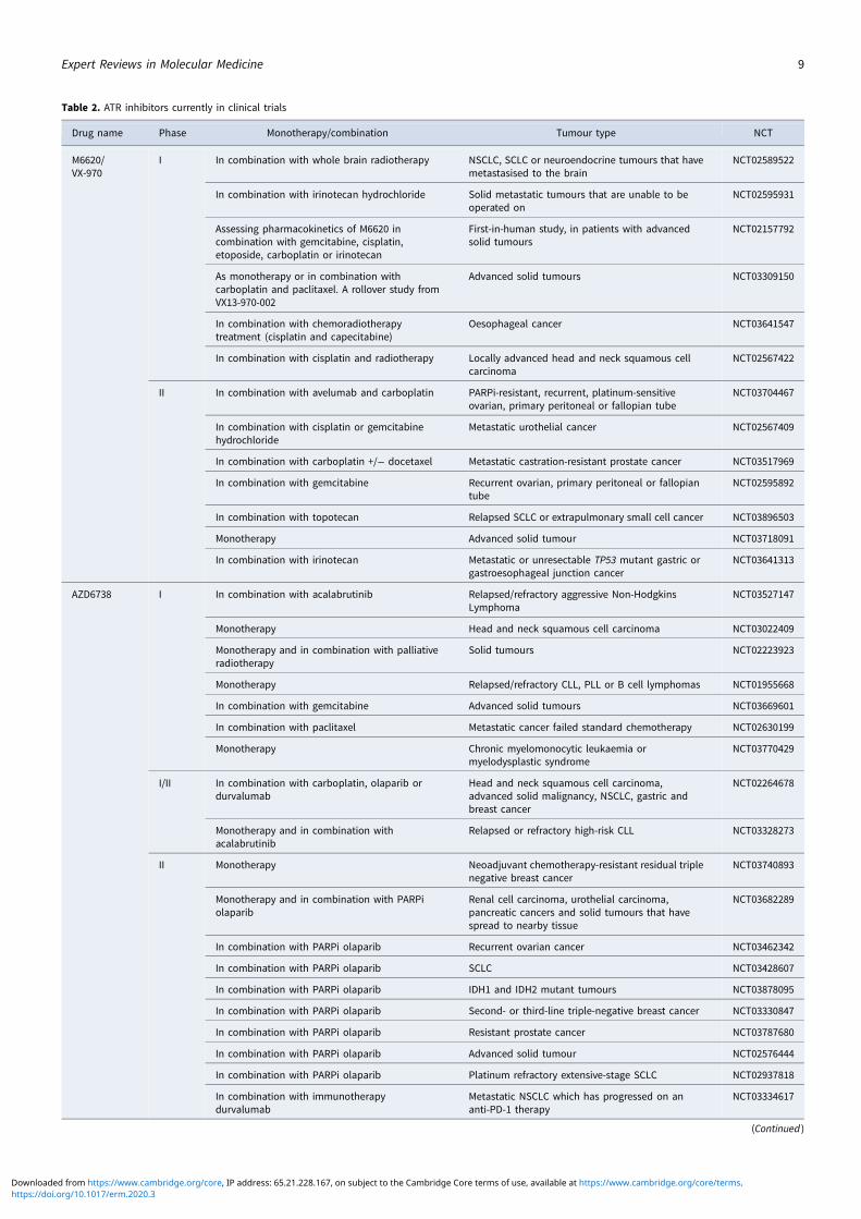
Table 2. ATR inhibitors currently in clinical trials
Drug name Phase Monotherapy/combination Tumour type NCT
M6620/VX-970
I In combination with whole brain radiotherapy NSCLC, SCLC or neuroendocrine tumours that havemetastasised to the brain
NCT02589522
In combination with irinotecan hydrochloride Solid metastatic tumours that are unable to beoperated on
NCT02595931
Assessing pharmacokinetics of M6620 incombination with gemcitabine, cisplatin,etoposide, carboplatin or irinotecan
First-in-human study, in patients with advancedsolid tumours
NCT02157792
As monotherapy or in combination withcarboplatin and paclitaxel. A rollover study fromVX13-970-002
Advanced solid tumours NCT03309150
In combination with chemoradiotherapytreatment (cisplatin and capecitabine)
Oesophageal cancer NCT03641547
In combination with cisplatin and radiotherapy Locally advanced head and neck squamous cellcarcinoma
NCT02567422
II In combination with avelumab and carboplatin PARPi-resistant, recurrent, platinum-sensitiveovarian, primary peritoneal or fallopian tube
NCT03704467
In combination with cisplatin or gemcitabinehydrochloride
Metastatic urothelial cancer NCT02567409
In combination with carboplatin +/− docetaxel Metastatic castration-resistant prostate cancer NCT03517969
In combination with gemcitabine Recurrent ovarian, primary peritoneal or fallopiantube
NCT02595892
In combination with topotecan Relapsed SCLC or extrapulmonary small cell cancer NCT03896503
Monotherapy Advanced solid tumour NCT03718091
In combination with irinotecan Metastatic or unresectable TP53 mutant gastric orgastroesophageal junction cancer
NCT03641313
AZD6738 I In combination with acalabrutinib Relapsed/refractory aggressive Non-HodgkinsLymphoma
NCT03527147
Monotherapy Head and neck squamous cell carcinoma NCT03022409
Monotherapy and in combination with palliativeradiotherapy
Solid tumours NCT02223923
Monotherapy Relapsed/refractory CLL, PLL or B cell lymphomas NCT01955668
In combination with gemcitabine Advanced solid tumours NCT03669601
In combination with paclitaxel Metastatic cancer failed standard chemotherapy NCT02630199
Monotherapy Chronic myelomonocytic leukaemia ormyelodysplastic syndrome
NCT03770429
I/II In combination with carboplatin, olaparib ordurvalumab
Head and neck squamous cell carcinoma,advanced solid malignancy, NSCLC, gastric andbreast cancer
NCT02264678
Monotherapy and in combination withacalabrutinib
Relapsed or refractory high-risk CLL NCT03328273
II Monotherapy Neoadjuvant chemotherapy-resistant residual triplenegative breast cancer
NCT03740893
Monotherapy and in combination with PARPiolaparib
Renal cell carcinoma, urothelial carcinoma,pancreatic cancers and solid tumours that havespread to nearby tissue
NCT03682289
In combination with PARPi olaparib Recurrent ovarian cancer NCT03462342
In combination with PARPi olaparib SCLC NCT03428607
In combination with PARPi olaparib IDH1 and IDH2 mutant tumours NCT03878095
In combination with PARPi olaparib Second- or third-line triple-negative breast cancer NCT03330847
In combination with PARPi olaparib Resistant prostate cancer NCT03787680
In combination with PARPi olaparib Advanced solid tumour NCT02576444
In combination with PARPi olaparib Platinum refractory extensive-stage SCLC NCT02937818
In combination with immunotherapydurvalumab
Metastatic NSCLC which has progressed on ananti-PD-1 therapy
NCT03334617
(Continued )
Expert Reviews in Molecular Medicine 9
https://doi.org/10.1017/erm.2020.3Downloaded from https://www.cambridge.org/core, IP address: 65.21.228.167, on subject to the Cambridge Core terms of use, available at https://www.cambridge.org/core/terms.

combination with gemcitabine and radiation showed promisingresults with good tolerability in patients with locally advancedpancreatic cancer (Ref. 197). The majority of clinical trials arestill recruiting, suggesting this kinase inhibitor has excitingpotential once trials have been completed and data is collectedand analysed (Table 4).
DNA checkpoint kinase and immune checkpoint inhibitorcombinations
Immune checkpoint inhibitors block the immunosuppressivemechanisms employed by cancers to prevent an effective anti-tumour immune response and have been found to be efficacious
in many types of cancer. There is increasing evidence that tumourmutational burden increases the immunogenicity of cancersthrough the production of mutation-associated neoantigens,including those associated with microsatellite instability fromdefective DNA mismatch repair (Ref. 198). Damaged cytosolicDNA may also directly activate the immune system by stimulatinginterferon via the STING pathway (Stimulation of InterferonGenes) leading to enhanced immune checkpoint inhibitorresponses in pre-clinical models. In ATM-deficient mice andpatients with ataxia telangiectasia enhanced interferon productionthrough the STING pathway has been observed (Ref. 198). ATMinhibition has recently been found to increase type 1 interferonsignalling in a STING independent manner (Ref. 199). Clinical
Table 2. (Continued.)
Drug name Phase Monotherapy/combination Tumour type NCT
In combination with immunotherapydurvalumab
NSCLC with PD-1 immune checkpoint inhibitorresistance
NCT03833440
In combination with immunotherapydurvalumab
Solid tumour, gastric cancer with failed secondarychemotherapy, melanoma patients
NCT03780608
BAY1895344 I Monotherapy (first-in-human) Advanced solid tumours and lymphomas NCT03188965
M4344/VX-803
I Monotherapy and in combination with cisplatin,carboplatin and gemcitabine
Advanced solid tumours NCT02278250
Table 3. CHK1 inhibitors currently in clinical trials
Drug name Phase Monotherapy/combination Tumour type NCT
MK-8776/SCH900776
I In combination with and without cytarabine Acute leukaemia NCT00907517
In combination with hydroxyurea Advanced solid tumours NCT01521299
II In combination with and without cytarabine Acute myeloid leukaemia NCT01870596
LY2606368/prexasertib
I In combination with desipramine, pemetrexed orgemcitabine
Drug interaction study NCT01358968
In combination with gemcitabine Solid tumours NCT01341457
In combination with pemetrexed or gemcitabine Advanced/metastatic solid tumours NCT01296568
In combination with pemetrexed Advanced/metastatic solid tumours NCT00415636
Monotherapy Japanese patients with advanced solidtumour
NCT02514603
Monotherapy Paediatric solid tumours NCT02808650
In combination with ralimetanib Advanced solid tumour NCT02860780
In combination with olaparib Advanced solid tumour NCT03057145
In combination with cisplatin, cetuximab orradiotherapy
Head and neck cancer NCT02555644
In combination with PD-L1 inhibitor Advanced solid tumours NCT03495323
In combination with cytarabine Chronic/acute myeloid leukaemia NCT02649764
I/II In combination with and without gemcitabine Pancreatic cancer NCT00839332
In combination with cisplatin or pemetrexed NSCLC NCT01139775
II Monotherapy Extensive stage SCLC NCT02735980
Monotherapy Platinum resistant ovarian cancer NCT03414047
Monotherapy Solid tumour with HRR defects or CCNE1amplification
NCT02873975
In combination with or without pemetrexed Advanced or metastatic NSCLC NCT00988858
Monotherapy BRCA mutant breast or ovarian cancerTriple-negative breast cancerHGSOCCastrate-resistant prostate cancer
NCT02203513
10 Hannah L. Smith et al.
https://doi.org/10.1017/erm.2020.3Downloaded from https://www.cambridge.org/core, IP address: 65.21.228.167, on subject to the Cambridge Core terms of use, available at https://www.cambridge.org/core/terms.

trials evaluating DNA damage repair inhibitors with immunecheckpoint inhibitors are ongoing, including PARP, ATR andCHK1 inhibitors (Table 2).
Concluding remarks and future directions
Targeting DDR checkpoint signalling has evolved to the clinicbased on sound scientific hypotheses and preclinical data.Early less specific inhibitors may have clouded the case fordevelopment but now more specific inhibitors are under inves-tigation both as monotherapy and in combination with conven-tional cytotoxic chemotherapy or novel agents. The preclinicaldata suggest that targeting the ATR-CHK1-WEE1 pathway islikely to be more fruitful than targeting ATM and CHK2 signal-ling. To date, the most active combinations for each class ofkinase inhibitor include ATR inhibitors with cis/carboplatinand CHK1 inhibitors with gemcitabine. A developing field is apotentiation with immune checkpoint inhibitors via severalmechanisms of action. Identification of predictive biomarkers,particularly for monotherapy, however, has been challenging,for example, whether the presence of TP53 mutations conferssensitivity.
The potential for some of these agents to be associated withsecond malignancy must not be forgotten, which is particularlya concern for young patients treated with these agents. Sincedefects in ATM and CHK2 are associated with tumours, butdefects in ATR, CHK1 and WEE1 are not, one might predictthat as single agents, inhibitors of the former might be asso-ciated with second malignancies but not the latter. However,in combination with cytotoxics already associated with secondmalignancies, the incidence is likely to be increased unlesslower doses of the primary cytotoxic can be used in combin-ation to achieve the same efficacy. Nevertheless, we mustremember that the malignancies for which these agents aremost likely to be used, especially in children, are the onesthat are most difficult to cure with current strategies inwhich cure has yet to be achieved for the majority at anycost rather than at least cost.
Acknowledgements. This work was partly funded by the Children’s Cancer& Leukaemia Group and Little Princess Trust and the JGWP foundation.
References
1. Weber AM and Ryan AJ (2015) ATM and ATR as therapeutic targets incancer. Pharmacology & Therapeutics 149(Suppl. C), 124–138.
2. Maréchal A and Zou L (2013) DNA damage sensing by the ATM andATR kinases. Cold Spring Harbor Perspectives in Biology 5, a012716.
3. Kozlov SV et al. (2011) Autophosphorylation and ATM activation: add-itional sites add to the complexity. The Journal of biological chemistry286, 9107–9119.
4. Tanya TP (2015) Mechanisms of ATM activation. Annual Review ofBiochemistry 84, 711–738.
5. Shiloh Y and Ziv Y (2013) The ATM protein kinase: regulating the cel-lular response to genotoxic stress, and more. Nature Reviews MolecularCell Biology 14, 197.
6. Zannini L, Delia D and Buscemi G (2014) CHK2 Kinase in the DNAdamage response and beyond. Journal of Molecular Cell Biology 6,442–457.
7. O’Connell MJ et al. (1997) Chk1 is a wee1 kinase in the G2 DNA dam-age checkpoint inhibiting cdc2 by Y15 phosphorylation. The EMBOJournal 16, 545–554.
8. Chow JPH and Poon RYC (2012) The CDK1 inhibitory kinase MYT1 inDNA damage checkpoint recovery. Oncogene 32, 4778.
9. Kastan MB et al. (1992) A mammalian cell cycle checkpoint pathwayutilizing p53 and GADD45 is defective in ataxia-telangiectasia. Cell 71,587–597.
10. Beamish H et al. (1996) Defect in multiple cell cycle checkpoints inataxia-telangiectasia postirradiation. Journal of Biological Chemistry271, 20486–20493.
11. Weber AM et al. (2016) Phenotypic consequences of somatic mutationsin the ataxia-telangiectasia mutated gene in non-small cell lung cancer.Oncotarget 7, 60807–60822.
12. Ou Y-H et al. (2005) P53 C-terminal phosphorylation by CHK1 andCHK2 participates in the regulation of DNA-damage-induced C-terminalacetylation. Molecular Biology of the Cell 16, 1684–1695.
13. Fischer M (2017) Census and evaluation of p53 target genes. Oncogene36, 3943.
14. Wade M, Wang YV and Wahl GM (2010) The p53 orchestra: Mdm2and Mdmx set the tone. Trends in cell Biology 20, 299–309.
15. Roos WP and Kaina B (2013) DNA damage-induced cell death: fromspecific DNA lesions to the DNA damage response and apoptosis.Cancer Letters 332, 237–248.
16. Abbas T and Dutta A (2009) P21 in cancer: intricate networks and mul-tiple activities. Nature Reviews. Cancer 9, 400–414.
17. Waldman T, Kinzler KW and Vogelstein B (1995) P21 Is necessary forthe p53-mediated G1 arrest in human cancer cells. Cancer Research 55,5187–5190.
Table 4. WEE1 inhibitors currently in clinical trials
Drug name Phase Monotherapy/combination Tumour type NCT
AZD1775/MK-1775
I Monotherapy Advanced/metastatic cancers NCT02610075
Monotherapy Ovarian cancer, SCLC, other solid tumours NCT02482311
In combination with carboplatin and paclitaxel Asian patients with advanced solid tumours NCT02341456
In combination with olaparib Refractory solid tumours NCT02511795
In combination with irinotecan RAS or BRAF mutated second-line metastaticcolorectal cancer
NCT02906059
In combination with either gemcitabine, cisplatinor carboplatin
Advanced solid tumours NCT00648648
In combination with either cisplatin andradiotherapy
Head and neck cancer NCT03028766
II Monotherapy Relapsed SCLC NCT02593019
In combination with docetaxel NSCLC NCT02087176
In combination with carboplatin-paclitaxel SCLC NCT02513563
In combination with cisplatin Triple-negative metastatic breast cancer NCT03012477
In combination with cytarabine Advanced acute myeloid leukaemia ormyelodysplastic syndrome
NCT02666950
Expert Reviews in Molecular Medicine 11
https://doi.org/10.1017/erm.2020.3Downloaded from https://www.cambridge.org/core, IP address: 65.21.228.167, on subject to the Cambridge Core terms of use, available at https://www.cambridge.org/core/terms.

18. Deng C et al. (1995) Mice lacking p21CIP1/WAF1 undergo normal devel-opment, but are defective in G1 checkpoint control. Cell 82, 675–684.
19. Bieging KT, Mello SS and Attardi LD (2014) Unravelling mechanismsof p53-mediated tumour suppression. Nature reviews. Cancer 14, 359–370.
20. Goodarzi AA, Jeggo P and Lobrich M (2010) The influence ofheterochromatin on DNA double strand break repair: getting the strong,silent type to relax. DNA Repair 9, 1273–1282.
21. Goodarzi AA et al. (2008) ATM signaling facilitates repair of DNAdouble-strand breaks associated with heterochromatin. Molecular Cell31, 167–177.
22. Scully R and Xie A (2013) Double strand break repair functions of histoneH2AX. Mutation Research/Fundamental and Molecular Mechanisms ofMutagenesis 750, 5–14.
23. Blackford AN and Jackson SP (2017) ATM, ATR, and DNA-PK: thetrinity at the heart of the DNA damage response. Molecular Cell 66,801–817.
24. Melander F et al. (2008) Phosphorylation of SDT repeats in the MDC1N terminus triggers retention of NBS1 at the DNA damage-modifiedchromatin. The Journal of Cell Biology 181, 213–226.
25. Spycher C et al. (2008) Constitutive phosphorylation of MDC1 physic-ally links the MRE11–RAD50–NBS1 complex to damaged chromatin.The Journal of Cell Biology 181, 227–240.
26. Panier S and Boulton SJ (2013) Double-strand break repair: 53BP1comes into focus. Nature Reviews Molecular Cell Biology 15, 7.
27. Greenberg RA (2008) Recognition of DNA double strand breaks by theBRCA1 tumor suppressor network. Chromosoma 117, 305–317.
28. Prakash R et al. (2015) Homologous recombination and human health:the roles of BRCA1, BRCA2, and associated proteins. Cold Spring HarborPerspectives in Biology 7, a016600–a00.
29. Chen X et al. (2011) Cell cycle regulation of DNA double-strand breakend resection by Cdk1-dependent Dna2 phosphorylation. NatureStructural & Molecular Biology 18, 1015–1019.
30. Ceccaldi R, Rondinelli B and D’Andrea AD (2016) Repair pathwaychoices and consequences at the double-strand break. Trends in CellBiology 26, 52–64.
31. Jazayeri A et al. (2006) ATM- and cell cycle-dependent regulation ofATR in response to DNA double-strand breaks. Nature Cell Biology 8,37–45.
32. Myers JS and Cortez D (2006) Rapid activation of ATR by ionizing radi-ation requires ATM and Mre11. Journal of Biological Chemistry 281,9346–9350.
33. Rothblum-Oviatt C et al. (2016) Ataxia telangiectasia: a review.Orphanet Journal of Rare Diseases 11, 159–159.
34. Choi M, Kipps T and Kurzrock R (2016) ATM mutations in cancer:therapeutic implications. Molecular Cancer Therapeutics 15, 1781–1791.
35. Wang C et al. (2017) ATM-deficient colorectal cancer cells are sensitiveto the PARP inhibitor Olaparib(). Translational Oncology 10, 190–196.
36. Abdel-Fatah TMA et al. (2014) Clinicopathological significance ofATM-Chk2 expression in sporadic breast cancers: a comprehensive ana-lysis in large cohorts. Neoplasia (New York, N.Y.) 16, 982–991.
37. Petersen LF et al. (2017) Loss of tumour-specific ATM protein expres-sion is an independent prognostic factor in early resected NSCLC.Oncotarget 8, 38326–38336.
38. Villaruz LC et al. (2016) ATM Protein is deficient in over 40% of lungadenocarcinomas. Oncotarget 7, 57714–57725.
39. Russell R et al. (2015) Loss of ATM accelerates pancreatic cancer forma-tion and epithelial-mesenchymal transition. Nature Communications 6,7677–7677.
40. Ouillette P et al. (2010) Aggressive chronic lymphocytic leukemia withelevated genomic complexity is associated with multiple gene defects inthe response to DNA double-strand breaks. Clinical Cancer Research16, 835–847.
41. Mlakar V et al. (2017) 11q Deletion in neuroblastoma: a review of bio-logical and clinical implications. Molecular cancer 16, 114.
42. Laake K et al. (1999) Loss of heterozygosity at 11q23. 1 and survival inbreast cancer: results of a large European study. Genes, Chromosomes andCancer 25, 212–221.
43. Monni O and Knuutila S (2001) 11q deletions in hematological malig-nancies. Leukemia & Lymphoma 40, 259–266.
44. Cybulski C et al. (2004) CHEK2 Is a multiorgan cancer susceptibilitygene. American Journal of Human Genetics 75, 1131–1135.
45. Lee SB et al. (2001) Destabilization of CHK2 by a missense mutationassociated with Li-Fraumeni syndrome. Cancer Research 61, 8062–8067.
46. Varley JM (2003) Germline TP53 mutations and Li-Fraumeni syndrome.Human Mutation 21, 313–320.
47. Lawrence MS et al. (2014) Discovery and saturation analysis of cancergenes across 21 tumour types. Nature 505, 495.
48. Bouaoun L et al. (2016) TP53 Variations in human cancers: new lessonsfrom the IARC TP53 database and genomics data. Human Mutation 37,865–876.
49. Hainaut P and Pfeifer GP (2016) Somatic TP53 mutations in the era ofgenome sequencing. Cold Spring Harbor Perspectives in Medicine 6,a026179.
50. Liu Y et al. (2016) Deletions linked to TP53 loss drive cancer throughp53-independent mechanisms. Nature 531, 471–475.
51. Fiskvik I et al. (2013) Karyotyping of diffuse large B-cell lymphomas:loss of 17p is associated with poor patient outcome. European Journalof Haematology 91, 332–338.
52. Hallek M (2017) Chronic lymphocytic leukemia: 2017 update on diagno-sis, risk stratification, and treatment. American Journal of Hematology 92,946–965.
53. Bartek J, Lukas J and Bartkova J (2007) DNA Damage response as ananti-cancer barrier: damage threshold and the concept of ‘conditionalhaploinsufficiency’. Cell Cycle 6, 2344–2347.
54. Barlow C et al. (1996) Atm-deficient mice: a paradigm of ataxia telangi-ectasia. Cell 86, 159–171.
55. Xu Y and Baltimore D (1996) Dual roles of ATM in the cellularresponse to radiation and in cell growth control. Genes & Development10, 2401–2410.
56. Jiang X et al. (2019) Current status and future prospects of PARP inhibi-tor clinical trials in ovarian cancer. Cancer Management and Research 11,4371.
57. Rabik CA, Njoku MC and Dolan ME (2006) Inactivation of O6-alkylguanine DNA alkyltransferase as a means to enhance chemotherapy.Cancer Treatment Reviews 32, 261–276.
58. Takai H et al. (2002) Chk2-deficient mice exhibit radioresistance anddefective p53-mediated transcription. The EMBO Journal 21, 5195–5205.
59. Niida H et al. (2010) Cooperative functions of Chk1 and Chk2 reducetumour susceptibility in vivo. The EMBO Journal 29, 3558–3570.
60. Brandsma I et al. (2017) Directing the use of DDR kinase inhibitors incancer treatment. Expert Opinion on Investigational Drugs 26, 1341–1355.
61. Hickson I et al. (2004) Identification and characterization of a novel andspecific inhibitor of the ataxia-telangiectasia mutated kinase ATM.Cancer Research 64, 9152–9159.
62. Biddlestone-Thorpe L et al. (2013) ATM kinase inhibition preferentiallysensitizes p53-mutant glioma to ionizing radiation. Clinical CancerResearch 19, 3189–3200.
63. Batey MA et al. (2013) Preclinical evaluation of a novel ATM inhibitor,KU59403, in vitro and in vivo in p53 functional and dysfunctionalmodels of human cancer. Molecular Cancer Therapeutics 12, 959–967.
64. Karlin J et al. (2018) Orally bioavailable and blood–brain barrier-penetrating ATM inhibitor (AZ32) radiosensitizes intracranial gliomasin mice. Molecular Cancer Therapeutics 17, 1637–1647.
65. Durant ST et al. (2018) The brain-penetrant clinical ATM inhibitorAZD1390 radiosensitizes and improves survival of preclinical braintumor models. Science Advances 4, eaat1719–eaeaat19.
66. Pike KG et al. (2018) The identification of potent, selective, and orallyavailable inhibitors of ataxia telangiectasia mutated (ATM) kinase: thediscovery of AZD0156 (8-{6-[3-(dimethylamino)propoxy]pyridin-3-yl}-3-methyl-1-(tetrahydro-2H-pyran-4-yl)-1,3-dihydro-2H-imidazo[4,5-c]quinolin-2-one). Journal of Medicinal Chemistry 61, 3823–3841.
67. Rainey MD et al. (2008) Transient inhibition of ATM kinase is sufficientto enhance cellular sensitivity to ionizing radiation. Cancer Research 68,7466–7474.
68. Dohmen AJC et al. (2017) Identification of a novel ATM inhibitor withcancer cell specific radiosensitization activity. Oncotarget 8, 73925–73937.
69. Ronco C et al. (2017) ATM, ATR, CHK1, CHK2 and WEE1 inhibitors incancer and cancer stem cells. Medicinal Chemistry Communications 8,295–319.
70. Zabludoff SD et al. (2008) AZD7762, A novel checkpoint kinase inhibi-tor, drives checkpoint abrogation and potentiates DNA-targeted therap-ies. Molecular Cancer Therapeutics 7, 2955–2966.
12 Hannah L. Smith et al.
https://doi.org/10.1017/erm.2020.3Downloaded from https://www.cambridge.org/core, IP address: 65.21.228.167, on subject to the Cambridge Core terms of use, available at https://www.cambridge.org/core/terms.

71. Arienti KL et al. (2005) Checkpoint kinase inhibitors: SAR and radio-protective properties of a series of 2-arylbenzimidazoles. Journal ofMedicinal Chemistry 48, 1873–1885.
72. Carlessi L et al. (2007) Biochemical and cellular characterization ofVRX0466617, a novel and selective inhibitor for the checkpoint kinaseChk2. Molecular Cancer Therapeutics 6, 935–944.
73. Jobson AG et al. (2009) Cellular inhibition of checkpoint kinase 2(Chk2) and potentiation of camptothecins and radiation by the novelChk2 inhibitor PV1019 [7-nitro-1H-indole-2-carboxylic acid {4-[1-(gua-nidinohydrazone)-ethyl]-phenyl}-amide]. Journal of Pharmacology andExperimental Therapeutics 331, 816–826.
74. Caldwell JJ et al. (2011) Structure-Based design of potent and selective2-(quinazolin-2-yl)phenol inhibitors of checkpoint kinase 2. Journal ofMedicinal Chemistry 54, 580–590.
75. Raso A et al. (2012) Characterization of glioma stem cells through mul-tiple stem cell markers and their specific sensitization to double-strandbreak-inducing agents by pharmacological inhibition of ataxia telangi-ectasia mutated protein. Brain Pathology 22, 677–688.
76. Anderson VE et al. (2011) CCT241533 is a potent and selective inhibitorof CHK2 that potentiates the cytotoxicity of PARP inhibitors. CancerResearch 71, 463–472.
77. Pires IM, Ward TH and Dive C (2010) Oxaliplatin responses in colorec-tal cancer cells are modulated by CHK2 kinase inhibitors. British Journalof Pharmacology 159, 1326–1338.
78. Sausville E et al. (2014) Phase I dose-escalation study of AZD7762, acheckpoint kinase inhibitor, in combination with gemcitabine in USpatients with advanced solid tumors. Cancer Chemotherapy andPharmacology 73, 539–549.
79. Saldivar J, Cortez D and Cimprich K (2017) The essential kinase ATR:ensuring faithful duplication of a challenging genome. Nature ReviewsMolecular Cell Biology 18, 622–636.
80. Thada V and Cortez D (2019) Common motifs in ETAA1 and TOPBP1required for ATR kinase activation. Journal of Biological Chemistry 294,8395–8402.
81. Rundle S et al. (2017) Targeting the ATR-CHK1 axis in cancer therapy.Cancers 9, 41.
82. Lewis CW et al. (2017) Prolonged mitotic arrest induced by Wee1inhibition sensitizes breast cancer cells to paclitaxel. Oncotarget 8,73705–73722.
83. Lin AB, McNeely SC and Beckmann RP (2017) Achieving precisiondeath with cell-cycle inhibitors that target DNA replication and repair.Clinical Cancer Research 23, 3232–3240.
84. Lee J, Kumagai A and Dunphy WG (2001) Positive regulation of wee1 byChk1 and 14-3-3 proteins. Molecular Biology of the Cell 12, 551–563.
85. Wyman C, Ristic D and Kanaar R (2004) Homologous recombination-mediated double-strand break repair. DNA Repair (Amst) 3, 827–833.
86. Brown JS et al. (2017) Targeting DNA repair in cancer: beyond PARPinhibitors. Cancer Discovery 7, 20–37.
87. Yazinski SA et al. (2017) ATR Inhibition disrupts rewired homologousrecombination and fork protection pathways in PARP inhibitor-resistantBRCA-deficient cancer cells. Genes & Development 31, 318–332.
88. Sørensen CS et al. (2005) The cell-cycle checkpoint kinase Chk1 isrequired for mammalian homologous recombination repair. NatureCell Biology 7, 195–201.
89. Buisson R et al. (2017) Coupling of homologous recombination and thecheckpoint by ATR. Molecular Cell 65, 336–346.
90. Krajewska M et al. (2012) Forced activation of Cdk1 via wee1 inhibitionimpairs homologous recombination. Oncogene 32, 3001.
91. de Klein A et al. (2000) Targeted disruption of the cell-cycle checkpointgene ATR leads to early embryonic lethality in mice. Current Biology 10,479–482.
92. Liu Q et al. (2000) Chk1 is an essential kinase that is regulated by Atrand required for the G(2)/M DNA damage checkpoint. Genes &Development 14, 1448–1459.
93. Tominaga Y et al. (2006) Murine Wee1 plays a critical role in cell cycleregulation and pre-implantation stages of embryonic development.International Journal of Biological Sciences 2, 161–170.
94. Murga M (2009) A mouse model of ATR-Seckel shows embryonic rep-licative stress and accelerated aging. Nature Genetics 41, 891–898.
95. Brown EJ and Baltimore D (2000) ATR Disruption leads to chromosomalfragmentation and early embryonic lethality. Genes & Development 14,397–402.
96. Macheret M and Halazonetis TD (2015) DNA replication stress as aHallmark of cancer. Annual Review of Pathology: Mechanisms ofDisease 10, 425–448.
97. Magdalou I et al. (2014) The causes of replication stress and theirconsequences on genome stability and cell fate. Seminars in Cell &Developmental Biology 30, 154–164.
98. Berti M and Vindigni A (2016) Replication stress: getting back on track.Nature Structural & Molecular Biology 23, 103–109.
99. Massague J (2004) G1 cell-cycle control and cancer. Nature 432, 298–306.
100. Kotsantis P, Petermann E and Boulton SJ (2018) Mechanisms ofoncogene-induced replication stress: jigsaw falling into place. CancerDiscovery 8, 537–555.
101. Hanahan D and Weinberg RA (2011) Hallmarks of cancer: the nextgeneration. Cell 144, 646–674.
102. Syljuåsen RG et al. (2005) Inhibition of human Chk1 causes increasedinitiation of DNA replication, phosphorylation of ATR targets, andDNA breakage. Molecular and Cellular Biology 25, 3553–3562.
103. Kitao H et al. (2018) DNA Replication stress and cancer chemotherapy.Cancer Science 109, 264–271.
104. Kim J-Y et al. (2015) Resveratrol analogue (E)-8-acetoxy-2-[2-(3, 4–dia-cetoxyphenyl) ethenyl]-quinazoline induces G2/M cell cycle arrestthrough the activation of ATM/ATR in human cervical carcinomaHeLa cells. Oncology Reports 33, 2639–2647.
105. Cliby WA et al. (1998) Overexpression of a kinase-inactive ATR proteincauses sensitivity to DNA-damaging agents and defects in cell cyclecheckpoints. The EMBO Journal 17, 159–169.
106. Hall-Jackson CA et al. (1999) ATR Is a caffeine-sensitive, DNA-activatedprotein kinase with a substrate specificity distinct from DNA-PK.Oncogene 18, 6707–6713.
107. Sarkaria JN et al. (1999) Inhibition of ATM and ATR kinase activities bythe radiosensitizing agent, Caffeine. Cancer Research 59, 4375–4382.
108. Nghiem P et al. (2002) ATR Is not required for p53 activation but syner-gizes with p53 in the replication checkpoint. Journal of BiologicalChemistry 277, 4428–4434.
109. Peasland A et al. (2011) Identification and evaluation of a potent novelATR inhibitor, NU6027, in breast and ovarian cancer cell lines. BritishJournal of Cancer 105, 372–381.
110. Toledo LI et al. (2011) A cell-based screen identifies ATR inhibitors withsynthetic lethal properties for cancer-associated mutations. NatureStructural & Molecular Biology 18, 721–727.
111. Teng P-n et al. (2015) Pharmacologic inhibition of ATR and ATM offersclinically important distinctions to enhancing platinum or radiationresponse in ovarian, endometrial, and cervical cancer cells. GynecologicOncology 136, 554–561.
112. Foote KM et al. (2013) Discovery of 4-{4-[(3R)-3-methylmorpholin-4-yl]-6-[1-(methylsulfonyl)cyclopropyl]pyrimidin-2-yl}-1H-indole (AZ20):a potent and selective inhibitor of ATR protein kinase with monotherapyin vivo antitumor activity. Journal of Medicinal Chemistry 56, 2125–2138.
113. Ma J et al. (2017) Mechanisms responsible for the synergistic antileuke-mic interactions between ATR inhibition and cytarabine in acute myeloidleukemia cells. Scientific Reports 7, 41950–41950.
114. Liu S et al. (2017) Inhibition of ATR potentiates the cytotoxic effect ofgemcitabine on pancreatic cancer cells through enhancement of DNAdamage and abrogation of ribonucleotide reductase induction by gemci-tabine. Oncology Reports 37, 3377–3386.
115. Foote KM et al. (2018) Discovery and characterization of AZD6738, apotent inhibitor of ataxia telangiectasia mutated and Rad3 related(ATR) kinase with application as an anticancer agent. Journal ofMedicinal Chemistry 61, 9889–9907.
116. Vendetti FP et al. (2015) The orally active and bioavailable ATR kinaseinhibitor AZD6738 potentiates the anti-tumor effects of cisplatin toresolve ATM-deficient non-small cell lung cancer in vivo. Oncotarget6, 44289–44305.
117. Dillon MT et al. (2017) Radiosensitization by the ATR inhibitor AZD6738through generation of acentric micronuclei.Molecular Cancer Therapeutics16, 25–34.
118. Min A et al. (2017) AZD6738, a novel oral inhibitor of ATR, induces syn-thetic lethality with ATM deficiency in gastric cancer cells. MolecularCancer Therapeutics 16, 566–577. research 78(13 Supplement), 838–38.
119. Reaper PM et al. (2011) Selective killing of ATM-or p53-deficient cancercells through inhibition of ATR. Nature Chemical Biology 7, 428–430.
Expert Reviews in Molecular Medicine 13
https://doi.org/10.1017/erm.2020.3Downloaded from https://www.cambridge.org/core, IP address: 65.21.228.167, on subject to the Cambridge Core terms of use, available at https://www.cambridge.org/core/terms.

120. Alsubhi N et al. (2016) Chk1 phosphorylated at serine345 is a predictorof early local recurrence and radio-resistance in breast cancer. MolecularOncology 10, 213–223.
121. Fujisawa H et al. (2015) VE-821, an ATR inhibitor, causes radiosensiti-zation in human tumor cells irradiated with high LET radiation.Radiation Oncology (London, England) 10, 175–175.
122. Šalovská B et al. (2018) Radio-sensitizing effects of VE-821 and beyond:distinct phosphoproteomic and metabolomic changes after ATR inhib-ition in irradiated MOLT-4 cells. PLoS One 13, e0199349–e49.
123. Jossé R et al. (2014) ATR Inhibitors VE-821 and VX-970 sensitize cancercells to topoisomerase i inhibitors by disabling DNA replication initiationand fork elongation responses. Cancer Research 74, 6968–6979.
124. Huntoon CJ et al. (2013) ATR inhibition broadly sensitizes ovarian can-cer cells to chemotherapy independent of BRCA status. Cancer Research73, 3683–3691.
125. Middleton FK et al. (2015) Common cancer-associated imbalances inthe DNA damage response confer sensitivity to single agent ATR inhib-ition. Oncotarget 6, 32396–32409.
126. Hall AB et al. (2014) Potentiation of tumor responses to DNA dam-aging therapy by the selective ATR inhibitor VX-970. Oncotarget 5,5674–5685.
127. Leszczynska KB et al. (2016) Preclinical testing of an Atr inhibitordemonstrates improved response to standard therapies for esophagealcancer. Radiotherapy and Oncology: Journal of the European Society forTherapeutic Radiology and Oncology 121, 232–238.
128. Shi Q et al. (2018) The identification of the ATR inhibitor VE-822 as atherapeutic strategy for enhancing cisplatin chemosensitivity in esopha-geal squamous cell carcinoma. Cancer Letters 432, 56–68.
129. Kurmasheva RT et al. (2018) Initial testing (stage 1) of M6620 (formerlyVX-970), a novel ATR inhibitor, alone and combined with cisplatin andmelphalan, by the Pediatric Preclinical Testing Program. Pediatric Blood& Cancer 65, e26825–n/a.
130. Fordham SE et al. (2018) Inhibition of ATR acutely sensitizes acute mye-loid leukemia cells to nucleoside analogs that target ribonucleotide reduc-tase. Blood Advances 2, 1157–1169.
131. Tu X et al. (2018) ATR inhibition is a promising radiosensitizing strategyfor triple-negative breast cancer. Molecular Cancer Therapeutics 17,2462–2472.
132. Nagel R et al. (2019) Inhibition of the replication stress response is a syn-thetic vulnerability in SCLC that acts synergistically in combination withcisplatin. Molecular Cancer Therapeutics 18, 762–770.
133. Schmitt A et al. (2017) ATM deficiency Is associated with sensitivity toPARP1- and ATR inhibitors in lung adenocarcinoma. Cancer Research77, 3040–3056.
134. Wengner AM et al. (2018) Synergistic in Vivo activity of the ATR inhibi-tor BAY 1895344 in combination with the targeted alpha therapyradium-223 dichloride in a preclinical tumor model mimicking bonemetastatic castration-resistant prostate cancer (mCRPC). CancerResearch 78(13 Suppl), Abstract nr 838.
135. Shapiro GI and Harper JW (1999) Anticancer drug targets: cell cycleand checkpoint control. The Journal of Clinical Investigation 104,1645–1653.
136. Graves PR et al. (2000) The Chk1 protein kinase and the Cdc25C regu-latory pathways are targets of the anticancer agent UCN-01. Journal ofBiological Chemistry 275, 5600–5605.
137. Xiao Y et al. (2013) Identification of preferred chemotherapeutics forcombining with a CHK1 inhibitor. Molecular Cancer Therapeutics 12,2285–2295.
138. Isono M et al. (2017) Checkpoint kinase inhibitor AZD7762 stronglysensitises urothelial carcinoma cells to gemcitabine. Journal ofExperimental & Clinical Cancer Research: CR 36, 1–1.
139. Xu H et al. (2011) Checkpoint kinase inhibitor synergizes withDNA-damaging agents in G1 checkpoint-defective neuroblastoma.International Journal of Cancer 129, 1953–1962.
140. Gadhikar MA et al. (2013) Chk1/2 inhibition overcomes the cisplatinresistance of head and neck cancer cells secondary to the loss of func-tional p53. Molecular Cancer Therapeutics 12, 1860–1873.
141. Bartucci M et al. (2011) Therapeutic targeting of Chk1 in NSCLC stemcells during chemotherapy. Cell Death and Differentiation 19, 768.
142. Blasina A et al. (2008) Breaching the DNA damage checkpoint viaPF-00477736, a novel small-molecule inhibitor of checkpoint kinase 1.Molecular Cancer Therapeutics 7, 2394–2404.
143. Bryant C, Rawlinson R and Massey AJ (2014) Chk1 inhibition as anovel therapeutic strategy for treating triple-negative breast and ovariancancers. BMC Cancer 14, 570–570.
144. Kim MK, James J and Annunziata CM (2015) Topotecan synergizeswith CHEK1 (CHK1) inhibitor to induce apoptosis in ovarian cancercells. BMC Cancer 15, 196–196.
145. Fang DD et al. (2013) Combined gemcitabine and CHK1 inhibitor treat-ment induces apoptosis resistance in cancer stem cell-like cells enrichedwith tumor spheroids from a non-small cell lung cancer cell line.Frontiers of Medicine 7, 462–476.
146. Zhang C et al. (2009) PF-00477736 mediates checkpoint kinase 1 signal-ing pathway and potentiates docetaxel-induced efficacy in Xenografts.Clinical Cancer Research 15, 4630–4640.
147. Busch C-J et al. (2013) HPV-positive HNSCC cell lines but not primaryhuman fibroblasts are radiosensitized by the inhibition of Chk1.Radiotherapy and Oncology 108, 495–499.
148. Restelli V et al. (2015) Characterization of a mantle cell lymphoma cellline resistant to the Chk1 inhibitor PF-00477736. Oncotarget 6, 37229–37240.
149. Nguyen T et al. (2015) Synergism between bosutinib (SKI-606) and theChk1 inhibitor (PF-00477736) in highly imatinib-resistant BCR/ABL+
leukemia cells. Leukemia Research 39, 65–71.150. Ferrao PT et al. (2011) Efficacy of CHK inhibitors as single agents in
MYC-driven lymphoma cells. Oncogene 31, 1661.151. Suzuki M et al. (2017) MK-8776, a novel Chk1 inhibitor, exhibits an
improved radiosensitizing effect compared to UCN-01 by exacerbatingradiation-induced aberrant mitosis. Translational Oncology 10, 491–500.
152. Zhou Z-R et al. (2017) The Chk1 inhibitor MK-8776 increases the radio-sensitivity of human triple-negative breast cancer by inhibiting autop-hagy. Acta Pharmacologica Sinica 38, 513–523.
153. Bridges KA et al. (2016) MK-8776, a novel chk1 kinase inhibitor, radio-sensitizes p53-defective human tumor cells. Oncotarget 7, 71660–71672.
154. Montano R et al. (2013) Sensitization of human cancer cells to gemcita-bine by the Chk1 inhibitor MK-8776: cell cycle perturbation and impactof administration schedule in vitro and in vivo. BMC Cancer 13, 604.
155. Herůdková J et al. (2017) Chk1 inhibitor SCH900776 effectively potenti-ates the cytotoxic effects of platinum-based chemotherapeutic drugs inhuman colon cancer cells. Neoplasia (New York, N.Y.) 19, 830–841.
156. Walton MI et al. (2012) CCT244747 Is a novel potent and selectiveCHK1 inhibitor with oral efficacy alone and in combination with geno-toxic anticancer drugs. Clinical Cancer Research: an Official Journal ofthe American Association for Cancer Research 18, 5650–5661.
157. Patel R et al. (2017) An orally bioavailable Chk1 inhibitor, CCT244747,sensitizes bladder and head and neck cancer cell lines to radiation.Radiotherapy and Oncology: Journal of the European Society forTherapeutic Radiology and Oncology 122, 470–475.
158. Booth L et al. (2018) The CHK1 inhibitor SRA737 synergizes withPARP1 inhibitors to kill carcinoma cells. Cancer Biology & Therapy 19,786–796.
159. King C et al. (2015) LY2606368 causes replication catastrophe and anti-tumor effects through CHK1-dependent mechanisms. Molecular CancerTherapeutics 14, 2004–2013.
160. Ghelli Luserna Di Rorà A et al. (2016) Prexasertib, a Chk1/Chk2 inhibi-tor, increases the effectiveness of conventional therapy in B-/T- cell pro-genitor acute lymphoblastic leukemia. Oncotarget 7, 53377–53391.
161. Sen T et al. (2017) CHK1 inhibition in small-cell lung cancer producessingle-agent activity in biomarker-defined disease subsets and combin-ation activity with Cisplatin or Olaparib. Cancer research 77, 3870–3884.
162. Lowery CD et al. (2017) The checkpoint kinase 1 inhibitor prexasertibinduces regression of preclinical models of human neuroblastoma.Clinical Cancer Research 23, 4354–4363.
163. Brill E et al. (2017) Prexasertib, a cell cycle checkpoint kinases 1 and 2inhibitor, increases in vitro toxicity of PARP inhibition by preventingRad51 foci formation in BRCA wild type high-grade serous ovariancancer. Oncotarget 8, 111026–40.
164. Yin Y et al. (2017) Chk1 inhibition potentiates the therapeutic efficacy ofPARP inhibitor BMN673 in gastric cancer. American Journal of CancerResearch 7, 473–483.
165. Hsu W-H et al. (2019) Checkpoint kinase 1 inhibition enhances cisplatincytotoxicity and overcomes cisplatin resistance in SCLC by promotingmitotic cell death. Journal of Thoracic Oncology 14, 1032–1045.
14 Hannah L. Smith et al.
https://doi.org/10.1017/erm.2020.3Downloaded from https://www.cambridge.org/core, IP address: 65.21.228.167, on subject to the Cambridge Core terms of use, available at https://www.cambridge.org/core/terms.

166. Wang Y et al. (2001) Radiosensitization of p53 mutant cells byPD0166285, a novel G2 checkpoint Abrogator. Cancer Research 61,8211–8217.
167. Mir SE et al. (2010) In silico analysis of kinase expression identifiesWEE1 as a gatekeeper against mitotic catastrophe in glioblastoma.Cancer Cell 18, 244–257.
168. PosthumaDeBoer J et al. (2011) WEE1 Inhibition sensitizes osteosar-coma to radiotherapy. BMC Cancer 11, 156–156.
169. de Gooijer MC et al. (2018) ATP-binding cassette transporters limit thebrain penetration of Wee1 inhibitors. Investigational New Drugs 36, 380–387.
170. Panek RL et al. (1997) In vitro pharmacological characterization of PD166285, a new nanomolar potent and broadly active protein tyrosine kinaseinhibitor. Journal of Pharmacology and Experimental Therapeutics 283,1433–1444.
171. Hirai H et al. (2009) Small-molecule inhibition of Wee1 kinase byMK-1775 selectively sensitizes p53-deficient tumor cells to DNA-damaging agents. Molecular Cancer Therapeutics 8, 2992–3000.
172. Kreahling JM et al. (2012) MK1775, A selective Wee1 inhibitor, showssingle-agent antitumor activity against sarcoma cells. Molecular CancerTherapeutics 11, 174–182.
173. Hirai H et al. (2010) MK-1775, a small molecule Wee1 inhibitor,enhances anti-tumor efficacy of various DNA-damaging agents, includ-ing 5-fluorouracil. Cancer Biology & Therapy 9, 514–522.
174. Rajeshkumar NV et al. (2011) MK-1775, a potent Wee1 inhibitor,synergizes with gemcitabine to achieve tumor regressions, selectively inp53-deficient pancreatic cancer xenografts. Clinical Cancer Research: anOfficial Journal of the American Association for Cancer Research 17,2799–2806.
175. Bridges KA et al. (2011) MK-1775, a novel Wee1 kinase inhibitor, radio-sensitizes p53-defective human tumor cells. Clinical Cancer Research: anOfficial Journal of the American Association for Cancer Research 17,5638–5648.
176. Sarcar B et al. (2011) Targeting radiation-induced G(2) checkpoint acti-vation with the Wee-1 inhibitor MK-1775 in glioblastoma cell lines.Molecular Cancer Therapeutics 10, 2405–2414.
177. Heijink AM et al. (2015) A haploid genetic screen identifies the G1/Sregulatory machinery as a determinant of Wee1 inhibitor sensitivity.Proceedings of the National Academy of Sciences of the United States ofAmerica 112, 15160–15165.
178. Sanjiv K et al. (2016) Cancer-specific synthetic lethality between ATRand CHK1 kinase activities. Cell Reports 14, 298–309.
179. Jin J et al. (2018) Combined inhibition of ATR and WEE1 as a noveltherapeutic strategy in triple-negative breast cancer. Neoplasia(New York, N.Y.) 20, 478–488.
180. Bukhari AB et al. (2019) Inhibiting Wee1 and ATR kinases producestumor-selective synthetic lethality and suppresses metastasis. TheJournal of Clinical Investigation 129, 1329–1344.
181. Chaudhuri L et al. (2014) CHK1 And WEE1 inhibition combine syner-gistically to enhance therapeutic efficacy in acute myeloid leukemia exvivo. Haematologica 99, 688–696.
182. Russell MR et al. (2013) Combination therapy targeting the Chk1 andWee1 kinases shows therapeutic efficacy in neuroblastoma. CancerResearch 73, 776–784.
183. Hauge S et al. (2017) Combined inhibition of Wee1 and Chk1 givessynergistic DNA damage in S-phase due to distinct regulation of CDKactivity and CDC45 loading. Oncotarget 8, 10966–10979.
184. Magnussen GI et al. (2015) Combined inhibition of the cell cycle relatedproteins Wee1 and Chk1/2 induces synergistic anti-cancer effect in mel-anoma. BMC Cancer 15, 462–462.
185. Chilà R et al. (2015) Combined inhibition of Chk1 and Wee1 as a newtherapeutic strategy for mantle cell lymphoma. Oncotarget 6, 3394–3408.
186. Mohni KN et al. (2015) A synthetic lethal screen identifies DNA repairpathways that sensitize cancer cells to combined ATR inhibition and cis-platin treatments. PLoS One 10, e0125482–e82.
187. Kim H et al. (2017) Targeting the ATR/CHK1 axis with PARP inhibitionresults in tumor regression in BRCA-mutant ovarian cancer models.Clinical Cancer Research: an Official Journal of the AmericanAssociation for Cancer Research 23, 3097–3108.
188. Booth L et al. (2013) PARP and CHK inhibitors interact to cause DNAdamage and cell death in mammary carcinoma cells. Cancer Biology &Therapy 14, 458–465.
189. Karnak D et al. (2014) Combined inhibition of Wee1 and PARP1/2 forradiosensitization in pancreatic cancer. Clinical Cancer Research: anOfficial Journal of the American Association for Cancer Research 20,5085–5096.
190. Parsels LA et al. (2018) PARP1 trapping and DNA replication stressenhance radiosensitization with combined WEE1 and PARP inhibitors.Molecular Cancer Research: MCR 16, 222–232.
191. Thomas A et al. (2018) Phase I study of ATR inhibitor M6620 in com-bination with topotecan in patients with advanced solid tumors. Journalof Clinical Oncology: Official Journal of the American Society of ClinicalOncology 36, 1594–1602.
192. Bono JSD et al. (2019) First-in-human trial of the oral ataxia telangiecta-sia and Rad3-related (ATR) inhibitor BAY 1895344 in patients (pts) withadvanced solid tumors. Journal of Clinical Oncology 37(15_suppl), 3007–3007.
193. Samadder P et al. (2017) Synthesis and profiling of a novel potent selectiveinhibitor of CHK1 kinase possessing unusual N-trifluoromethylpyrazolepharmacophore resistant to metabolic N-dealkylation. Molecular CancerTherapeutics 16, 1831–1842.
194. Leijen S et al. (2016) Phase II study of WEE1 inhibitor AZD1775 pluscarboplatin in patients With TP53-mutated ovarian cancer refractoryor resistant to first-line therapy within 3 months. Journal of ClinicalOncology 34, 4354–4361.
195. Oza AM et al. (2015) An international, biomarker-directed, randomized,phase II trial of AZD1775 plus paclitaxel and carboplatin (P/C) for thetreatment of women with platinum-sensitive, TP53–mutant ovarian can-cer. Journal of Clinical Oncology 33(15_suppl), 5506–5506.
196. Lheureux S et al. (2015) A randomized, placebo-controlled phase II trialcomparing gemcitabine monotherapy to gemcitabine in combinationwith AZD 1775 (MK 1775) in women with recurrent, platinum-resistantepithelial ovarian, primary peritoneal, or Fallopian tube cancers: trial ofprincess Margaret, Mayo, Chicago, and California consortia. Journal ofClinical Oncology 33(15_suppl), TPS5613–TPTPS13.
197. Cuneo KC et al. (2019) Dose escalation trial of the wee1 inhibitorAdavosertib (AZD1775) in combination with gemcitabine and radiationfor patients with locally advanced pancreatic cancer. Journal of ClinicalOncology 37, 2643–2650.
198. Bever KM and Le DT (2018) DNA repair defects and implications forimmunotherapy. The Journal of Clinical Investigation 128, 4236–4242.
199. Zhang Q et al. (2019) Inhibition of ATM increases interferon signalingand sensitizes pancreatic cancer to immune checkpoint blockade therapy.Cancer research 79, 3940–3951.
Expert Reviews in Molecular Medicine 15
https://doi.org/10.1017/erm.2020.3Downloaded from https://www.cambridge.org/core, IP address: 65.21.228.167, on subject to the Cambridge Core terms of use, available at https://www.cambridge.org/core/terms.