docosahexaenoic acid utilization during rod photoreceptor … · · 2003-09-23docosahexaenoic...
TRANSCRIPT

The Journal of Neuroscience, July 1990, 70(7): 2190-2202
Docosahexaenoic Acid Utilization During Rod Photoreceptor Cell Renewal
William C. Gordon’ and Nicolas G. Bazan1a2
Louisiana State University ‘Eye Center and ‘Neuroscience Center, New Orleans, Louisiana 70112
The supply of docosahexaenoic acid (22:6) to the frog retina, and its subsequent use by retinal cells, was studied by au- toradiography and biochemical methods. Different delivery routes of 3H-22:6 were evaluated. Predominant uptake by the neural retina, mainly in ganglion cell axons, outer synaptic layer, and Muller cells, was observed when the radiolabeled fatty acid was given intravitreally or by short-term incuba- tions of eyecups. In short-term eyecup incubations, Muller cells preferentially labeled, suggesting their involvement as a transient storage site. After intravenous or dorsal lymph sac injections of 3H-22:6, most of the retinal label was seen in rod photoreceptor cells. Two different labeling patterns were found in rod outer segments (ROS) as a function of postinjection time: an overall diffuse labeling pattern, as well as a dense-label region at the ROS base. This dense-label region expanded until it reached the apex of the ROS after about 30 d. HPLC analysis of fatty acid methyl esters from retinal lipid extracts showed that 3H-22:6 comprised essen- tially all of the label until after day 46, indicating lack of metabolic recycling of this molecule. Lipid-extracted retinal residue was devoid of radioactivity, demonstrating that pro- tein did not contain significant covalently bound label. 3H- 22:6 acylated to phospholipids in photoreceptor membranes moved apically, as evidenced by the expanding labeled re- gion from the base of the ROS. Oil droplets in both the pig- ment epithelium and the cone photoreceptors labeled heavi- ly, suggesting that 22:6 may be transiently stored. ROS tips that were phagocytosed by the pigment epithelium con- tained label similar in density to that of the outer segments, demonstrating that 22:6-phospholipids, at least in part, cycle through the pigment epithelial cells during visual cell renew- al. In parallel experiments in frogs injected with 3H-leucine and maintained under the same experimental conditions, well-defined, narrow protein bands were observed. Since the leading edge of the3H-leucine-labeled band (rhodopsin), and that of the dense-label region of 3H-22:6 migrated at the same rate, reaching the rod tips at the same time, we suggest that the 3H-22:6-labeled phospholipids giving this profile are a unique molecular species noncovalently associated with rhodopsin.
Received Aug. 29, 1989; revised Dec. 26, 1989; accepted Feb. 2, 1990. This work was supported by USPHS grants EY02377 and EY04428 from the
National Eve Institute. Bethesda. MD. Correspdndence shduld be addressed to Nicolas G. Bazan, Louisiana State
University Eye Center, 2020 Gravier Street, Suite B, New Orleans, LA 70112.
Copyright 0 1990 Society for Neuroscience 0270-6474/90/072190-13$03.00/O
The continuous renewal of rod cell photoreceptor membranes involves the addition of disc membranes at the base of the outer segment with ongoing displacement of discs towards the apical tip. Thereafter, discs are shed and phagocytosed by the retinal pigment epithelium. Photomembrane biogenesis and shedding are tightly regulated in such a way that these 2 processes are equal, thus maintaining a constant rod outer segments (ROS) length (Young, 1967; Bok, 1985).
Using tritium-labeled amino acids, autoradiography, and bio- chemical techniques, it was shown that rhodopsin, an integral membrane protein of ROS, remains associated with disc mem- branes during renewal (Hall et al., 1969). Unlike proteins, lipids in ROS move between discs, as well as between discs and the plasma membrane. This conclusion is based on diffuse auto- radiographic labeling profiles in the ROS using tritiated pal- mitic, stearic, and arachidonic acids, and contrasts greatly with the distinct migrating band that is formed when tritiated amino acids are used to label ROS proteins (Bibb and Young, 1974a). The use of 3H-glycerol to label the backbone of phospholipids produces a band at the ROS base that rapidly diffuses throughout the outer segment, giving a final image that resembles the la- beling pattern of fatty acids (Bibb and Young, 1974b). More- over, biochemical studies on the incorporation and distribution of radiolabeled glycerol, serine, and ethanolamine in ROS in- dicated that lipid turnover in the photoreceptor is different from that of protein (Anderson et al., 198Oa-c).
Docosahexaenoic acid, although enriched in phospholipids of ROS (Fliesler and Anderson, 1983; Bazan and Reddy, 1985) has not been studied in relation to visual cell renewal. In fact, very little is understood about the cell biology and biochemistry of this fatty acid. Docosahexaenoic acid belongs to the 18:3, n-3 fatty acid family, which animals cannot synthesize. In addition to ROS, neuronal cell membranes of both retina and brain con- tain by far the largest concentration of 22:6 (Fliesler and An- derson, 1983; Bazan and Reddy, 1985). Although the function of this fatty acid is not clearly defined, experiments involving dietary deprivation of its precursor suggest specific roles, since there is avid retention of 22:6 even after very prolonged periods of deprivation, as well as a gradual decrease in visual acuity, and behavioral alterations (Neuringer and Connor, 1986).
This study examined the uptake and distribution of 3H-22:6 in the frog retina using autoradiographic and biochemical tech- niques and compared them to leucine uptake in parallel exper- iments. Our findings suggest, unlike previous studies, that among 22:6-containing phospholipids, some molecular species become noncovalently associated with rhodopsin in the ROS and mi- grate during photomembrane renewal with this protein. An ab-

The Journal of Neuroscience, July 1990, lO(7) 2191
stract has reported part of these observations (Gordon and Ba- zan, 1989). 2000
Materials and Methods Animals. Ram pipiens (4-8 gm, J. M. Hazen, Albert, VT) were kept at 25°C in transparent plastic boxes and fed crickets (Fluker’s Cricket Farm, Baton Rouge, LA) once a week. Incubators were set on a 14L: 10D photoregime. Animals were cycled in fluorescent light (Philips, 40 W, cool white), which was maintained at 20 pE/m* set (monitored with a LI-COR quantum/radiometer/photometer LI- 185B and a quantum sen- sor LI-190SD measuring from 400 to 700 nm), or 1.2 x lOI quanta/ cm2 sec.
‘H-Docosahexaenoic acid and ‘H-leucine labeling. Frogs were anes- thetized by immersion in a 0.05% aqueous solution of m-aminobenzoic acid ethyl ester (MS-222, Sigma, St. Louis, MO) and then placed on damp cloths under a dissecting microscope. Four routes oflabeling were compared: intravitreal, intravenous, and dorsal lymph sac injections, as well as short-term incubations of eyecups with )H-22:6.
For intravenous injections, a slit was made through the abdominal skin, exposing a large vein just under the surface ofthe lateral abdominal musculature. Glass micropipettes (tip diameter, 50-100 Km) were used to inject the radiolabeled compounds. The skin was then sutured and the animals placed on their backs in finger bowls to recover. Dorsal lymph sac or intravitreal injections were made under a dissecting mi- croscope with 10 ~1 syringes fitted with 0.5 inch, 32 gauge needles (Hamilton).
)H-22:6 (4, 7, 10, 13, 16, 19 [4, 5-3H (N)]; New England Nuclear, Inc.: 17.9 Ci/mmol. 0.69 mCi/ml) was dried under N, and resusuended in lb ~1 ethanol (the amount of radioactivity varied: as noted In each experiment, from 30-80 j&XFl) for intravenous and dorsal lymph sac administration, or in 1 ~1 ethanol for intravitreal injections (30 &XI~l).
3H-leu (L-leucine [4, 5-‘H (N)]; American Radiolabeled Chemicals, Inc.; 60 Ci/mmol, 1 mCi/ml) was dried down and resuspended in 10 J water for intravenous and dorsal lymph sac injections, and in 1 ~1 water for intravitreal administration. The injected 3H-leu activity was equivalent to the ‘H-22:6 activity in each experiment.
Frogs were then maintained individually in 4 inch finger bowls con- taining 20 ml water in light-cycled incubators. Wash water was changed and counted daily. Figure l(right) illustrates the radioactivity shed daily into the holding-water throughout a 19 day period for a 22:6- and for a leucine-iniected frog. Totals of 180 uCi of 22:6 (23% of total iniected) and 480 WC! of leuciie (69% of totalinjected) were found. At 1:5, 2.5: 5, 7, 9, 19, 46, and 67 d after injection, frogs were killed; 25 animals were used throughout the in vivo studies. Of these, 8 died following venous delivery of the 22:6. An additional 9 animals were used for in vitro incubations and 2 1 animals for the leucine control experiments.
Retinal labeling by diJ%erent routes of.<H-22.6 administration. Labeled 22:6 was introduced intravitreally or from the systemic circulation (Fig. 2) and the retina examined by autoradiography. Intravitreal injection resulted in a high density of label in the neural retina. Heaviest labeling occurred in the ganglion cell axon/inner limiting membrane region, demonstrating extensive uptake by the neural retina, while the photo- receptor cells showed little or no uptake. Counting the radioactivity in the contralateral eye revealed very low labeling, showing that if any activity was released to the circulatory system, it was not taken up appreciably by the uninjected eye (Fig. 2, A, B).
To label the retina by way of the systemic circulation (choriocapil- laris), 3H-22:6 was injected either intravenously or into the dorsal lymph sac. In both instances, most of the retinal label was found in the pho- toreceptor cells. Five days after injection of the radiolabeled fatty acid, the pigment epithelium and the photoreceptor cell outer segments were more heavily labeled than the neural retina, and dense label had ac- cumulated at the base of the ROS. Fewer silver grains were observed in the inner segments, photoreceptor synaptic terminals, and nerve fiber layer (Fig. 2, C, D). Prolonged exposure times revealed ganglion cell axon/inner limiting membrane labeling as well.
Incubation of eyecups for 3 hr in medium containing ‘H-22:6 resulted in massive uptake from the vitreal surface, with very high labeling
21. 0 5 10 15 20
DAYS
GF GF F F R R R OS R R R R 25 50 75 100 100 1:l
FIXATION SOLUTIONS
Figure I. Radioactivity detected in retinal fixation solutions (left) and frog wash water (inset) for a typical experiment. Left, Following dorsal lymph sac injections of 500 @Zi 1H-22:6 (open circles) or 500 &i of iH-leucine (filled circles) and 19 d of incorporation in the frog, the amount of tritium in each 5 ml aliquot of fixation or dehydration so- lution was measured. Throughout fixation, 0.118 r.&Zi 22:6 (0.024% of total injected) and 0.508 PCi leucine (0.102% of total injected) were removed from each retina. Radioactivity was lost only during the pri- mary fixation steps; counts dropped to background levels in the first buffer rinses, and remained there. G/F, glutaraldehyde/formaldehyde; F, formaldehyde; R, sodium cacodylate rinsing buffer; OS, osmium tetroxide; 25, 50, 75, 100, percent acetone used in a rapid dehydration series; 1: 1, equal parts of 100% acetone and plastic. Inset, Dorsal lymph sac iniections of 800 uCi 2H-22:6 or 700 uCi 3H-leucine were made. Each animal was maintained in 20 ml water in a finger bowl for 19 d. Water was sampled and then changed once each day. A total of 180 PCi 22:6 (23% of total injected) and 480 PCi leucine (69% of total injected) was found in the wash water.
bling oil droplets. These labeled columns and spheres could be high- lighted by overexposure of the autoradiograms. Prolonged autoradio- graphic exposures of frog retinas labeled by other routes did not often produce these Miiller-like profiles or their associated “oil droplets.”
After comparison of the results of the different routes of labeling, dorsal lymph sac injections were chosen to study the utilization of 22:6 during rod photoreceptor cell renewal because of the simplicity of the injection technique and the specificity of this method for the labeling of photoreceptors.
Triggeringofphotoreceptorsheddingandphagocytosis. Animals main- tained on a 24 hr (14L: 10D) photoregime were placed for 24 hr in constant light (20 pE/m2 set). Then they were placed in darkness for 1 hr (dark priming), followed by 1 hr of light to induce shedding. Tissue was prepared as described below and phagosomes were viewed in the pigment epithelium (Gordon and Keith, 1987). The shedding response was large, with 52-91 phagosomes produced from 100 ROS. Control animals maintained in constant light did not produce phagosomes.
Tissue preparation. Animals were decapitated and pithed, and then retinas were either fixed for light microscopy and autoradiography or lipid-extracted. The left eyes were used for lipid analysis (see below). Right eyes were removed, opened by slitting the corneas, and fixed in 2% glutaraldehyde and 2% formaldehyde in 0.1 M sodium cacodylate buffer, pH 7.3,4”C. After 1 hr the cornea was removed, the lens excised,
around the inner limiting membrane, and minimal labeling in the ROS and the eyecup cut into smaller pieces. Primary fixation continued over- (Fig. 3A). Dense, radial bands were often observed to extend from the night. Tissue was rinsed 3 times (15 min each) in 0.1 M sodium caco- vitreous into the inner nuclear layer (Fig. 3B). These radial patterns dylate buffer, 4°C postfixed for 1 hr in 1% OsO,, 4°C and rinsed again. match Miiller cell profiles (Fig. 3C) described by Ramon y Cajal Dehydration through a stepped 20% ethanol series was followed by (1893). Within the inner nuclear layer and near these labeled processes acetone rinses and embedding in a Polybed/Araldite plastic mixture. were frequently seen small-diameter silver grain-filled spheres resem- Throughout tissue preparation, radioactivity in the fixative buffers and

2192 Gordon and Bazan - 22:6 Pathways During Visual Cell Renewal
Figure 2. Comparative retinal labeling patterns using different routes of )H-22:6 administration. Autoradiograms illustrating ‘H-22:6 labeling in retina, 5 d after injection. A and B, Contralateral and intravitreally injected eyes, respectively. C, Intravenously injected animal. D, Dorsal lymph sac-injected animal. The white UYYOWS point to green rods. Details as in Materials and Methods. Scale bar, 20 pm.
dehydration solutions (maintained at 5 ml) was counted (Fig. 1, left). During fixation. 0.118 uCi 22:6 (0.024% of total iniectedl and 0.508 PCi leicine (O.lb2% of total injecied) were lost from-each retina. Only initially was tritium label released into the fixative. Following primary fixation, no tritium was lost to any of the solutions. Retinal pieces were oriented so that sections could be taken from the dorsal or ventral halves of the eye along a nasotemporal plane. After the optic nerve was re- moved, retinal pieces were oriented so that ROS could be viewed lon- gitudinally. Sections from these areas (usually the ventral portions of the eye) were prepared for autoradiography.
Autoradiogruphy. Plastic sections (115 pm thick) of eyecups were placed on glass slides, dinned in NT-B2 emulsion (Kodak). and exnosed for I-Sweeks. Some skctions on each slide were count&stained with to- luidine blue (Polysciences, Inc., Warrington, PA) and destained in eth- anol. Both stained and unstained materials were examined and pho- tographed with a Zeiss Axioskop (Carl Zeiss, Inc., New York, NY) light microscope. Autoradiographic analysis was done in 2 ways: (1) silver grain positions were transferred manually to acetate overlays, and graphs were constructed to represent grain densities after subtraction of back- ground (grains/unit area); (2) images of outer segments were digitized and then analyzed by computer. The second procedure was carried out
as follows. Large photographs of labeled retinas were prepared, showing 15-20 outer segments. All photographs were printed at the same mag- nification. Individual ROS were digitized and analyzed using a Zeiss IBAS 2000 Image Analyzer. An outer segment from the photograph was aligned on the monitor and digitized. The image was then outlined and the surrounding field discarded. Autoradiographic silver grains were selected and marked by adjusting the color-intensity scale on the screen. The resulting field, containing the single ROS with its silver grains, was then fragmented into windows, within each of which a measurement was taken of the total area of the silver grains and of the cytoplasmic area represented by that portion of the ROS. The ratio of silver grain area to outer segment area was printed and the next field measured. Outer segments (8-12) were measured in this manner and plotted singly as well as averaged. The migrating fronts of the labeled molecules could be easily distinguished and measured from the resulting plots (see Re- sults).
Lipid extraction and HPLC analysis. Liver, brain, and retina plus pigment epithelium of left eyes were extracted with chloroform: methanol (2 : 1) in glass-on-glass homogenizers. Lipid extracts were stored at -40°C under nitrogen prior to purification (Marcheselli et al., 1988). Then, aliquots oftotal lipid extracts were methanolyzed with BF,-meth-

The Journal of Neuroscience, July 1990, 70(7) 2193
Figure 3. Eyecups incubated for 3 hr with medium containing 3H-22:6. A, Portion of eyecup showing a labeling gradient that is greatest near the vitreous and least at the rod outer segments. B, Overexposure of autoradiograms sometimes reveals labeled columns (arrows) that extend from the vitreous (bottom) into the inner nuclear layer. These columns resemble Miiller cells in size, shape, and distribution. The large, flattened structure with heavy labeling near the base of this section is a surface blood vessel at the vitreal-retinal interface. C, Mtiller cells and photoreceptor cells are traced (from Ramon y Cajal, 1893) to scale for comparison with labeled columns in B. Scale bars, 50 pm.
anol followed by HPLC analysis (Marcheselli et al., 1988). Lipid-free tissue residues were solubilized in 1 N NaOH and analyzed in a liquid scintillation counter.
Results Uptake and time course of labeling of photoreceptor cells, retinal pigment epithelium, and neural retina by jH-22:6 or -‘H-leucine Retinas labeled by these tritiated compounds were examined at 1.5, 2.5, 5, 7, 9, 19, and 46 d after injection. 3H-leucine was used as a spaciotemporal marker of the assembly of newly syn- thesized rhodopsin into ROS discs. Figure 4 illustrates a typical progression of leucine-labeled protein through the outer seg- ments. Initial labeling (recorded 2 d postinjection) was at the base of the outer segment. Subsequent disc morphogenesis and membrane addition at the ROS base displaced the labeled band apically. Labeled disc membranes reached the ROS tip in about 30 d, after which they were shed and phagocytosed by the pig- ment epithelium.
Tritiated 22:6 also initially formed a dense region at the base of the ROS, as well as diffuse labeling over the entire ROS. The densely labeled region at the base continually thickened and gradually filled the ROS, reaching the pigment epithelium in about 30 d (Fig. 5). The distances from the base of the ROS to the migrating fronts of the leucine band and the dense 22:6 region were measured and plotted as a function of time. Typical grain-density scans of ROS throughout the labeling periods are shown in Figure 6. Measurements from these plots show a close correlation between the displacement of the fronts of both labels (Fig. 7). ROS length bears no relationship to the frontal dis- placement of the band; the protein marker (leucine) and the 22:6 fatty acid comigrated, reaching the tips in 28 d.
At 46 d after injection of 3H-22:6 a slight decrease was ap- parent in silver grain density in the basal area of the ROS.
Examination of retinas at longer time periods would indicate whether this observation represented a reduction in the 3H-22:6 pool that is being utilized for membrane synthesis, but this has not yet been completed.
Photoreceptor cells appeared to have the greatest affinity for 22:6 uptake, retaining label in the outer segments for the life of the disc membranes. While all plots of tritium displacement have been made from data obtained from red rods, very similar kinds of profiles were observed for green rods. Band position in green rods was also noted for both leucine (Fig. 4, 5 and 9 d) and 22:6 (Figs. 20, and 5, 5 d). With both these labels, displacement distance was slightly less than that observed for red rods. Since green rods have shorter outer segments than red rods, estimates of total migration rates through the outer seg- ment of both photoreceptor types are similar. However, not enough observations of green ROS have been made at the longer time intervals to make a conclusive statement.
Cone photoreceptors also incorporate 22:6 into their photo- membranes. However, because of the small size of cone outer segments, they cannot be adequately analyzed by light micro- scopic autoradiography. Our current investigations at the elec- tron microscopic level will permit resolution of this issue.
Other regions ofthe retina also incorporate labeled 22:6. The nerve cell fibers of the ganglion cells avidly took up 22:6, pro- ducing a dense band of silver grains at the retina/vitreous in- terface. Oil droplets within the photoreceptors, the pigment ep- ithelium, and the Mtiller cells also labeled heavily. In the frog, red-sensitive cone cells all possess a small oil droplet, and each pigment epithelial cell has a large nucleus-size oil droplet. These all labeled heavily (Fig. 8) and only gradually lost label through- out the time course of these experiments.
When photoreceptors were induced to shed, the phagosomes in the pigment epithelial cells contained label (Fig. 8). Phago- somes produced early in the labeling experiments (Fig. 8A, 5 d)

2194 Gordon and Bazan * 22:6 Pathways During Visual Cell Renewal
Figure 4. Labeling pattern as a func- tion of time after injecting ‘H-leucine. The labeling period is indicated in days to the right of each autoradiogram. Dense leucine bands mark newly syn- thesized protein opsin near the base of the ROS after 1.5 d (bottom). The dis- tance from the ROS base to the front of the newly synthesized disc mem- branes (silver grain band) is indicated by pairs of black arrows at the right. White arrow3 indicate examples ofgreen rods. Scale bar, 20 pm.
displayed diffuse 3H-22:6 label, while 28 d phagosomes from densely labeled photoreceptors were heavily labeled (Fig. 8B). Leucine-injected animals, when induced to shed at day 28, pro- duced phagosomes with a tritium band (Fig. 8E), indicating that under our experimental conditions, discs reach the pigment ep- ithelium in 28 d (Fig. 7). Interestingly, the cytoplasm of the pigment epithelium contained relatively few silver grains, even when shedding had not been induced (Fig. 8, C, D).
The amount of radioactivity within the retina was calculated from 7 d postinjection retinas. When 800 rCi 22~6 was injected via the lymphatic system, a total of 2.56 PCi (0.32% of total label) was detected in the left eye of 2.5 d animals, while 2.28
19d
9d
5d
1.5d
&i (0.23% of total label) was detected in 19 d animals. The right eye was prepared for light autoradiography and sections analyzed. A sample strip of retina extending from the pigment epithelium to the vitreous and 15 rod cells wide was used. The total number of silver grains was counted, normalized to lOO%, and the amount of label within each layer converted to a per- centage. An indication of the relative labeling of 22:6 uptake at the cellular level for these 2 time points is illustrated in Table 1. These values provide an indication of the relative labeling and were obtained by assuming that the 2 eyes label in a quan- titatively similar manner and that the biochemical analysis of the left eye can be used as a base for the calculations of the

The Journal of Neuroscience, July 1990, IO(7) 2195
46d
19d
9d
5d
2.5 d
Figure 5. Labeling pattern as a func- tion of time after injecting 3H-22:6. Times from dorsal lymph sac injection to sampling are noted in days at the right. Notice that the ROS label dif- fusely. In addition, a dense region forms at the base of the ROS. This region thickens continually until it reaches the overlying pigment epithelium. The dis- tance from the base of the outer seg- ment to the front of this dense region is indicated for each time point by a pair of arrows at the right. By day 46, the front of the dense region has moved past the receptor tips. The white arrows indicate green ROS. Scale bar, 20 pm.

2196 Gordon and Bazan * 22:6 Pathways During Visual Cell Renewal
60 -
46 DAYS autoradiographic profile of the right eye. Another assumption used in the calculations is that the diffuse label is uniform in the ROS and that there is a constant and identical rate of disc synthesis throughout the retina. While the potential error is significant, Table 1 shows increasing label of the dense basal clusters and the inner segments from 2.5 to 19 d, with a reduc- tion of the ROS diffuse label.
19 DAYS
HPLC analysis of 22:6 in the retina and liver
Throughout the labeling period, more than 95% of the retinal label was 22:6. Even though densely labeled photomembranes began appearing in phagosomes of the pigment epithelium after 30 d as a result of daily photomembrane turnover (Fig. 8), 46 d samples contained 3H-22:6 exclusively. Only when the 67 d retinas were examined did other labeled fatty acids appear (Fig. 9). These included small amounts of 22:5 and 16:0, as well as 2 other unidentified fatty acids.
80 - The only radioactivity detected in the liver through 46 d after
injection was that of 3H-22:6. In addition, less than 0.5% of the total retinal radioactivity was present in lipid-free tissue resi-
60 - dues, indicating no significant labeling of protein.
s Discussion
ii 40 -
i This study demonstrates that systemically injected 3H-22:6 is
2 20-
avidly taken up from the choriocapillaris by rod photoreceptor cells in the frog. This fatty acid is used in the inner segments of
p _ .,,,,,,! a&+ the visual cells for the synthesis of docosahexaenoyl-phospho-
0 lipids. It is shown here for the first time that the movement of , I 1 1 I I Y I some phospholipids correlates with that of newly synthesized
> rhodopsin during ROS renewal. No other lipid precursors have k loo- 9 DAYS allowed the establishment ofthis correlation (see Bibb and Young, co
5 00-
1974a, b; Anderson et al., 198Oa-c; Fliesler and Basinger, 1987). Moreover, 3H-labeled docosahexaenoyl-phospholipids do not
P move as a narrow band during rod outer segment renewal, as z $ 60-
3H-leucine does (Young, 1967; Hollyfield, 1979), nor do they form an initial dense basal region which soon diffuses into the
Q ROS, as has been shown for glycerol (Bibb and Young, 1974b)
40 - and for vitamin A (Defoe and Bok, 1983). The labeling profile of 22:6 is unlike that obtained by using other phospholipid precursors (Bibb and Young, 1974a, b; Anderson et al., 198Oa-
20 - c; Mercurio and Holtzman, 1982; Matheke and Holtzman, 1984), resulting in 2 distinct regions along the ROS. Silver grains form
o-
Figure 6. Quantitative assessment of the distribution of silver grains in ROS. Representative plots of digitized ROS autoradiograms of 3 cells each from single retinas at selected time points showing the amount of tritium label along the length of the ROS. The base of the ROS at the
80 - .A.E ROS TIC left is represented by zero, while the tip is positioned to the right, usually
-__ g&r!A,4:~ 3 near 35. 2216 is represented by filled circles and solid lines; leucine is
shown with open circles and dashed lines. Leucine labels are illustrated for days 5 and 19. Each data point represents a window at which the density of silver grains per area has been measured. The density (vertical axis) has been arbitrarily set so that the maximum response equals approximately 100. No attempt was made to correct for variations in ROS length since absolute distance from ROS base is critical. Each ROS base is set at 0 (horizontal axis). As experimental time progresses, it can be seen clearly that the front of the dense 22:6 region coincides with the leucine marker (days 5 and 19). By day 46, the 22:6 label has begun to disperse basally, suggesting a depletion of the original dense region.
0 i The insets are similar plots made by hand-counting autoradiographic
I 1 I silver grains of single ROS against a grid overlay. The original tracing 0 10 20 30 40 is shown below each plot. The vertical axis represents the number of
ROD OUTER SEGMENT LENGTH silver grains. The lower inset represents 22:6 and leucine profiles after I d; upper inset, a 2216 profile after 46 d.
The Journal of Neuroscience, July 1990, 70(7) 2197
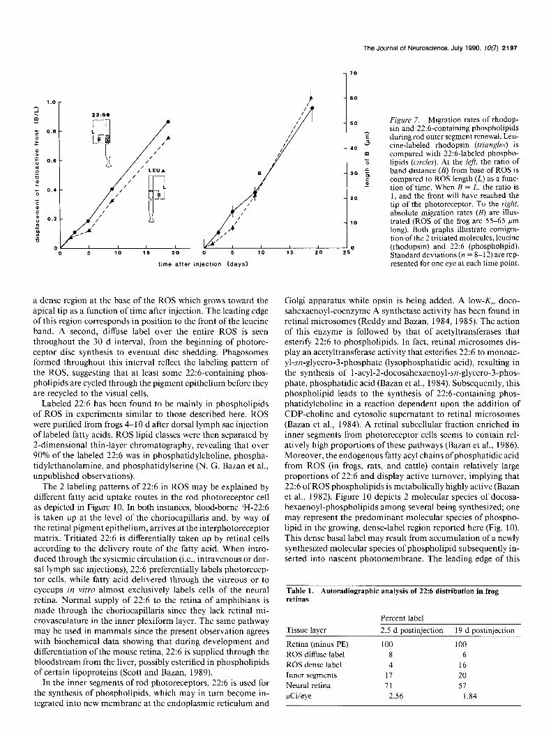
Figure 7. Migration rates of rhodop- sin and 22:6-containing phospholipids during rod outer segment renewal. Leu- tine-labeled rhodopsin (triangles) is compared with 22:6-labeled phospho- lipids (circles). At the left, the ratio of band distance (B) from base of ROS is compared to ROS length (L) as a func- tion of time. When B = L, the ratio is 1, and the front will have reached the tip of the photoreceptor. To the right, absolute migration rates (B) are illus- trated (ROS of the frog are 55-65 Frn long). Both graphs illustrate comigra- tion ofthe 2 tritiated molecules, leucine (rhodopsin) and 22:6 (phospholipid). Standard deviations (n = 8- 12) are rep- resented for one eye at each time point. time after injection (days)
a dense region at the base of the ROS which grows toward the apical tip as a function of time after injection. The leading edge of this region corresponds in position to the front of the leucine band. A second, diffuse label over the entire ROS is seen throughout the 30 d interval, from the beginning of photore- ceptor disc synthesis to eventual disc shedding. Phagosomes formed throughout this interval reflect the labeling pattern of the ROS, suggesting that at least some 22:6-containing phos- pholipids are cycled through the pigment epithelium before they are recycled to the visual cells.
Labeled 22:6 has been found to be mainly in phospholipids of ROS in experiments similar to those described here. ROS were purified from frogs 4-l 0 d after dorsal lymph sac injection of labeled fatty acids. ROS lipid classes were then separated by 2-dimensional thin-layer chromatography, revealing that over 90% of the labeled 22:6 was in phosphatidylcholine, phospha- tidylethanolamine, and phosphatidylserine (N. G. Bazan et al., unpublished observations).
The 2 labeling patterns of 22:6 in ROS may be explained by different fatty acid uptake routes in the rod photoreceptor cell as depicted in Figure 10. In both instances, blood-borne 3H-22:6 is taken up at the level of the choriocapillaris and, by way of the retinal pigment epithelium, arrives at the interphotoreceptor matrix. Tritiated 22:6 is differentially taken up by retinal cells according to the delivery route of the fatty acid. When intro- duced through the systemic circulation (i.e., intravenous or dor- sal lymph sac injections), 22:6 preferentially labels photorecep- tor cells, while fatty acid delivered through the vitreous or to eyecups in vitro almost exclusively labels cells of the neural retina. Normal supply of 22:6 to the retina of amphibians is made through the choriocapillaris since they lack retinal mi- crovasculature in the inner plexiform layer. The same pathway may be used in mammals since the present observation agrees with biochemical data showing that during development and differentiation of the mouse retina, 22:6 is supplied through the bloodstream from the liver, possibly esterified in phospholipids of certain lipoproteins (Scott and Bazan, 1989).
In the inner segments of rod photoreceptors, 22:6 is used for the synthesis of phospholipids, which may in turn become in- tegrated into new membrane at the endoplasmic reticulum and
Golgi apparatus while opsin is being added. A low-K, doco- sahexaenoyl-coenzyme A synthetase activity has been found in retinal microsomes (Reddy and Bazan, 1984, 1985). The action of this enzyme is followed by that of acetyltransferases that esterify 22:6 to phospholipids. In fact, retinal microsomes dis- play an acetyltransferase activity that esterifies 22:6 to monoac- yl-sn-glycero-3-phosphate (lysophosphatidic acid), resulting in the synthesis of 1 -acyl-2-docosahexaenoyl-sn-glycero-3-phos- phate, phosphatidic acid (Bazan et al., 1984). Subsequently, this phospholipid leads to the synthesis of 22:6-containing phos- phatidylcholine in a reaction dependent upon the addition of CDP-choline and cytosolic supernatant to retinal microsomes (Bazan et al., 1984). A retinal subcellular fraction enriched in inner segments from photoreceptor cells seems to contain rel- atively high proportions of these pathways (Bazan et al., 1986). Moreover, the endogenous fatty acyl chains of phosphatidic acid from ROS (in frogs, rats, and cattle) contain relatively large proportions of 22:6 and display active turnover, implying that 22:6 of ROS phospholipids is metabolically highly active (Bazan et al., 1982). Figure 10 depicts 2 molecular species of docosa- hexaenoyl-phospholipids among several being synthesized; one may represent the predominant molecular species of phospho- lipid in the growing, dense-label region reported here (Fig. 10). This dense basal label may result from accumulation of a newly synthesized molecular species of phospholipid subsequently in- serted into nascent photomembrane. The leading edge of this
Table 1. Autoradiographic analysis of 22:6 distribution in frog retinas
Percent label
Tissue layer 2.5 d postiniection 19 d postiniection
Retina (minus PE) 100 100 ROS diffuse label 8 6 ROS dense label 4 16 Inner segments 17 20 Neural retina 71 51 &i/eye 2.56 1.84

2198 Gordon and Bazan l 22:6 Pathways During Visual Cell Renewal
Figure 8. Light micrographs depicting grain distribution throughout the ROS and pigment epithelium during shed- ding and phagosome formation. De- scription of the triggering of shedding and phagocytosis is in Materials and Methods. A, Retina induced to shed 5 d after injection of )H-22:6. A thin re- gion at the base of the ROS shows the dense silver grain region, while the rest of the ROS has labeled diffusely. Pha- gosomes (arrows) within the pigment epithelium contain diffuse label. The large, spherical structures in the pig- ment epithelium are heavily labeled oil droplets. B, Retina induced to shed 28 d after injection of ‘H-22:6. Discs that were formed when tritium was intro- duced into the animal are now being shed (see Fig. 7). By 28 d, ROS have become completely filled with the dense form of the label; the newly shed phago- somes (arrows) also contain this la- bel. C, Retina that was not induced to shed at day 28. While ROS are heavily labeled with 3H-22:6, no phagosomes appear in the very lightly labeled pig- ment epithelium. D, Unstained retinal autoradiogram at day 28. All contrast is due to the presence of autoradio- graphic silver. Note that the oil droplets of both the pigment epithelium and the cones are heavily labeled. This labeling pattern with ‘H-22:6 occurs throughout these experiments; oil droplets initially label heavily, but then gradually di- minish as a function of postinjection time. No such labeling pattern is seen with 3H-leucine (see E). E, Retina in- duced to shed 28 d after the injection of ‘H-leucine. This demonstrates that at 28 d, the initially labeled discs have just reached the pigment epithelium and are being shed. The newly formed phagosomes (arrows) contain a band of 3H-leucine labeled rhodopsin. Scale bar, 20 pm.

The Journal of Neuroscience, July 19% 10(7) 2199
22:6
I 22:5
16:O
Figure 9. HPLC analysis of fatty acid methyl esters from retinas sampled at 7, 46, and 67 d after injection of 3H- 22:6 into the dorsal lymph sac. Doco- sahexaenoic acid (22:6) is the only la- beled fatty acid through day 46. Our analysis conditions (lipid extraction, methanolysis, partition, on-line HPLC radioactive detector) provide for recov- ery of 85-90% of labeled fatty acids. Some conversion begins to occur by day 67, but only small amounts of other fatty acids demonstrate tritium label- ing. HPLC retention time is denoted in minutes along the horizontal axis.
6 days
35 40
time (min)
dense label coincides with the leucine band, reaching the pig- ment epithelium in about 30 d. This implies that some 22:6- containing phospholipids move with the newly synthesized rho- dopsin. Additionally, this dense region thickens from the base toward the apex, as if it were constantly being synthesized or added to, maintaining a steady progress toward the pigment epithelium at the same rate as a labeled protein band. HPLC analysis of the retina confirms that most of the label is 22:6, even 45 d after systemic injection. Docosahexaenoic acid-con- taining phospholipids comprise a significant portion of the rho- dopsin microenvironment (Dratz and Deese, 1986). We suggest that the 22:6-enriched phospholipids of the dense label at the base of the outer segment are physically associated with rho- dopsin of the newly synthesized membrane and remain so until reaching the apex of the photoreceptor. There is no direct evi- dence for an association between a molecular species of phos- pholipid-containing 22:6 with rhodopsin. However, the coin- ciding, moving fronts of the 3H-leucine band and the ‘H-22:6 dense label suggest this possibility. Recent data show that certain molecular species of phosphatidylcholine-containing 22:6 are not extracted from ROS by the usual procedures and require hexane extraction due to a tight interaction with rhodopsin (Av- eldano, 1988, see below). These studies have been conducted in isolated ROS and the 22:6-containing molecular species of phosphatidylcholine have been characterized by gas chroma- tography-mass spectrometry (Aveldano and Sprecher, 1987; Aveldano, 1988). To date, no direct biochemical studies of the 22:6-containing phospholipids of the base of the ROS have been conducted. The slight decrease in silver grain density near the
ROS base by day 46 suggests, by extrapolation, that dilution with nonradioactive 22:6 has begun by about day 60. According to our hypothesis, greater or lesser amounts of initial label (?H- 22:6) would affect only the intensity of the dense label, not the rate at which it decreases. Therefore, if final dissipation of the densely labeled region can be extrapolated to about 60 d, then the molecular species of 22:6-containing phospholipids (asso- ciated with rhodopsin) must turn over roughly every outer seg- ment cycle (i.e., 30 d).
Since the overall, diffuse labeling pattern (filled triangles in Fig. 10) accumulates rapidly, it likely involves lipid exchange in the ROS, perhaps by a phospholipid exchange protein (Dud- ley and Anderson, 1978). Figure 10 depicts 2 possible mecha- nisms to explain the diffuse silver grains in the ROS. In Figure 1 OA, a rapid exchange stemming from the dense-label region is shown, and in Figure IOB, the diffuse silver grains are shown arising from 22:6 arriving from the interphotoreceptor matrix and bypassing lipid synthesis mechanisms or membrane assem- bly at the inner segment. Interestingly, the monkey interpho- toreceptor matrix contains endogenous 22:6 noncovalently bound to interphotoreceptor retinoid-binding protein, as well as to other proteins (Bazan et al., 1985). Available data cannot con- clusively exclude either model A or B. Our results are consistent with a diversity of pathways for lipid routing in rod photore- ceptor cells (Holtzman and Mercurio, 1980; Mercurio and Holtzman, 1982; Matheke and Holtzman, 1984; Fliesler and Basinger, 1987). However, it is not yet known if the molecular species of phospholipid resulting in the dense-label region is utilized in photoreceptor membrane biogenesis along the path-

2200 Gordon and Bazan * 22:6 Pathways During Visual Cell Renewal
- DIFFUSE LABELING
- DENSE LABELING
INNER SEGMENT
A o c-22:
0
UNLABELED B
RHODOPSIN PHOSPHOLIPIDS PHOSPHOLIPIDS
Figure 10. Schematic representation of the supply of 22:6 and labeling patterns in rod photoreceptor cells 1 week after )H-22:6 injection. The drawing of the photoreceptor cell depicts a stack of discs within the ROS. The squares are “top” views of disc membrane from the areas indicated by the open arrows. The ellipses represent rhodopsin, while the circles and triangles denote phospholipids of the membrane bilayer. Filled circles represent 3H-22:6-containing phospholipids, noncovalently associated with rhodopsin. Disc membranes of apical regions of the photoreceptor contain sparingly labeled phospholipids (black triangles) that are apparently not associated with membrane proteins, arriving either by rapid exchange from the dense grain region (A) or from the extracellular matrix (B). The lowest arrow in both A and B indicates a possible supply of 22: 6 lipids from the adjacent Miiller cells (see Fig. 3).
way followed by opsin through the Golgi apparatus, specialized The retina (Aveldano de Caldironi and Bazan, 1977, 1980; membrane vesicles, and discrete domains at the inner segment Aveldano et al., 1983) and ROS (Miljanich et al., 1979; Avel- plasma membrane adjacent to the connecting cilium (Paper- dano and Bazan, 1983; Louie et al., 1988) contain unique mo- master and Schneider, 1982; Besharse, 1986). Furthermore, our lecular species of supraenoic phospholipids with 22:6 esterified results showing that the leading front of the dense-label region in both C, and C,. The molecular species of phospholipid tightly of 3H-22:6 coincides with that of the 3H-leucine band are the associated with rhodopsin may be a phosphatidylcholine with first to indicate a close association of a phospholipid with rho- 22:6 esterified at C, of the glycerol backbone, and a very long- dopsin. This is unlike available evidence that suggests that phos- chain polyunsaturated fatty acid with 6 double bonds derived pholipids of photoreceptor cells are transported toward mem- from 22:6 at C,. This unique molecular species of phosphati- brane biogenesis sites and undergo turnover independent of dylcholine is selectively associated with rhodopsin, as shown opsin (Hall et al., 1969; Bibb and Young, 1974a, b; Anderson by differential hexane lipid extraction of ROS (Aveldano, 1988). et al., 198Oa-c; Fliesler and Basinger, 1987). The 3H-22:6 may The rapidly diffusible label may correspond to a phospholipid label specific molecular species of phospholipids. containing 22:6 at C,, and other fatty acids at C,, a molecular

The Journal of Neuroscience, July 1990, IO(7) 2201
species not preferentially associated with rhodopsin (Aveldano, 1987, 1988; Aveldano and Sprecher, 1987). Our solubilized, lipid-free retinas demonstrate no radioactivity associated with protein. This is in agreement with O’Brien et al. (1987), who have shown that no 22:6 can be detected by gas chromatogra- phy-mass spectrometry in covalent association with rhodopsin.
In conclusion, the major fatty acyl chain of phospholipids in photoreceptor membranes, 22:6, does not behave like other lipids during visual cell renewal in the frog. A dense silver grain region at the base of the ROS expands toward the tip of the photoreceptor outer segment during renewal. The front of this region coincides with the front of the 3H-leucine band (a rho- dopsin marker) that results when the radioactive amino acid is injected, suggesting an association between the 22:6-containing phospholipid and the protein. Furthermore, 2 molecular species of phospholipids (or 2 groups of molecular species), both con- taining docosahexaenoyl chains, may explain the labeling profile arising from 3H-22:6. A unique molecular species of phospho- lipid tightly associated with rhodopsin may give rise to the densely labeled region. A second, rapidly labeled, diffuse pattern throughout the ROS may be due to a different molecular species (also containing 22:6) of phospholipids. Phagosomes containing 3H-22:6-phospholipids are formed throughout the disc mem- brane turnover cycle, suggesting that both the diffuse and the dense form of label leave the ROS and then cycle through the pigment epithelium before being reutilized. Oil droplets in the pigment epithelium and the cone cells label rapidly, suggesting that some 22:6 is temporarily stored in neutral lipids. This is supported by work showing the rapid accumulation of another polyunsaturated fatty acid, arachidonic acid, in triacylglycerols in the in vitro bovine retina (Bazan and Bazan, 1975). Since label gradually diminishes over long periods oftime, those drop- lets may serve to gradually resupply 22:6 to the photoreceptors or other cells of the retina. Finally, the supply of 22:6 to the rod photoreceptor cells is made from the choriocapillaris.
References Anderson RE, Kelleher PA, Maude MB (I 980a) Metabolism of phos-
phatidylethanolamine in the frog retina. Biochim Biophys Acta 620: 227-235.
Anderson RE, Kelleher PA, Maude MB, Maida TM (1980b) Synthesis and turnover of lipid and protein components of frog retinal rod outer segments. Neurochem In; 1:2942.
Anderson RE. Maude MB. Kelleher PA. Maida TM. Basineer SF (1980~) Metabolism of phosphatidylcholine in the frogretina.Bioch- im Biophys Acta 620~212-226.
Aveldano MI (1987) A novel group of very long chain polyenoic fatty acids in dipolyunsaturated phosphatidylcholines from vertebrate ret- ina. J Biol Chem 262: 1172-l 179.
Aveldano MI (1988) Phospholipid species containing long and very long polyenoic fatty acids remain with rhodopsin after hexane ex- traction of photoreceptor membranes. Biochemistry 27: 1229- 1239.
Aveldano MI, Bazan NG (1983) Molecular species of phosphatidyl- choline, -ethanolamine, -serine, and -inositol in microsomal and pho- toreceptor membranes of bovine retina. J Lipid Res 24:620-627.
Aveldano MI, Sprecher H (1987) Very long chain (C,, to C,,) polyenoic fatty acids of the n-3 and n-6 series in dipolyunsaturated phospha- tidylcholines from bovine retina. J Biol Chem 262: 1180-l 186.
Aveldano MI, Pasquare de Garcia SJ, Bazan NC (1983) Biosynthesis of molecular species of inositol, choline, serine, and ethanolamine glycerophospholipids in the bovine retina. J Lipid Res 24:628-638.
Aveldano de Caldironi MI, Bazan NG (1977) Acyl groups, molecular species, and labeling by 14C-glycerol and 3H-arachidonic acid of ver- tebrate retina glycerolipids. Adv Exp Med Biol 83:3971104.
Aveldano de Caldironi MI, Bazan NG (1980) Composition and bio- synthesis of molecular species of retina phosphoglycerides. Neuro- them Int 1:381-392.
Bazan HEP, Bazan NG (1975) Incorporation of (-‘H)-arachidonic acid into cattle retina lipids: high uptake in triacylglycerols, diacylglycerols, phosphatidylcholine and phosphatidylinositol. Life Sci 17: 167 l-l 678.
Bazan HEP, Sprecher H, Bazan NG (1984) De novo biosynthesis of docosahexaenoyl phosphatidic acid in bovine retinal membranes. Biochim Biophys Acta 796: 11-19.
Bazan HEP, Ridenour B, Birkle DL, Bazan NG (1986) Unique met- abolic features of docosahexaenoate metabolism related to functional roles in brain and retina. Phospholipid Research and the Nervous System. Biochemical and Molecular Pharmacology, FIDIA Res. Ser. Vol 4 (Horrocks L, Freysz L, Toffano G, eds), pp 67-78. Padua: Liviana Press.
Bazan NG, Reddy TS (1985) Retina. In: Handbook of neurochemistry, Vol 8 (Lajtha A, ed), pp 507-575. New York: Plenum.
Bazan NG. di Fazio de Escalante MS. Careaaa MM. Bazan HEP. Giusto NM (1982) High content of 22:6’(doco&hexaenoate) and active [2- ‘HIglycerol metabolism of phosphatidic acid from photoreceptor membranes. Biochim Biophys Acta 7 12:702-706.
Bazan NG, Reddy TS, Redmond TM, Wiggert B, Chader GJ (1985) Endogenous fatty acids are covalently and noncovalently bound to interphotoreceptor retinoid-binding protein in the monkey retina. J Biol Chem 260:13677-13680.
Besharse JC (1986) Photosensitive membrane turnover: differentiated membrane domains and cell-cell interaction. In: The retina (Adler R, Farber D, eds), pp 297-35 1. Orlando, FL: Academic.
Bibb C, Young RW (1974a) Renewal of fatty acids in the membranes of the visual cell outer segments. J Cell Biol 61:327-343.
Bibb C, Young RW (1974b) Renewal of glycerol in the visual cells and pigment epithelium of the frog retina. J Cell Biol 62:378-389.
Bok D (1985) Retinal photoreceptor-pigment epithelium interactions. Invest Ophthalmol Vis Sci 26: 1659-l 694.
Defoe DM, Bok D (1983) Rhodopsin chromophore exchanges among opsin molecules in the dark. Invest Ophthalmol Vis Sci 24: 121 l- 1226.
Dratz EA, Deese AJ (1986) The role of docosahexaenoic acid (22:6 omega-3) in biological membranes: examples from photoreceptors and model membrane bilayers. In: Health effects of polyunsaturated fatty acids in seafood (Simopoulos AP, Kifer R, Martin RE, eds), pp 3 19-35 1. New York: Academic.
Dudley PA, Anderson RE (1978) Phospholipid transfer protein from bovine retina with high activity towards retinal rod disc membranes. FEBS Lett 95:57-60.
Fliesler SJ, Anderson RE (1983) Chemistry and metabolism of lipids in the vertebrate retina. Proa Linid Res 22:79-l 3 1.
Fliesler SJ, Basinger SF (1987) Monensin stimulates glycerolipid in- corporation into rod outer segment membranes. J Biol Chem 262: 17516-17523.
Gordon WC, Bazan NG (1989) Docosahexaenoic acid in rod outer segment renewal (abstr). Invest Ophthalmol Vis Sci (Suppl) 30:294.
Gordon WC. Keith ME (1987) Tubocurarine chloride inhibits rod outer segment shedding in thefrog retina. Invest Ophthalmol Vis Sci 28:1030-1032.
Hall MO, Bok D, Bacharach ADE (1969) Biosynthesis and assembly of the rod outer segment membrane system. Formation and fate of visual pigment in the frog retina. J Mol Biol45:397-406.
Hollyfield JG (1979) Membrane addition to photoreceptor outer seg- ments: progressive reduction in the stimulatory effect of light with increased temperature. Invest Ophthalmol Vis Sci 18:977-98 1.
Holtzman E, Mercurio AM (1980) Membrane circulation in neurons and photoreceptors: some unresolved issues. Int Ret Cytol 67: l-67.
Louie K, Wiegand RD, Anderson RE (1988) Docosahexaenoate-con- taining molecular species of glycerophospholipids from frog retinal rod outer segments show different rates of biosynthesis and turnover. Biochemistry 27:9014-9020.
Marcheselli VL, Scott BL, Reddy TS, Bazan NG (1988) Quantitative analysis of acyl group composition of brain phospholipids, neutral lipids, and free fatty acids. Neuromethods 7:83-l 10.
Matheke ML, Holtzman E (1984) The effects of monensin and of puromycin on transport of membrane components in the frog retinal photoreceptor. II. Electron microscopic autoradiography of proteins and glycerolipids. J Neurosci 4:1093-l 103.
Mercurio AM, Holtzman E (1982) Ultrastructural localization ofglyc- erolipid synthesis in rod cells of the isolated frog retina. J Neurocytol 11:295-322.
Miljanich GP, Sklar LA, White DL, Dratz EA (1979) Disaturated
2202 Gordon and Bazan - 22:6 Pathways During Visual Cell Renewal
dipolyunsaturated phospholipids in the bovine retina rod outer seg- ment disc membrane. Biochim Biophys Acta 55294-306.
Neuringer M, Connor WE (1986) N-3 fatty acids in the brain and retina: evidence for their essentiality. Nutrition Rev 44:285-294.
O’Brien PJ, St Jules R, Reddy TS, Bazan NG, Zatz M (1987) Acylation of disc membrane rhodopsin may be non-enzymatic. J Biol Chem 262:5210-5215.
Papermaster DS, Schneider BG (1982) Biosynthesis and morphogen- esis of outer segment membranes in vertebrate photoreceptor cells. In: Cell biology of the eye (McDevit DS, ed), pp 477-5 3 1. New York: Academic.
Ramon y Cajal S (1973) The vertebrate retina. In: The vertebrate retina. Principles of structure and function (Rodieck RW, ed), pp 772-904. San Francisco: WH Freeman.
Reddy TS, Bazan NG (1984) Synthesis of arachidonoyl coenzyme A and docosahexaenoyl coenzyme A in retina. Curr Eye Res 3:1225- 1232.
Reddy TS, Bazan NG (1985) Synthesis of docosahexaenoyl-, arachi- donoyl-, and palmitoyl-coenzyme A in ocular tissues. Exp Eye Res 41:87-95.
Scott BL, Bazan NG (1989) Membrane docosahexaenoate is supplied to the develonina brain and retina bv the liver. Proc Nat1 Acad Sci USA 86:2903-2907.
Young RW (1967) The renewal of photoreceptor cell outer segments. J Cell Biol 33:61-72.