double-balloon enteroscopy: a descriptive study of 50...
TRANSCRIPT

ABSTRACT
Aim: to evaluate the utility of double-balloon enteroscopy forsmall-bowel disease.
Design: a prospective study of 50 consecutive enteroscopiesperformed from December 2004 to July 2005 to analyze diag-noses and treatments.
Patients: 44 patients (33 had undergone a previous capsuleendoscopy) with indications for obscure digestive hemorrhage, an-giodysplasia, Peutz-Jeghers syndrome, ulcer, suspected Crohn´sdisease, tumors, and refractory celiac disease.
Results: we carried out enteroscopy studies in 44 patients bythe oral route and, in 6 additional patients, by both the oral andanal routes. We reached the ileon with the oral route in all casesbut one (jejunal stenosis), and in 4 cases out of 7 with the analroute, with an average duration of 73 minutes. We found an-giodysplasia in 19 cases, as well as NSAID-related enteropathy,Crohn’s disease, diverticulosis, and Waldenström’s disease. Weperformed biopsies in 31% of cases with diagnoses of adenocarci-noma, lymphangiectasia secondary to tumor in celiac disease, andWhipple’s disease. We treated 19 patients with angiodysplasia (1to 20 synchronous lesions) with argon, and 4 patients with polypsusing polipectomy (sporadic polyps or Peutz-Jeghers syndrome).A retained capsule in one patient with stenosis was removed.
Conclusions: double-balloon enteroscopy is a useful and ef-fective technique in the diagnosis and treatment of small intestinediseases, thus complementing capsule endoscopy. More studiesare needed to analyze its impact on the management of this con-dition.
Key words: Double-balloon enteroscopy. Occult gastrointestinalbleeding. Angiodysplasia.
Pérez-Cuadrado E, Más P, Hallal H, Shanabo J, Muñoz E, Or-tega I, López Martín A, Torrella E, López Higueras A, MartínA, Carballo F. Double-balloon enteroscopy: a descriptive studyof 50 explorations. Rev Esp Enferm Dig 2006; 98: 73-81.
INTRODUCTION
The small intestine is nowadays fully accessible withcapsule endoscopy (1), a first-line, non-invasive diagnosticprocedure that works better than small-bowel follow-through (2). This “wireless” device has opened an oral ap-proach to the small intestine, with an explosion of know-ledge about endoscopic lesions (3). Despite the highdiagnostic effectiveness of capsule endoscopy (around75%) (4), we need a pathological and/or therapeutic classi-fication in most cases. Oral push enteroscopy (OPE) was astep forward in both ways (5), but it could not examine thewhole small intestine despite the help of the overtube,which increased range with no significant increase in diag-nosis yield. If capsule endoscopy reveals a lesion out ofOPE’s reach, this lesion may only be accessible by surgeryand/or intraoperative enteroscopy (IOE), which is deemedthe “gold standard” for small intestine. However, an effec-tiveness lower than expected has been shown in IOE incase of obscure gastrointestinal hemorrhage (OGH), withrelapses in 40% of patients (6). Recently, Yamamoto et al.(7) have developed a fine enteroscope that can be fittedinto an special, flexible overtube with a balloon with mon-itored pressure at its distal end. This double-balloon en-teroscopy (DBE), by means of a smart push-pull mecha-nism, makes the SI fold like a telescope, so that it can befully examined with an adequate channel for biopsy andtherapy measures, thus being better than OPE.
There are no publications so far about DBE in ourcountry.
METHODS
Material
A videoenteroscope (EN-450. Fujinon. Japan) 2000mm long with a 2.2 mm biopsy channel, and a flexible
Double-balloon enteroscopy: a descriptive study of 50explorations
E. Pérez-Cuadrado, P. Más, H. Hallal, J. Shanabo, E. Muñoz, I. Ortega, A. López Martín, E. Torrella,A. López Higueras, A. Martín and F. Carballo
Unit of Small Bowel. Section of Digestive Diseases. Hospital General Universitario Morales Meseguer. Murcia, Spain
1130-0108/2006/98/2/73-81REVISTA ESPAÑOLA DE ENFERMEDADES DIGESTIVASCopyright © 2006 ARÁN EDICIONES, S. L.
REV ESP ENFERM DIG (Madrid)Vol. 98. N.° 2, pp. 73-81, 2006
Recibido: 04-03-05.Aceptado: 08-08-05.
Correspondencia: Enrique Pérez-Cuadrado. C/ Delfos, 19. 30007 Murcia.e-mail: [email protected]
ORIGINAL PAPERS

overtube (TS-12140 Fujinon. Japan) 1500 mm long withan inner wash channel (Fig. 1). Both devices have bal-loons fitted in their distal end that are filled with air andconnected by catheters to a manometer (PB-10. Fujinon.Japan) for pressure monitoring, which has an acoustic-vi-sual security system in case of overpressure. A videoprocessor with a magnification of 2:1 (EVE 400. Fujinon.Japan). Disposable ancillary material with a caliber lowerthan 2.2 mm and specifications similar to those of con-ventional endoscopy (biopsy forceps, polypectomysnares, injection needles, Dormia baskets). It is connect-ed to an argon and coagulation unit (APC 300/ICC 200.Erbe. Germany).
Inclusion protocol
1. OGH with a diagnosis –by means of capsule en-doscopy– of angiodysplasia, active bleeding, ulcer, polyp(sporadic or Peutz Jeghers) or tumor of the small intes-tine.
2. OGH with no etiological filiation after capsule en-doscopy.
3. Celiac disease with a poor outcome and no signifi-cant findings during capsule endoscopy.
4. A procedure previous to DBE.All patients signed an agreement and were informed
about the technique and anesthesia. Preanesthetic exami-nation: nothing to eat or drink for 12 hours. In case ofrectal insertion, preparation with an oral cleansing solu-tion (Fosfosoda®). Where appropriate, the patient was he-modynamically stabilized.
5. DBE technique: oral insertion with a mouth gagand/or rectal insertion (Fig. 2) in left lateral decubitus un-der deep sedation, moving forward the overtube and en-doscope with alternative push-pull movements, with thehelp of the sequential inflation and deflation of their re-
spective balloons, making the small intestine fold outsidethe overtube and endoscope.
—Oral DBE: theendoscope and overtube were insert-ed with deflated balloons approaching the jejunum,sucking the gastric cavity until they could not push for-ward. After the inflation of the overtube balloon, the in-sertion of the endoscope was carried out, and when itcould not move forward, the inflation of the endoscopeballoon and the deflation of the overtube balloon insert-ing the overtube later were carried out. These move-ments were alternatively repeated until no more couldbe inserted, and then both balloons were inflated (endo-scope and overtube balloons) and then removed “enbloc” until they became resistant to pull or manometricoverpressure. After this simultaneous pull, they stayedin the same place of the small intestine, so handles wererectified; after this, previous movements were repeated(8). After examining the small intestine, the endoscopeand overtube balloons stayed half-inflated in order toavoid an abrupt movement of the enteroscope duringwithdrawal.
—Rectal DBE: the endoscope and overtube were in-serted with deflated balloons through the colon in a waysimilar to that orally used until the Bahuin valve wasreached, making the way through easier in prone decubi-tus. The small intestine was examined using movementssimilar to those carried out during oral insertion.
6. Lesion topography in small intestine: the jejunumwas considered to be the two proximal fifths of thesmall intestine from the ligament of Treitz to the Bahuinvalve, and the ileum the three distal fifths. Jejunum andileum were divided into proximal, middle and distal inthree equal parts. Radiological control and the instilla-tion of a water-soluble contrast were occasionally car-ried out (Fig. 2).
7. Diagnostic procedures:—Biopsy: it was carried out with forceps in order to
obtain tissue samples for histological evaluation, poly-merase chain reaction, electronic microscopy, etc.
74 E. PÉREZ-CUADRADO ET AL. REV ESP ENFERM DIG (Madrid)
REV ESP ENFERM DIG 2006; 98(2): 73-81
Fig. 1.- Outward appearance of a double-balloon enteroscope (withboth balloons inflated, at the enteroscope and the overtube).Aspecto externo del enteroscopio de doble balón (con los dos baloneshinchados del enteroscopio y del sobretubo).
Fig. 2.- Oral insertion of the enteroscope approaching the Bahuin valve(left), and rectal insertion.Introducción oral del enteroscopio hasta cerca de la válvula de Bahuin(izquierda) y anal a través de la misma.

—Indian-ink staining: in case of finding SI lesionssubject to surgery or bleeding relapse after DBE thera-peutics. Besides, after oral insertion, the most distal areawas examined aiming at finding it in case of later instruc-tions of rectal insertion, so that a complete examinationof the SI was guaranteed.
—Chromoendoscopy: magnification (2:1). It was usedto allow a better examination of the villous standard encases of patchy atrophy, in an easy way using water or in-digo carmine dye as instilled through a catheter.
8. Therapeutic procedures:—Disposable argon catheters were used in cases of an-
giodysplasia, with a maximum flow of 0.5 L/m, 40 W,and tangential impact on the lesion (Fig. 3).
—Polyps were removed by means of diathermy snares,and foreign bodies were removed using a Dormia basket.
9. Statistical study:In the descriptive study, quantitative variables have
been expressed as average plus/less standard deviation (a± SD) values, and qualitative variables as total percent-age. The statistical program used was SPSS, version9.0.1, by SPSS Inc.
RESULTS
Sample characteristics
From December 2004 to July 2005, 44 patients, 23 menand 21 women with an average age of 57.7 ± 19 years,
were consecutively included, and a total of 50 examina-tions using DBE were performed. Six patients underwentDBE both rectally and orally.
The indication for DBE was OGH in 32 cases, with aprevious diagnosis by capsule endoscopy in 18 and with-out it in 14 cases –suspicion of Crohn’s disease (n: 4),Peutz Jegher’s syndrome (n: 3), tumor (n: 3), and refracto-ry celiac disease (one case); 33 patients (75%) had a previ-ous capsule endoscopy performed. CE diagnoses included:in 22 patients (50%) angiodysplasia, 3 suspected cases ofCrohn’s disease, two stenosis, two polyps, and two ulcers.Oral insertion (n: 44) always led to reach the distal-middleileum (one case close to Bahuin) except for one tumor andone jejunal stenosis. The rectum was reached in one caseof complementary insertion by jejunostomy. Rectal inser-tion (n: 6) reached the proximal ileum (the middle part in 3cases, and the distal part in one case). The way through theBahuin valve was found difficult in one case, and in anoth-er case the examination was given up in the sigmoid colonafter having some complications. Mean duration for theoral and rectal DBE was 73.2 ± 25.7 (25-165) and 73.1 ±27.0 (10-100) minutes, respectively.
Diagnostic-therapeutic DBE
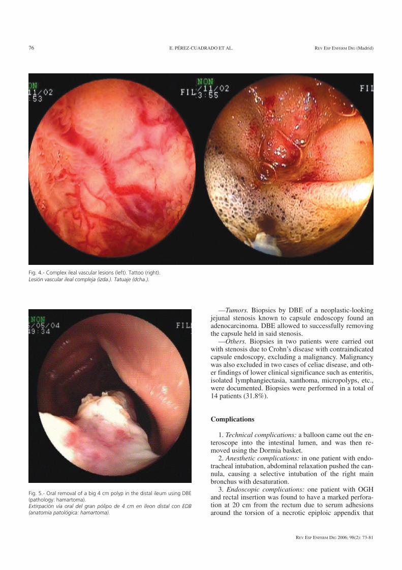
—Angiodysplasia. In the 19 patients suffering fromangiodysplasia an average of 5.05 ± 5.61 (1-20) lesionswere found, which were all treated using argon with nocomplications. The most complex vascular lesions weretattoos (Fig. 4).
—Peutz Jeghers. In two patients who underwent previ-ous laparotomies for small intestine obstruction (1 to 6surgical resections) DBE confirmed the findings of cap-sule endoscopy, and big ileal polyps of 3.5 and 2 cm insize were removed (Fig. 5). In another case, capsule en-doscopy detected 6 polyps, while with DBE 15 polypsbigger than 1 cm were found and removed with no com-plications (hamartomas).
—Jejunal diverticula. In two patients jejunal divertic-ula were found, which had remained unnoticed to capsuleendoscopy.
—Polyps. One patient diagnosed as having an ulcer bycapsule endoscopy had actually an ulcered polyp, whichwas removed.
—Diffuse lymphangiectasia. One patient sufferingfrom refractory celiac disease developed a secondary dif-fuse lymphangiectasia close to one celiac tumor (adeno-carcinoma) detected by capsule endoscopy.
—Whipple’s disease. In one case of segmentary sponta-neous bleeding during capsule endoscopy in several sec-tions, DBE ruled out associated conditions and allowed toobtain histological material for polymerase chain reactionand EM (Fig. 6), which confirmed the disease.
—Ulcers. One case with Waldenström’s disease andOGH had multiple ulcers. Two cases of ulcers caused byNSAIDs were also found.
Vol. 98. N.° 2, 2006 DOUBLE BALLOON ENTEROSCOPY: A DESCRIPTIVE STUDY 75OF 50 EXPLORATIONS
REV ESP ENFERM DIG 2006; 98(2): 73-81
Fig. 3.- Jejunal angiodysplasia: treatment with argon plasma.Angiodisplasia de yeyuno: tratamiento con argón plasma.
—Tumors. Biopsies by DBE of a neoplastic-lookingjejunal stenosis known to capsule endoscopy found anadenocarcinoma. DBE allowed to successfully removingthe capsule held in said stenosis.
—Others. Biopsies in two patients were carried outwith stenosis due to Crohn’s disease with contraindicatedcapsule endoscopy, excluding a malignancy. Malignancywas also excluded in two cases of celiac disease, and oth-er findings of lower clinical significance such as enteritis,isolated lymphangiectasia, xanthoma, micropolyps, etc.,were documented. Biopsies were performed in a total of14 patients (31.8%).
Complications
1. Technical complications: a balloon came out the en-teroscope into the intestinal lumen, and was then re-moved using the Dormia basket.
2. Anesthetic complications: in one patient with endo-tracheal intubation, abdominal relaxation pushed the can-nula, causing a selective intubation of the right mainbronchus with desaturation.
3. Endoscopic complications: one patient with OGHand rectal insertion was found to have a marked perfora-tion at 20 cm from the rectum due to serum adhesionsaround the torsion of a necrotic epiploic appendix that
76 E. PÉREZ-CUADRADO ET AL. REV ESP ENFERM DIG (Madrid)
REV ESP ENFERM DIG 2006; 98(2): 73-81
Fig. 4.- Complex ileal vascular lesions (left). Tattoo (right).Lesión vascular ileal compleja (izda.). Tatuaje (dcha.).
Fig. 5.- Oral removal of a big 4 cm polyp in the distal ileum using DBE(pathology: hamartoma).Extirpación vía oral del gran pólipo de 4 cm en íleon distal con EDB(anatomía patológica: hamartoma).

had not been previously diagnosed. The immediate oper-ation showed an extraluminal lesion in that loop. As thepreparation of the colon for DBE was good, a simple su-ture was carried out and the patient was discharged after3 days.
DISCUSSION
The small intestine is nowadays fully accessible for apathological and/or therapeutic diagnosis using DBE, ei-ther orally or rectally, if necessary. This technique hasbeen shown to be better than OPE in terms of length ofsmall intestine explored (8).
One of the contraindications of capsule endoscopy isthe known stenosis of the small intestine, but in caseswhere this is not suspected, capsules may be held within.DBE allows the pathological classification of stenosisand capsule removal, as was the case in one of our pa-tients, the first one with OGH, who had a jejunal adeno-carcinoma.
Capsule endoscopy and DBE have been shown in ourstudy to be complementary techniques; the fairly harm-lessness and non-invasivity of capsule endoscopy ren-dered it a first-line study for diagnosis, and DBE was car-ried out in cases requiring a histological diagnosis orendoscopic therapy. Capsule endoscopy can lead the way(orally or rectally) for DBE.
In OGH, capsule endoscopy has previously shown ahigh diagnostic effectiveness related to the early carryingout of the technique in connection with the time of thebleeding episode (4). In cases with no clear diagnosis bycapsule endoscopy (as when visualizing bleeding in thesmall intestine with no detectable pathology), DBE hasthe advantage of allowing the washing and aspirating ofblood remnants, which makes diagnosis and treatmenteasy.
As for angiodysplasia, the most frequently found le-sion in our study, the number of lesions found by DBEhas always been the same or higher than the one found bycapsule endoscopy in all cases, which is obviously due tothe insufflations, mobility, possibility of mucus washing,and higher number of pictures taken per second of DBE,with the disadvantage of causing alterations because ofinstrumental friction, which requires assessment andtreatment at insertion (not at removal). The usefulness ofDBE would be a reduction of transfusion requirementsby treating as many lesions as possible, which would di-rectly affect OPE (9). Another difficulty of these“evanescent” lesions is that they tend to be multiple(some synchronic lesions may go unnoticed) and mayalso develop metachronically; therefore, follow-up usingcapsule endoscopy seems an obvious option, and shouldnew findings arrive then DBE would be carried out again.
On the other hand, some hereditary diseases such asPeutz Jeghers syndrome require a mapping by capsuleendoscopy, which has been proved to be better than bari-um examinations in a study by Bordas et al. (10) wherecapsule endoscopy showed the number, size and locationof polyps. After capsule endoscopy, a therapeutic DBEcan be carried out, and following the resection of polypscapsule endoscopy (less invasive) can be used to controlmetachronic lesions before they become serious byblocking the lumen and folding the affected loop, whichis important in patients with several previous laparo-tomies. That way, DBE has allowed in our study the re-moval of big-sized and multiple polyps, in one case with6 previous laparotomies (Fig. 5), and the removal of 15polyps in the same lesion in another patient.
Inflammatory intestinal disease has several difficultylevels in the small intestine. Crohn’s disease can only af-fect the small intestine or even only the jejunum, andOPE has limited usefulness (11). On the other hand, inde-terminate colitis where lesions in the small intestine are
Vol. 98. N.° 2, 2006 DOUBLE BALLOON ENTEROSCOPY: A DESCRIPTIVE STUDY 77OF 50 EXPLORATIONS
REV ESP ENFERM DIG 2006; 98(2): 73-81
Fig. 6.- Whipple’s disease on capsule endoscopy (left), double-balloon enteroscopy (center) (arrow: biopsy collection). Electronic microscope study oftissue obtained (right).Enfermedad de Whipple en cápsula endoscópica (izquierda), enteroscopia de doble balón (centro) (flecha: toma de biopsias). Microscopio electrónicodel material obtenido (derecha).

found by capsule endoscopy requires a DBE with biopsycollection for its diagnosis.
It seems only obvious that IOE requiring laparotomyand IOE having a limited examination area of the smallintestine will give way to this new technique, as DBE hasbeen shown in our study to be an easy, safe, and effectivetechnique for the treatment of bleeding lesions, and forthe performance of biopsies in areas not accessible toother techniques; it has been seen to even offer a newmacroscopic diagnosis after capsule endoscopy. We thinkthat DBE is a basic tool in many cases after a first-linenon-invasive study of the small intestine with capsule en-doscopy, and this endoscopic technique can be designat-ed the diagnostic and therapeutic “gold standard” in thesmall intestine.
REFERENCES
1. Iddan G, Meron G, Glukhovsky A, Swain P. Wireless capsule en-doscopy. Nature 2000; 405 (6785): 417.
2. Costamagna G, Shah SK, Riccioni ME, Foschia F, Mutignani M, Pe-rri V, et al. A prospective trial comparing small bowel radiographs
and video capsule endoscopy for suspected small bowel disease. Gas-troenterology 2002; 123 (4): 999-1005.
3. Pérez-Cuadrado E. Acceso oral al intestino delgado. Rev Esp EnfermDig 2002; 94 (7): 423-6.
4. Pennazio M. Outcome of patients with obscure gastrointestinal bleed-ing after capsule endoscopy: report of 100 consecutive cases. Gas-troenterology 2004, 126: 643-53.
5. Pérez-Cuadrado E. Videoenteroscopia oral: estudio prospectivo de 30casos. Rev Esp Enferm Dig 1996; 88: 9-15.
6. Ress AM, Benacci JC, Sarr MG. Efficacy of intraoperative en-teroscopy in diagnosis and prevention of recurrent, occult gastroin-testinal bleeding. Am J Surg 1992; 163: 94-8.
7. Yamamoto H, Sekine Y, Sato Y, Higashizawa T, Miyata T, Iino S, etal. Total enteroscopy with a nonsurgical steerable double-balloonmethod. Gastrointest Endosc 2001; 53: 216-20.
8. May A, Nachbar L, Shneider M, Neuman M, Ell C. Push-and-pull en-teroscopy using the double-balloon technique: method os assessingdepth os insertion and training of the enteroscopy technique using theerlangen endo-trainer. Endoscopy 2005; 37 (1): 66-70.
9. Pérez-Cuadrado E, Molina E, Lamas D. Diagnostic and therapeuticefficacy in a push enteroscopy series. Endoscopy 2000; 32 (Supl. 1)(E 58).
10. Mata A, Llach J, Castells A, Rovira JM, Pellise M, Gines A, et al. Aprospective trial comparing wireless capsule endoscopy and bariumcontrast series for small-bowel surveillance in hereditary GI polypo-sis syndromes. Gastrointest Endosc 2005; 61: 721-5.
11. Pérez-Cuadrado E, Macenlle R, Iglesias J. Usefulness of oral videopush enteroscopy in Crohn´s disease. Endoscopy 1997; 29: 745-7.
78 E. PÉREZ-CUADRADO ET AL. REV ESP ENFERM DIG (Madrid)
REV ESP ENFERM DIG 2006; 98(2): 73-81