double trouble chimerism in twin a - heart of america ... · double trouble chimerism in twin a ....
TRANSCRIPT

DOUBLE TROUBLE CHIMERISM IN TWIN A

CASE STUDY
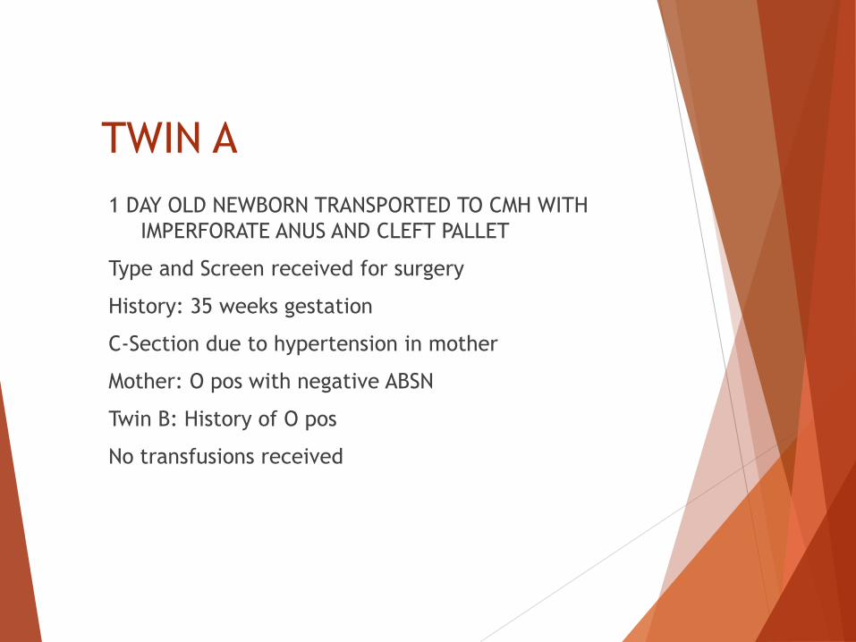
TWIN A
1 DAY OLD NEWBORN TRANSPORTED TO CMH WITH
IMPERFORATE ANUS AND CLEFT PALLET
Type and Screen received for surgery
History: 35 weeks gestation
C-Section due to hypertension in mother
Mother: O pos with negative ABSN
Twin B: History of O pos
No transfusions received

SEROLOGY RESULTS
Tube Method
Specimen #2 and #3
Anti-A 2+ Mixed Field
Anti-B 0
Anti-D 4+
A cells: Not performed
B cells: Not performed
Antibody Screen:
Negative
DAT: Negative
Specimen #1
Anti-A: 0
Anti-B: 0
Anti-D: 4+
A cells: Not performed
B cells: Not performed
Antibody Screen:
Negative
DAT: Negative

Confirmation ABO did not match
initial type. Not enough
specimen to repeat testing on
sample #1.
Sent to CBC Reference Lab

IRL Results
Results: ABO Discrepancy
Blood group: Indeterminate
Repeat Blood type in 1 month for repeat typing
or genotyping to determine blood type.
Transfuse: O positive RBCs and AB plasma.
Crossmatch: IS in tube

Mixed Field: Double Trouble
for the Blood Bank
What is the patient’s blood type?
What is the origin of the mixed field
results?

What Causes Mixed Field
Reactions?

POSSIBILITIES:
1. Maternal-Fetal hemorrhage
2. Twin - Twin hemorrhage
3. Twin A is a Twin Chimera
4. Blood Transfusion
5. Subgroup of A
6. Mosaicism

Based on patient history #4 is
excluded.
Other than the A subgroup and
mosaicism, all possibilities
involve a type of chimera.

What is a Chimera?

Chimera
A person who has more than one
distinct cell line that originates
from two or more zygotes.1
Originally believed to be rare, twin
chimeras have been detected in 8%
of twins and 21% of triplets.2

Mosaic vs. Chimera
Both terms refer to one organism with two or
more populations of cells.
Both have more than one genetically distinct
population of cells.
A Mosaic forms from the same zygote and a
chimera forms from two zygotes.
Mosaics arise when a mutation occurs early in
development and results in a mixture of cells,
some with the mutation and some without.
HISTORY OF CHIMERAS
1945: Owen discovers that RBCs of
dizygotic twin cattle are a mixture of the
two separate twins.3
1953: First Described in humans by
Dunsford et al. Mrs. McK had 60% O cells
and 40% A cells.4
1975: Race and Sanger divided Chimeras
into either twin or dispermic.1
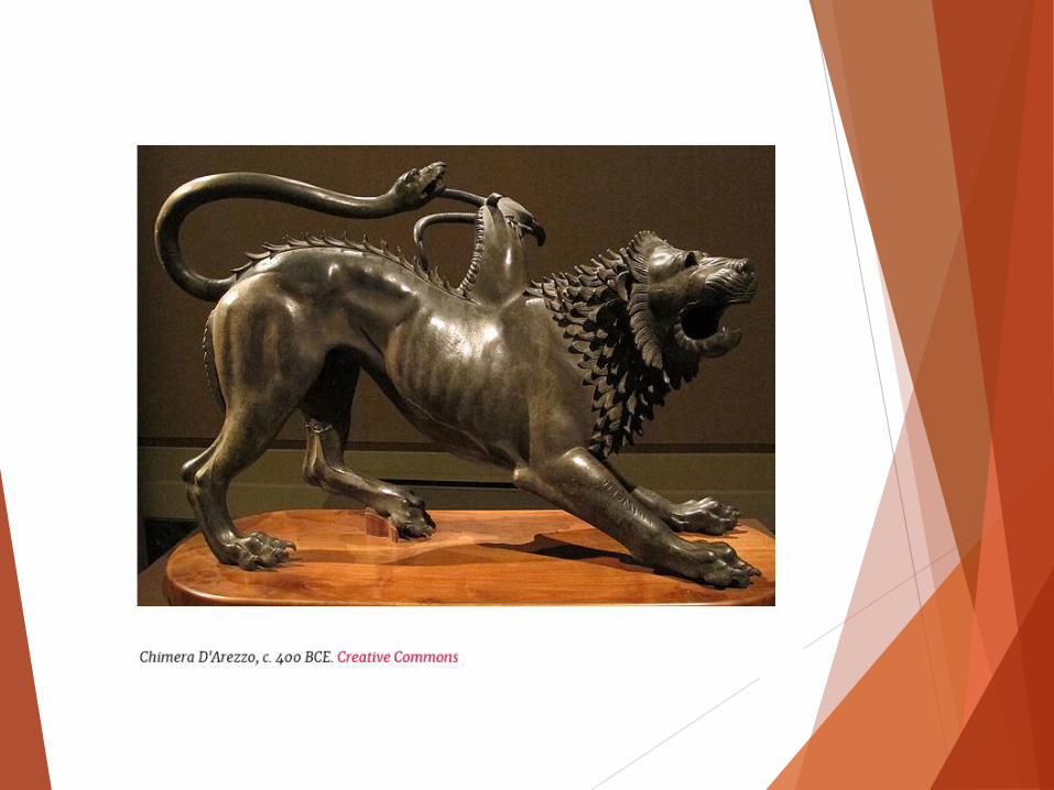
Greek Mythology
CHIMERA:
A mythological monster with the head of a
lion, body of a goat, and the tail of a
serpent. This symbol has been the symbol
of an organism whose cells derive from
two or more distinct zygotes.1
CLASSIFICATION
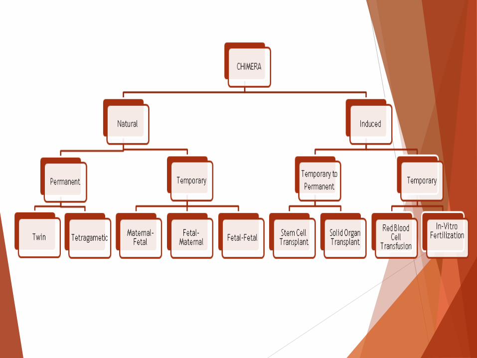
2 TYPES OF CHIMERAS
1.NATURAL
2. INDUCED

NATURAL CHIMERA
1. PERMANENT
2. TEMPORARY

PERMANENT NATURAL
1. Twin Chimera
2. Tetragametic Chimera

TWIN CHIMERAS
Caused by blood vessel anastomoses of
nonidentical twins, resulting in exchange
of blood cells, including hematopoietic
stem cells. These blood cells travel to
each others bone marrow. Due to the
inability of the fetus to recognize foreign
antigens, both cell lines are produced
within each individual twin.
Isohemagglutinins may or may not be
produced.5,6

Blood Vessel Anastomosis
http://www.slideshare.net/jrmmdod/twins-29613296

Monochorionic vs. Dichorionic
Placenta
http://hdydi.com/category/multiple-types/fraternal

Single Fetus
ZYGOTE: The cell formed by the union of a male sperm
and a female ovum.
http://themidwifeisin.com/post/102982437630/are-conjoined-twins-always-identical-or-can-they

Monozygotic vs. Dizygotic
Pregnancy
Identical Fraternal
http://themidwifeisin.com/post/102982437630/are-conjoined-twins-always-identical-or-can-they
TETRAGAMETIC CHIMERAS
(Dispermic dizygotic) Fertilization of 2 maternal nuclei by 2 sperm.
These 2 zygotes fuse into one person.
If a male and female zygote are involved, the individual
will have a combination of XX/XY chromosomes.
This is a whole-body permanent chimera.
May have patchy skin color, different colored eyes, and
hermaphrodism.
Which organs contain which DNA is random.
Known as self-contained or vanishing twins.
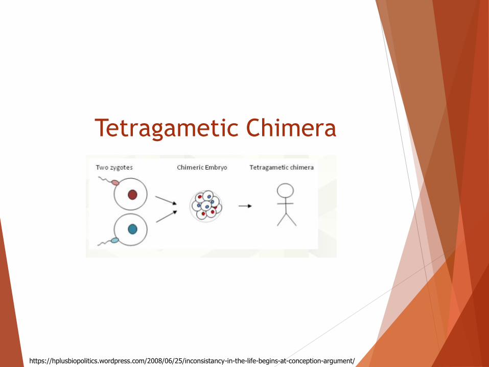
Tetragametic Chimera
https://hplusbiopolitics.wordpress.com/2008/06/25/inconsistancy-in-the-life-begins-at-conception-argument/

TEMPORARY NATURAL
1. MATERNAL-FETAL
2. FETAL-MATERNAL
3. FETAL-FETAL

INDUCED CHIMERAS
1. Induced “Permanent”
2. Induced Temporary
INDUCED PERMANENT CHIMERAS
1. ALLOGENEIC STEM CELL TRANSPLANT
2. SOLID ORGAN TRANSPLANT

Allogenic Stem Cell
Transplants 1. Standard myeloablative is
transplantation where the donor’s marrow
is destroyed.
2. Nonmyeloablative transplantation
allows both cell lines to coexist.
Solid Organ Transplants
Results in “Microchimerism”
Amount of donor cells are too small to be seen in
serologic testing.
An interaction between donor and recipient
leukocytes takes place.
Dendritic donor cells have been found in the
recipients lymph nodes, peripheral blood, skin,
intestine and heart.7
This balance can result in rejection, GVHD, or
tolerance.
INDUCED TEMPORARY CHIMERA
1. Blood Transfusion
2. In-Vitro Fertilization
RBC Transfusions
Chronic transfusions and massive
transfusions will create temporary
chimeras and the donor cells will
last for weeks.

In-Vitro Fertilization
IVF places more than one embryo into the
mother.
Results in a 33.9% increase in dizygotic
twin deliveries when 2 embryos are
implanted.8
Causes an increase in multiple births with
chimeras, twin chimeras, and
tetragametic chimeras.

Back to our Case Study
Original Results: Anti-A 2+ Mixed Field
Anti-B 0
Anti-D 4+
A cells: Not performed
B cells: Not performed
Antibody Screen: Negative
DAT: Negative

3 Months Later:
Patient returns for additional surgery.
Type and screen is ordered.

Results:
Anti-A 3+
Anti-B 0
Anti-D 4+
A cells: 0*
B cells: 0*
Antibody Screen: Not performed
DAT: Not performed
*Common reaction in children under 6 months

No Mixed Field!

Blood Group A positive!
Our patient does not appear to
be a twin chimera or a subgroup
of A.
What caused the mixed field
in the initial sample?

Most Probable:
Maternal-Fetal hemorrhage
Fetal-fetal hemorrhage

Looking Back:

Original specimen sent to CMH
Cytogenetics Lab for Microarray
Analysis. These results support
the conclusion that this patient is
not a twin chimera.
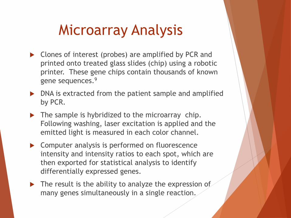
Microarray Analysis
Clones of interest (probes) are amplified by PCR and
printed onto treated glass slides (chip) using a robotic
printer. These gene chips contain thousands of known
gene sequences.9
DNA is extracted from the patient sample and amplified
by PCR.
The sample is hybridized to the microarray chip.
Following washing, laser excitation is applied and the
emitted light is measured in each color channel.
Computer analysis is performed on fluorescence
intensity and intensity ratios to each spot, which are
then exported for statistical analysis to identify
differentially expressed genes.
The result is the ability to analyze the expression of
many genes simultaneously in a single reaction.

Illustration of a DNA GeneChip (Affymetrix)
http://grf.lshtm.ac.uk/microarrayoverview.htm
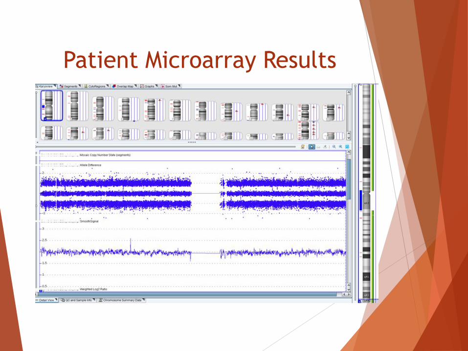
Patient Microarray Results

INTERPRETATION
Variant of Unknown Significance - Gain
arr 4q27(122,544,755-123,261,107)x3
Microarray analysis shows an ~716 kb gain within chromosome band 4q27 that contains ANXA5, TMEM155, PP12613, EXOSC9, CCNA2, BBS7, TRPC3 genes, and exons 1 through 70 of KIAA1109 gene. There are three copies of this region instead of two copies (normal) per diploid genome.
Duplication of the region as identified in this patient has not been previously reported in the literature.
Of note, two of the genes within the duplicated region are known to be disease-associated. TRPC3 is considered a candidate gene for adult-onset cerebellar ataxia, and a heterozygous missense sequence variant was reported in an individual with this disease (1). However, the clinical significance of a duplication of TRPC3, as identified in this patient, is unknown. Homozygous or compound heterozygous mutations in the BBS7 gene are associated with Bardet-Biedl syndrome (OMIM: 615984). The clinical significance of a duplication of BBS7, as identified in this patient, is unknown.
The clinical significance of duplication of the remainder of the genes in this region is also unknown.
A single benign copy number gain has been reported for the duplicated region in the Database of Genomic Variants.
Parental testing may be useful to help determine the significance of this variant. Quantitative PCR analysis is available, as needed.

Microarray of a Chimera

Chimera Microarray:
Patient Microarray:
What if the mixed field
was still present at 3
months?

Short Tandem Repeat Analysis
would be performed.
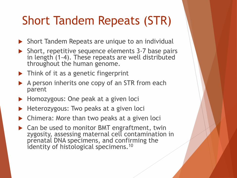
Short Tandem Repeats (STR)
Short Tandem Repeats are unique to an individual
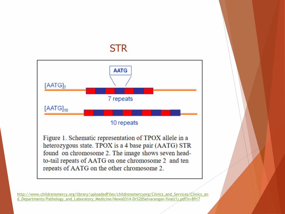
Short, repetitive sequence elements 3-7 base pairs in length (1-4). These repeats are well distributed throughout the human genome.
Think of it as a genetic fingerprint
A person inherits one copy of an STR from each parent
Homozygous: One peak at a given loci
Heterozygous: Two peaks at a given loci
Chimera: More than two peaks at a given loci
Can be used to monitor BMT engraftment, twin zygosity, assessing maternal cell contamination in prenatal DNA specimens, and confirming the identity of histological specimens.10

STR Process: Sample collection (EDTA blood or bone
marrow)
DNA extraction
PCR amplification of DNA
Color coded separation of fluorescein labeled STR alleles by capillary electrophoresis
Data and profile interpretation using data collection software
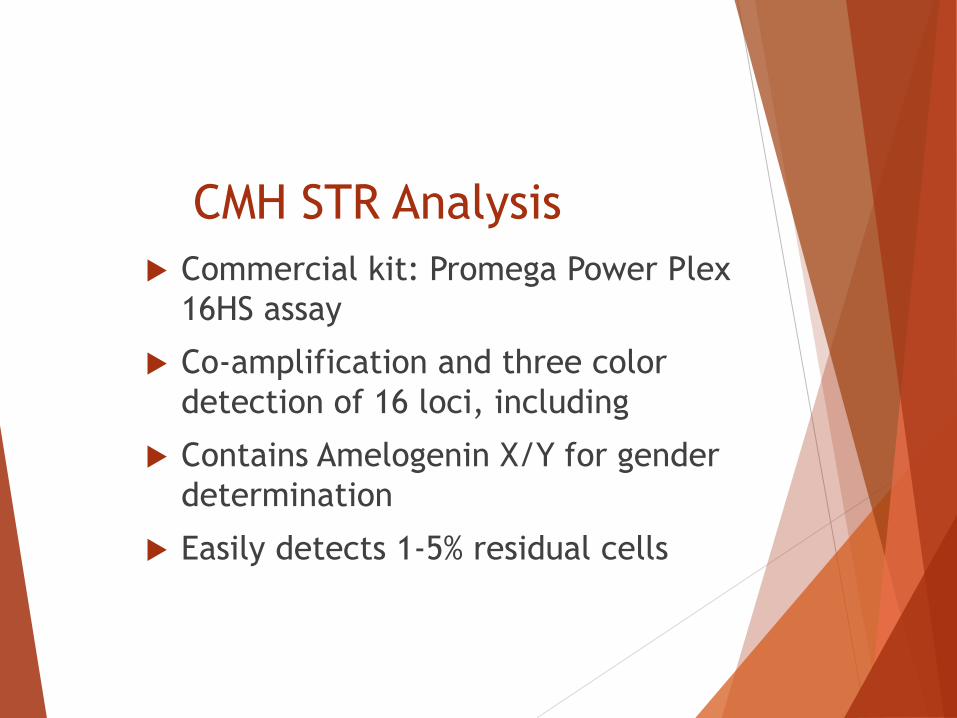
CMH STR Analysis
Commercial kit: Promega Power Plex
16HS assay
Co-amplification and three color
detection of 16 loci, including
Contains Amelogenin X/Y for gender
determination
Easily detects 1-5% residual cells
http://www.childrensmercy.org/library/uploadedFiles/childrensmercyorg/Clinics_and_Services/Clinics_an
d_Departments/Pathology_and_Laboratory_Medicine/News0314-Dr%20Selvarangan-final(1).pdf?n=8917
STR

STR Electopheragram Example:

A Closer Look:
http://projects.nfstc.org/fse/01/01-10.html

Heterozygous vs. Homozygous

Normal vs. Chimera
Normal
Chimera

The Future of Chimeras
Due to the increase of artificial chimerism, via in-vitro
fertilization, assisted fertility, hematopoietic stem cell
and solid donor transplants, the occurrence of chimeras
in transfusion services will continue to increase. These
chimera patients provide a unique set of challenges for
both the clinical blood bank and blood centers.
Communication with the physician for accurate patient
history is extremely important when investigating mixed
field reactions.
The future of Genetics:
Next Generation Sequencing
High-throughput sequencing technologies that
screen large amounts of genetic material at lower
cost than traditional sequencing technologies,
such as automated Sanger sequencing.12
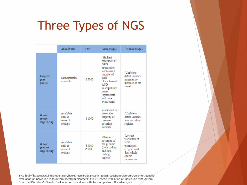
Three Types of NGS
<a href="http://www.intechopen.com/books/recent-advances-in-autism-spectrum-disorders-volume-i/genetic-evaluation-of-individuals-with-autism-spectrum-disorders" title="Genetic Evaluation of Individuals with Autism Spectrum Disorders">Genetic Evaluation of Individuals with Autism Spectrum Disorders</a>

Genetic testing is the key to
identifying Chimeras!

Interesting Chimera Examples
Case Study #1:
6 yr. old female having surgery
Pre-operative Type and Screen shows Mixed Field
Anti-A: 4+
Anti-B: Mixed Field
A cells: 0
B cells:0
Phenotype reveals mixed field reactions with Anti-E
Patient History
Patient is a dizygotic triplet (one girl and two
boys) born at 32 weeks after IVF.
Three quadricellular embryos were implanted.

Results of genetic testing:
Both males blood type: A
The propositus female blood type: AB
All three children revealed dual population of A and AB
RBCs with the same proportion (90% A and 10% AB).
The female patient was shown to have more male DNA
than female DNA(90% vs. 10%)! She was normal and
showed no signs of hermaphrodism.
Three zygotes were implanted. One survived (female),
one split to form identical twins (males), and one did
not survive.
Female is a triplet chimera
following In-Vitro fertilization.13

Case Study #2
70-year old woman admitted to the hospital for surgery
Presurgical Type and Antibody Screen ordered.
Anti-A: 2+ Mixed Field
Anti-B: 0
A cells: 0
B cells:3+
RhD: 1+ Mixed Field
RhE: 1+ Mixed Field
Patient History
No history of transfusion or
transplantation
7 previous pregnancies

Patient was interviewed for
more information and…
She had a twin brother
that died in infancy!

Results of Genetic Testing:
Genotyping was performed and the ABO type was an
A2/O chimera.
STR analysis was performed and more than two alleles
were present in 6 of 21 loci, confirming chimerism.
Genomic DNA analysis for the presence of SRY (Sex
determining Region Y) was performed and found
positive for male DNA.

Patient is a Twin Chimera.14

Case Study #3
39-year old normal Korean male
Demonstrated mixed field hemagglutination on B cell
typing at the time of blood donation
Genotyping was performed to identify the ABO subtype
B3. This subtype is relatively common in Korea

Patient History
Morphological normal male and father of one child
No history of transfusion or transplants
No history of a twin
Results of Genetic Testing:
Direct sequencing of exons 6 and 7 of the ABO gene
showed two distinct RBC populations, group B
and group O and not the B3 subgroup
STR analysis demonstrated more than two alleles in 4 of
9 loci
STR analysis of the parents and propositus demonstrated
a pattern consistent with a double paternal DNA
contribution
His karyotype revealed a mosaic pattern: 32/50
metaphases were 46XY and 18/50 metaphases
demonstrated 47XYY

STR Electropheragram Results
Father
Propositus
Mother

These results indicate the
propositus is most likely a
dispermic chimera resulting from
the division of one ovum and its
fertilization by two
spermatozoa.15

I LEAVE YOU WITH….
There may be a link with microchimerism and some autoimmune diseases. (Body sees the foreign DNA and attacks itself).18
Women with sons have been found to contain some Y chromosomes in peripheral blood mononuclear cells for decades.19
Stem cells passing from fetus to mother have been shown to have a healing affect. For example, fetal stem cells have been found in a pregnant mother’s diseased heart.20
Researchers studied the brains of 59 women ages 32 to 101 and found male DNA in 63% of the women’s brains.21
DEFINITIONS:
ZYGOTE: The cell formed by the union of a male sperm and a
female ovum.
MONOZYGOTE: Developed from a single fertilized ovum
(identical twins)
DIZYGOTE: Developed from two fertilized ovum (fraternal
twins)
TETRAGAMETIC CHIMERA: Two fertilized eggs fuse together
DISPERMIC CHIMERA: One ovum splits and fertilized by two
sperm
early in development and one baby forms.
MOSAIC: Two or more populations of cells with different
genotypes in one individual who has developed from a single
(zygote).
ANASTAMOSIS: Interconnecting blood vessels
Hermaphrodite: Containing both male and female DNA
Microchimerism: The presence of a small number of cells that
originate from another individual (zygote)
A Special Thanks to Dr. Elena Repnikova!
Assistant Director, Cytogenetics/Molecular Genetics Laboratory Children's Mercy Hospital and Clinics Assistant Professor, Departments Pathology and Pediatrics, University of Missouri-Kansas city School of Medicine
References:
1. Race R.R., and Sanger R.: Blood groups in man. Oxford (England): Blackwell Scientific Publications, 1975.
2. Van Dijk B.A., Boomsma D.I., and De Man A.J.M.: Blood group chimerism in human multiple births is not rare. Am J Med Genet 1996; 61: pp. 264-268
3. Owen RD. 1945. Immunogenetic consequences of vascular anastomoses between bovine twins. Science 102: 400– 407. Palmer JF, ed. 1835.
4. Dunsford I., Bowley C.C., Hutchinson A.M., et al: A human blood-group chimera. Br Med J 1953; 2: pp.80-81.
5. Bluth M. Reid M., Manny N. Chimerism in the Immunohematology Laboratory in the Molecular Biology Era: Transfusion Medicine Reviews, 2007-04-01, Volume 21, pp. 134-46.
6. Tipett P.: Blood group chimeras, A review. Vox Sang 1983; 44: pp.333-359.
Cross Ref (http://dx.doi.org/10.1111/j.1423-0410. 1983tb06357.x)
7. Starzl TE, Demetris AJ, Murase N, et al. Chimerism after organ transplantation. Current opinion in nephrology and hypertension. 1997;6(3):292-298.
8. Centers for Disease Control and Prevention, American Society for Reproductive Medicine, 2009 Assisted Reproductive Technology Success Rates: National Summary and Fertility Clinic Reports. Atlanta: U.S. Department of Health and Human Services; 2011. <http://www.cdc.gov/art/ART2009/PDF/ART_2009_Full.pdf>
9. http://grf.lshtm.ac.uk/microarrayoverview.htm
10. Butler J.: Short tandem repeat typing technologies used in human identity testing. BioTechniques; 43: Sii-Sv.
11.http://www.childrensmercy.org/library/uploadedFiles/childrensmercyorg/Clinics_and_Services/Clinics_and_Departments/Pathology_and_Laboratory_Medicine/News0314-Dr%20Selvarangan-final(1).pdf?n=8917
12. 15. http://www.illumina.com/content/dam/illumina-marketing/documents/products/illumina_sequencing_introduction.pdf
13. Kuhl-Burmeister R., Simeoni E., Weber-Matthiesen K., et al: Equal distribution of congenital blood cell chimerism in dizygotic triplets after in-vitro fertilization. Hum Reprod 2000; 15 pp. 1200-1204
14. Sharpe C., Lane, D., Cote J., et al: Mixed field reactions in ABO and Rh typing chimerism likely resulting from twin haematopoiesis. Blood Transfus 2014: 12: pp. 608-610.
References:
15. Duck C., Sang K., Yazer, M., et al: A dispermic chimera with mixed field blood group B and Mosaic 46, XY/47,XYY Karyotype: J Korean Med Sci 2007; 22: 553-6
16. Lo Y. M.D., Corbetta N., Chamberlain P.F., et al: Presence of fetal DNA in maternal plasma and serum. Lancet 1997; 350:pp. 485-487 Cross ref (http://dx.doi.org/10.1016/S0140-6736(97)02174-0)
17. http://genetics.thetech.org/ask/ask208
18. Adams K. M., and Nelson J.L.: Microchimerism: An investigative frontier in autoimmunity and transplantation. JAMA 2004; 291: pp. 1127-1131
19. Evans P.C., Lambert N., Maloney S., et al: Long-term fetal microchimerism in peripheral blood mononuclear cell subsets in healthy women and women with scleroderma. Blood 1999; 93:pp. 2033-2037.
20. Rina J. Kara et al. “Fetal Cells Traffic to Injured Maternal Myocardium and Undergo Cardiac Differentiation.Circulation Research, published online November 14, 2011. DOI: 10.1161/
21. http://www.independent.co.uk/news/science/male-dna-found-in-womens-brains-8181068.html
22. Bianchi D.W., Zickwolf G.K., Weil G.J., et al: Male fetal progenitor cells persist in maternal blood for as long as 27 years postpartum. Proc Natl Acad Sci USA 1996;93 pp. 705-708

Questions?