Download - Acute Lymphoblastic Leukaemia
Seminar
1030 www.thelancet.com Vol 371 March 22, 2008
Acute lymphoblastic leukaemiaChing-Hon Pui, Leslie L Robison, A Thomas Look
Acute lymphoblastic leukaemia, a malignant disorder of lymphoid progenitor cells, a! ects both children and adults, with peak prevalence between the ages of 2 and 5 years. Steady progress in development of e! ective treatments has led to a cure rate of more than 80% in children, creating opportunities for innovative approaches that would preserve past gains in leukaemia-free survival while reducing the toxic side-e! ects of current intensive regimens. Advances in our understanding of the pathobiology of acute lymphoblastic leukaemia, fuelled by emerging molecular technologies, suggest that drugs specifi cally targeting the genetic defects of leukaemic cells could revolutionise management of this disease. Meanwhile, studies are underway to ascertain the precise events that take place in the genesis of acute lymphoblastic leukaemia, to enhance the clinical application of known risk factors and antileukaemic agents, and to identify treatment regimens that might boost the generally low cure rates in adults and subgroups of children with high-risk leukaemia.
IntroductionAddition of acute lymphoblastic leukaemia to the growing list of cancers that have succumbed to e! ective treatment is tempting. The decision would be easy to justify in view of data showing cure rates higher than 80% for children treated in modern centres, most of whom will lead healthy productive lives as long-term cancer survivors.1–3 Thus, the future management of acute lymphoblastic leukaemia might be viewed as simply tweaking existing protocols and devising alternative regimens for the fi fth of patients who respond poorly to available agents. This scenario, however attractive, must be rejected on several grounds. It does not accommodate the poor prognosis for adults with acute lymphoblastic leukaemia or the complexity, expense, and toxic e! ects of contemporary multiagent treatments.1,4 Most importantly, it overlooks our rapidly increasing ability to analyse the genetic and epigenetic abnormalities of leukaemic cells and to translate them into enhanced diagnostic methods and molecularly targeted therapy.5,6 Although the molecular medicine approach is still in its investigative stage, with many new obstacles to overcome, it holds enormous promise. Put simply, we are about to enter an era in which leukaemia patients will probably receive individualised treatment based on the genetic features of their malignant cells and their own unique genetic make-up (so-called pharmacogenomics).7 Our intent in this Seminar is to review advances in both the fundamental understanding and clinical management of acute lymphoblastic leukaemia in children and adults.
Epidemiology and causeThe precise pathogenetic events leading to development of acute lymphoblastic leukaemia are unknown. Only a few cases (<5%) are associated with inherited, predisposing genetic syndromes, such as Down’s syndrome, Bloom’s syndrome, ataxia-telangiectasia, and Nijmegen breakage syndrome, or with ionising radiation or exposure to specifi c chemotherapeutic drugs. Although accumulating published work on high birthweight as a risk factor for childhood acute lymphoblastic leukaemia is becoming increasingly convincing,8 there exists an
extensive list of confl icting or isolated reports of factors purported to confer an increased risk for this disease, including parental occupation, maternal reproductive history, parental tobacco or alcohol use, maternal diet, prenatal vitamin use, exposure to pesticides or solvents, and exposure to the highest levels (>0·3 or 0·4 "T) of residential, power-frequency magnetic fi elds.9,10
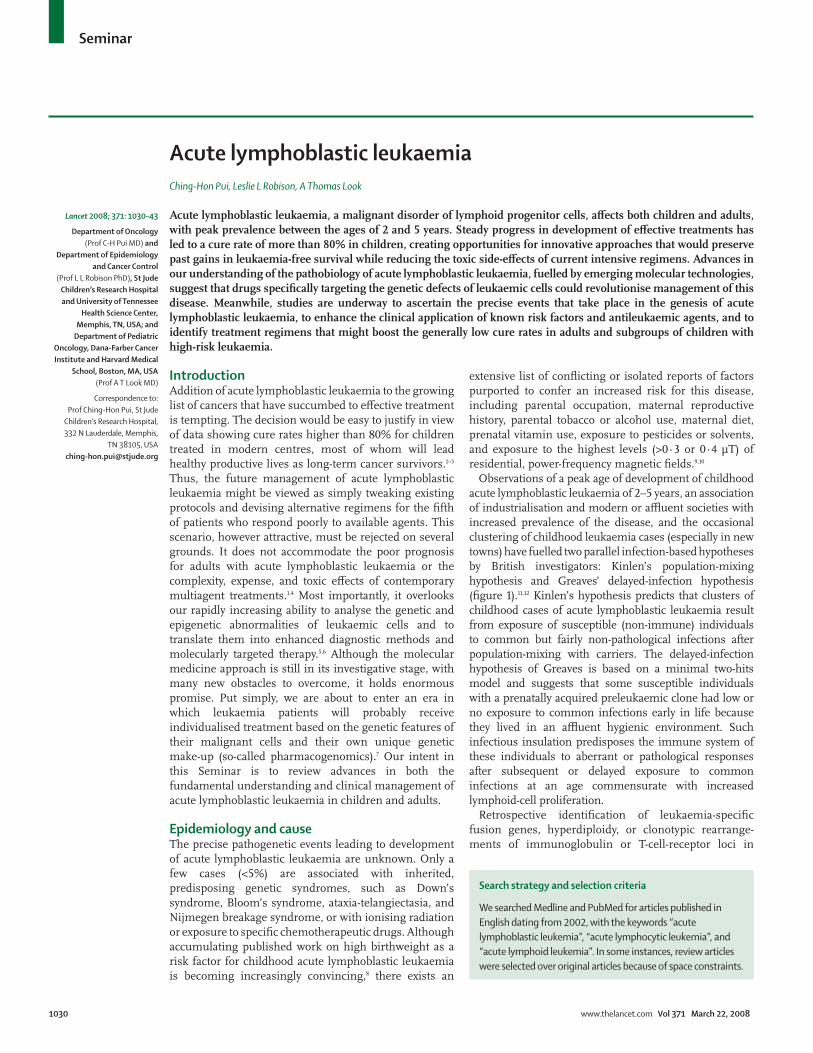
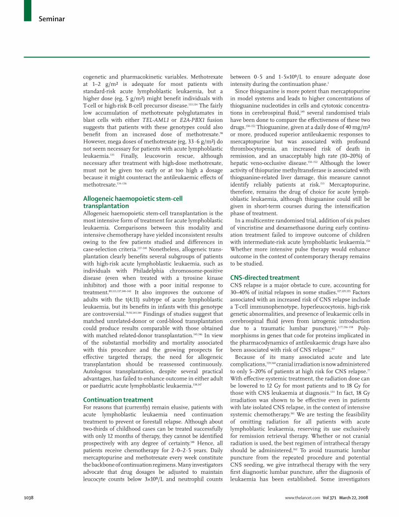
Observations of a peak age of development of childhood acute lymphoblastic leukaemia of 2–5 years, an association of industrialisation and modern or a# uent societies with increased prevalence of the disease, and the occasional clustering of childhood leukaemia cases (especially in new towns) have fuelled two parallel infection-based hypotheses by British investigators: Kinlen’s population-mixing hypothesis and Greaves’ delayed-infection hypothesis (fi gure 1).11,12 Kinlen’s hypothesis predicts that clusters of childhood cases of acute lymphoblastic leukaemia result from exposure of susceptible (non-immune) individuals to common but fairly non-pathological infections after population-mixing with carriers. The delayed-infection hypothesis of Greaves is based on a minimal two-hits model and suggests that some susceptible individuals with a prenatally acquired preleukaemic clone had low or no exposure to common infections early in life because they lived in an a# uent hygienic environment. Such infectious insulation predisposes the immune system of these individuals to aberrant or pathological responses after subsequent or delayed exposure to common infections at an age commensurate with increased lymphoid-cell proliferation.
Retrospective identifi cation of leukaemia-specifi c fusion genes, hyperdiploidy, or clonotypic rearrange-ments of immunoglobulin or T-cell-receptor loci in
Lancet 2008; 371: 1030–43
Department of Oncology (Prof C-H Pui MD) and
Department of Epidemiology and Cancer Control
(Prof L L Robison PhD), St Jude Children’s Research Hospital and University of Tennessee
Health Science Center, Memphis, TN, USA; and
Department of Pediatric Oncology, Dana-Farber Cancer Institute and Harvard Medical
School, Boston, MA, USA (Prof A T Look MD)
Correspondence to:Prof Ching-Hon Pui, St Jude
Children’s Research Hospital, 332 N Lauderdale, Memphis,
TN 38105, [email protected]
Search strategy and selection criteria
We searched Medline and PubMed for articles published in English dating from 2002, with the keywords “acute lymphoblastic leukemia”, “acute lymphocytic leukemia”, and “acute lymphoid leukemia”. In some instances, review articles were selected over original articles because of space constraints.
Seminar
www.thelancet.com Vol 371 March 22, 2008 1031
archived neonatal blood spots (Guthrie cards) and studies of leukaemia in monozygotic twins indicate clearly a prenatal origin for some childhood leukaemias.12–15 Screening of neonatal cord-blood samples has revealed a putative leukaemic clone with the TEL-AML1 fusion gene (also known as ETV6-RUNX1) in 1% of newborn babies, a frequency 100 times higher than the prevalence of acute lymphoblastic leukaemia defi ned by this fusion gene later in childhood.16 The variable incubation period and clinical outcome of such cases, and the 10% concordance rate of leukaemia in identical twins with this genotype, support the notion that additional postnatal events are needed for full leukaemic transformation.17 A recent study further established the presence of a preleukaemic clone with the TEL-AML1 fusion.15
Investigations have also focused on the genetic variability in xenobiotic metabolism, DNA repair pathways, and cell-cycle checkpoint functions that might interact with environmental, dietary, maternal, and other external factors to a! ect development of acute lymphoblastic leukaemia. Although the number of investigations and sample sizes are limited, data exist to support a possible causal role for polymorphisms in genes encoding cytochrome P450, NAD(P)H quinone oxidoreductase, glutathione S-transferases, methyl-enetetahydrofolate reductase, thymidylate synthase, serine hyroxymethyltransferase, and cell-cycle inhi-bitors.18–23 To date, however, no direct gene-environment interactions have been established convincingly.
In view of the scarcity of causal insights from large-scale epidemiological studies, some investigators have adopted a strategy that focuses on distinct subtypes of childhood acute lymphoblastic leukaemia. An important example is the study of infant acute lymphoblastic leukaemia with MLL rearrangement,24 a genetic abnormality that has also been associated with secondary leukaemia after exposure to a topoisomerase II inhibitor.25 Thus, dietary, medical, and environmental exposures to substances that inhibit topoisomerases, and the reduced ability of fetuses or their mothers to detoxify such agents, could lead to development of infant leukaemia.26,27
PathobiologyAcute lymphoblastic leukaemia is thought to originate from various important genetic lesions in blood-progenitor cells that are committed to di! erentiate in the T-cell or B-cell pathway, including mutations that impart the capacity for unlimited self-renewal and those that lead to precise stage-specifi c developmental arrest.6,28 In some cases, the fi rst mutation along the multistep pathway to overt acute lymphoblastic leukaemia might arise in a haemopoietic stem cell possessing multilineage develop-mental capacity.29 The cells implicated in acute lymphoblastic leukaemia have clonal rearrangements in their immunoglobulin or T-cell receptor genes and express antigen-receptor molecules and other
di! erentiation-linked cell-surface glycoproteins that largely recapitulate those of immature lymphoid progenitor cells within the early developmental stages of normal T and B lymphocytes.6,28,30 The dominant theme of contemporary research in pathobiology of acute lymphoblastic leukaemia is to understand the outcomes of frequently arising genetic lesions, in terms of their e! ects on cell proliferation, di! erentiation, and survival, and then to devise selectively targeted treatments against the altered gene products to which the leukaemic clones have become addicted.31
Chromosomal translocationsChromosomal translocations that activate specifi c genes are a defi ning characteristic of human leukaemias and of acute lymphoblastic leukaemia in particular.6,28 Gene-expression patterns studied in large series of newly diagnosed leukaemias have substantiated the idea that specifi c chromosomal translocations identify unique subtypes of the disease.32–35 Usually, translocations activate transcription-factor genes, which in many cases can control cell di! erentiation (rather than cell division per se), are developmentally regulated, and frequently encode proteins at the apex of important transcriptional cascades.28 These so-called master oncogenic transcription factors, which can exert either positive or negative control over downstream responder genes, are expressed aberrantly in leukaemic cells as one gene product or as a unique fusion protein combining elements from two di! erent transcription factors.6,28
About 25% of cases of B-cell precursor acute lymphoblastic leukaemia, the most frequent form of acute leukaemia in children, harbour the TEL-AML1 fusion gene—generated by the t(12;21)(p13;q22) chromosomal translocation.6 Although the molecular pathogenesis of TEL-AML1-positive leukaemia remains unclear, fi ndings in mice establish the Tel gene as an
Fetal development Birth Infancy – early childhood
Low exposureto pathogens
resulting indecreased
proliferativestress
Rapid cellproliferation
Exposure topathogens during
period ofincreased lymphoid
proliferation
Exposure to the new pathogens as a result ofincreased population mixing
Delayed exposure to common pathogens
Low or noexposure to
non-endemicpathogens
Live in an environmentwith decreased
exposure to commonpathogens
Randommutations
Grea
ves’
hypo
thes
isKi
nlen
’shy
poth
esis
Figure !: Infection-based models of leukaemia development
Seminar
1032 www.thelancet.com Vol 371 March 22, 2008
important regulator of haemopoietic-cell development, essential for defi nitive haemopoiesis.36 Similarly, Aml1 gene is essential for defi nitive embryonic haemo-poiesis.37,38 Thus, the presence of the TEL-AML1 fusion protein in B-cell progenitors seems to lead to disordered early B-lineage lymphocyte development, a hallmark of leukaemic lymphoblasts. Analysis of TEL-AML1-induced cord blood cells suggests that the fusion gene serves as a fi rst-hit mutation by endowing the preleukemic cell with altered self-renewal and survival properties.15
In adults, the most frequent chromosomal translocation is t(9;22), or the Philadelphia chromosome, which causes fusion of the BCR signalling protein to the ABL non-receptor tyrosine kinase, resulting in constitutive tyrosine kinase activity and complex interactions of this fusion protein with many other transforming elements,
such as the signalling pathway for RAS (GTP-binding protein that activates target genes involved in cell di! erentiation, proliferation, and survival).39 As an activated kinase, BCR-ABL o! ers an attractive therapeutic target, and imatinib mesilate, a small-molecule inhibitor of the ABL kinase, has proven e! ective against leukaemias that express BCR-ABL.40
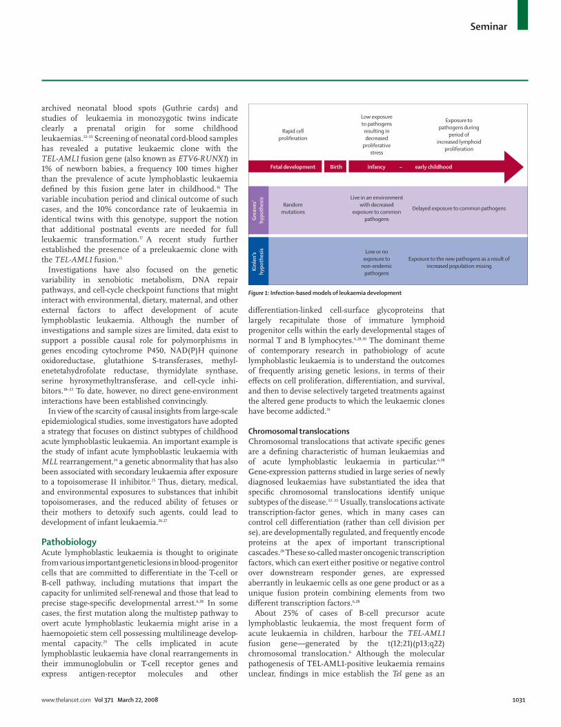
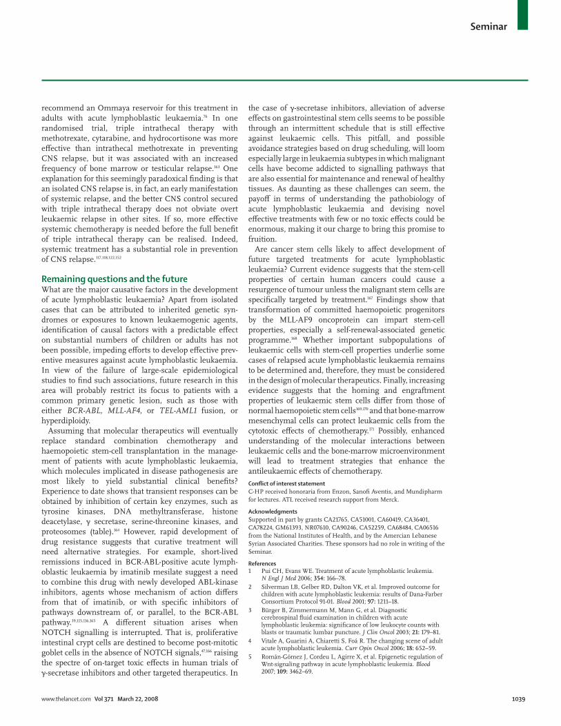
More than 50% of cases of T-cell acute lymphoblastic leukaemia have activating mutations that involve NOTCH1,41 a gene encoding a transmembrane receptor that regulates normal T-cell development.42 NOTCH receptors become activated when ligands of the Delta-Serrate-Lag2 family of proteins bind to the extracellular portion of the transmembrane molecule. This interaction initiates a cascade of proteolytic cleavages, terminating in $-secretase generation of intracellular
pre-Notch
Notch ICN
Heterodimerisation
Glycosylation
Fucosylation and endoplasmic reticulum exit
Ubiquitination and proteosome degradation
!-secretase complex
S3 cleavage S2
cleavage
S1 Cleavage
ICN
GSI ICN
ICN
ICN
ICN
MINT dnMAML NRARP
Notch
ADAM metalloprotease
pre-Notch
OFUT 1
Fringe
Fringe
Numb
Furin
DLL
Jagged
SEL 10
CSL
CSL CoA
MAML
n300
CoR
Deltex?
NEURL
MIB
HES 1 PRE T" DELTEX NRARP CD25 MYC
Receiving cell
Nucleus
Golgi
Endoplasmicreticulum
Ligandendocytosis
anddegradation
Sending cell
A
B
C
E
F
G
Figure ": NOTCH signalling in normal thymocytesThe NOTCH signalling pathway is complex and involves the coordinated activities of many di# erent molecules. Briefl y, NOTCH is synthesised in the endoplasmic reticulum (A) as one protein consisting of an extracellular domain (pre-Notch) and an intracellular domain (ICN), which are transported in tandem to the Golgi (B), where several post-translational modifi cations take place, including a proteolytic cleavage (S1) that separates the two domains from each other. The resultant heterodimer is then transported to the cell membrane, where NOTCH interacts with ligands (C) and is cleaved twice (D) by the ADAM protease (S2) and a !-secretase complex (S3), enabling the liberated ICN domain to translocate to the nucleus (E). Nuclear ICN forms a binding/activator complex with a group of cooperating proteins (F), resulting in transcriptional activation of several functionally important genes, including MYC and pre-T". Hyperphosphorylation of ICN via interaction of its PEST domain (polypeptide enriched in proline, glutamate, serine, and threonine) with CDK8, MAML, and p300 facilitates ubiquitylation (G) by SEL1 family members, targeting ICN to the proteosome. ADAM=a disintegrin and metalloproteinase domain. DLL=Delta ligand. hes1=hairy/enhancer of split. GSI=!-secretase inhibition. MAML=mastermind-like proteins. MIB=mindbomb. NEURL=neuralised-like. Nrarp=gene encoding NOTCH-regulated ankyrin-repeat protein. OFUT1=O-fucosyltransferase1. Pre-T"=pre-TCR". Deltex=positive regulator of Notch signalling pathway. CS1=DNA binding component. CoA=co-activators. CoR=co-repressors. Fringe=regulator of Notch ligand. SEL10=positive regulator of Notch. Numb=negative receptor of Notch.
Seminar
www.thelancet.com Vol 371 March 22, 2008 1033
NOTCH1, which translocates to the nucleus and regulates by transcription a diverse set of responder genes, including the MYC oncogene (fi gure 2).43,44 The precise mechanisms by which aberrant NOTCH signalling (due to mutational activation) causes T-cell acute lymphoblastic leukaemia are still unclear but probably entail constitutive expression of oncogenic responder genes, such as MYC, and cooperation with other signalling pathways (pre-TCR [T-cell receptor for antigen] and RAS, for example). Interference with NOTCH signalling by small-molecule inhibition of $-secretase activity has the potential to induce remission of T-cell acute lymphoblastic leukaemia.
Evidence suggests that the MYC oncoprotein is an important downstream mediator of the pro-growth e! ects of NOTCH1 signalling in developing thym-ocytes.28,45 However, results of retroviral insertional mutagenesis in murine models of transgenic T-cell acute lymphoblastic leukaemia show that Notch1 mutations, with outcomes similar to those in primary human T-cell acute lymphoblastic leukaemias, can potentiate the e! ects of pre-existing MYC overexpression,42,46 suggesting that NOTCH1 must have important transformational targets other than MYC. Activating mutations in NOTCH1 su% cient to produce constitutive NOTCH1 signalling can induce T-cell acute lymphoblastic leukaemia in experimental models and could be the instigating event in most human T-cell leukaemias.28,42 $-secretase, a multicomponent membrane-associated enzyme, is needed for NOTCH1 signalling through mutant NOTCH receptors in T-cell acute lymphoblastic leukaemia, providing an attractive target for therapeutic intervention with newly developed $-secretase inhibitors.28,47
Cooperating mutationsAlthough chromosomal abnormalities are a hallmark of pathogenesis of acute lymphoblastic leukaemia, evidence suggests that they must act in concert with several other genetic lesions to induce overt leukaemia. A prime example is the biallelic deletion or epigenetic silencing of the cyclin-dependent kinase inhibitor 2A gene (CDKN2A), which encodes both the tumour suppressors p16INK4A and p14ARF and whose inactivation neutralises both the TP53 and retinoblastoma pathways in most cases of T-cell and many cases of B-cell precursor acute lymphoblastic leukaemia.6 In a genome-wide analysis of 242 cases of paediatric acute lymphoblastic leukaemia using high-resolution single nucleotide polymorphism arrays, deletions, amplifi cations, point mutations, and other structural rearrangements were identifi ed in genes encoding regulators of B-lymphocyte development in 40% of cases of B-cell precursor acute lymphoblastic leukaemia.48 The PAX5 gene was the most frequent target of somatic mutation, being altered in almost a third of cases. Deletions were also detected in other B-cell developmental genes, such as TCF3 (E2A), EBF1 (EBF),
LEF1, IKZF1 (Ikaros), and IKZF3 (Aiolos). Finally, in T-cell acute lymphoblastic leukaemia, at least fi ve multistep mutational pathways leading to frank leukaemia have been identifi ed, and in some cases these pathways entail fi ve or more documented genetic lesions.28,30,42
Ongoing research to defi ne the oncogenic contributions of various classes of genetic lesions relies heavily on animal models that accurately recapitulate the molecular pathogenesis of B-cell precursor or T-cell acute lymphoblastic leukaemia.49 Most studies undertaken to date have used genetically engineered mice to elucidate the multistep transformation pathways leading to T-cell acute lymphoblastic leukaemia.50 Such models depend on breeding strategies to combine one or more genetic lesions and show synergy in transformation,51 whereas some investigators have also capitalised on the use of retroviral insertional mutagenesis screens to uncover collaborating oncogenes.46,52 New models of T-cell acute lymphoblastic leukaemia in the zebrafi sh o! er a powerful alternative vertebrate system for leukaemia research, whose unique advantages complement those of extant murine models.53 Currently available zebrafi sh models of acute lymphoblastic leukaemia include a myc transgene-driven system, in which lymphoblasts faithfully reproduce the multistep oncogenic pathway noted in up to 60% of human T-cell acute lymphoblastic leukaemias,54,55 and a transgenic zebrafi sh model, in which the TEL-AML1 oncoprotein induces B-cell precursor leukaemia.56
The challenge now is to understand how these cooperative genetic lesions and their a! ected pathways interact to alter the proliferation, di! erentiation, and survival of lymphocyte progenitors leading to their leukaemic conversion. This research will undoubtedly provide the molecular rationales needed to select new therapeutic targets and to develop interfering small molecules or antibodies with high levels of antileukaemic specifi city and activity.57 The table provides a partial list of molecularly targeted drugs now in clinical testing.
DiagnosisPhenotypeImmunophenotyping of leukaemic lymphoblasts by fl ow cytometry is essential to establish the correct diagnosis and defi ne cell lineage. Although acute lymphoblastic leukaemia can be readily subclassifi ed according to the many steps of normal B-cell and T-cell di! erentiation, the only fi ndings with therapeutic importance are T-cell, mature B-cell, and B-cell precursor phenotypes.26,58
Myeloid-associated antigen expression can be detected in as many as half the cases of acute lymphoblastic leukaemia. However, with contemporary treatment, this so-called aberrant antigen expression has no prognostic implications but can be used to distinguish leukaemic cells from normal progenitor cells, thereby enabling detection of minimal (ie, submicroscopic) residual leukaemia.6,26
Seminar
1034 www.thelancet.com Vol 371 March 22, 2008
GenotypeAlthough chromosomal analysis is still an integral component of initial work-up of acute lymphoblastic leukaemia, other highly specifi c and sensitive techniques—such as RT-PCR, fl uorescence in-situ hybridisation, and fl ow cytometry—are increasingly used to detect specifi c fusion transcripts, gain or loss of cellular DNA content, or specifi c chromosomes with prognostic or therapeutic relevance.26,58 Although still a research technique, gene-expression profi ling can not only identify accurately the major subtypes of acute lymphoblastic leukaemia but also implicate single genes or signalling pathways as important determinants of clinical outcome.32–34,59–61 Once this method has been refi ned and made cost e! ective, it will undoubtedly replace many current diagnostic techniques.
PharmacogeneticsGenetic polymorphisms of drug transporters, receptors, targets, and drug-metabolising enzymes can a! ect the e! ectiveness and toxic e! ects of antineoplastic drugs.7,62
Traditionally, pharmacogenetic studies have focused on single genes identifi ed on the basis of their infl uence on the pharmacokinetics and pharmacological e! ects of anticancer drugs. Findings of global gene-expression profi ling studies have identifi ed a growing number of genomic determinants of treatment responses that could allow development of polygenic models for optimisation of treatment for acute lymphoblastic leukaemia.6,7,63
Despite the promise of pharmacogenetic studies to enhance treatment outcome in acute lymphoblastic leukaemia, only polymorphisms and the activity of thiopurine methyltransferase—an enzyme that catalyses S-methylation (inactivation) of thiopurines such as mercaptopurine and thioguanine—have been useful in clinical practice.7,64,65 About 10% of the total population inherit one wild-type gene encoding thiopurine methyltransferase and one non-functional variant allele, resulting in intermediate enzyme activity, whereas 1 in 300 people inherit two non-functional variant alleles with no enzyme activity. When treated with conventional doses of thiopurines, up to half of patients with the heterozygous defi ciency and all homozygous-defi cient patients develop haemopoietic toxic e! ects, which can be fatal in the homozygous group.7 The enzyme defi ciency also confers a high risk of developing therapy-related acute myeloid leukemia7 and radiation-induced brain tumours, in the context of intensive thiopurine treatment.66 Conversely, patients with high levels of enzyme activity might be at greater risk of relapse owing to decreased exposure of leukaemic cells to active drug metabolites.65 In most centres, studies of thiopurine methyltransferase activity are undertaken only in people with poor tolerance to antimetabolite-based therapy, and the result is used to guide reductions in drug dosage.67 We use a fairly high dose of mercaptopurine and, thus, study this enzyme prospectively in all patients, lowering the dose of mercaptopurine in individuals with enzyme defi ciency.64
Risk assessmentCareful assessment of the risk of relapse in individual patients ensures that very intensive treatment is given only to high-risk cases, thus sparing people at lower risk from undue toxic e! ects. Although enhanced treatment has abolished the prognostic strength of many clinical and biological risk factors identifi ed in the past, we would stress that even so-called low-risk patients need a certain degree of treatment intensifi cation to avoid unacceptable rates of relapse. Findings have shown that adolescents and young adults who were treated on adult protocols fared signifi cantly worse than the same age-groups treated on paediatric protocols.68–70 The superior outcome achieved with paediatric regimens has been attributed to more e! ective treatment and to better adherence by patients, parents, and doctors.68–72 To understand the actual basis for this di! erence in
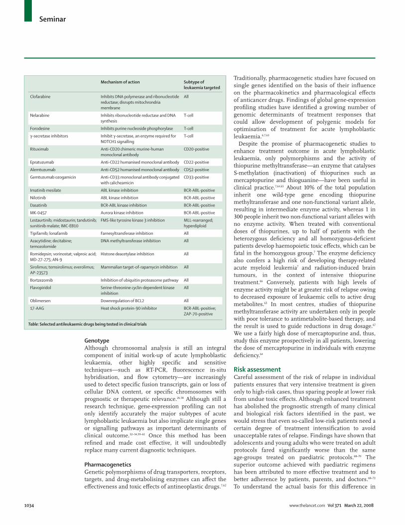
Mechanism of action Subtype of leukaemia targeted
Clofarabine Inhibits DNA polymerase and ribonucleotide reductase; disrupts mitochrondria membrane
All
Nelarabine Inhibits ribonucleotide reductase and DNA synthesis
T-cell
Forodesine Inhibits purine nucleoside phosphorylase T-cell
!-secretase inhibitors Inhibit !-secretase, an enzyme required for NOTCH1 signalling
T-cell
Rituximab Anti-CD20 chimeric murine-human monoclonal antibody
CD20-positive
Epratuzumab Anti-CD22 humanised monoclonal antibody CD22-positive
Alemtuzumab Anti-CD52 humanised monoclonal antibody CD52-positive
Gemtuzumab ozogamicin Anti-CD33 monoclonal antibody conjugated with calicheamicin
CD33-positive
Imatinib mesilate ABL kinase inhibition BCR-ABL-positive
Nilotinib ABL kinase inhibition BCR-ABL-positive
Dasatinib BCR-ABL kinase inhibition BCR-ABL-positive
MK-0457 Aurora kinase inhibition BCR-ABL-positive
Lestaurtinib; midostaurin; tandutinib; sunitinib malate; IMC-EB10
FMS-like tyrosine kinase 3 inhibition MLL-rearranged; hyperdiploid
Tipifarnib; lonafarnib Farnesyltransferase inhibition All
Azacytidine; decitabine; temozolomide
DNA methyltransferase inhibition All
Romidepsin; vorinostat; valproic acid; MD-27-275; AN-9
Histone deacetylase inhibition All
Sirolimus; temsirolimus; everolimus; AP-23573
Mammalian target-of-rapamycin inhibition All
Bortzezomib Inhibition of ubiquitin proteasome pathway All
Flavopiridol Serine-threonine cyclin-dependent kinase inhibition
All
Oblimersen Downregulation of BCL2 All
17-AAG Heat shock protein-90 inhibitor BCR-ABL-positive; ZAP-70-positive
Table: Selected antileukaemic drugs being tested in clinical trials
Seminar
www.thelancet.com Vol 371 March 22, 2008 1035
outcome, several combined adult and paediatric consortia are using common regimens to treat patients aged 1–50 years.
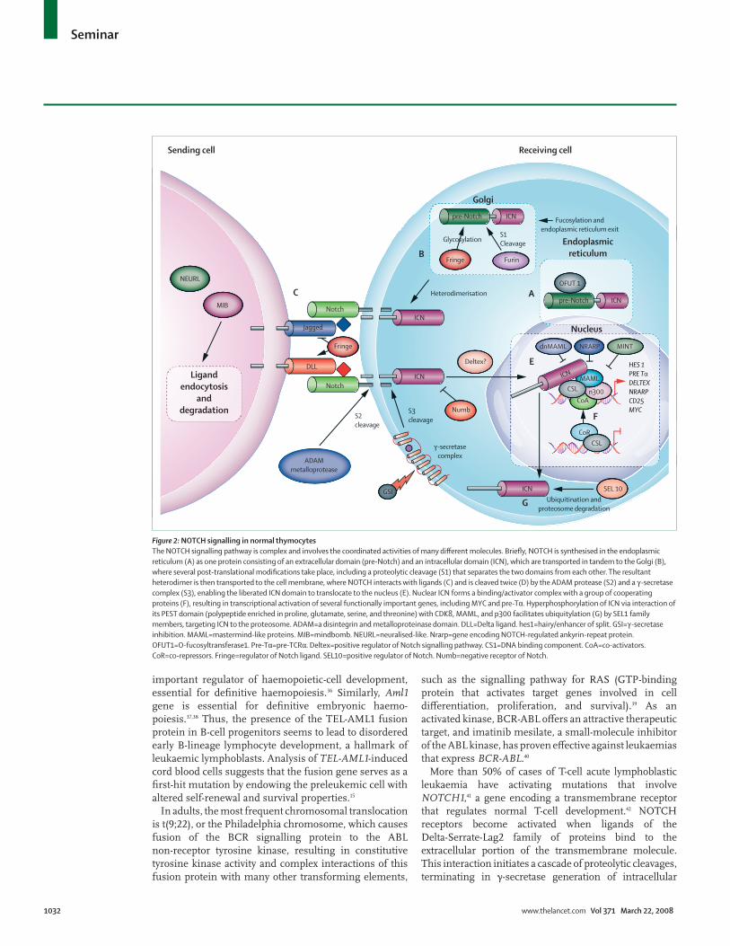
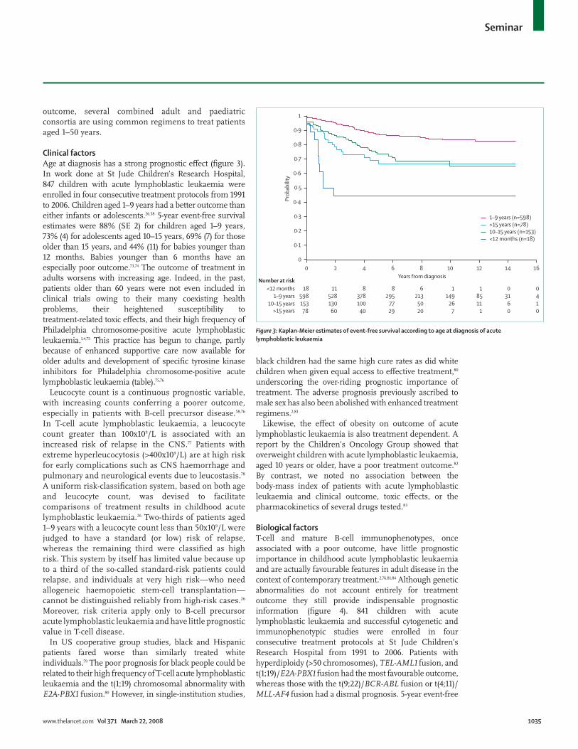
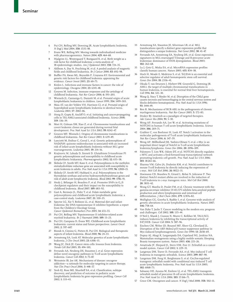
Clinical factorsAge at diagnosis has a strong prognostic e! ect (fi gure 3). In work done at St Jude Children’s Research Hospital, 847 children with acute lymphoblastic leukaemia were enrolled in four consecutive treatment protocols from 1991 to 2006. Children aged 1–9 years had a better outcome than either infants or adolescents.26,58 5-year event-free survival estimates were 88% (SE 2) for children aged 1–9 years, 73% (4) for adolescents aged 10–15 years, 69% (7) for those older than 15 years, and 44% (11) for babies younger than 12 months. Babies younger than 6 months have an especially poor outcome.73,74 The outcome of treatment in adults worsens with increasing age. Indeed, in the past, patients older than 60 years were not even included in clinical trials owing to their many coexisting health problems, their heightened susceptibility to treatment-related toxic e! ects, and their high frequency of Philadelphia chromosome-positive acute lymphoblastic leukaemia.1,4,75 This practice has begun to change, partly because of enhanced supportive care now available for older adults and development of specifi c tyrosine kinase inhibitors for Philadelphia chromosome-positive acute lymphoblastic leukaemia (table).75,76
Leucocyte count is a continuous prognostic variable, with increasing counts conferring a poorer outcome, especially in patients with B-cell precursor disease.58,76 In T-cell acute lymphoblastic leukaemia, a leucocyte count greater than 100x109/L is associated with an increased risk of relapse in the CNS.77 Patients with extreme hyperleucocytosis (>400x109/L) are at high risk for early complications such as CNS haemorrhage and pulmonary and neurological events due to leucostasis.78 A uniform risk-classifi cation system, based on both age and leucocyte count, was devised to facilitate comparisons of treatment results in childhood acute lymphoblastic leukaemia.26 Two-thirds of patients aged 1–9 years with a leucocyte count less than 50x109/L were judged to have a standard (or low) risk of relapse, whereas the remaining third were classifi ed as high risk. This system by itself has limited value because up to a third of the so-called standard-risk patients could relapse, and individuals at very high risk—who need allogeneic haemopoietic stem-cell transplantation—cannot be distinguished reliably from high-risk cases.26 Moreover, risk criteria apply only to B-cell precursor acute lymphoblastic leukaemia and have little prognostic value in T-cell disease.
In US cooperative group studies, black and Hispanic patients fared worse than similarly treated white individuals.79 The poor prognosis for black people could be related to their high frequency of T-cell acute lymphoblastic leukaemia and the t(1;19) chromosomal abnormality with E2A-PBX1 fusion.80 However, in single-institution studies,
black children had the same high cure rates as did white children when given equal access to e! ective treatment,80 underscoring the over-riding prognostic importance of treatment. The adverse prognosis previously ascribed to male sex has also been abolished with enhanced treatment regimens.2,81
Likewise, the e! ect of obesity on outcome of acute lymphoblastic leukaemia is also treatment dependent. A report by the Children’s Oncology Group showed that overweight children with acute lymphoblastic leukaemia, aged 10 years or older, have a poor treatment outcome.82 By contrast, we noted no association between the body-mass index of patients with acute lymphoblastic leukaemia and clinical outcome, toxic e! ects, or the pharmacokinetics of several drugs tested.83
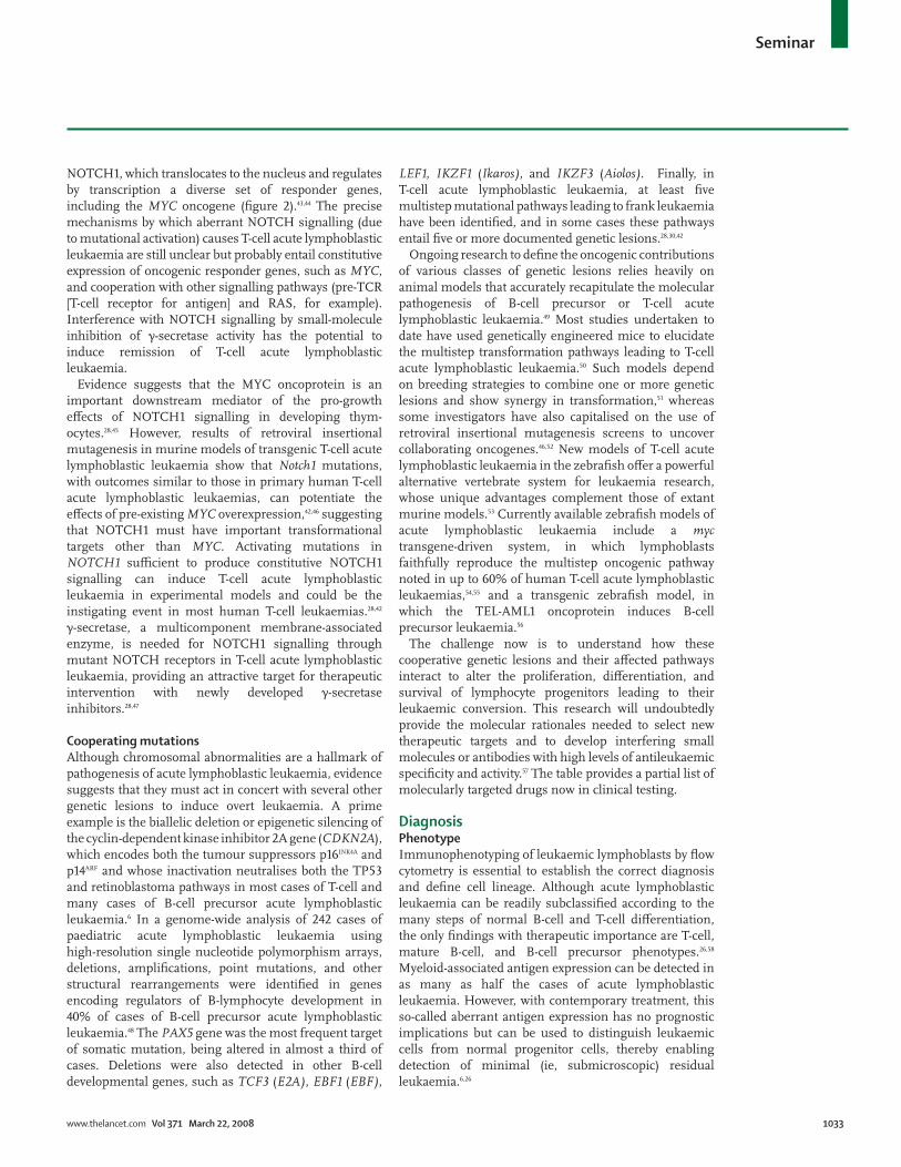
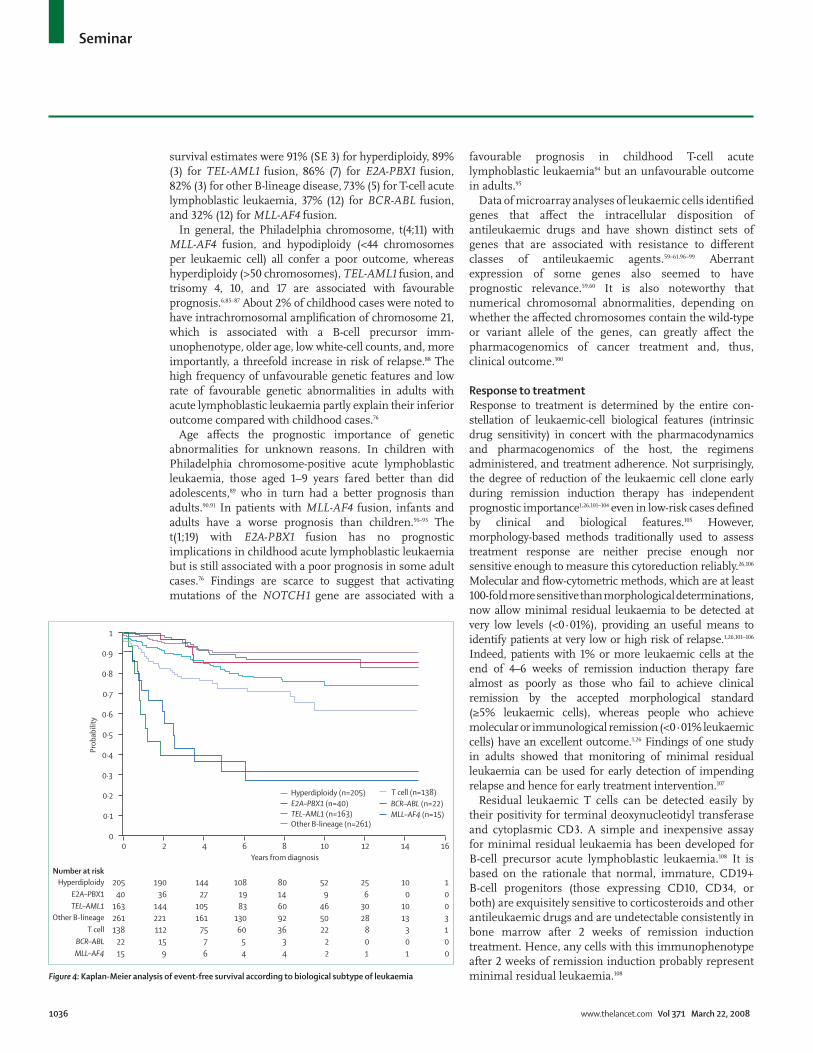
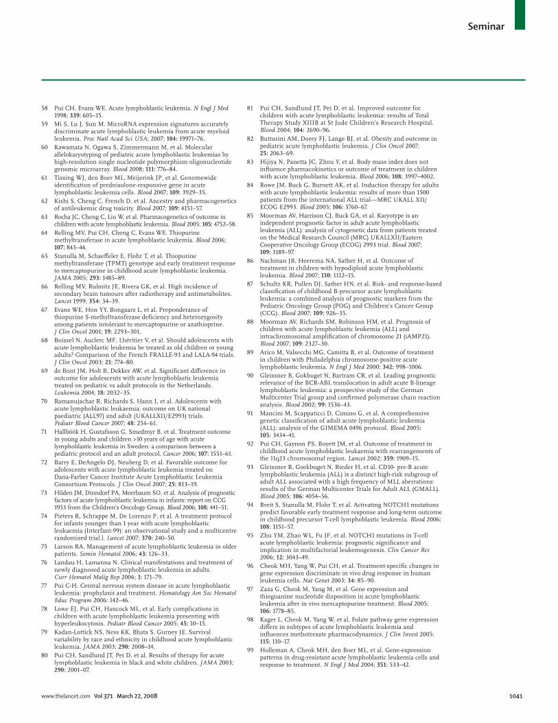
Biological factorsT-cell and mature B-cell immunophenotypes, once associated with a poor outcome, have little prognostic importance in childhood acute lymphoblastic leukaemia and are actually favourable features in adult disease in the context of contemporary treatment.2,76,81,84 Although genetic abnormalities do not account entirely for treatment outcome they still provide indispensable prognostic information (fi gure 4). 841 children with acute lymphoblastic leukaemia and successful cytogenetic and immunophenotypic studies were enrolled in four consecutive treatment protocols at St Jude Children’s Research Hospital from 1991 to 2006. Patients with hyperdiploidy (>50 chromosomes), TEL-AML1 fusion, and t(1;19)/E2A-PBX1 fusion had the most favourable outcome, whereas those with the t(9;22)/BCR-ABL fusion or t(4;11)/MLL-AF4 fusion had a dismal prog nosis. 5-year event-free
00
0·1
0·2
0·3
0·4
0·5
0·6
0·7
0·8
0·9
1
2 4 6 8Years from diagnosis
10 12 14 16
18<12 monthsNumber at risk
1–9 years10–15 years
>15 years
<12 months (n=18)
1–9 years (n=598)
10–15 years (n=153)>15 years (n=78)
11 8 8 6 1 1 0 0598 528 378 295 213 149 85 31 4153 130 100 77 50 26 11 6 178 60 40 29 20 7 1 0 0
Prob
abili
ty
Figure #: Kaplan-Meier estimates of event-free survival according to age at diagnosis of acute lymphoblastic leukaemia
Seminar
1036 www.thelancet.com Vol 371 March 22, 2008
survival estimates were 91% (SE 3) for hyperdiploidy, 89% (3) for TEL-AML1 fusion, 86% (7) for E2A-PBX1 fusion, 82% (3) for other B-lineage disease, 73% (5) for T-cell acute lymphoblastic leukaemia, 37% (12) for BCR-ABL fusion, and 32% (12) for MLL-AF4 fusion.
In general, the Philadelphia chromosome, t(4;11) with MLL-AF4 fusion, and hypodiploidy (<44 chromosomes per leukaemic cell) all confer a poor outcome, whereas hyperdiploidy (>50 chromosomes), TEL-AML1 fusion, and trisomy 4, 10, and 17 are associated with favourable prognosis.6,85–87 About 2% of childhood cases were noted to have intrachromosomal amplifi cation of chromosome 21, which is associated with a B-cell precursor imm-unophenotype, older age, low white-cell counts, and, more importantly, a threefold increase in risk of relapse.88 The high frequency of unfavourable genetic features and low rate of favourable genetic abnormalities in adults with acute lymphoblastic leukaemia partly explain their inferior outcome compared with childhood cases.76
Age a! ects the prognostic importance of genetic abnormalities for unknown reasons. In children with Philadelphia chromosome-positive acute lymphoblastic leukaemia, those aged 1–9 years fared better than did adolescents,89 who in turn had a better prognosis than adults.90,91 In patients with MLL-AF4 fusion, infants and adults have a worse prognosis than children.91–93 The t(1;19) with E2A-PBX1 fusion has no prognostic implications in childhood acute lymphoblastic leukaemia but is still associated with a poor prognosis in some adult cases.76 Findings are scarce to suggest that activating mutations of the NOTCH1 gene are associated with a
favourable prognosis in childhood T-cell acute lymphoblastic leukaemia94 but an unfavourable outcome in adults.95
Data of microarray analyses of leukaemic cells identifi ed genes that a! ect the intracellular disposition of antileukaemic drugs and have shown distinct sets of genes that are associated with resistance to di! erent classes of antileukaemic agents.59–61,96–99 Aberrant expression of some genes also seemed to haveprognostic relevance.59,60 It is also noteworthy that numerical chromosomal abnormalities, depending on whether the a! ected chromosomes contain the wild-type or variant allele of the genes, can greatly a! ect the pharmacogenomics of cancer treatment and, thus, clinical outcome.100
Response to treatmentResponse to treatment is determined by the entire con-stellation of leukaemic-cell biological features (intrinsic drug sensitivity) in concert with the pharmaco dynamics and pharmacogenomics of the host, the regimens administered, and treatment adherence. Not surprisingly, the degree of reduction of the leukaemic cell clone early during remission induction therapy has independent prognostic importance1,26,101–104 even in low-risk cases defi ned by clinical and biological features.105 However, morphology-based methods traditionally used to assess treatment response are neither precise enough nor sensitive enough to measure this cytoreduction reliably.26,106 Molecular and fl ow-cytometric methods, which are at least 100-fold more sensitive than morpho logical determinations, now allow minimal residual leukaemia to be detected at very low levels (<0·01%), providing an useful means to identify patients at very low or high risk of relapse.1,26,101–106 Indeed, patients with 1% or more leukaemic cells at the end of 4–6 weeks of remission induction therapy fare almost as poorly as those who fail to achieve clinical remission by the accepted morpho logical standard (&5% leukaemic cells), whereas people who achieve molecular or immunological remission (<0·01% leukaemic cells) have an excellent outcome.1,26 Findings of one study in adults showed that monitoring of minimal residual leukaemia can be used for early detection of impending relapse and hence for early treatment intervention.107
Residual leukaemic T cells can be detected easily by their positivity for terminal deoxynucleotidyl transferase and cytoplasmic CD3. A simple and inexpensive assay for minimal residual leukaemia has been developed for B-cell precursor acute lymphoblastic leukaemia.108 It is based on the rationale that normal, immature, CD19+ B-cell progenitors (those expressing CD10, CD34, or both) are exquisitely sensitive to corticosteroids and other antileukaemic drugs and are undetectable consistently in bone marrow after 2 weeks of remission induction treatment. Hence, any cells with this immunophenotype after 2 weeks of remission induction probably represent minimal residual leukaemia.108
Years from diagnosis
0
0·1
0·2
0·3
0·4
0·5
0·6
0·7
0·8
0·9
1
0 2 4 6 8 10 12 14 16
205Hyperdiploidy
Prob
abili
ty
Hyperdiploidy (n=205)E2A–PBX1 (n=40)
Number at risk
TEL–AML1
TEL–AML1 (n=163)
E2A–PBX1
Other B-lineage
Other B-lineage (n=261)
190 144 108 80 52 25 10 140 36 27 19 14 9 6 0 0
163 144 105 83 60 46 30 10 0261 221 161 130 92 50 28 13 3138T cell
T cell (n=138)
BCR–ABL
BCR–ABL (n=22)
MLL–AF4
MLL–AF4 (n=15)
112 75 60 36 22 8 3 122 15 7 5 3 2 0 0 015 9 6 4 4 2 1 1 0
Figure $: Kaplan-Meier analysis of event-free survival according to biological subtype of leukaemia
Seminar
www.thelancet.com Vol 371 March 22, 2008 1037
TreatmentWith the exception of patients with mature B-cell acute lymphoblastic leukaemia, who are treated with short-term intensive chemotherapy (including high-dose methotrexate, cytarabine, and cyclophosphamide),109–111 treatment for acute lymphoblastic leukaemia typically consists of a remission-induction phase, an intensifi cation (or consolidation) phase, and continuation therapy to eliminate residual disease. Treatment is also directed to the CNS early in the clinical course to prevent relapse attributable to leukaemic cells sequestered in this site.1 The drugs currently in use for these phases were developed and tested between the 1950s and 1970s, but e! orts to identify new antileukaemic agents have begun to intensify (table).
Remission-induction phaseThe goal of remission-induction treatment is to eradicate more than 99% of the initial leukaemic cell burden and to restore normal haemopoiesis and healthy performance status. This approach typically includes administration of a glucocorticoid (prednisone or dexamethasone), vincristine, and at least a third drug (asparaginase, anthracycline, or both). A three-drug induction regimen seems su% cient for most standard-risk cases provided they receive intensifi ed post-remission treatment. Children with high-risk or very high-risk acute lymph-oblastic leukaemia, and virtually all adult cases of the disease, are treated with four or more drugs for remission induction.1 We measure levels of minimal residual leukaemia after 2 weeks of remission induction and we intensify treatment in patients with high amounts of residual blasts (>1%). Clinical remission can now be induced in 96–99% of children and 78–93% of adults.1 Although no induction regimen is clearly superior to any others, addition of cyclophosphamide and intensive treatment with asparaginase are widely considered benefi cial to patients with T-cell acute lymphoblastic leukaemia,2,76 and imatinib mesilate has greatly enhanced the remission-induction rate, duration of disease-free survival, and quality of life of patients with Philadelphia chromosome-positive acute lymphoblastic leukaemia.112–114 Whether the cure rate of this subtype of leukaemia can be raised with imatinib or the newly developed, more potent, tyrosine kinase inhibitors nilotinib and dasatinib remains unknown.115,116
Presumably because of its longer half-life and increased penetration into the CNS, dexamethasone has been deemed more e! ective than either prednisone or prednisolone for treatment of acute lymphoblastic leukaemia.117,118 However, fi ndings of a small randomised study showed that an augmented dose of prednisolone produced results comparable with those achieved with dexamethasone in the context of other intensive treatment.119 Similarly, the pharmacodynamics of asparaginase di! er by formulation,120 and in terms of leukaemia control, the dose intensity and duration of
asparaginase treatment (ie, the amount of asparagine depletion) are far more important than the type of asparaginase used.1,2 Compared with Escherichia coli asparaginase, Erwinia asparaginase was associated with inferior antileukaemic response but fewer toxic e! ects,121,122 a fi nding now attributed to use of inadequate doses of the Erwinia drug. In some current protocols, polyethylene glycol-conjugated asparaginase—a long-acting and less allergenic form—has replaced the native product in initial treatment.123–126 Many comp lications recorded during remission induction are attributable to the synergistic e! ects of corticosteroid and asparaginase. In the context of multiagent treatment, a fairly small increase in dose of dexame thasone or asparaginase can result in excessive toxic e! ects and death, especially in older children and adults.
Consolidation (intensifi cation) treatmentWith the restoration of normal haemopoiesis and body function, intensifi cation treatment is generally used to eradicate drug-resistant residual leukaemic cells, thus reducing the risk of relapse. For example, patients with TEL-AML1-positive disease have an especially good outcome in clinical trials of intensive post-remission therapy with corticosteroids, vincristine, and aspara-ginase.81,127 Although the importance of this treatment phase is rarely disputed, consensus is scarce on the best regimens and duration of treatment. Frequently used strategies include high-dose methotrexate plus mercaptopurine, reinduction treatment with the same agent that was given initially, frequent pulses of vincristine and corticosteroid plus high-dose asparaginase for 20–30 weeks, and an augmented regimen consisting of reinduction treatment and additional doses of vincristine, asparaginase, and intravenous methotrexate during periods of myelosuppression.1–3,128 For patients with high-risk or very high-risk acute lymphoblastic leukaemia, incorporation of high-dose methotrexate plus mercaptopurine into a regimen based on intensive asparaginase treatment could be desirable. Findings of ongoing studies will establish if these approaches in children are e! ective and tolerable in adults.
Reinduction treatment has become an integral component of contemporary protocols. In one randomised study of intermediate-risk acute lymph oblastic leukaemia, double reinduction further enhanced treatment outcome, whereas additional pulses of vincristine and prednisone after one reinduction course were not benefi cial, suggesting that the increased dose-intensity of other drugs—such as asparaginase—led to the noted improvement.129 Although a standard intensifi cation regimen for adult acute lymphoblastic leukaemia is absent, post-remission treatment with cytarabine, cyclophosphamide, anthra-cyclines, and methotrexate has improved outcome in some non-randomised studies.130–132
The best dose of methotrexate depends on the leukaemic-cell genotype and phenotype and host pharma-
Seminar
1038 www.thelancet.com Vol 371 March 22, 2008
cogenetic and pharmacokinetic variables. Methotrexate at 1–2 g/m' is adequate for most patients with standard-risk acute lymphoblastic leukaemia, but a higher dose (eg, 5 g/m') might benefi t individuals with T-cell or high-risk B-cell precursor disease.133,134 The fairly low accumulation of methotrexate polyglutamates in blast cells with either TEL-AML1 or E2A-PBX1 fusion suggests that patients with these genotypes could also benefi t from an increased dose of methotrexate.98 However, mega doses of methotrexate (eg, 33·6 g/m') do not seem necessary for patients with acute lymphoblastic leukaemia.135 Finally, leucovorin rescue, although necessary after treatment with high-dose methotrexate, must not be given too early or at too high a dosage because it might counteract the antileukaemic e! ects of methotrexate.134–136
Allogeneic haemopoietic stem-cell transplantationAllogeneic haemopoietic stem-cell transplantation is the most intensive form of treatment for acute lymphoblastic leukaemia. Comparisons between this modality and intensive chemotherapy have yielded inconsistent results owing to the few patients studied and di! erences in case-selection criteria.137–140 Nonetheless, allogeneic trans-plantation clearly benefi ts several subgroups of patients with high-risk acute lymph oblastic leukaemia, such as individuals with Philadelphia chromosome-positive disease (even when treated with a tyrosine kinase inhibitor) and those with a poor initial response to treatment.89,113,137,140–142 It also improves the outcome of adults with the t(4;11) subtype of acute lymphoblastic leukaemia, but its benefi ts in infants with this genotype are controversial.74,92,143,144 Findings of studies suggest that matched unrelated-donor or cord-blood transplanta tion could produce results comparable with those obtained with matched related-donor transplantation.145,146 In view of the substantial morbidity and mortality associated with this procedure and the growing prospects for e! ective targeted therapy, the need for allogeneic transplantation should be reassessed continuously. Autologous transplantation, despite several practical advantages, has failed to enhance outcome in either adult or paediatric acute lymphoblastic leukaemia.138,147
Continuation treatmentFor reasons that (currently) remain elusive, patients with acute lymphoblastic leukaemia need continuation treatment to prevent or forestall relapse. Although about two-thirds of childhood cases can be treated successfully with only 12 months of therapy, they cannot be identifi ed prospectively with any degree of certainty.148 Hence, all patients receive chemotherapy for 2·0–2·5 years. Daily mercaptopurine and methotrexate every week constitute the backbone of continuation regimens. Many investigators advocate that drug dosages be adjusted to maintain leucocyte counts below 3(10)/L and neutrophil counts
between 0·5 and 1·5(10)/L to ensure adequate dose intensity during the continuation phase.1
Since thioguanine is more potent than mercaptopurine in model systems and leads to higher concentrations of thioguanine nucleotides in cells and cytotoxic concentra-tions in cerebrospinal fl uid,149 several randomised trials have been done to compare the e! ectiveness of these two drugs.150–152 Thioguanine, given at a daily dose of 40 mg/m' or more, produced superior antileukaemic responses to mercaptopurine but was associated with profound thrombocytopenia, an increased risk of death in remission, and an unacceptably high rate (10–20%) of hepatic veno-occlusive disease.150–152 Although the lower activity of thiopurine methyltransferase is associated with thioguanine-related liver damage, this measure cannot identify reliably patients at risk.153 Mercaptopurine, therefore, remains the drug of choice for acute lymph-oblastic leukaemia, although thioguanine could still be given in short-term courses during the intensifi cation phase of treatment.
In a multicentre randomised trial, addition of six pulses of vincristine and dexamethasone during early continu-ation treatment failed to improve outcome of children with intermediate-risk acute lymphoblastic leukaemia.154 Whether more intensive pulse therapy would enhance outcome in the context of contemporary therapy remains to be studied.
CNS-directed treatment CNS relapse is a major obstacle to cure, accounting for 30–40% of initial relapses in some studies.117,129,155 Factors associated with an increased risk of CNS relapse include a T-cell immunophenotype, hyperleucocytosis, high-risk genetic abnormalities, and presence of leukaemic cells in cerebrospinal fl uid (even from iatrogenic introduction due to a traumatic lumbar puncture).3,77,156–158 Poly-morphisms in genes that code for proteins implicated in the pharmacodynamics of antileukaemic drugs have also been associated with risk of CNS relapse.63
Because of its many associated acute and late complications,159,160 cranial irradiation is now administered to only 5–20% of patients at high risk for CNS relapse.77 With e! ective systemic treatment, the radiation dose can be lowered to 12 Gy for most patients and to 18 Gy for those with CNS leukaemia at diagnosis.133 In fact, 18 Gy irradiation was shown to be e! ective even in patients with late isolated CNS relapse, in the context of intensive systemic chemotherapy.161 We are testing the feasibility of omitting radiation for all patients with acute lymphoblastic leukaemia, reserving its use exclusively for remission retrieval therapy. Whether or not cranial radiation is used, the best regimen of intrathecal therapy should be administered.162 To avoid traumatic lumbar puncture from the repeated procedure and potential CNS seeding, we give intrathecal therapy with the very fi rst diagnostic lumbar puncture, after the diagnosis of leukaemia has been established. Some investigators
Seminar
www.thelancet.com Vol 371 March 22, 2008 1039
recommend an Ommaya reservoir for this treatment in adults with acute lymphoblastic leukaemia.76 In one randomised trial, triple intrathecal therapy with methotrexate, cytarabine, and hydrocortisone was more e! ective than intrathecal methotrexate in preventing CNS relapse, but it was associated with an increased frequency of bone marrow or testicular relapse.163 One explanation for this seemingly paradoxical fi nding is that an isolated CNS relapse is, in fact, an early manifestation of systemic relapse, and the better CNS control secured with triple intrathecal therapy does not obviate overt leukaemic relapse in other sites. If so, more e! ective systemic chemotherapy is needed before the full benefi t of triple intrathecal therapy can be realised. Indeed, systemic treatment has a substantial role in prevention of CNS relapse.117,118,122,152
Remaining questions and the futureWhat are the major causative factors in the development of acute lymphoblastic leukaemia? Apart from isolated cases that can be attributed to inherited genetic syn-dromes or exposures to known leukaemogenic agents, identifi cation of causal factors with a predictable e! ect on substantial numbers of children or adults has not been possible, impeding e! orts to develop e! ective prev-entive measures against acute lymphoblastic leukaemia. In view of the failure of large-scale epidemiological studies to fi nd such associations, future research in this area will probably restrict its focus to patients with a common primary genetic lesion, such as those with either BCR-ABL, MLL-AF4, or TEL-AML1 fusion, or hyperdiploidy.
Assuming that molecular therapeutics will eventually replace standard combination chemotherapy and haemopoietic stem-cell transplantation in the manage-ment of patients with acute lymphoblastic leukaemia, which molecules implicated in disease pathogenesis are most likely to yield substantial clinical benefi ts? Experience to date shows that transient responses can be obtained by inhibition of certain key enzymes, such as tyrosine kinases, DNA methyltransferase, histone deacetylase, $ secretase, serine-threonine kinases, and proteosomes (table).164 However, rapid development of drug resistance suggests that curative treatment will need alternative strategies. For example, short-lived remissions induced in BCR-ABL-positive acute lymph-oblastic leukaemia by imatinib mesilate suggest a need to combine this drug with newly developed ABL-kinase inhibitors, agents whose mechanism of action di! ers from that of imatinib, or with specifi c inhibitors of pathways downstream of, or parallel, to the BCR-ABL pathway.39,115,116,165 A di! erent situation arises when NOTCH signalling is interrupted. That is, proliferative intestinal crypt cells are destined to become post-mitotic goblet cells in the absence of NOTCH signals,47,166 raising the spectre of on-target toxic e! ects in human trials of $-secretase inhibitors and other targeted therapeutics. In
the case of $-secretase inhibitors, alleviation of adverse e! ects on gastrointestinal stem cells seems to be possible through an intermittent schedule that is still e! ective against leukaemic cells. This pitfall, and possible avoidance strategies based on drug scheduling, will loom especially large in leukaemia subtypes in which malignant cells have become addicted to signalling pathways that are also essential for maintenance and renewal of healthy tissues. As daunting as these challenges can seem, the payo! in terms of understanding the pathobiology of acute lymphoblastic leukaemia and devising novel e! ective treatments with few or no toxic e! ects could be enormous, making it our charge to bring this promise to fruition.
Are cancer stem cells likely to a! ect development of future targeted treatments for acute lymphoblastic leukaemia? Current evidence suggests that the stem-cell properties of certain human cancers could cause a resurgence of tumour unless the malignant stem cells are specifi cally targeted by treatment.167 Findings show that transformation of committed haemopoietic prog enitors by the MLL-AF9 oncoprotein can impart stem-cell properties, especially a self-renewal-associated genetic programme.168 Whether important subpopulations of leukaemic cells with stem-cell properties underlie some cases of relapsed acute lymphoblastic leukaemia remains to be determined and, therefore, they must be considered in the design of molecular therapeutics. Finally, increasing evidence suggests that the homing and engraftment properties of leukaemic stem cells di! er from those of normal haemopoietic stem cells169,170 and that bone-marrow mesenchymal cells can protect leuk aemic cells from the cytotoxic e! ects of chemotherapy.171 Possibly, enhanced understanding of the molecular interactions between leukaemic cells and the bone-marrow microenvironment will lead to treatment strategies that enhance the antileukaemic e! ects of chemotherapy.Confl ict of interest statementC-HP received honoraria from Enzon, Sanofi Aventis, and Mundipharm for lectures. ATL received research support from Merck.
AcknowledgmentsSupported in part by grants CA21765, CA51001, CA60419, CA36401, CA78224, GM61393, NR07610, CA90246, CA52259, CA68484, CA06516 from the National Institutes of Health, and by the Amercian Lebanese Syrian Associated Charities. These sponsors had no role in writing of the Seminar.
References1 Pui CH, Evans WE. Treatment of acute lymphoblastic leukemia.
N Engl J Med 2006; 354: 166–78.2 Silverman LB, Gelber RD, Dalton VK, et al. Improved outcome for
children with acute lymphoblastic leukemia: results of Dana-Farber Consortium Protocol 91-01. Blood 2001; 97: 1211–18.
3 Bürger B, Zimmermann M, Mann G, et al. Diagnostic cerebrospinal fl uid examination in children with acute lymphoblastic leukemia: signifi cance of low leukocyte counts with blasts or traumatic lumbar puncture. J Clin Oncol 2003; 21: 179–81.
4 Vitale A, Guarini A, Chiaretti S, Foá R. The changing scene of adult acute lymphoblastic leukemia. Curr Opin Oncol 2006; 18: 652–59.
5 Román-Gómez J, Cordeu L, Agirre X, et al. Epigenetic regulation of Wnt-signaling pathway in acute lymphoblastic leukemia. Blood 2007; 109: 3462–69.
Seminar
1040 www.thelancet.com Vol 371 March 22, 2008
6 Pui CH, Relling MV, Downing JR. Acute lymphoblastic leukemia. N Engl J Med 2004; 350: 1535–48.
7 Evans WE, Relling MV. Moving towards individualized medicine with pharmacogenomics. Nature 2004; 429: 464–68.
8 Hjalgrim LL, Westergaard T, Rostgaard K, et al. Birth weight as a risk factor for childhood leukemia: a meta-analysis of 18 epidemiologic studies. Am J Epidemiol 2003; 158: 724–35.
9 Ahlbom A, Day N, Feychting M, et al. A pooled analysis of magnetic fi elds and childhood leukaemia. Br J Cancer 2000; 83: 692–98.
10 Bu# er PA, Kwan ML, Reynolds P, Urayama KY. Environmental and genetic risk factors for childhood leukemia: appraising the evidence. Cancer Invest 2005; 23: 60–75.
11 Kinlen L. Infections and immune factors in cancer: the role of epidemiology. Oncogene 2004; 23: 6341–48.
12 Greaves M. Infection, immune responses and the aetiology of childhood leukaemia. Nat Rev Cancer 2006; 6: 193–203.
13 Wiemels JL, Cazzaniga G, Daniotti M, et al. Prenatal origin of acute lymphoblastic leukaemia in children. Lancet 1999; 354: 1499–503.
14 Maia AT, van der Velden VH, Harrison CJ, et al. Prenatal origin of hyperdiploid acute lymphoblastic leukemia in identical twins. Leukemia 2003; 17: 2002–06.
15 Hong D, Gupta R, Ancli! O, et al. Initiating and cancer-propagating cells in TEL-AML1-associated childhood leukemia. Science 2008; 319: 336–39.
16 Mori H, Colman SM, Xiao Z, et al. Chromosome translocations and covert leukemic clones are generated during normal fetal development. Proc Natl Acad Sci USA 2002; 99: 8242–47.
17 Greaves MF, Wiemels J. Origins of chromosome translocations in childhood leukaemia. Nat Rev Cancer 2003; 3: 639–49.
18 Lanciotti M, Dufour C, Corral L, et al. Genetic polymorphism of NAD(P)H: quinone oxidoreductase is associated with an increased risk of infant acute lymphoblastic leukemia without MLL gene rearrangements. Leukemia 2005; 19: 214–16.
19 Krajinovic M, Labuda D, Sinnett D. Glutathione S-transferase P1 genetic polymorphisms and susceptibility to childhood acute lymphoblastic leukaemia. Pharmacogenetics 2002; 12: 655–58.
20 Skibola CF, Smith MT, Kane E, et al. Polymorphisms in the methylenetetrahydrofolate reductase gene are associated with susceptibility to acute leukemia in adults. Proc Natl Acad Sci USA 1999; 96: 12810–15.
21 Skibola CF, Smith MT, Hubbard A, et al. Polymorphisms in the thymidylate synthase and serine hydroxymethyltransferase genes and risk of adult acute lymphocytic leukemia. Blood 2002; 99: 3786–91.
22 Healy J, Bélanger H, Beaulieu P, et al. Promoter SNPs in G1/S checkpoint regulators and their impact on the susceptibility to childhood leukemia. Blood 2007; 109: 683–92.
23 Gast A, Bermejo JL, Flohr T, et al. Folate metabolic gene polymorphisms and childhood acute lymphoblastic leukemia: a case-control study. Leukemia 2007; 21: 320–25.
24 Spector LG, Xie Y, Robison LL, et al. Maternal diet and infant leukemia: the DNA topoisomerase II inhibitor hypothesis: a report from the Children’s Oncology Group. Cancer Epidemiol Biomarkers Prev 2005; 14: 651–55.
25 Pui CH, Relling MV. Topoisomerase II inhibitor-related acute myeloid leukaemia. Br J Haematol 2000; 109: 13–23.
26 Pui CH, Campana D, Evans WE. Childhood acute lymphoblastic leukaemia: current status and future perspectives. Lancet Oncol 2001; 2: 597–607.
27 Biondi A, Cimino G, Pieters R, Pui CH. Biological and therapeutic aspects of infant leukemia. Blood 2000; 96: 24–33.
28 Armstrong SA, Look AT. Molecular genetics of acute lymphoblastic leukemia. J Clin Oncol 2005; 23: 6306–15.
29 Wang JC, Dick JE. Cancer stem cells: lessons from leukemia. Trends Cell Biol 2005; 15: 494–501.
30 Ferrando AA, Neuberg DS, Staunton J, et al. Gene expression defi ne novel oncogenic pathways in T-cell acute lymphoblastic leukemia. Cancer Cell 2002; 1: 75–87.
31 Weinstein IB, Joe AK. Mechanisms of disease: oncogene addiction—a rationale for molecular targeting in cancer therapy. Nat Clin Pract Oncol 2006; 3: 448–57.
32 Yeoh EJ, Ross ME, Shurtle! SA, et al. Classifi cation, subtype discovery, and prediction of outcome in pediatric acute lymphoblastic leukemia by gene expression profi ling. Cancer Cell 2002; 1: 133–43.
33 Armstrong SA, Staunton JE, Silverman LB, et al. MLL translocations specify a distinct gene expression profi le that distinguishes a unique leukemia. Nat Genet 2002; 30: 41–47.
34 Ferrando AA, Armstrong SA, Neuberg DS, et al. Gene expression signatures in MLL-rearranged T-lineage and B-precursor acute leukemias: dominance of HOX dysregulation. Blood 2003; 102: 262–68.
35 Lu J, Getz G, Miska EA, et al. MicroRNA expression profi les classify human cancers. Nature 2005; 435: 834–38.
36 Hock H, Meade E, Medeiros S, et al. Tel/Etv6 is an essential and selective regulator of adult hematopoietic stem cell survival. Genes Dev 2004; 18: 2336–41.
37 Okuda T, van Deursen J, Hiebert SW, Grosveld G, Downing JR. AML1, the target of multiple chromosomal translocations in human leukemia, is essential for normal fetal liver hematopoiesis. Cell 1996; 84: 321–30.
38 Wang Q, Stacy T, Binder M, et al. Disruption of the Cbfa2 gene causes necrosis and hemorrhaging in the central nervous system and blocks defi nitive hematopoiesis. Proc Natl Acad Sci USA 1996; 93: 3444–49.
39 Ren R. Mechanisms of BCR-ABL in the pathogenesis of chronic myelogenous leukaemia. Nat Rev Cancer 2005; 5: 172–83.
40 Druker BJ. Imatinib as a paradigm of targeted therapies. Adv Cancer Res 2004; 91: 1–30.
41 Weng AP, Ferrando AA, Lee W, et al. Activating mutations of NOTCH1 in human T cell acute lymphoblastic leukemia. Science 2004; 306: 269–71.
42 Grabher C, von Boehmer H, Look AT. Notch 1 activation in the molecular pathogenesis of T-cell acute lymphoblastic leukaemia. Nat Rev Cancer 2006; 6: 347–59.
43 Weng AP, Millholland JM, Yashiro-Ohtani Y, et al. c-Myc is an important direct target of Notch1 in T-cell acute lymphoblastic leukemia/lymphoma. Genes Dev 2006; 20: 2096–109.
44 Palomero T, Lim WK, Odom DT, et al. NOTCH1 directly regulates c-MYC and activates a feed-forward-loop transcriptional network promoting leukemic cell growth. Proc Natl Acad Sci USA 2006; 103: 18 261–66.
45 Sharma VM, Calvo JA, Draheim KM, et al. Notch1 contributes to mouse T-cell leukemia by directly inducing the expression of c-myc. Mol Cell Biol 2006; 26: 8022–31.
46 Hoemann CD, Beaulieu N, Girard L, Rebai N, Jolicoeur P. Two distinct Notch1 mutant alleles are involved in the induction of T-cell leukemia in c-myc transgenic mice. Mol Cell Biol 2000; 20: 3831–42.
47 Wong GT, Manfra D, Poulet FM, et al. Chronic treatment with the gamma-secretase inhibitor LY-411,575 inhibits beta-amyloid peptide production and alters lymphopoiesis and intestinal cell di! erentiation. J Biol Chem 2004; 279: 12 876–82.
48 Mullighan CG, Goorha S, Radtke I, et al. Genome-wide analysis of genetic alterations in acute lymphoblastic leukaemia. Nature 2007; 446: 758–64.
49 Van Dyke T, Jacks T. Cancer modeling in the modern era: progress and challenges. Cell 2002; 108: 135–44.
50 O’Neil J, Shank J, Cusson N, Murre C, Kelliher M. TAL1/SCL induces leukemia by inhibiting the transcriptional activity of E47/HEB. Cancer Cell 2004; 5: 587–96.
51 Eischen CM, Weber JD, Roussel MF, Sherr CJ, Cleveland JL. Disruption of the ARF-Mdm2-p53 tumor suppressor pathway in Myc-induced lymphomagenesis. Genes Dev 1999; 13: 2658–69.
52 Dupuy AJ, Akagi K, Largaespada DA, Copeland NG, Jenkins NA. Mammalian mutagenesis using a highly mobile somatic Sleeping Beauty transposon system. Nature 2005: 436: 221–26.
53 Amatruda JF, Shepard JL, Stern HM, Zon LI. Zebrafi sh as a cancer model system. Cancer Cell 2002; 1: 229–31.
54 Langenau DM, Traver D, Ferrando AA, et al. Myc-induced T cell leukemia in transgenic zebrafi sh. Science 2003; 299: 887–90.
55 Langenau DM, Feng H, Berghmans S, et al. Cre/lox-regulated transgenic zebrafi sh model with conditional myc-induced T cell acute lymphoblastic leukemia. Proc Natl Acad Sci USA 2005; 102: 6068–73.
56 Sabaawy HE, Azuma M, Embree LJ, et al. TEL-AML1 transgenic zebrafi sh model of precursor B cell acute lymphoblastic leukemia. Proc Natl Acad Sci USA 2006; 103: 15 166–71.
57 Croce CM. Oncogenes and cancer. N Engl J Med 2008; 358: 502–11.
Seminar
www.thelancet.com Vol 371 March 22, 2008 1041
58 Pui CH, Evans WE. Acute lymphoblastic leukemia. N Engl J Med 1998; 339: 605–15.
59 Mi S, Lu J, Sun M. MicroRNA expression signatures accurately discriminate acute lymphoblastic leukemia from acute myeloid leukemia. Proc Natl Acad Sci USA; 2007; 104: 19971–76.
60 Kawamata N, Ogawa S, Zimmermann M, et al. Molecular allelokaryotyping of pediatric acute lymphoblastic leukemias by high-resolution single nucleotide polymorphism oligonucleotide genomic microarray. Blood 2008; 111: 776–84.
61 Tissing WJ, den Boer ML, Meijerink JP, et al. Genomewide identifi cation of prednisolone-responsive gene in acute lymphoblastic leukemia cells. Blood 2007; 109: 3929–35.
62 Kishi S, Cheng C, French D, et al. Ancestry and pharmacogenetics of antileukemic drug toxicity. Blood 2007; 109: 4151–57.
63 Rocha JC, Cheng C, Liu W, et al. Pharmacogenetics of outcome in children with acute lymphoblastic leukemia. Blood 2005; 105: 4752–58.
64 Relling MV, Pui CH, Cheng C, Evans WE. Thiopurine methyltransferase in acute lymphoblastic leukemia. Blood 2006; 107: 843–44.
65 Stanulla M, Schae! eler E, Flohr T, et al. Thiopurine methyltransferase (TPMT) genotype and early treatment response to mercaptopurine in childhood acute lymphoblastic leukemia. JAMA 2005; 293: 1485–89.
66 Relling MV, Rubnitz JE, Rivera GK, et al. High incidence of secondary brain tumours after radiotherapy and antimetabolites. Lancet 1999; 354: 34–39.
67 Evans WE, Hon YY, Bongaars L, et al. Preponderance of thiopurine S-methyltransferase defi ciency and heterozygosity among patients intolerant to mercaptopurine or azathioprine. J Clin Oncol 2001; 19: 2293–301.
68 Boissel N, Auclerc MF, Lhéritier V, et al. Should adolescents with acute lymphoblastic leukemia be treated as old children or young adults? Comparison of the French FRALLE-93 and LALA-94 trials. J Clin Oncol 2003; 21: 774–80.
69 de Bont JM, Holt B, Dekker AW, et al. Signifi cant di! erence in outcome for adolescents with acute lymphoblastic leukemia treated on pediatric vs adult protocols in the Netherlands. Leukemia 2004; 18: 2032–35.
70 Ramanujachar R, Richards S, Hann I, et al. Adolescents with acute lymphoblastic leukaemia: outcome on UK national paediatric (ALL97) and adult (UKALLXII/E2993) trials. Pediatr Blood Cancer 2007; 48: 254–61.
71 Hallböök H, Gustafsson G, Smedmyr B, et al. Treatment outcome in young adults and children >10 years of age with acute lymphoblastic leukemia in Sweden: a comparison between a pediatric protocol and an adult protocol. Cancer 2006; 107: 1551–61.
72 Barry E, DeAngelo DJ, Neuberg D, et al. Favorable outcome for adolescents with acute lymphoblastic leukemia treated on Dana-Farber Cancer Institute Acute Lymphoblastic Leukemia Consortium Protocols. J Clin Oncol 2007; 25: 813–19.
73 Hilden JM, Dinndorf PA, Meerbaum SO, et al. Analysis of prognostic factors of acute lymphoblastic leukemia in infants: report on CCG 1953 from the Children’s Oncology Group. Blood 2006; 108: 441–51.
74 Pieters R, Schrappe M, De Lorenzo P, et al. A treatment protocol for infants younger than 1 year with acute lymphoblastic leukaemia (Interfant-99): an observational study and a multicentre randomised trial.l. Lancet 2007; 370: 240–50.
75 Larson RA. Management of acute lymphoblastic leukemia in older patients. Semin Hematol 2006; 43: 126–33.
76 Landau H, Lamanna N. Clinical manifestations and treatment of newly diagnosed acute lymphoblastic leukemia in adults. Curr Hematol Malig Rep 2006; 1: 171–79.
77 Pui C-H. Central nervous system disease in acute lymphoblastic leukemia: prophylaxis and treatment. Hematology Am Soc Hematol Educ Program 2006: 142–46.
78 Lowe EJ, Pui CH, Hancock ML, et al. Early complications in children with acute lymphoblastic leukemia presenting with hyperleukocytosis. Pediatr Blood Cancer 2005; 45: 10–15.
79 Kadan-Lottick NS, Ness KK, Bhata S, Gurney JE. Survival variability by race and ethnicity in childhood acute lymphoblastic leukemia. JAMA 2003; 290: 2008–14.
80 Pui CH, Sandlund JT, Pei D, et al. Results of therapy for acute lymphoblastic leukemia in black and white children. JAMA 2003; 290: 2001–07.
81 Pui CH, Sandlund JT, Pei D, et al. Improved outcome for children with acute lymphoblastic leukemia: results of Total Therapy Study XIIIB at St Jude Children’s Research Hospital. Blood 2004; 104: 2690–96.
82 Butturini AM, Dorey FJ, Lange BJ, et al. Obesity and outcome in pediatric acute lymphoblastic leukemia. J Clin Oncol 2007; 25: 2063–69.
83 Hijiya N, Panetta JC, Zhou Y, et al. Body mass index does not infl uence pharmacokinetics or outcome of treatment in children with acute lymphoblastic leukemia. Blood 2006; 108: 3997–4002.
84 Rowe JM, Buck G, Burnett AK, et al. Induction therapy for adults with acute lymphoblastic leukemia: results of more than 1500 patients from the international ALL trial—MRC UKALL XII/ECOG E2993. Blood 2005; 106: 3760–67.
85 Moorman AV, Harrison CJ, Buck GA, et al. Karyotype is an independent prognostic factor in adult acute lymphoblastic leukemia (ALL): analysis of cytogenetic data from patients treated on the Medical Research Council (MRC) UKALLXII/Eastern Cooperative Oncology Group (ECOG) 2993 trial. Blood 2007; 109: 3189–97.
86 Nachman JB, Heerema NA, Sather H, et al. Outcome of treatment in children with hypodiploid acute lymphoblastic leukemia. Blood 2007; 110: 1112–15.
87 Schultz KR, Pullen DJ, Sather HN, et al. Risk- and response-based classifi cation of childhood B-precursor acute lymphoblastic leukemia: a combined analysis of prognostic markers from the Pediatric Oncology Group (POG) and Children’s Cancer Group (CCG). Blood 2007; 109: 926–35.
88 Moorman AV, Richards SM, Robinson HM, et al. Prognosis of children with acute lymphoblastic leukemia (ALL) and intrachromosomal amplifi cation of chromosome 21 (iAMP21). Blood 2007; 109: 2327–30.
89 Arico M, Valsecchi MG, Camitta B, et al. Outcome of treatment in children with Philadelphia chromosome-positive acute lymphoblastic leukemia. N Engl J Med 2000; 342: 998–1006.
90 Gleissner B, Gokbuget N, Bartram CR, et al. Leading prognostic relevance of the BCR-ABL translocation in adult acute B-lineage lymphoblastic leukemia: a prospective study of the German Multicenter Trial group and confi rmed polymerase chain reaction analysis. Blood 2002; 99: 1536–43.
91 Mancini M, Scappaticci D, Cimino G, et al. A comprehensive genetic classifi cation of adult acute lymphoblastic leukemia (ALL): analysis of the GIMEMA 0496 protocol. Blood 2005; 105: 3434–41.
92 Pui CH, Gaynon PS, Boyett JM, et al. Outcome of treatment in childhood acute lymphoblastic leukaemia with rearrangements of the 11q23 chromosomal region. Lancet 2002; 359: 1909–15.
93 Gleissner B, Goekbuget N, Rieder H, et al. CD10- pre-B acute lymphoblastic leukemia (ALL) is a distinct high-risk subgroup of adult ALL associated with a high frequency of MLL aberrations: results of the German Multicenter Trials for Adult ALL (GMALL). Blood 2005; 106: 4054–56.
94 Breit S, Stanulla M, Flohr T, et al. Activating NOTCH1 mutations predict favorable early treatment response and long-term outcome in childhood precursor T-cell lymphoblastic leukemia. Blood 2006; 108: 1151–57.
95 Zhu YM, Zhao WL, Fu JF, et al. NOTCH1 mutations in T-cell acute lymphoblastic leukemia: prognostic signifi cance and implication in multifactorial leukemogenesis. Clin Cancer Res 2006; 12: 3043–49.
96 Cheok MH, Yang W, Pui CH, et al. Treatment-specifi c changes in gene expression discriminate in vivo drug response in human leukemia cells. Nat Genet 2003; 34: 85–90.
97 Zaza G, Cheok M, Yang M, et al. Gene expression and thioguanine nucleotide disposition in acute lymphoblastic leukemia after in vivo mercaptopurine treatment. Blood 2005; 106: 1778–85.
98 Kager L, Cheok M, Yang W, et al. Folate pathway gene expression di! ers in subtypes of acute lymphoblastic leukemia and infl uences methotrexate pharmacodynamics. J Clin Invest 2005; 115: 110–17.
99 Holleman A, Cheok MH, den Boer ML, et al. Gene-expression patterns in drug-resistant acute lymphoblastic leukemia cells and response to treatment. N Engl J Med 2004; 351: 533–42.
Seminar
1042 www.thelancet.com Vol 371 March 22, 2008
100 Cheng Q, Yang W, Raimondi SC, et al. Karyotypic abnormalities create discordance of germline genotype and cancer cell phenotypes. Nat Genet 2005; 37: 878–82.
101 Panzer-Grümayer ER, Schneider M, Panzer S, et al. Rapid molecular response during early induction chemotherapy predicts a good outcome in childhood acute lymphoblastic leukemia. Blood 2000; 95: 790–94.
102 Dworzak MN, Fröschl G, Printz D, et al. Prognostic signifi cance and modalities of fl ow cytometric minimal residual disease detection in childhood acute lymphoblastic leukemia. Blood 2002; 99: 1952–58.
103 Zhou J, Goldwasser MA, Li A, et al. Quantitative analysis of minimal residual disease predicts relapse in children with B-lineage acute lymphoblastic leukemia in DFCI ALL Consortium Protocol 95-01. Blood 2007; 110: 1607–11.
104 Bruggemann M, Ra! T, Flohr T, et al. Clinical signifi cance of minimal residual disease quantifi cation in adult patients with standard-risk acute lymphoblastic leukemia. Blood 2006; 107: 1116–23.
105 Chauvenet AR, Martin PL, Devidas M, et al. Antimetabolite therapy for lesser-risk B-lineage acute lymphoblastic leukemia of childhood: a report from Children’s Oncology Group Study P9201. Blood 2007; 110: 1105–11.
106 Coustan-Smith E, Sancho J, Behm FG, et al. Prognostic importance of measuring early clearance of leukemic cells by fl ow cytometry in childhood acute lymphoblastic leukemia. Blood 2002; 100: 52–58.
107 Ra! T, Gökbüget N, Luschen S, et al. Molecular relapse in adult standard-risk ALL patients detected by prospective MRD monitoring during and after maintenance treatment: data from the GMALL 06/99 and 07/03 trials. Blood 2007; 109: 910–15.
108 Coustan-Smith E, Ribeiro RC, Stow P, et al. A simplifi ed fl ow cytometric assay identifi es children with acute lymphoblastic leukemia who have a superior clinical outcome. Blood 2006; 108: 97–102.
109 Patte C, Auperin A, Michon J, Behrendt H. The Société Française d’Oncologie Pédiatrique LMB89 protocol: highly e! ective multiagent chemotherapy tailored to the tumor burden and initial response in 561 unselected children with B-cell lymphomas and L3 leukemia. Blood 2001; 97: 3370–79.
110 Woessmann W, Seidemann K, Mann G, et al. The impact of the methotrexate administration schedule and dose in the treatment of children and adolescents with B-cell neoplasms: a report of the BFM Group Study NHL-BFM95. Blood 2005; 105: 948–58.
111 Lee EJ, Petroni GR, Schi! er CA, et al. Brief-duration high-intensity chemotherapy for patients with small noncleaved-cell lymphoma or FAB L3 acute lymphocytic leukemia: results of cancer and leukemia group B study 9251. J Clin Oncol 2001; 19: 4014–22.
112 Yanada M, Takeuchi J, Sugiura I, et al. High complete remission rate and promising outcome by combination of imatinib and chemotherapy for newly diagnosed BCR-ABL-positive acute lymphoblastic leukemia: a phase II study by the Japan Adult Leukemia Study Group. J Clin Oncol 2006; 24: 460–66.
113 de Labarthe A, Rousselot P, Huguet-Rigal F, et al. Imatinib combined with induction or consolidation chemotherapy in patients with de novo Philadelphia chromosome-positive acute lymphoblastic leukemia: results of the GRAAPH-2003 study. Blood 2007; 109: 1408–13.
114 Delannoy A, Delabesse E, Lheritier V, et al. Imatinib and methylprednisolone alternated with chemotherapy improve the outcome of elderly patients with Philadelphia-positive acute lymphoblastic leukemia: results of the GRAALL AFR09 study. Leukemia 2006; 20: 1526–32.
115 Kantarjian H, Giles F, Wunderle L, et al. Nilotinib in imatinib-resistant CML and Philadelphia chromosome-positive ALL. N Engl J Med 2006; 354: 2542–51.
116 Talpaz M, Shah NP, Kantarjian H, et al. Dasatinib in imatinib-resistant Philadelphia chromosome-positive leukemias. N Engl J Med 2006; 354: 2531–41.
117 Bostrom BC, Sensel MR, Sather HN, et al. Dexamethasone versus prednisone and daily oral versus weekly intravenous mercaptopurine for patients with standard-risk acute lymphoblastic leukemia: a report from the Children’s Cancer Group. Blood 2003; 101: 3809–17.
118 Mitchell CD, Richards SM, Kinsey SE, et al. Benefi t of dexamethasone compared with prednisolone for childhood acute lymphoblastic leukaemia: results of the UK Medical Research Council ALL97 randomized trial. Br J Haematol 2005; 129: 734–45.
119 Igarashi S, Manabe A, Ohara A, et al. No advantage of dexamethasone over prednisolone for the outcome of standard- and intermediate-risk childhood acute lymphoblastic leukemia in the Tokyo Children’s Cancer Study Group L95-14 protocol. J Clin Oncol 2005; 23: 6489–98.
120 Pinheiro JP, Boos J. The best way to use asparaginase in childhood acute lymphoblastic leukemia: still to be defi ned? Br J Haematol 2004; 125: 117–27.
121 Duval M, Suciu S, Ferster A, et al. Comparison of Escherichia coli-asparaginase with Erwinia-asparaginase in the treatment of childhood lymphoid malignancies: results of a randomized European Organisation for Research and Treatment of Cancer-Children’s Leukemia Group phase 3 trial. Blood 2002; 99: 2734–39.
122 Moghrabi A, Levy DE, Asselin B, et al. Results of the Dana-Farber Cancer Institute ALL Consortium Protocol 95-01 for children with acute lymphoblastic leukemia. Blood 2007; 109: 896–904.
123 Avramis VI, Sencer S, Periclou AP, et al. A randomized comparison of native Escherichia coli asparaginase and polyethylene glycol conjugated asparaginase for treatment of children with newly diagnosed standard-risk acute lymphoblastic leukemia: a Children’s Cancer Group study. Blood 2002; 99: 1986–94.
124 Rizzari C, Citterio M, Zucchetti M, et al. A pharmacological study on pegylated asparaginase used in front-line treatment of children with acute lymphoblastic leukemia. Haematologica 2006; 91: 24–31.
125 Douer D, Yampolsky H, Cohen LJ, et al. Pharmacodynamics and safety of intravenous pegaspargase during remission induction in adults aged 55 years or younger with newly diagnosed acute lymphoblastic leukemia. Blood 2007; 109: 2744–50.
126 Wetzler M, Sanford BL, Kurtzberg J, et al. E! ective asparagine depletion with pegylated asparaginase results in improved outcomes in adult acute lymphoblastic leukemia: Cancer and Leukemia Group B Study 9511. Blood 2007; 109: 4164–67.
127 Loh ML, Goldwasser MA, Silverman LB, et al. Prospective analysis of TEL/AML1-positive patients treated on Dana-Farber Cancer Institute Consortium Protocol 95-01. Blood 2006; 107: 4508–13.
128 Nachman JB, Sather HN, Sensel MG, et al. Augmented post-induction therapy for children with high-risk acute lymphoblastic leukemia and a slow response to initial therapy. N Engl J Med 1998; 338: 1663–71.
129 Lange BJ, Bostrom BC, Cherlow JM, et al. Double-delayed intensifi cation improves event-free survival for children with intermediate-risk acute lymphoblastic leukemia: a report from the Children’s Cancer Group. Blood 2002; 99: 825–33.
130 Hoelzer D, Gokbuget N. New approaches to lymphoblastic leukemia in adults: where do we go? Semin Oncol 2000; 27: 540–59.
131 Kantarjian H, Thomas D, O’Brien S, et al. Long-term follow-up results of hyperfractionated cyclophosphamide, vincristine, doxorubicin, and dexamethasone (Hyper-CVAD), a dose-intensive regimen, in adult acute lymphocytic leukemia. Cancer 2004; 101: 2788–801.
132 Linker C, Damon L, Ries C, Navarro W. Intensifi ed and shortened cyclical chemotherapy for adult acute lymphoblastic leukemia. J Clin Oncol 2002; 20: 2464–71.
133 Schrappe M, Reiter A, Ludwig WD, et al. Improved outcome in childhood acute lymphoblastic leukemia despite reduced use of anthracyclines and cranial radiotherapy: results of trial ALL-BFM 90. Blood 2000; 95: 3310–22.
134 Pui CH, Relling MV, Evans WE. Is mega dose of methotrexate benefi cial to patients with acute lymphoblastic leukemia? Leuk Lymphoma 2006; 47: 2431–32.
135 Nathan PC, Whitcomb T, Wolters PL, et al. Very high-dose methotrexate (33·6 g/m2) as central nervous system preventive therapy for childhood acute lymphoblastic leukemia: results of National Cancer Institute/Children’s Cancer Group trials CCG-191P, CCG-134P, and CCG-144P. Leuk Lymphoma 2006, 47: 2488–504.
136 Skärby TV, Anderson H, Heldrup J, et al. High leucovorin doses during high-dose methotrexate treatment may reduce the cure rate in childhood acute lymphoblastic leukemia. Leukemia 2006; 20: 1955–62.
137 Hunault M, Harousseau JL, Delain M, et al. Better outcome of adult acute lymphoblastic leukemia after early genoidentical allogeneic bone marrow transplantation (BMT) than after late high-dose therapy and autologous BMT: a GOELAMS trial. Blood 2004; 104: 3028–37.
Seminar
www.thelancet.com Vol 371 March 22, 2008 1043
138 Ribera JM, Ortega JJ, Oriol A et al. Comparison of intensive chemotherapy, allogeneic or autologous stem cell transplantation as postremission treatment, for children with very high-risk acute lymphoblastic leukemia: PETHEMA ALL-93 Trial. J Clin Oncol 2007; 25: 16–24.
139 Balduzzi A, Valsecchi MG, Uderzo C, et al. Chemotherapy versus allogeneic transplantation for very-high-risk childhood acute lymphoblastic leukaemia in fi rst complete remission: comparison by genetic randomisation in an international prospective study. Lancet 2005; 366: 635–42.
140 Thomas X, Boiron JM, Huguet F, et al. Outcome of treatment in adults with acute lymphoblastic leukemia: analysis of the LALA-94 trial. J Clin Oncol 2004; 22: 4075–86.
141 Schrauder A, Reiter A, Gadner H, et al. Superiority of allogeneic hematopoietic stem cell transplantation compared with chemotherapy alone in high-risk childhood T-cell acute lymphoblastic leukemia: results from ALL-BFM 90 and 95. J Clin Oncol 2006; 24: 5742–49.
142 Vey N, Thomas X, Picard C, et al. Allogeneic stem cell transplantation improves the outcome of adults with t(1;19)/E2A-PBX1 and t(4;11)/MLL-AF4 positive B-cell acute lymphoblastic leukemia: results of the prospective multicenter LALA-94 study. Leukemia 2006; 20: 2155–61.
143 Kosaka Y, Koh K, Kinukawa N, et al. Infant acute lymphoblastic leukemia with MLL gene rearrangements: outcome following intensive chemotherapy and hematopoietic stem cell transplantation. Blood 2004; 104: 3527–34.
144 Sanders JE, Im HJ, Ho! meister PA, et al. Allogeneic hematopoietic cell transplantation for infants with acute lymphoblastic leukemia. Blood 2005; 105: 3749–56.
145 Rocha V, Labopin M, Sanz G, et al. Transplants of umbilical-cord blood or bone marrow from unrelated donors in adults with acute leukemia. N Engl J Med 2004; 351: 2276–85.
146 Eapen M, Rubinstein P, Zhang MJ, et al. Outcomes of transplantation of unrelated donor umbilical cord blood and bone marrow in children with acute leukaemia: a comparison study. Lancet 2007; 369: 1947–54.
147 Dhedin N, Dombret H, Thomas X, et al. Autologous stem cell transplantation in adults with acute lymphoblastic leukemia in fi rst complete remission: analysis of the LALA-85, -87 and -94 trials. Leukemia 2006; 20: 336–44.
148 Toyoda Y, Manabe A, Tsuchida M, et al. Six months of maintenance chemotherapy after intensifi ed treatment for acute lymphoblastic leukemia of childhood. J Clin Oncol 2000; 18: 1508–16.
149 Jacobs SS, Stork LC, Bostrom BC, et al. Substitution of oral and intravenous thioguanine for mercaptopurine in a treatment regimen for children with standard risk acute lymphoblastic leukemia: a collaborative Children’s Oncology Group/National Cancer Institute pilot trial (CCG-1942). Pediatr Blood Cancer 2007; 49: 250–55.
150 Harms DO, Gobel U, Spaar HJ, et al. Thioguanine o! ers no advantage over mercaptopurine in maintenance treatment of childhood ALL: results of the randomized trial COALL-92. Blood 2003; 102: 2736–40.
151 Stork LC, Sather H, Hutchinson RJ, et al. Comparison of mercaptopurine (MP) with thioguanine (TG) and IT methotrexate (ITM) with IT “triples” (ITT) in children with SR-ALL: results of CCG-1952. Blood 2002; 100: 156a.
152 Vora A, Mitchell CD, Lennard L, et al. Toxicity and e% cacy of 6-thioguanine versus 6-mercaptopurine in childhood lymphoblastic leukaemia: a randomised trial. Lancet 2006; 368: 1339–48.
153 Lennard L, Richards S, Cartwright CS, et al. The thiopurine methyltransferase genetic polymorphism is associated with thioguanine-related veno-occlusive disease of the liver in children with acute lymphoblastic leukemia. Clin Pharmacol Ther 2006; 80: 375–83.
154 Conter V, Valsecchi MG, Silvestri D, et al. Pulses of vincristine and dexamethasone in addition to intensive chemotherapy for children with intermediate-risk acute lymphoblastic leukaemia: a multicentre randomised trial. Lancet 2007; 369: 123–31.
155 Hutchinson RJ, Gaynon PS, Sather H, et al. Intensifi cation of therapy for children with lower-risk acute lymphoblastic leukemia: long-term follow-up of patients treated on Children’s Cancer Group Trial 1881. J Clin Oncol 2003; 21: 1790–97.
156 Gajjar A, Harrison PL, Sandlund JT, et al. Traumatic lumbar puncture at diagnosis adversely a! ects outcome in childhood acute lymphoblastic leukemia. Blood 2000; 96: 3381–84.
157 te Loo DM, Kamps WA, Van der Does-van den Berg AV, Van Wering ER, de Graaf SS. Prognostic signifi cance of blasts in the cerebrospinal fl uid without pleiocytosis or a traumatic lumbar puncture in children with acute lymphoblastic leukemia: the experience of the Dutch Childhood Oncology Group. J Clin Oncol 2006; 24: 2332–36.
158 Lazarus HM, Richards SM, Chopra R, et al. Central nervous system involvement in adult acute lymphoblastic leukemia at diagnosis: results from the international ALL trial MRC UKALL XII/ECOG E2993. Blood 2006; 108: 465–72.
159 Pui CH, Cheng C, Leung W, et al. Extended follow-up of long-term survivors of childhood acute lymphoblastic leukemia. N Engl J Med 2003; 349: 640–49.
160 Hijiya N, Hudson MM, Lensing S, et al. Cumulative incidence of secondary neoplasms as a fi rst event after childhood acute lymphoblastic leukemia. JAMA 2007; 297: 1207–15.
161 Barredo JC, Devidas M, Lauer SJ, et al. Isolated CNS relapse of acute lymphoblastic leukemia treated with intensive systemic chemotherapy and delayed CNS radiation: a pediatric oncology group study. J Clin Oncol 2006; 24: 3142–49.
162 Pui CH. Toward optimal central nervous system-directed treatment in childhood acute lymphoblastic leukemia. J Clin Oncol 2003; 21: 179–81.
163 Matloub Y, Lindemulder S, Gaynon PS, et al. Intrathecal triple therapy decreases central nervous system relapse but fails to improve event-free survival when compared with intrathecal methotrexate: results of the Children’s Cancer Group (CCG) 1952 study for standard-risk acute lymphoblastic leukemia, reported by the Children’s Oncology Group. Blood 2006; 108: 1165–73.
164 Pui CH, Jeha S. New therapeutic strategies for the treatment of acute lymphoblastic leukaemia. Nat Rev Drug Discov 2007; 6: 149–65.
165 Burgess MR, Skaggs BJ, Shah NP, Lee FY, Sawyers CL. Comparative analysis of two clinically active BCR-ABL kinase inhibitors reveals the role of conformation-specifi c binding in resistance. Proc Natl Acad Sci U S A 2005; 102: 3395–400.
166 van Es JH, van Gijn ME, Riccio O, et al. Notch/gamma-secretase inhibition turns proliferative cells in intestinal crypts and adenomas into goblet cells. Nature 2005; 435: 959–63.
167 Jordan CT, Guzman ML, Noble M. Cancer stem cells. N Engl J Med 2006; 355: 1253–61.
168 Krivtsov AV, Twomey D, Feng Z, et al. Transformation from committed progenitor to leukaemia stem cell initiated by MLL-AF9. Nature 2006; 442: 818–22.
169 Krause DS, Lazarides K, von Andrian UH, van Etten RA. Requirement for CD44 in homing and engraftment of BCR-ABL-expressing leukemic stem cells. Nat Med 2006; 12: 1175–80.
170 Jin L, Hope KJ, Zhai Q, Smadja-Jo! e F, Dick JE. Targeting of CD44 eradicates human acute myeloid leukemic stem cells. Nat Med 2006; 12: 1167–74.
171 Iwamoto S, Mihara K, Downing JR, Pui CH, Campana D. Mesenchymal cells regulate the response of acute lymphoblastic leukemia cells to asparaginase. J Clin Invest 2007; 117: 1049–57.


