Download - Brain Imaging in Colloid Cyst

12/1/13 Brain Imaging in Colloid Cyst
emedicine.medscape.com/article/337686-overview 1/5
Brain Imaging in Colloid Cyst
Author: Andrew L Wagner; Chief Editor: L Gill Naul, MD more...
Updated: Aug 28, 2013
Overview
Colloid cysts are benign, congenital epithelium-lined cysts that almost always arise in the anterior third ventricle.However, rare reports describe cysts in other locations. The cysts are believed to derive from either primitive
neuroepithelium of the tela choroidea or from endoderm.[1] See the images of colloid cysts below.
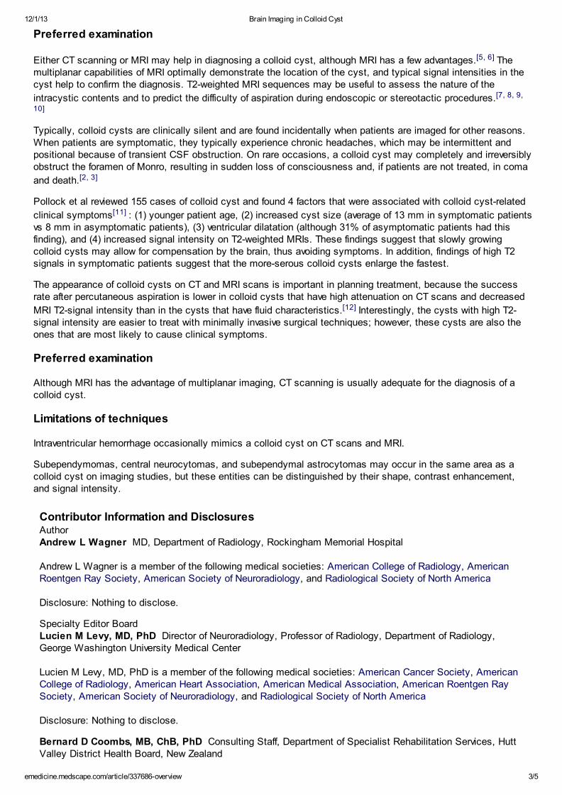
Sagittal nonenhanced T1-w eighted magnetic resonance image. This image demonstrates a round area of increased signal intensity in the
anterosuperior portion of the third ventricle (arrow ).
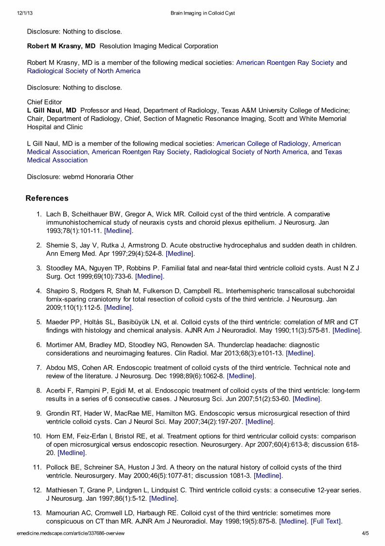
Axial f luid-attenuated inversion recovery magnetic resonance image. This image show s a bright mass.
Today NewsReferenceEducationLog Out My AccountDr. M El-Faresy

12/1/13 Brain Imaging in Colloid Cyst
emedicine.medscape.com/article/337686-overview 2/5
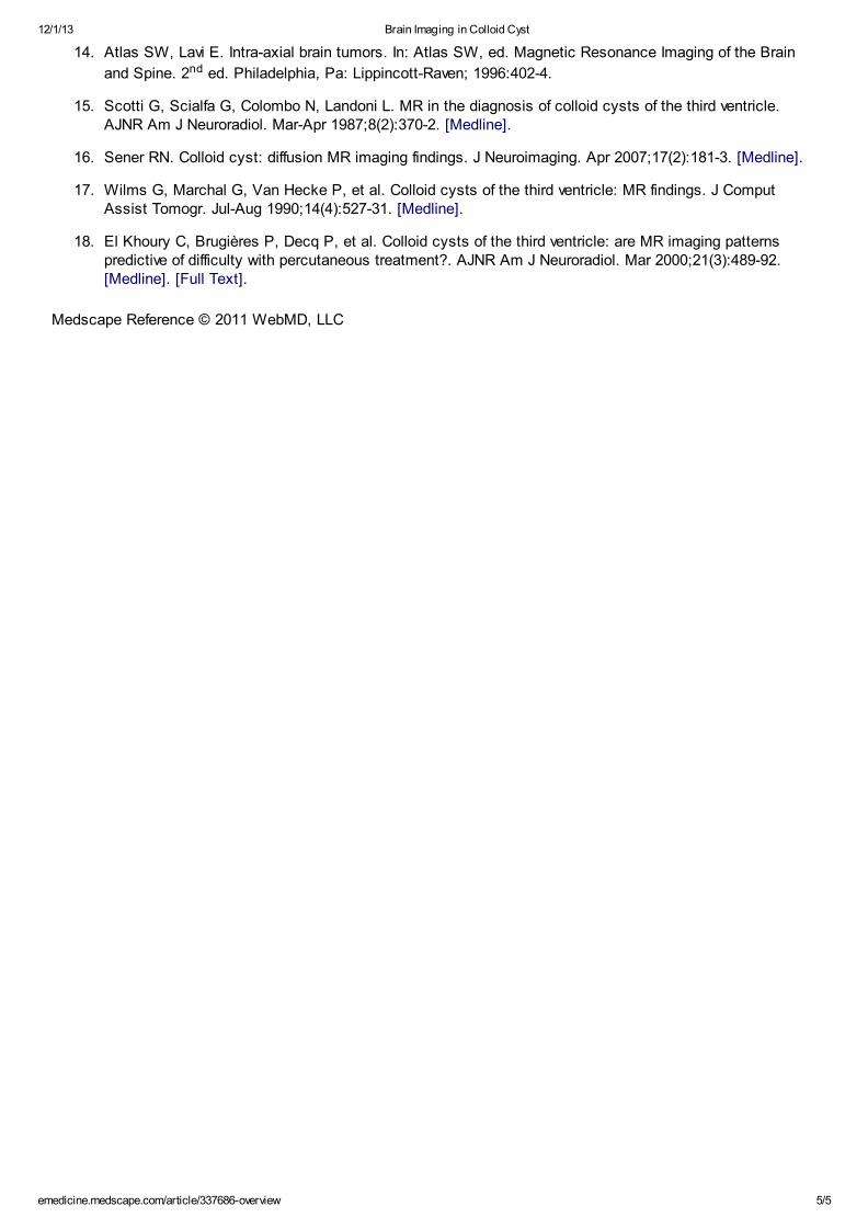
Axial contrast-enhanced T1-w eighted magnetic resonance image. This image demonstrates a small amount of peripheral enhancement
but no discernible central enhancement. Note the location of the colloid cyst near the foramina of Monro.
Computed tomography scan in a 65-year-old man w ho had acute onset of headache. This image demonstrates a round area of
increased attenuation at the foramina of Monro, w ith hydrocephalus. The image is degraded because of motion artifact, as the patient
w as in severe pain at the time of imaging.
Axial computed tomography scan in a 50-year-old man w ho w as transported to the emergency department after falling dow n w hile
lif ting w eights; he later had cardiopulmonary arrest. This image demonstrates a hyperattenuating colloid cyst at the foramina of Monro,
w ith marked hydrocephalus.
The diagnosis is usually made by assessing the typical location and appearance of the cyst. Colloid cystsaccount for approximately 1% of all intracranial tumors and are the most common type of the neuroepithelialcysts, as well as the most common tumor in the third ventricle. Typically, patients are asymptomatic, althoughcolloid cysts may cause symptoms by obstructing the foramen of Monro, which results in sudden death in rare
cases.[2, 3]
Shapiro et al described the long-term results of an interhemispheric, transcallosal, subchoroidal, fornix-sparingapproach to gross-total resection of colloid cysts. In 57 colloid cysts, total removal was achieved via a 3 x 3-inparamedian craniotomy flap and a microscopic interhemispheric, transcallosal, subchoroidal approach sparing theipsilateral fornix. According to the investigators, at 1 year after surgery, computed tomography (CT) scanning ormagnetic resonance imaging (MRI) confirmed gross-total resection, with no infection, hemiparesis, seizures, ordisconnection syndrome. There were no deaths or recurrences. The authors noted that the results were superior to
those seen with endoscopy.[4]
12/1/13 Brain Imaging in Colloid Cyst
emedicine.medscape.com/article/337686-overview 3/5
Preferred examination
Either CT scanning or MRI may help in diagnosing a colloid cyst, although MRI has a few advantages.[5, 6] Themultiplanar capabilities of MRI optimally demonstrate the location of the cyst, and typical signal intensities in thecyst help to confirm the diagnosis. T2-weighted MRI sequences may be useful to assess the nature of the
intracystic contents and to predict the difficulty of aspiration during endoscopic or stereotactic procedures.[7, 8, 9,
10]
Typically, colloid cysts are clinically silent and are found incidentally when patients are imaged for other reasons.When patients are symptomatic, they typically experience chronic headaches, which may be intermittent andpositional because of transient CSF obstruction. On rare occasions, a colloid cyst may completely and irreversiblyobstruct the foramen of Monro, resulting in sudden loss of consciousness and, if patients are not treated, in coma
and death.[2, 3]
Pollock et al reviewed 155 cases of colloid cyst and found 4 factors that were associated with colloid cyst-related
clinical symptoms[11] : (1) younger patient age, (2) increased cyst size (average of 13 mm in symptomatic patientsvs 8 mm in asymptomatic patients), (3) ventricular dilatation (although 31% of asymptomatic patients had thisfinding), and (4) increased signal intensity on T2-weighted MRIs. These findings suggest that slowly growingcolloid cysts may allow for compensation by the brain, thus avoiding symptoms. In addition, findings of high T2signals in symptomatic patients suggest that the more-serous colloid cysts enlarge the fastest.
The appearance of colloid cysts on CT and MRI scans is important in planning treatment, because the successrate after percutaneous aspiration is lower in colloid cysts that have high attenuation on CT scans and decreased
MRI T2-signal intensity than in the cysts that have fluid characteristics.[12] Interestingly, the cysts with high T2-signal intensity are easier to treat with minimally invasive surgical techniques; however, these cysts are also theones that are most likely to cause clinical symptoms.
Preferred examination
Although MRI has the advantage of multiplanar imaging, CT scanning is usually adequate for the diagnosis of acolloid cyst.
Limitations of techniques
Intraventricular hemorrhage occasionally mimics a colloid cyst on CT scans and MRI.
Subependymomas, central neurocytomas, and subependymal astrocytomas may occur in the same area as acolloid cyst on imaging studies, but these entities can be distinguished by their shape, contrast enhancement,and signal intensity.
Contributor Information and DisclosuresAuthorAndrew L Wagner MD, Department of Radiology, Rockingham Memorial Hospital
Andrew L Wagner is a member of the following medical societies: American College of Radiology, AmericanRoentgen Ray Society, American Society of Neuroradiology, and Radiological Society of North America
Disclosure: Nothing to disclose.
Specialty Editor BoardLucien M Levy, MD, PhD Director of Neuroradiology, Professor of Radiology, Department of Radiology,George Washington University Medical Center
Lucien M Levy, MD, PhD is a member of the following medical societies: American Cancer Society, AmericanCollege of Radiology, American Heart Association, American Medical Association, American Roentgen RaySociety, American Society of Neuroradiology, and Radiological Society of North America
Disclosure: Nothing to disclose.
Bernard D Coombs, MB, ChB, PhD Consulting Staff, Department of Specialist Rehabilitation Services, HuttValley District Health Board, New Zealand
12/1/13 Brain Imaging in Colloid Cyst
emedicine.medscape.com/article/337686-overview 4/5
Disclosure: Nothing to disclose.
Robert M Krasny, MD Resolution Imaging Medical Corporation
Robert M Krasny, MD is a member of the following medical societies: American Roentgen Ray Society andRadiological Society of North America
Disclosure: Nothing to disclose.
Chief EditorL Gill Naul, MD Professor and Head, Department of Radiology, Texas A&M University College of Medicine;Chair, Department of Radiology, Chief, Section of Magnetic Resonance Imaging, Scott and White MemorialHospital and Clinic
L Gill Naul, MD is a member of the following medical societies: American College of Radiology, AmericanMedical Association, American Roentgen Ray Society, Radiological Society of North America, and TexasMedical Association
Disclosure: webmd Honoraria Other
References
1. Lach B, Scheithauer BW, Gregor A, Wick MR. Colloid cyst of the third ventricle. A comparativeimmunohistochemical study of neuraxis cysts and choroid plexus epithelium. J Neurosurg. Jan1993;78(1):101-11. [Medline].
2. Shemie S, Jay V, Rutka J, Armstrong D. Acute obstructive hydrocephalus and sudden death in children.Ann Emerg Med. Apr 1997;29(4):524-8. [Medline].
3. Stoodley MA, Nguyen TP, Robbins P. Familial fatal and near-fatal third ventricle colloid cysts. Aust N Z JSurg. Oct 1999;69(10):733-6. [Medline].
4. Shapiro S, Rodgers R, Shah M, Fulkerson D, Campbell RL. Interhemispheric transcallosal subchoroidalfornix-sparing craniotomy for total resection of colloid cysts of the third ventricle. J Neurosurg. Jan2009;110(1):112-5. [Medline].
5. Maeder PP, Holtås SL, Basibüyük LN, et al. Colloid cysts of the third ventricle: correlation of MR and CTfindings with histology and chemical analysis. AJNR Am J Neuroradiol. May 1990;11(3):575-81. [Medline].
6. Mortimer AM, Bradley MD, Stoodley NG, Renowden SA. Thunderclap headache: diagnosticconsiderations and neuroimaging features. Clin Radiol. Mar 2013;68(3):e101-13. [Medline].
7. Abdou MS, Cohen AR. Endoscopic treatment of colloid cysts of the third ventricle. Technical note andreview of the literature. J Neurosurg. Dec 1998;89(6):1062-8. [Medline].
8. Acerbi F, Rampini P, Egidi M, et al. Endoscopic treatment of colloid cysts of the third ventricle: long-termresults in a series of 6 consecutive cases. J Neurosurg Sci. Jun 2007;51(2):53-60. [Medline].
9. Grondin RT, Hader W, MacRae ME, Hamilton MG. Endoscopic versus microsurgical resection of thirdventricle colloid cysts. Can J Neurol Sci. May 2007;34(2):197-207. [Medline].
10. Horn EM, Feiz-Erfan I, Bristol RE, et al. Treatment options for third ventricular colloid cysts: comparisonof open microsurgical versus endoscopic resection. Neurosurgery. Apr 2007;60(4):613-8; discussion 618-20. [Medline].
11. Pollock BE, Schreiner SA, Huston J 3rd. A theory on the natural history of colloid cysts of the thirdventricle. Neurosurgery. May 2000;46(5):1077-81; discussion 1081-3. [Medline].
12. Mathiesen T, Grane P, Lindgren L, Lindquist C. Third ventricle colloid cysts: a consecutive 12-year series.J Neurosurg. Jan 1997;86(1):5-12. [Medline].
13. Mamourian AC, Cromwell LD, Harbaugh RE. Colloid cyst of the third ventricle: sometimes moreconspicuous on CT than MR. AJNR Am J Neuroradiol. May 1998;19(5):875-8. [Medline]. [Full Text].
12/1/13 Brain Imaging in Colloid Cyst
emedicine.medscape.com/article/337686-overview 5/5
Medscape Reference © 2011 WebMD, LLC
14. Atlas SW, Lavi E. Intra-axial brain tumors. In: Atlas SW, ed. Magnetic Resonance Imaging of the Brain
and Spine. 2nd ed. Philadelphia, Pa: Lippincott-Raven; 1996:402-4.
15. Scotti G, Scialfa G, Colombo N, Landoni L. MR in the diagnosis of colloid cysts of the third ventricle.AJNR Am J Neuroradiol. Mar-Apr 1987;8(2):370-2. [Medline].
16. Sener RN. Colloid cyst: diffusion MR imaging findings. J Neuroimaging. Apr 2007;17(2):181-3. [Medline].
17. Wilms G, Marchal G, Van Hecke P, et al. Colloid cysts of the third ventricle: MR findings. J ComputAssist Tomogr. Jul-Aug 1990;14(4):527-31. [Medline].
18. El Khoury C, Brugières P, Decq P, et al. Colloid cysts of the third ventricle: are MR imaging patternspredictive of difficulty with percutaneous treatment?. AJNR Am J Neuroradiol. Mar 2000;21(3):489-92.[Medline]. [Full Text].


