Download - Figure 6.4b Muscle cells

Copyright © 2009 Pearson Education, Inc.
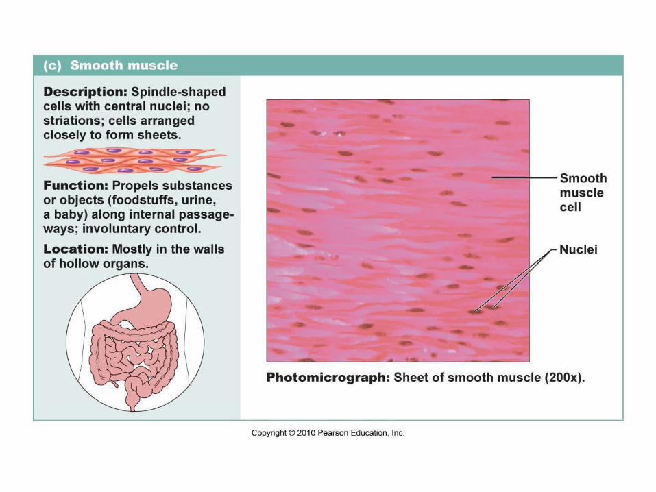
Figure 6.4b Muscle cells.


Copyright © 2009 Pearson Education, Inc.
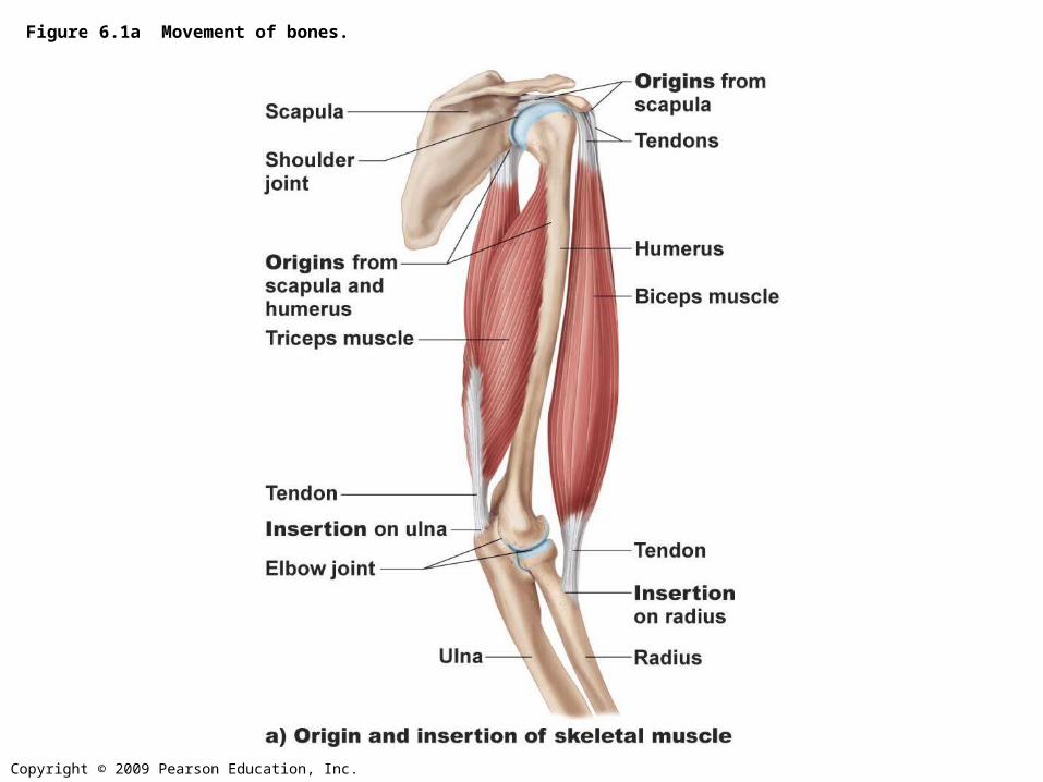
Figure 6.1a Movement of bones.

Copyright © 2009 Pearson Education, Inc.
Figure 6.1b Movement of bones.

Copyright © 2009 Pearson Education, Inc.
Figure 6.3 Muscle structure.

Copyright © 2010 Pearson Education, Inc.
Figure 9.1a Connective tissue sheaths of skeletal muscle: epimysium, perimysium, and endomysium.
Bone
Endomysium(between individualmuscle fibers)
Muscle fiber
Fascicle(wrapped by perimysium)
Epimysium
Tendon
Blood vessel

Copyright © 2010 Pearson Education, Inc.
NucleusLight I bandDark A band
Sarcolemma
Mitochondrion
(b) Diagram of part of a muscle fiber showing the myofibrils. Onemyofibril is extended afrom the cut end of the fiber.
Myofibril
Figure 9.2b Microscopic anatomy of a skeletal muscle fiber.

Copyright © 2009 Pearson Education, Inc.
Figure 6.5 Structure of a myofibril.
Copyright © 2010 Pearson Education, Inc.
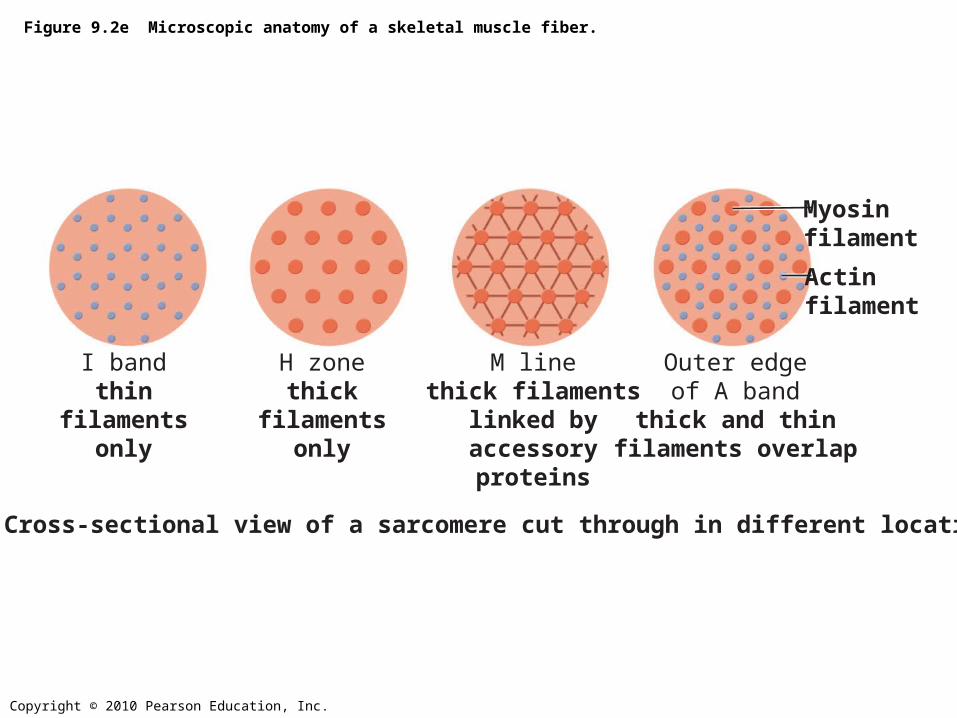
Figure 9.2e Microscopic anatomy of a skeletal muscle fiber.
I bandthin
filamentsonly
Actinfilament
Myosinfilament
H zonethick
filamentsonly
M linethick filaments
linked byaccessoryproteins
Outer edgeof A band
thick and thinfilaments overlap
(e) Cross-sectional view of a sarcomere cut through in different locations.

Copyright © 2009 Pearson Education, Inc.
Figure 6.5d Structure of a myofibril.
Copyright © 2009 Pearson Education, Inc.
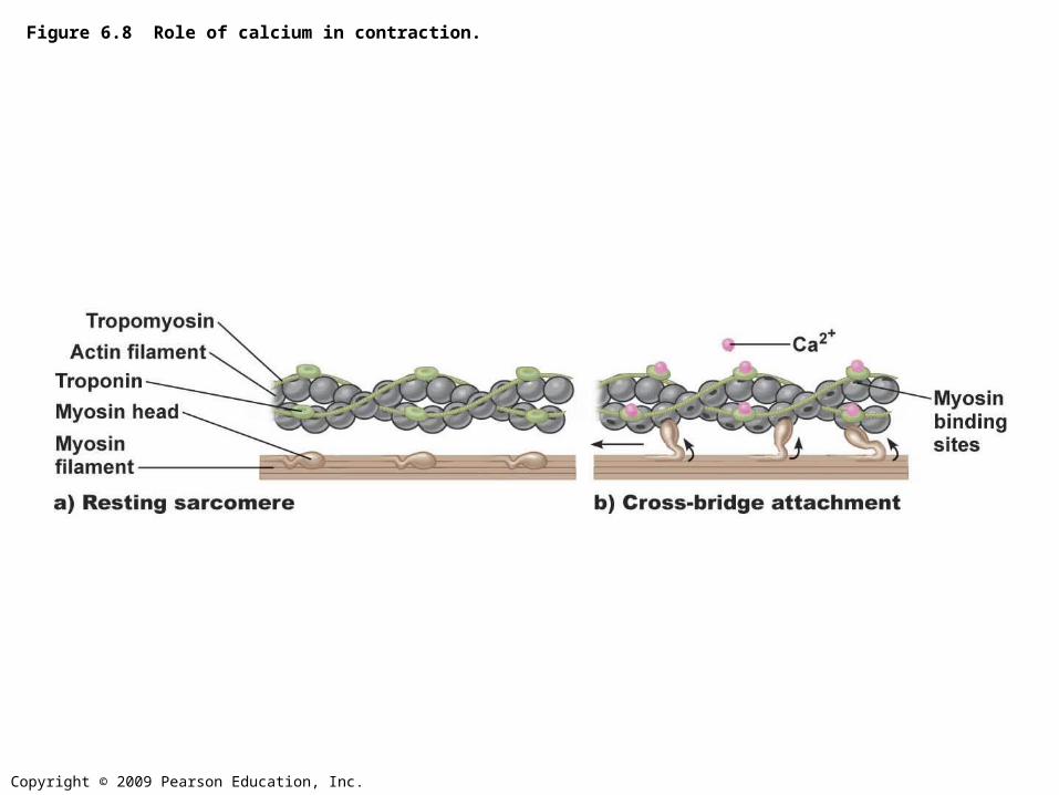
Figure 6.8 Role of calcium in contraction.

Copyright © 2010 Pearson Education, Inc.
Figure 9.12 Cross Bridge Cycle
Actin
Cross bridge formation.
Cocking of myosin head. The power (working)stroke.
Cross bridgedetachment.
Ca2+
1
2
3
4
Myosinhead
Thickfilament
Thin filament
ADP
Myosin
P i
ADP
P iATPhydrolysis
ADP
P i
ATP
ATP
Copyright © 2010 Pearson Education, Inc.
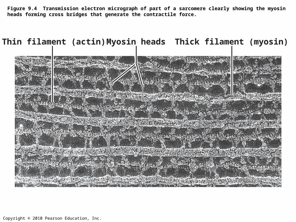
Figure 9.4 Transmission electron micrograph of part of a sarcomere clearly showing the myosin heads forming cross bridges that generate the contractile force.
Thin filament (actin) Thick filament (myosin)Myosin heads

Copyright © 2009 Pearson Education, Inc.
Figure 6.9a Motor units.

Copyright © 2010 Pearson Education, Inc.
Figure 9.8 Events at the Neuromuscular Junction (1 of 4)
Nucleus
Action potential(AP)
Myelinated axonof motor neuron
Axon terminal ofneuromuscular junction
Sarcolemmaof the musclefiber

Copyright © 2009 Pearson Education, Inc.
Figure 6.9b Motor units.

Copyright © 2010 Pearson Education, Inc.
Acetylcholine, a neurotransmitter, diffuses across the synaptic cleft and binds to receptors in the sarcolemma.
Figure 9.8 Events at the Neuromuscular Junction (2 of 4)
Ca2+
Axon terminalof motor neuron
Synaptic vesiclecontaining ACh
Synaptic cleft
Fusingsynaptic vesicles
ACh
Sarcoplasm of muscle fiber
Ca2+
Copyright © 2010 Pearson Education, Inc.
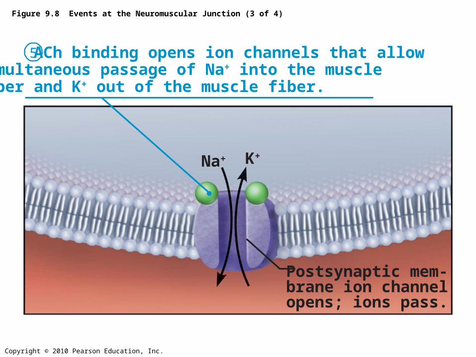
Figure 9.8 Events at the Neuromuscular Junction (3 of 4)
Postsynaptic mem-brane ion channel opens; ions pass.
Na+ K+
5 ACh binding opens ion channels that allowsimultaneous passage of Na+ into the muscle fiber and K+ out of the muscle fiber.

• Resting Potential of muscle cells – – Potassium higher inside– Sodium higher outside– A voltage difference of about 90 mvolts
• Action Potential – – A wave of depolarization that propogates from
the point of stimulation over the entire membrane, followed by a wave of repolarization.

Copyright © 2010 Pearson Education, Inc.
Figure 9.9 Summary of events in the generation and propagation of an action potential in a skeletal muscle fiber.
Na+ K+
Axon terminal
Synapticcleft
ACh–
ACh
1 Local depolarization:
Na+
Na+
Open Na+
ChannelClosed K+
Channel
K+
K+
K+
2 Generation and propagation ofthe action potential (AP)
3 Repolarization
Sarcoplasm of muscle fiber
Na+

Copyright © 2010 Pearson Education, Inc.
Figure 9.5 Relationship of the sarcoplasmic reticulum and T tubules to myofibrils of skeletal muscle.
Myofibrils
Sarcolemma
Mitochondria
Terminal cisternae of SR
T tubuleTriad
Copyright © 2010 Pearson Education, Inc.
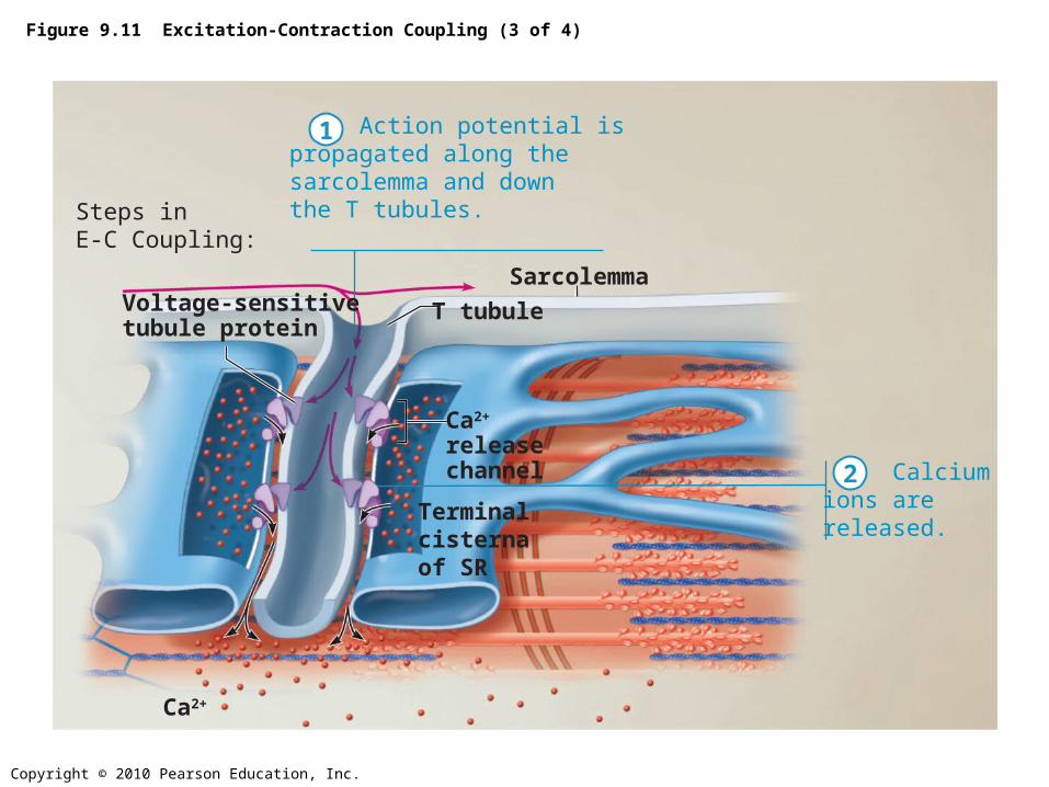
Figure 9.11 Excitation-Contraction Coupling (3 of 4)
Calciumions arereleased.
Steps inE-C Coupling:
Terminalcisterna of SR
Voltage-sensitivetubule protein
T tubule
Ca2+
releasechannel
Ca2+
Sarcolemma
Action potential ispropagated along thesarcolemma and downthe T tubules.
1
2


