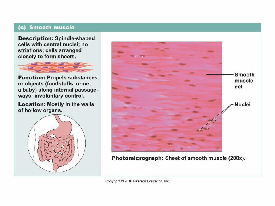
figure 6.4b muscle cells
DESCRIPTION
Figure 6.4b Muscle cells. Figure 6.1a Movement of bones. Figure 6.1b Movement of bones. Figure 6.3 Muscle structure. Epimysium. Bone. Tendon. Blood vessel. Fascicle (wrapped by perimysium). Endomysium (between individual muscle fibers). Muscle fiber. - PowerPoint PPT PresentationTRANSCRIPT

Copyright © 2009 Pearson Education, Inc.
Figure 6.4b Muscle cells.


Copyright © 2009 Pearson Education, Inc.
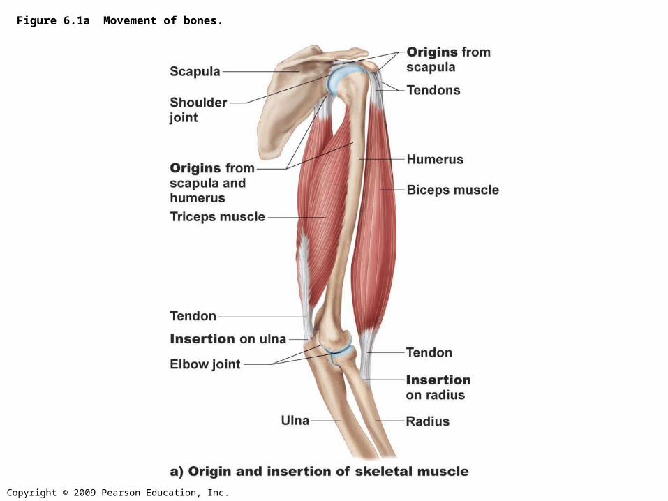
Figure 6.1a Movement of bones.

Copyright © 2009 Pearson Education, Inc.
Figure 6.1b Movement of bones.

Copyright © 2009 Pearson Education, Inc.
Figure 6.3 Muscle structure.

Copyright © 2010 Pearson Education, Inc.
Figure 9.1a Connective tissue sheaths of skeletal muscle: epimysium, perimysium, and endomysium.
Bone
Endomysium(between individualmuscle fibers)
Muscle fiber
Fascicle(wrapped by perimysium)
Epimysium
Tendon
Blood vessel

Copyright © 2010 Pearson Education, Inc.
NucleusLight I bandDark A band
Sarcolemma
Mitochondrion
(b) Diagram of part of a muscle fiber showing the myofibrils. Onemyofibril is extended afrom the cut end of the fiber.
Myofibril
Figure 9.2b Microscopic anatomy of a skeletal muscle fiber.

Copyright © 2009 Pearson Education, Inc.
Figure 6.5 Structure of a myofibril.
Copyright © 2010 Pearson Education, Inc.
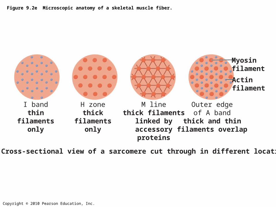
Figure 9.2e Microscopic anatomy of a skeletal muscle fiber.
I bandthin
filamentsonly
Actinfilament
Myosinfilament
H zonethick
filamentsonly
M linethick filaments
linked byaccessoryproteins
Outer edgeof A band
thick and thinfilaments overlap
(e) Cross-sectional view of a sarcomere cut through in different locations.

Copyright © 2009 Pearson Education, Inc.
Figure 6.5d Structure of a myofibril.
Copyright © 2009 Pearson Education, Inc.
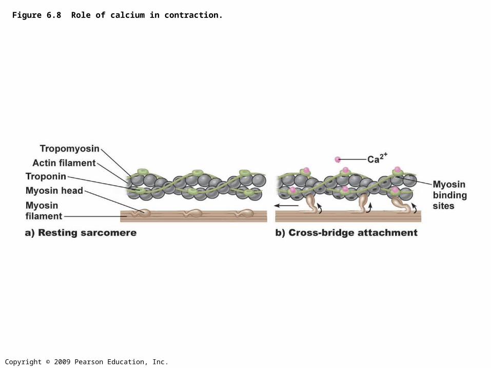
Figure 6.8 Role of calcium in contraction.

Copyright © 2010 Pearson Education, Inc.
Figure 9.12 Cross Bridge Cycle
Actin
Cross bridge formation.
Cocking of myosin head. The power (working)stroke.
Cross bridgedetachment.
Ca2+
1
2
3
4
Myosinhead
Thickfilament
Thin filament
ADP
Myosin
P i
ADP
P iATPhydrolysis
ADP
P i
ATP
ATP
Copyright © 2010 Pearson Education, Inc.
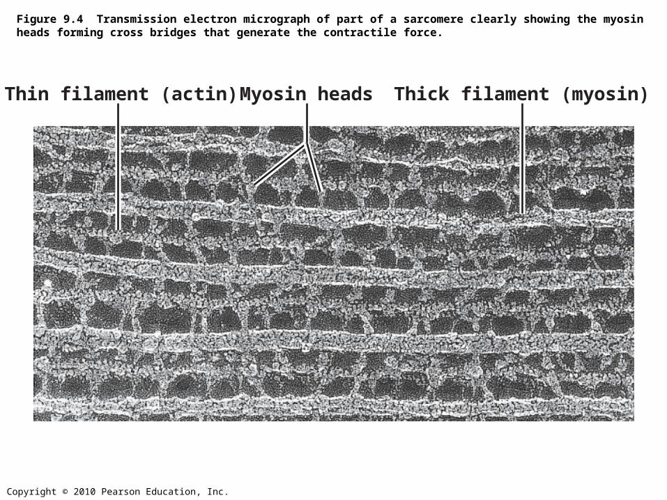
Figure 9.4 Transmission electron micrograph of part of a sarcomere clearly showing the myosin heads forming cross bridges that generate the contractile force.
Thin filament (actin) Thick filament (myosin)Myosin heads

Copyright © 2009 Pearson Education, Inc.
Figure 6.9a Motor units.

Copyright © 2010 Pearson Education, Inc.
Figure 9.8 Events at the Neuromuscular Junction (1 of 4)
Nucleus
Action potential(AP)
Myelinated axonof motor neuron
Axon terminal ofneuromuscular junction
Sarcolemmaof the musclefiber

Copyright © 2009 Pearson Education, Inc.
Figure 6.9b Motor units.

Copyright © 2010 Pearson Education, Inc.
Acetylcholine, a neurotransmitter, diffuses across the synaptic cleft and binds to receptors in the sarcolemma.
Figure 9.8 Events at the Neuromuscular Junction (2 of 4)
Ca2+
Axon terminalof motor neuron
Synaptic vesiclecontaining ACh
Synaptic cleft
Fusingsynaptic vesicles
ACh
Sarcoplasm of muscle fiber
Ca2+
Copyright © 2010 Pearson Education, Inc.
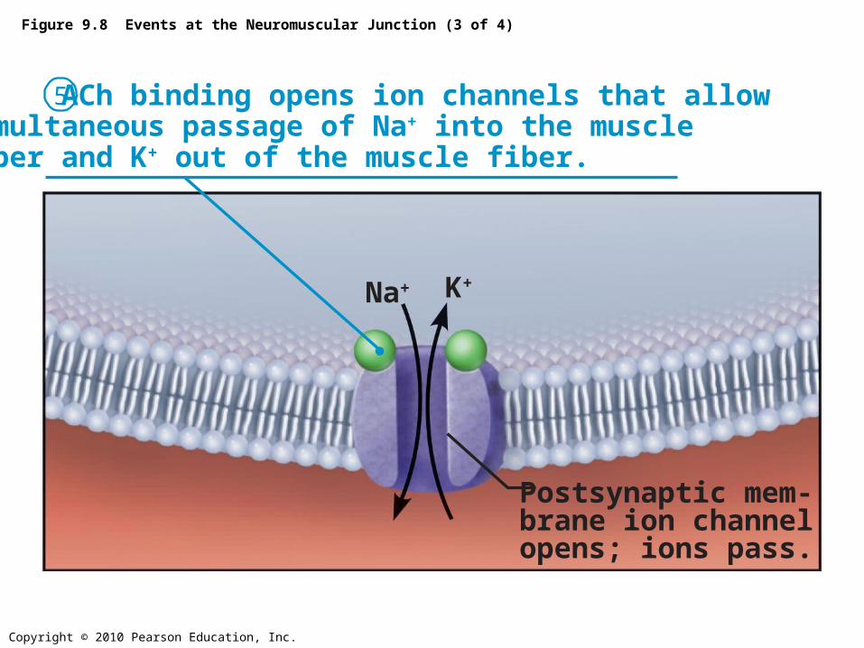
Figure 9.8 Events at the Neuromuscular Junction (3 of 4)
Postsynaptic mem-brane ion channel opens; ions pass.
Na+ K+
5 ACh binding opens ion channels that allowsimultaneous passage of Na+ into the muscle fiber and K+ out of the muscle fiber.

• Resting Potential of muscle cells – – Potassium higher inside– Sodium higher outside– A voltage difference of about 90 mvolts
• Action Potential – – A wave of depolarization that propogates from
the point of stimulation over the entire membrane, followed by a wave of repolarization.

Copyright © 2010 Pearson Education, Inc.
Figure 9.9 Summary of events in the generation and propagation of an action potential in a skeletal muscle fiber.
Na+ K+
Axon terminal
Synapticcleft
ACh–
ACh
1 Local depolarization:
Na+
Na+
Open Na+
ChannelClosed K+
Channel
K+
K+
K+
2 Generation and propagation ofthe action potential (AP)
3 Repolarization
Sarcoplasm of muscle fiber
Na+

Copyright © 2010 Pearson Education, Inc.
Figure 9.5 Relationship of the sarcoplasmic reticulum and T tubules to myofibrils of skeletal muscle.
Myofibrils
Sarcolemma
Mitochondria
Terminal cisternae of SR
T tubuleTriad
Copyright © 2010 Pearson Education, Inc.
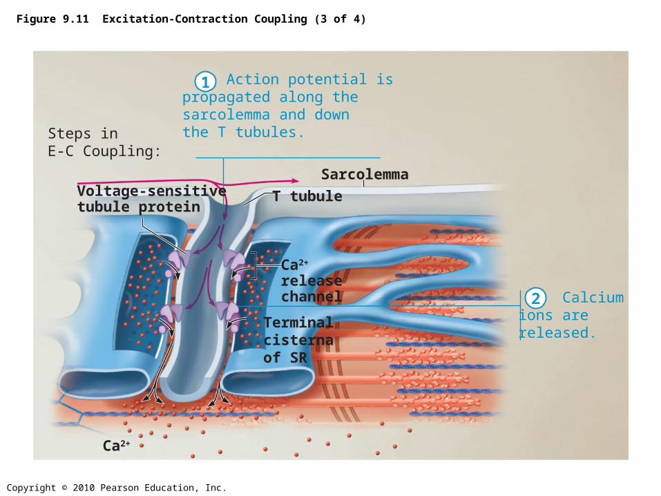
Figure 9.11 Excitation-Contraction Coupling (3 of 4)
Calciumions arereleased.
Steps inE-C Coupling:
Terminalcisterna of SR
Voltage-sensitivetubule protein
T tubule
Ca2+
releasechannel
Ca2+
Sarcolemma
Action potential ispropagated along thesarcolemma and downthe T tubules.
1
2