IMPORTANCE OF THE
RADIOGRAPH IN ENDODONTICS
DR. NEMES JÚLIA

THE OBJECTIVE OF OBTURATION:
is to create a complete seal along the
length of the root canal, from coronal
opening to the apical determination.
Radiograph
THE RADIOGRAPH IN ENDODONTICS
1. Why is it recommended to make radiograph
during the rootcanal treatment?
2. What kind of radiographs recommended?
3. When is it recommended to make radiographs?

ANATOMIE OF THE APICAL THIRD OF ROOTCANAL
THE LENGTH OF THE OBTURATION
Requirement:
foramen physiologicum (3)
(physiological apex)
Apical third of rootcanal
-apical constriction (3)
-foramen apicale (2)
(foramen anatomicum)
-radiological apex (1)
There is a different distance between 1-3
(1)
(2)
(3)
End of the
cement cement

THE APICAL THIRD OF ROOTCANAL
Christine Haugseth

THE ROLE OF THE RADIOGRAPH IN
ENDODONTICS
1. Why it is recommended to make radiograph
during the rootcanal treatment?
2. What kind of radiographs recommended?
3. When it is recommended to make radiographs?

What kind of radiograph?
Periapical radiograph, with
paralleling technique
with longtube
film and tooth axel is
parallel
(filmholder)
longtube technique
X-ray parallel

X-ray arrives the film/sensor at about 90°
Film or
Sensor

CONE (TUBE)-IMAGE SHIFT
Can be :
Orthoradiale (facial) pro-
jection (basic!)
Excentric (2O-30˚)
-mesial-excentric
-distal-excentric
(mesial, or distal projection)
Reveals the third dimension!
F M
D

UPPER FIRST PREMOLAR
SLOB rule : same lingual, opposite buccal
(tube movement)
mesial
facial
The lingual root moves in the same direction as the cone,and
the buccal root moves in the opposite direction.
M F D

ORTORADIALE and EXCENTRIC RADIOGRAPH
Facial and mesial or distal projection
Mesial projection Distal projection
Facial projection
SLOB rule

Other technic
X-RAY
Film and tooth are not
parallel!
BISECTING technique
MODIFIED PARALLELING
technique: neither
parallel, nor bisecting
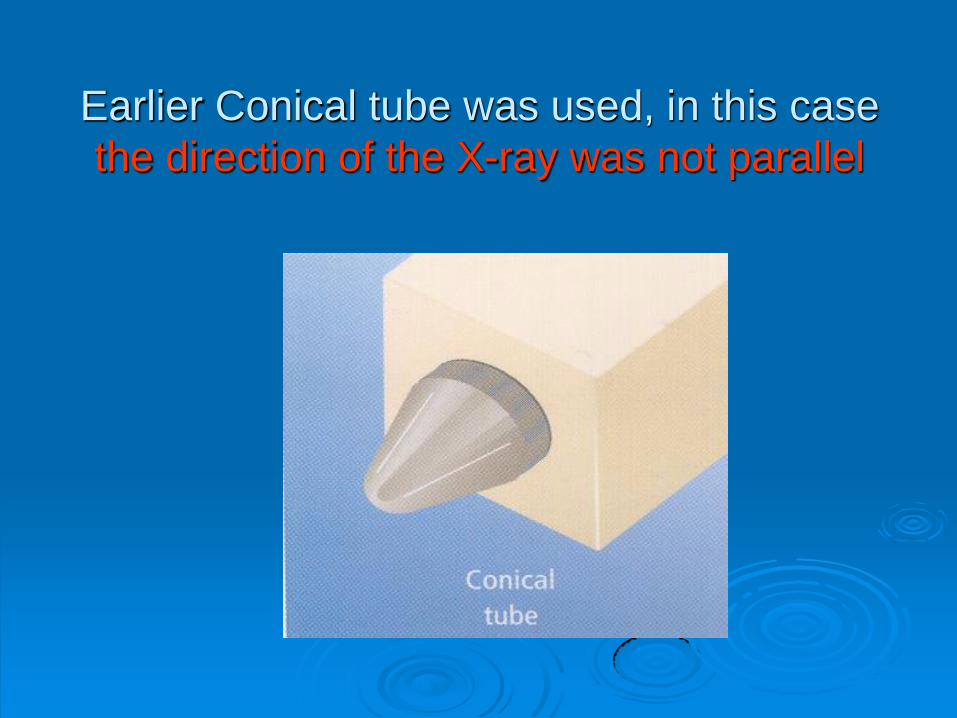
Earlier Conical tube was used, in this case
the direction of the X-ray was not parallel
ROLE OF THE RADIOGRAPH IN ENDODONTICS
Why is it recommended to make radiograph during
the rootcanal treatment?
What kind of radiograph recommended?
3. When it is recommended to make radiograph?
-befor the treatment (diagnostic radiograph)
-during the treatment (working length determination)
-after the treatment (controll radiograph)
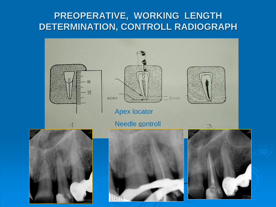
PREOPERATIVE, WORKING LENGTH
DETERMINATION, CONTROLL RADIOGRAPH
Apex locator
Needle controll

1. PREOPERATIVE or DIAGNOSTIC
RADIOGRAPH
AIM:
Anatomic structures
extension of pulp chamber, curved or
not curved root
Diagnostic information (periapical process)
Estimated working length
determination
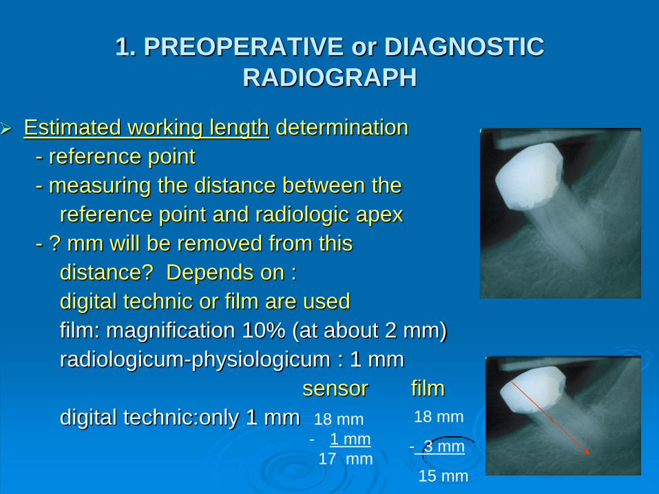
1. PREOPERATIVE or DIAGNOSTIC
RADIOGRAPH
Estimated working length determination
- reference point
- measuring the distance between the
reference point and radiologic apex
- ? mm will be removed from this
distance? Depends on :
digital technic or film are used
film: magnification 10% (at about 2 mm)
radiologicum-physiologicum : 1 mm
sensor film
digital technic:only 1 mm 18 mm
- 3 mm
15 mm
18 mm
- 1 mm
17 mm

2. WORKING LENGTH DETERMINATION
(needel-controll or apex-locator)
Aim:
to check the estimated
working length
PRECONDITION
-Reference point
-Silicon stop
-Stable file in canal
(mindestens ISO 15)
-Endoblock or ruler Correction!
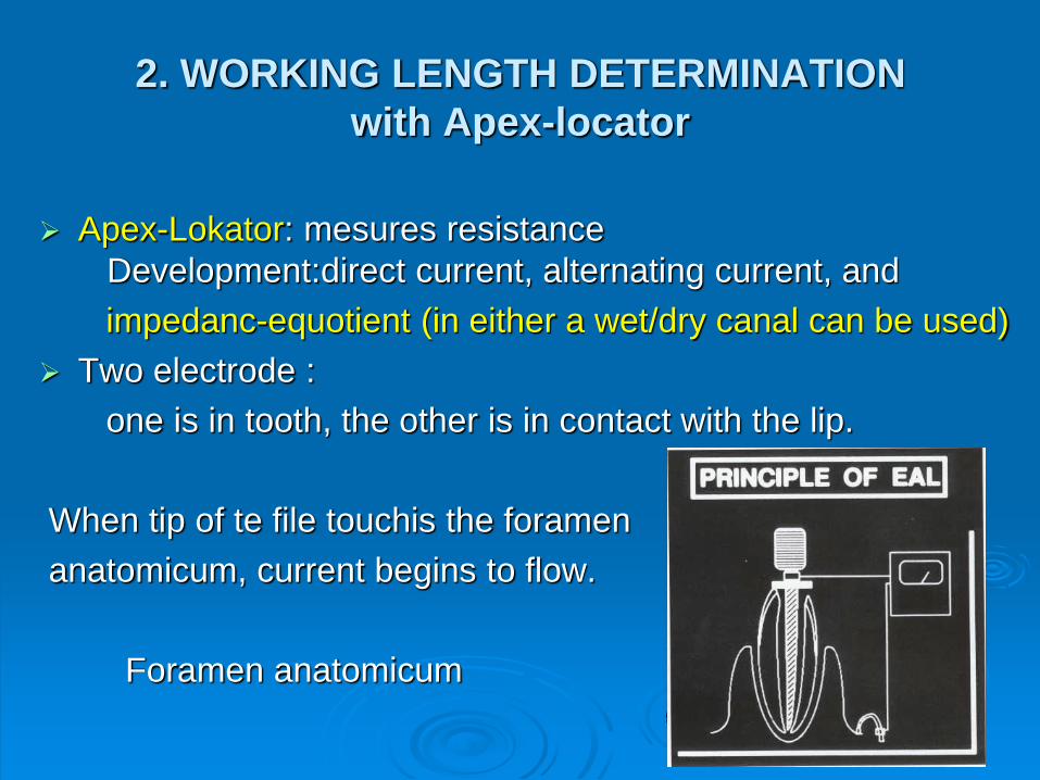
2. WORKING LENGTH DETERMINATION
with Apex-locator
Apex-Lokator: mesures resistance
Development:direct current, alternating current, and
impedanc-equotient (in either a wet/dry canal can be used)
Two electrode :
one is in tooth, the other is in contact with the lip.
When tip of te file touchis the foramen
anatomicum, current begins to flow.
Foramen anatomicum
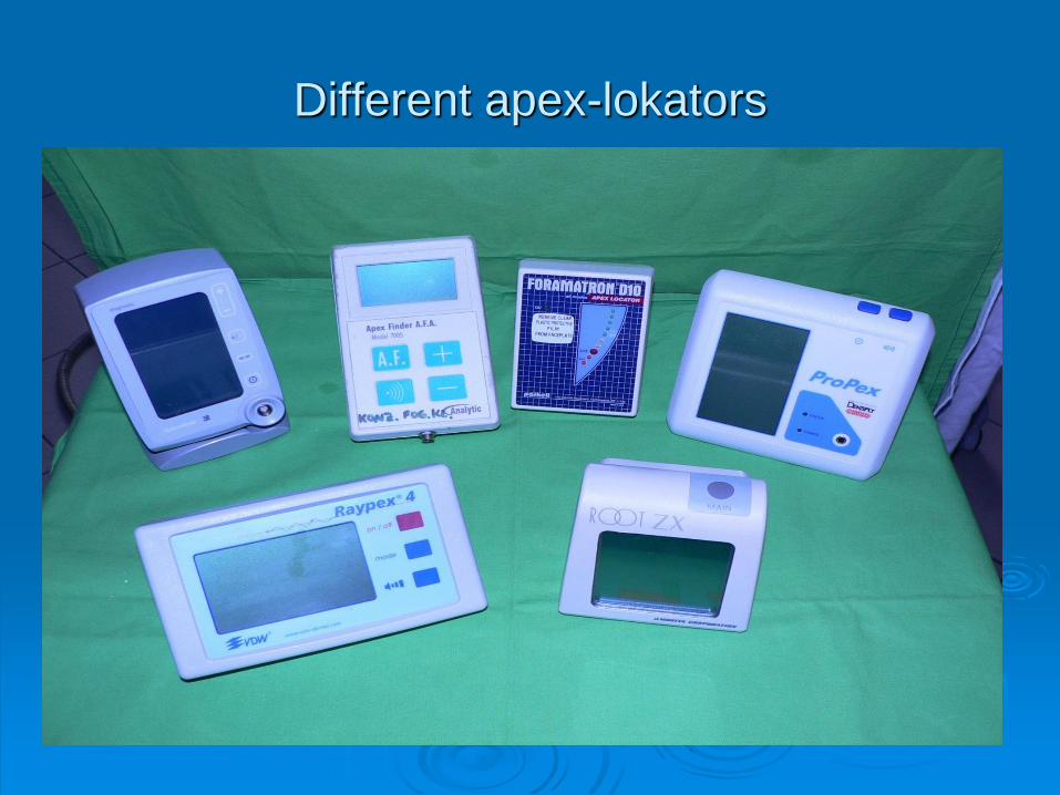
Different apex-lokators
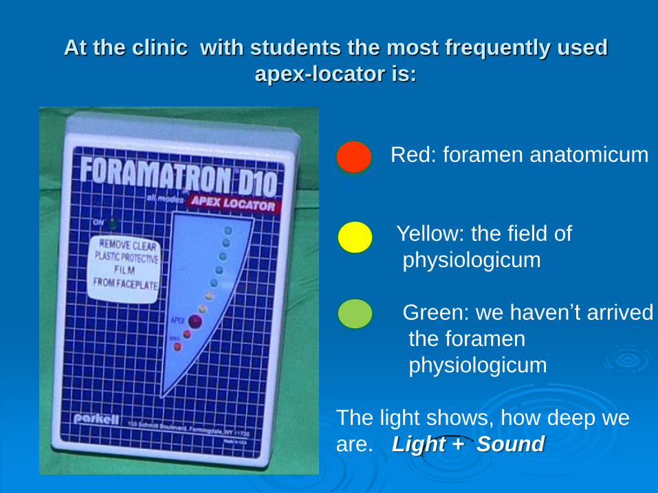
At the clinic with students the most frequently used
apex-locator is:
Red: foramen anatomicum
Yellow: the field of
physiologicum
Green: we haven’t arrived
the foramen
physiologicum
The light shows, how deep we
are. Light + Sound

EVALUATION OF WORKING LENGTH
DETERMINATION (needel-controll or apex
locator)
The working length is good. There is 1-2 mm distance between the end of the needel and the radiological apex.
Apex-locator: (sound, color, mm)
(Foramatron: for.anat.= red, above yellow, green )
The working length is to short.
The working length is too long.
If the distance more than 3 mm, in this case has to make a second radiograph with the korrekted working length.

CALCULATION
LT length of the tooth ?
LIT length of the instrument
LTR length of the tooth on RTG
LIR length of the instrument on RTG
LT = LTR x LIT
LIR

3. EVALUATION OF OBTURATION
The length of the rootcanal filling
(good, short, long)
The density of the rootcanal filling
Uniform density from coronal to apical
(voids, bubble)

Shape: It should be tapered from coronal
to apical region.
Coronal removal: material remains or not
remain in the chamber
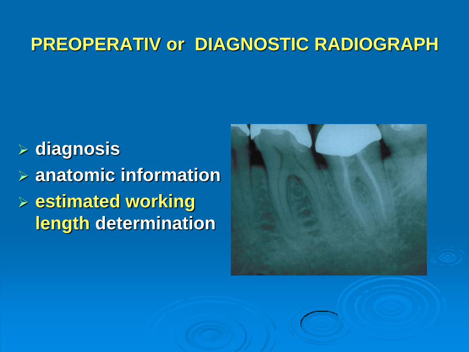
PREOPERATIV or DIAGNOSTIC RADIOGRAPH
diagnosis
anatomic information
estimated working
length determination

WORKING LENGTH DETERMINATION or
NEEDEL CONTROLL
rubber dam
silicon stop
reference point
Stable file in canal
(at least ISO 15)
endoblock or ruler

CONTROLL, AFTER THE OBTURATION
the length
(good, short, or long)
density
(bubble, homogene)
The shape
(taper)
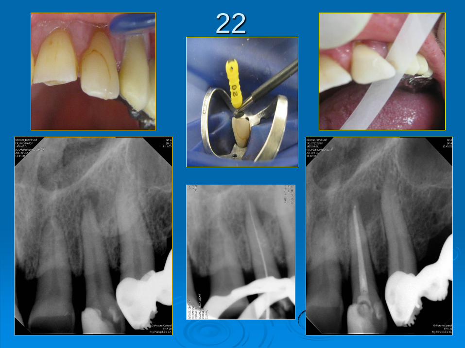
22
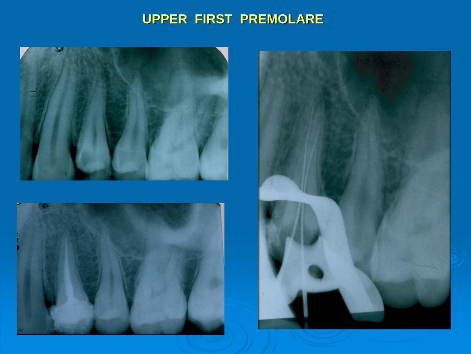
UPPER FIRST PREMOLARE

UPPER FIRST PREMOLAR

UPPER FIRST MOLAR

UPPER FIRST MOLAR

LOWER SECOND MOLAR
Pulp chamber ?

LOWER SECOND MOLAR

IMPORTANCE OF THE RADIOGRAPH IN
ENDODONTICS
1. Why it is recommended to make radiograph
during the rootcanal treatment?
2. What kind of radiographs recommended?
3. When it is recommended to make radiographs?


