
HAEMATURIAKen Chow

What is haematuria? Macroscopic
Visible haematuria Pink or red
Microscopic Gold standard – Microscopy
○ Presence of >3 RBCs per high-powered field Dipsticks
○ Positive – >1 RBCs per high-powered field○ Higher false positives
Symptomatic vs. non-symptomatic○ Lower Urinary Tract Symptoms (LUTS)
Eg. Dysuria, hesitancy, frequency, urgency
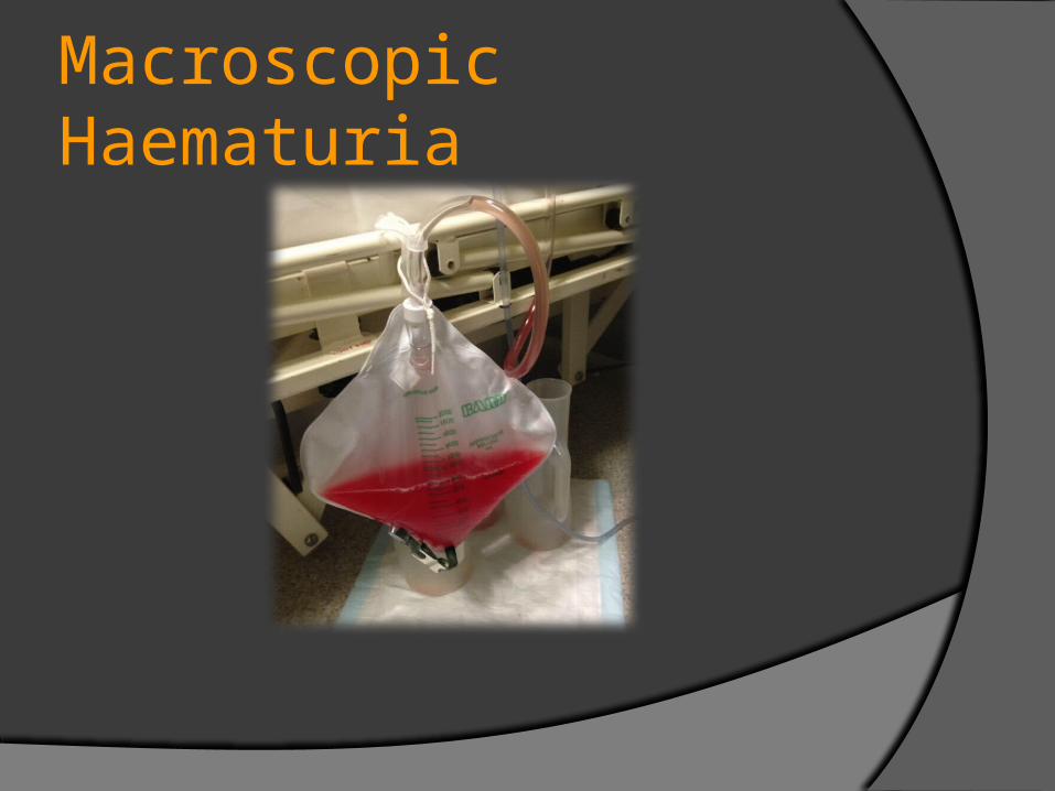
Macroscopic Haematuria
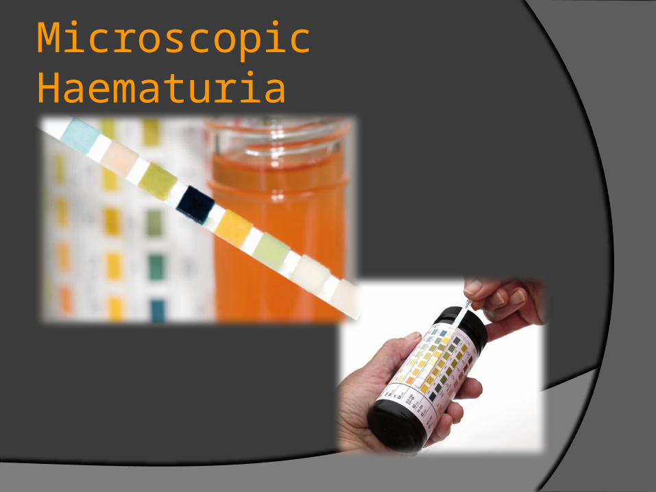
Microscopic Haematuria
Causes Renal
Malignant renal mass Benign renal mass Glomerular bleeding Structural diseases Pyelonephritis Hydronephrosis Hypercalcinuria / Hyperuricosuria Renal vein thrombosis Renal artery embolism Arteriovenous malformation
Ureteric Malignancy Calculi Strictures Fibroepithelial polyp Fistulas
Bladder Malignancy Radiation Cystitis
Prostate/Urethra Benign prostatic hyperplasia Prostate carcinoma Catheterisation Urethritis

History and Examination History
Time course Infective symptoms Urinary symptoms Associated symptoms Past history Social history Family history
Examination Vital signs Abdominal DRE
Workup Significant haematuria:
Single episode of macroscopic haematuria Single episode of symptomatic microscopic haematuria Persistent non-symptomatic microscopic haematuria
Initial investigations Exclude transient causes
○ Eg. UTIs, exercise induced, trauma, menstruation○ Urine cultures
Serum creatinine and eGFR Measure for proteinuria
○ Protein:Creatinine ratio (PCR)○ Albumin:Creatinine ratio (ACR)
Blood pressure

Urological Referral All patients with macroscopic haematuria All patients with symptomatic microscopic haematuria All patients with asymptomatic microscopic haematuria who
are aged 35 and over
Nephrological Referral Consideration of nephrological referral
Declining GFR○ >10ml/min within the previous 5 years○ >5ml/min within the last 1 year
Stage 4 or 5 CKD (eGFR <30) Significant proteinuria
○ PCR ≥50mg/mmol○ ACR ≥30mg/mmol
Isolated haematuria with hypertension who are aged ≤40 Haematuria with coinciding intercurrent infection
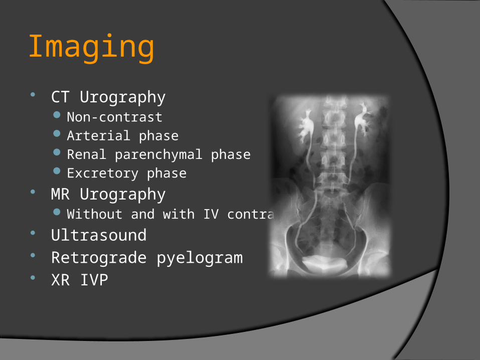
Imaging CT Urography
Non-contrast Arterial phase Renal parenchymal phase Excretory phase
MR Urography Without and with IV contrast
Ultrasound Retrograde pyelogram XR IVP
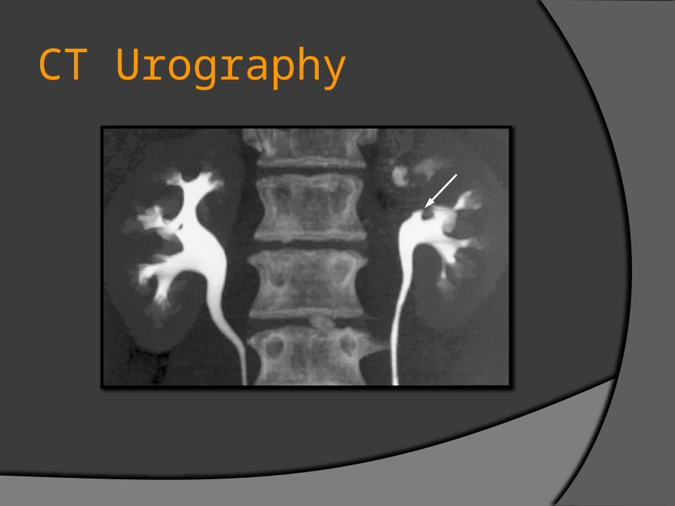
CT Urography

Procedure Cystoscopy
Full visualisation of the bladder, prostate and urethra All haematuria patients aged 35 years and over All patients with risk factors for urinary tract malignancy

Risk Factors Male gender Aged 35 and over Past or current smoker Occupational exposure to chemicals Analgesic abuse History of gross haematuria History of urologic disorder or disease History of irritative voiding symptoms History of pelvic irradiation History of chronic urinary tract infections History of exposure to known carcinogens or chemotherapy History of chronic indwelling foreign body

Procedure
Rigid cystoscopy
Negative Urological Workup Annual assessment
Creatinine / eGFR PCR / ACR Blood pressure
Monitor Voiding LUTS Macrohaematuria Significant proteinuria Worsening renal function
Repeat full urological work-up if persistent haematuria Consider nephrological referral
Follow up not required 2x consecutive negative annual urinalyses
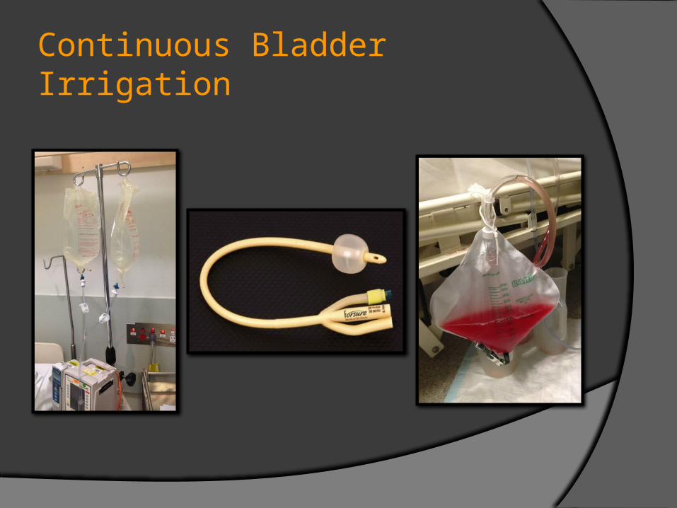
Continuous Bladder Irrigation
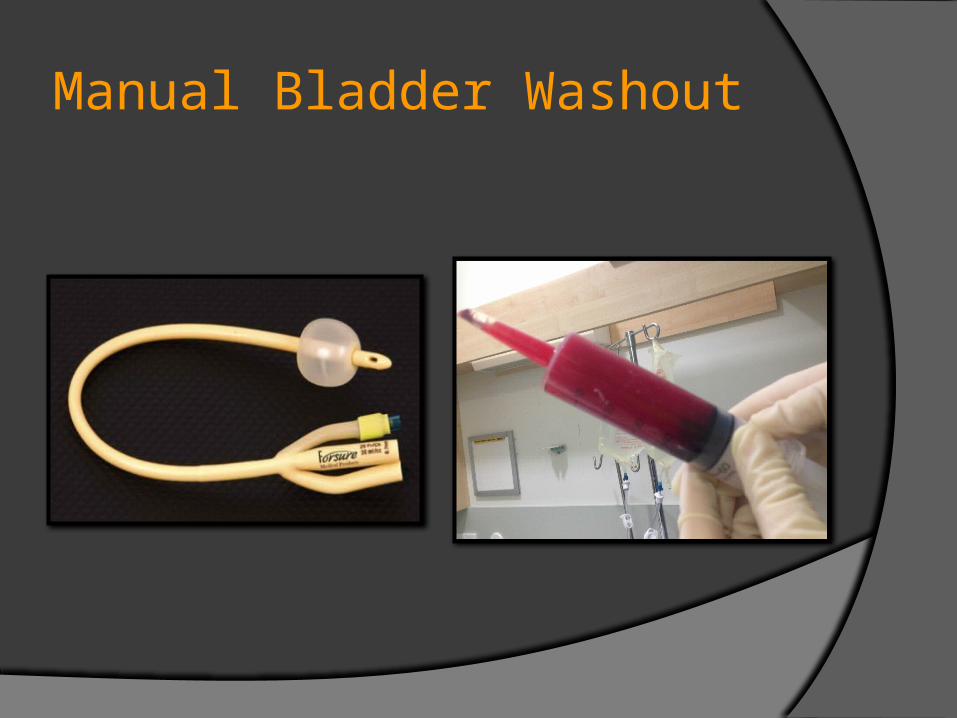
Manual Bladder Washout
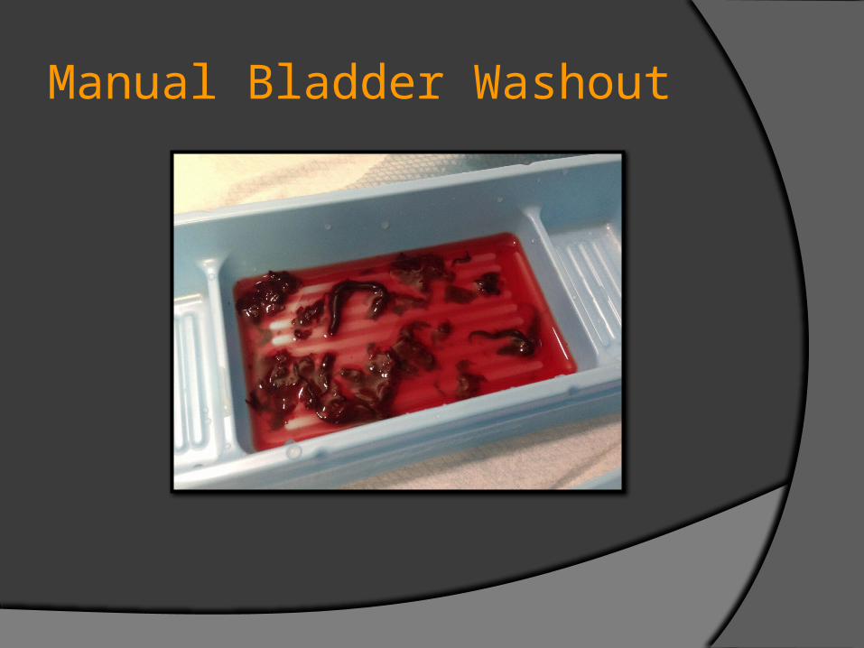
Manual Bladder Washout

Thank you


