
American Journal of Clinical and Experimental Medicine 2015; 3(5): 314-321
Published online December 3, 2015 (http://www.sciencepublishinggroup.com/j/ajcem)
doi: 10.11648/j.ajcem.20150305.31
ISSN: 2330-8125 (Print); ISSN: 2330-8133 (Online)
Lipoprotein(a) Binds to Recombinant Nontypeable Haemophilus influenzae Aspartase
Wenlong Li1, Liping Xu
2, Yakun Zhang
1, Wencheng Bai
1, Lulei Zhou
1, Yuxin Li
1, Na Liu
1,
Ling Liu1, Runlin Han
1, 3, *
1Research Center of Plasma Lipoprotein Immunology, Inner Mongolia Agricultural University, Huhhot, China 2College of Basic Medicine, Inner Mongolia Medical University, Huhhot, China 3Key Laboratory of Animal Clinic Diagnosis and Treatment (Ministry of Agriculture of the People’s Republic of China), Inner Mongolia
Agricultural University, Huhhot, China
Email address: [email protected] (Runlin Han)
To cite this article: Wenlong Li, Liping Xu, Yakun Zhang, Wencheng Bai, Lulei Zhou, Yuxin Li, Na Liu, Ling Liu, Runlin Han. Lipoprotein(a) Binds to
Recombinant Nontypeable Haemophilus influenzae Aspartase. American Journal of Clinical and Experimental Medicine.
Vol. 3, No. 5, 2015, pp. 314-321. doi: 10.11648/j.ajcem.20150305.31
Abstract: The respiratory pathogen nontypeable Haemophilus influenzae (NTHi) can recruit plasminogen (Plg) on the cell
surface by its Plg receptor aspartase (ASP) and utilize host Plg and fibrinolytic system to achieve its adherence and immune
invasion. Lipoprotein(a) [Lp(a)] consists of one molecule low-density lipoprotein (LDL) and one molecule apolipoprotein(a)
[Apo(a)]. Apo(a) shares a high degree of homology with the human Plg, and both of them contain lysine-binding sites (LBS),
which enables them to interact with various cell-surface receptors or fibrin(ogen). However, the definite physiological function
of Lp(a) remains vague. Here, we present evidence that Lp(a) via its Apo(a) may bind to the Plg receptor ASP. Recombinant
aspartase (rASP) and C-terminal lysine-deleted variant of ASP (rASP∆K) were used in the current study. The rASP specifically
bound to Lp(a), but rASP∆K did not, indicating that C-terminal lysine residue of rASP was responsible for the interaction. In
addition, rASP interacted with Lp(a), but not with LDL, revealing that LBS of Apo(a) was involved in the binding. Our results
also showed that Lp(a) could inhibit the binding of Plg to rASP. Plasma Lp(a) might play a role in anti-NTHi infection by binding
to its Plg receptor ASP.
Keywords: Lipoprotein(a), Apolipoprotein(a), Nontypeable Haemophilus influenzae, Plasminogen, Recombinant Aspartase
1. Introduction
Plasminogen (Plg) and the fibrinolytic system can be
utilized and exploited by a number of Gram-positive and
Gram-negative bacteria, including Streptococcus pneumoniae,
Streptococcus pyogens, Helicobacter pylori, Neisseria
meningitides, and Haemophilus influenzae (H. influenzae),
for dissemination and invasion within the host [1-4].
Aspartase (ASP) and protein E (PE) have been identified as
two plasminogen receptors (PlgRs) on the surface of H.
influenzae [5,6]. ASP stimulates the activation of Plg,
mediated by tissue-type plasminogen activators (tPA), and
then contributes to the formation of bacterium-bound
proteolytic plasmin (Pm) activity in the spread of H.
influenzae through tissue barriers [2,5]. H. influenzae can be
subtyped into encapsulated and unencapsulated strains [7].
Unencapsulated strains, referred to Nontypeable H.
influenzae (NTHi), are normal inhabitants of human
nasopharynx and rarely cause systemic infection, but they are
responsible for otitis media, sinusitis, chronic bronchitis, and
other milder infections [7-9].
Lipoprotein(a) [Lp(a)] contains one molecule of
apolipoprotein(a) [Apo(a)] covalently bound to
apolipoprotein B 100 (apoB 100) of low-density lipoprotein
(LDL) via a disulfide bridge [10]. Lp(a) acts as a unique risk
factor for cardiovascular diseases and the elevated plasma
concentration of Lp(a) leads to atherogenesis and thrombosis
since it contains abundant LDL [11-13].
The apo(a) component of Lp(a) confers distinctive
structural properties to this lipoprotein and exhibits high
sequence similarity to zymogen plasminogen, both
possessing weak and strong LBS in their kringle domains
[1,14,15]. Apo(a) contains two types of plasminogen-like
kringle domains (KIV and KV), followed by a serine

315 Wenlong Li et al.: Lipoprotein(a) Binds to Recombinant Nontypeable Haemophilus influenzae Aspartase
protease domain which is catalytically inactive [14, 16]. The
KIV-like domain in Apo(a) can be classified into ten types
(KIV1 to KIV10), and KIV10 have strong LBS that resembles
Kringle 4 in Plg [17,18]. Both Plg and Apo(a) bind to various
cell-surface receptors or fibrin(ogen) surface by using their
LBS in the kringle domains [19-22]. Research has
demonstrated that Lp(a) inhibits the binding of plasminogen
to fibrin and tPA-fibrin compound, and suppresses the
activation of Plg [23-25]. The Plg activation plays an
important role in host fibrinolytic system [26]. We previously
observed that the competitive Plg binding between Lp(a) and
several bacterial PlgRs, like inosine 5'-monophosphate
dehydrogenase (IMPDH) of Staphyylococcus aureus and
glyceraldehyde-3-phosphate dehydrogenase (GAPDH) of
group A Streptococcus [27, 28].
Pathogen infection and inflammation exert a great effect
on the plasma lipoproteins metabolism [29]. We have
hypothesized that Lp(a) might be a potent anti-infective
molecule in the human immune system since Lp(a) might
competitively inhibit pathogens from employing host Plg
[30]. However, there has been no research focused on the
relationship between host Lp(a) and NTHi infection.
The present study demonstrates that human plasma Lp(a)
binds specifically to the recombinant NTHi aspartase (rASP)
via its Apo(a) component, which results in partial inhibition
of the binding of rASP to Plg.
2. Materials and Methods
2.1. Bacterial Strain and Growth Conditions
The strain of nontypeable Haemophilus influenzae (ATCC
49247) was obtained from the National Center for Clinical
Laboratories (Beijing, China). The cultivation of NTHi was
performed as reported previously [31]. Briefly, NTHi, stored
at -70°C in 25% glycerin, was subcultured onto chocolate
agar and incubated in a humid atmosphere at 37 °C and 5%
CO2 for 24 h. The bacterial lawn was inoculated into
brain-heart infusion (BHI) liquid broth supplemented with 4
µg/mL β-NAD (Sigma-Aldrich, Saint Louis, USA) and 10
µg/mL Hemin (Sigma-Aldrich, Saint Louis, USA). The
bacterial culture was grown at 37 °C, with shaking at 200
rpm. Escherichia coli (E. coli ) BL21 (DE3) was grown in
Luria-Bertani (LB) broth. The bacterial strains used in the
study are listed in Table 1.
2.2. Expression of Recombinant Proteins in E. coli
Recombinant NTHi aspartase (rASP) (~53 kDa) and
C-terminal lysine-deleted variant of ASP (rASP∆K) were
expressed in E. coli BL21 (DE3) and purified by using
6×Histidine-tag expression and purification system
(IBA-GmbH, Goettingen, Germany), as described previously
[32]. In brief, genome DNA was extracted from NTHi
(ATCC 49247). The DNA fragments encoding ASP (Genbank:
NC009566; Region: 57522..58940) (or C-terminal
lysine-deleted variant of ASP) were amplified by PCR,
digested with BsaI, and cloned into the plasmid
pASK-IBA37, designed for periplasmic expression. The
plasmid and specific primers for PCR are listed in Table 1.
E.coli BL21 (DE3) harboring plasmid constructs were
grown in the presence of ampicillin (Bio Basic Inc.,
Markham, Canada). Protein expression was induced with
anhydrotetracycline for 3 h, and the recombinant proteins
were extracted from the periplasm by FastBreak Cell Lysis
Reagent (Promega, Madison, USA) and were purified by
affinity chromatography with TALON® Metal Affinity Resins
(Clontech laboratories Inc., Mountain View, USA). The
purity of recombinant proteins was analyzed by
SDS-ployacrylamid gel electrophoresis (SDS-PAGE), and
the protein concentration was quantified with Pierce BCA
Protein Assay Kit (Thermo Scientific, Rockford, USA).
Table 1. Strains, plasmids, and primers used in the present study.
Strains, plasmids, or primers Source or application
Stains
nontypeable Haemophilus influenzae (ATCC 49247) National Center for Clinical Laboratories (Beijing, China)
E.coli BL21 (DE3) Shanghai Sangon
Plasmid
pASK-IBA37 IBA-GmbH
Primers
ASPF: 5′-atggtaggtctcagcgcatgactcaatttagaaaagaagtagat-3′ for ASP Cloning
ASPR: 5′-atggtaggtctcatatcatttatttaatttcgctttgtaagttgg-3′ for ASP Cloning
ASP∆KF: same as ASPF for ASP∆K Cloning
ASP∆KR: 5′-atggtaggtctcatatcaatttaatttcgctttgtaagttgg-3′ for ASP∆K Cloning
2.3. Assay of Aspartase Activity of Recombinant Proteins
Aspartase activity was measured by determination of
fumarate formation using the method described previously by
Williams and Lartigue [33], with several modifications. In
brief, purified recombinant protein (1 µg dissolved in 1 µL
PBS) was added to 299 µL reaction medium (pH 7.0)
(containing 50 mM Sodium L-aspartate, 50 mM Tris, 2 mM
MgSO4·7H2O, and 0.1 mM EDTA). An aliquot of 300 µL
reaction medium was used as a control group. The
absorbance at 240 nm was determined by a Multi-Detection
Microplate Reader (model Synergy HT, BioTek, Winooski,
USA). The molar extinction coefficient of fumarate was
reported by Emery to be 2.53×10³
M-1
·cm-1
[34]. A unit of
aspartase was defined as the amount producing 1 µmol of
fumarate per min at 30°C. Aspartase specific activity was
American Journal of Clinical and Experimental Medicine 2015; 3(5): 314-321 316
expressed as units per mg of protein under the standard assay
conditions.
2.4. Enzyme-Linked Immunosorbent Assay (ELISA)
Purified rASP or rASP∆K solution (2 µg in 100 µL PBS)
was immobilized onto a 96-well microtiter plate (Greiner
Bio-One, Frickenhausen, Germany) at room temperature for
1.5 h. After washing three times with 200 µL TBST
(Tris-buffered saline containing 0.05% Tween 20, pH 7.4),
aliquots of 200 µL blocking buffer (1% BSA in TBST) were
added, and the samples were incubated for 1.5 h at room
temperature. Then, different amounts (100 ng, 50 ng, and 10
ng) of Lp(a) (Biomedical Technologies Inc., Stoughton, USA)
in 100 µL TBST were supplemented into the wells. After
incubation for 1.5 h at room temperature, the unbound Lp(a)
was removed by washing three times with TBST, while the
bound Lp(a) was identified by a goat polyclonal anti-Apo(a)
antibody (Biomedical Technologies Inc., Stoughton,
USA)(1:4000 diluted in 100 µL TBST). After three washes
with TBST, HRP-conjugated donkey anti-goat IgG (R&D
Systems, Minneapolis, USA) (1:1000 diluted in 100 µL
TBST) was used as a secondary antibody for the detection.
The reaction of color development with the addition of TMB
substrate (Promega, Madison, USA) was stopped by the
addition of the stop solution (8.5 M acetic acid, 2.5 M
H2SO4). The absorbance at 450 nm was determined by using
the Multi-Detection Microplate Reader.
The binding of rASP and rASP∆K to human plasma [the
concentration of plasma Lp(a) was 20 mg/L)] was aslo
analyzed by ELISA. Plasma Lp(a) [1:20 diluted in TBST,
plasma Lp(a) employed at a concentration of 100 ng/100 µL]
bound to recombinant proteins was examined by the same
method described above.
The binding of rASP to different amounts of LDL (100 ng,
50 ng and 10 ng) was performed as described above. LDL
(Biomedical Technologies Inc., Stoughton, USA), bound to
immobilized rASP, was identified by a goat anti-LDL
polyclonal antibody (Sigma-Aldrich, Saint Louis, USA) and
HRP-conjugated donkey anti-goat IgG secondary antibody.
The binding of rASP and rASP∆K to different amounts of
Plg (100ng, 50 ng and 10ng) were also investigated. Plg
(R&D Systems, Minneapolis, USA), bound to immobilized
rASP, was identified after incubation with a mouse anti-Plg
monoclonal antibody (Abcam, Cambridge, UK) (1:2000
dilution in TBST). HRP-conjugated goat anti-mouse IgG
(Bios, Beijing, China) was employed as a secondary antibody
(dilution 1:2000 in TBST). The procedures that followed
were performed as described above.
2.5. Asp-affinity Pull Down Assay and Western Blot
Affinity-chromatography columns were packed with 0.2
mL of the TALON® metal resin and equilibrated with buffer
W (50 mM NaH2PO4, 300 mM NaCl, 0.01% NaN3, pH 8.0).
Purified rASP and rASP∆K (100 µg in 150 µL Buffer W)
were applied respectively onto two columns to allow binding
through C-terminal affinity tags. Besides, 150 µL PBS was
applied onto another column. After washing with 8 column
volumes (8 CV) of buffer W, aliquots of 2 mL human plasma
were passed over the three columns. The columns were
washed with 12 CV of buffer W. The complexes of rASP
protein and their ligands were eluted in 300µL fractions with
1.5 CV of buffer E (50 mM NaH2PO4, 300 mM NaCl, 150
mM Imidazole, 0.01% NaN3, pH 7.0). “rASP + Plasma”,
“rASP∆K + Plasma”, and “PBS + Plasma” represented the
elution fraction of the different columns. The total protein
present in each sample was precipitated with 10%
trichloroacetic acid (TCA) for 1 h on ice. After centrifugation
at 4 °C, the pellets were resuspended in 1 M Tris-HCl buffer
(pH 8.0). Plasma Lp(a) were separated by 6% SDS-PAGE,
and then immunoblotting was conducted.
For immunodetection of Apo(a), elution samples were
subjected to 6% SDS-PAGE and transferred to a
nitrocellulose membrane at 400 mA for 3 h (wet blotting).
The nitrocellulose membrane was blocked with TBST
supplemented with 5% skim milk at room temperature for 2 h
and washed with TBST. Bound Apo(a) was detected with a
goat anti-Apo(a) antibody (1:20000 diluted in TBST,
supplemented with 2% skim milk) followed by
HRP-conjugated donkey anti-goat IgG as a secondary
antibody (1:5000 diluted in TBST, supplemented with 2%
skim milk). Bound Apo(a) was detected with
HRP-conjugated Tactin (Thermo Scientific, Rockford, USA)
(1:8000 diluted in TBST). Detection of reactive antibodies
was performed by using a chemiluminescence substrate
(Tiangen, Beijing, China), and the chemiluminescence
signals were visualized on Syngene G:Box Chemi XT4
(Syngene, Cambridge, UK).
2.6. Binding-Inhibition ELISA
The effect of 6-aminocaproic acid (EACA) (Sigma-Aldrich,
Saint Louis, USA) on Lp(a) binding to immobilized rASP was
analyzed by binding-inhibition ELISA. EACA, utilized at
different concentrations (0 mM, 0.1mM, and 1mM), were
combined with a constant quantity (100 ng) of Lp(a) in 100 µL
TBST. Then the mixtures were added to immobilized rASP.
After incubation and extensive washing, each bound Lp(a) was
identified by specific antibodies.
In addition, Lp(a), utilized at different amounts (0 ng, 50 ng,
and 100 ng), was combined with a constant quantity (100 ng)
of Plg in 100 µL TBST. Then, the mixtures were added to
immobilized rASP for the determination of the effect of Lp(a)
on the binding of Plg to rASP. After incubation and extensive
washing, each bound Plg was identified by specific antibodies.
3. Statement of Ethical Standards
The blood was taken from volunteers after the provision of
written informed consent and authorization in compliance
with the regulation of the Inner Mongolia Agricultural
University (Hohhot, China). The experiments with human
plasma were approved by the Academic Board and the
Science and Technology Department of the Inner Mongolia
Agricultural University. All participants signed a written
317 Wenlong Li et al.: Lipoprotein(a) Binds to Recombinant Nontypeable Haemophilus influenzae Aspartase
informed consent form. The concentration of Lp(a) was
determined in the Inner Mongolia Autonomous Region
People’s Hospital.
4. Statistical Analyses of the Data
All tests were performed in triplicates, and the results were
presented as mean ± standard deviation. Unless stated
otherwise, the statistical significance of the results was
established by Student’s t-test for paired data (GraphPad
Software, San Diego, USA). Differences with p<0.05 were
considered statistically significant (*P<0.05, **P<0.01,
***P<0.001).
5. Results
5.1. Purification of rASP and rASP∆K
rASP and rASP∆K were purified by affinity
chromatography with TALON® Metal Affinity Resins. The
samples collected from different purification procedures were
analyzed by 12% SDS-PAGE. The results indicated that both
rASP and rASP∆K were purified successfully (Fig. 1).
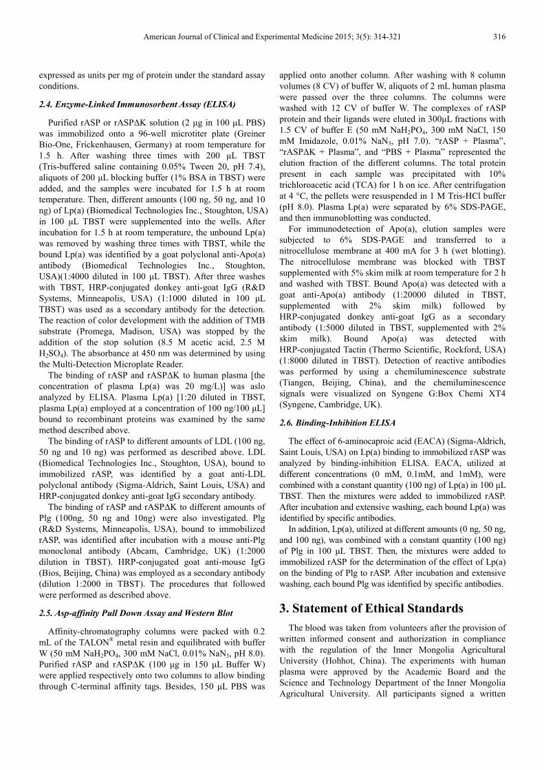
Fig. 1. Purification of recombinant proteins.
a The purification of rASP. b The purification of rASP∆K. “M” represents
the molecular mass markers (kDa); “Sup” refers to the supernatant after lysis
by E.coli BL21; “F-T” denotes the resolution passed over the column; “W1,
W10” represent the 1st, 10th wash fraction; “E1, E2, E3, E7” refer to the 1st,
2nd, 3rd, and 7th eluate fraction, respectively. The common ~53 kDa bands
depicted the target proteins.
5.2. ASP Activity Assay of Recombinant Proteins
Aspartase activity of the purified recombinant proteins was
determined spectrophotometrically at 240 nm. The specific
activity of rASP and rASP∆K were 59.81 ± 6.30 U/mg and
104.38 ± 18.08 U/mg, respectively. The results showed that
the recombinant proteins had the ability to catalyze the
formation of fumarate and possessed efficient biological
activity of ASP. The specific activity of the mutant ASP is
higher than the wildtype protein, which has been reported by
early researches [35].
5.3. Lp(a) Binds to C-terminal Lysine of rASP via LBS
To test the hypothesis that ASP binds to Apo(a), binding of
rASP to LDL or Lp(a) was assayed by ELISA, respectively.
rASP was incubated with different amounts of Lp(a) or LDL.
The results evidenced that Lp(a) bound to rASP in a
dose-dependent manner. In contrast, LDL did not associate
with rASP (Fig. 2). This indicated that Lp(a) binds to rASP
via Apo(a), but not LDL.
In order to determine the region of rASP responsible for
binding to Lp(a), a variant of ASP (rASP∆K) which lacked
C-terminal lysine residue was also expressed and purified.
The binding of rASP∆K to Lp(a) was analyzed by using
rASP as a positive control. The results revealed that rASP∆K
cannot bind to Lp(a) (Fig. 3a). Thus, as we speculated, the
C-terminal lysine residue of ASP plays a key role in this
process.
To further characterize the relevance of rASP for Lp(a)
binding, we assayed binding of both purified Lp(a) and
human plasma. The data showed that they had no difference
in the binding of recombinant proteins (Fig. 3b). The results
indicated that the source of Lp(a) exerted no effect on the
Lp(a) binding assay, and rASP∆K did not bind to Lp(a).
Human plasma was also used for stimulating the
physiological environment in vitro.
These data were further confirmed by Asp-affinity pull
down assay and western blot analysis. Lp(a) was present only
in the elution of “rASP + Plasma” (Fig. 3c); the elution of
“PBS + plasma” was used as a negative control. These
findings implied that rASP binds to plasma Lp(a) via its
C-terminal lysine.
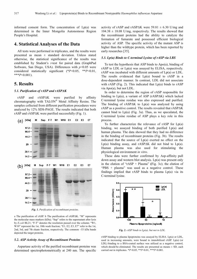
Fig. 2. rASP binds to Lp(a), but not to LDL.
rASP binding to plasma lipoproteins was assayed by ELISA. Lp(a) or LDL,
used in increasing amounts, were bound to immobilized rASP. Lp(a) (or
LDL) binding to a BSA-coated surface was utilized as a negative control,
which should be eliminated. The results are presented as means ± SD, each
carried out in triplicates. *P<0.05, **P<0.01, ***P<0.001.
American Journal of Clinical and Experimental Medicine 2015; 3(5): 314-321 318
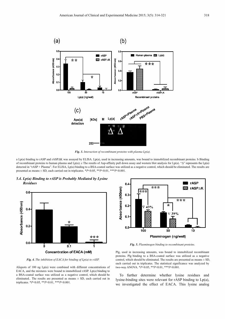
Fig. 3. Interaction of recombinant proteins with plasma Lp(a).
a Lp(a) binding to rASP and rASP∆K was assayed by ELISA. Lp(a), used in increasing amounts, was bound to immobilized recombinant proteins. b Binding
of recombinant proteins to human plasma and Lp(a); c The results of Asp-affinity pull down assay and western blot analysis for Lp(a). “∆” represents the Lp(a)
detected in “rASP + Plasma”. For ELISA, Lp(a) binding to a BSA-coated surface was utilized as a negative control, which should be eliminated. The results are
presented as means ± SD, each carried out in triplicates. *P<0.05, **P<0.01, ***P<0.001.
5.4. Lp(a) Binding to rASP is Probably Mediated by Lysine
Residues
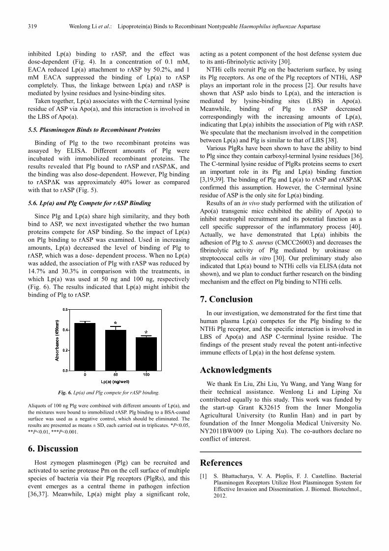
Fig. 4. The inhibition of EACA for binding of Lp(a) to rASP.
Aliquots of 100 ng Lp(a) were combined with different concentrations of
EACA, and the mixtures were bound to immobilized rASP. Lp(a) binding to
a BSA-coated surface was utilized as a negative control, which should be
eliminated.. The results are presented as means ± SD, each carried out in
triplicates. *P<0.05, **P<0.01, ***P<0.001.
Fig. 5. Plasminogen binding to recombinant proteins.
Plg, used in increasing amounts, was bound to immobilized recombinant
proteins. Plg binding to a BSA-coated surface was utilized as a negative
control, which should be eliminated. The results are presented as means ± SD,
each carried out in triplicates. The statistical significance was analyzed by
two-way ANOVA. *P<0.05, **P<0.01, ***P<0.001.
To further determine whether lysine residues and
lysine-binding sites were relevant for rASP binding to Lp(a),
we investigated the effect of EACA. This lysine analog
319 Wenlong Li et al.: Lipoprotein(a) Binds to Recombinant Nontypeable Haemophilus influenzae Aspartase
inhibited Lp(a) binding to rASP, and the effect was
dose-dependent (Fig. 4). In a concentration of 0.1 mM,
EACA reduced Lp(a) attachment to rASP by 50.2%, and 1
mM EACA suppressed the binding of Lp(a) to rASP
completely. Thus, the linkage between Lp(a) and rASP is
mediated by lysine residues and lysine-binding sites.
Taken together, Lp(a) associates with the C-terminal lysine
residue of ASP via Apo(a), and this interaction is involved in
the LBS of Apo(a).
5.5. Plasminogen Binds to Recombinant Proteins
Binding of Plg to the two recombinant proteins was
assayed by ELISA. Different amounts of Plg were
incubated with immobilized recombinant proteins. The
results revealed that Plg bound to rASP and rASP∆K, and
the binding was also dose-dependent. However, Plg binding
to rASP∆K was approximately 40% lower as compared
with that to rASP (Fig. 5).
5.6. Lp(a) and Plg Compete for rASP Binding
Since Plg and Lp(a) share high similarity, and they both
bind to ASP, we next investigated whether the two human
proteins compete for ASP binding. So the impact of Lp(a)
on Plg binding to rASP was examined. Used in increasing
amounts, Lp(a) decreased the level of binding of Plg to
rASP, which was a dose- dependent process. When no Lp(a)
was added, the association of Plg with rASP was reduced by
14.7% and 30.3% in comparison with the treatments, in
which Lp(a) was used at 50 ng and 100 ng, respectively
(Fig. 6). The results indicated that Lp(a) might inhibit the
binding of Plg to rASP.
Fig. 6. Lp(a) and Plg compete for rASP binding.
Aliquots of 100 ng Plg were combined with different amounts of Lp(a), and
the mixtures were bound to immobilized rASP. Plg binding to a BSA-coated
surface was used as a negative control, which should be eliminated. The
results are presented as means ± SD, each carried out in triplicates. *P<0.05,
**P<0.01, ***P<0.001.
6. Discussion
Host zymogen plasminogen (Plg) can be recruited and
activated to serine protease Pm on the cell surface of multiple
species of bacteria via their Plg receptors (PlgRs), and this
event emerges as a central theme in pathogen infection
[36,37]. Meanwhile, Lp(a) might play a significant role,
acting as a potent component of the host defense system due
to its anti-fibrinolytic activity [30].
NTHi cells recruit Plg on the bacterium surface, by using
its Plg receptors. As one of the Plg receptors of NTHi, ASP
plays an important role in the process [2]. Our results have
shown that ASP aslo binds to Lp(a), and the interaction is
mediated by lysine-binding sites (LBS) in Apo(a).
Meanwhile, binding of Plg to rASP decreased
correspondingly with the increasing amounts of Lp(a),
indicating that Lp(a) inhibits the association of Plg with rASP.
We speculate that the mechanism involved in the competition
between Lp(a) and Plg is similar to that of LBS [38].
Various PlgRs have been shown to have the ability to bind
to Plg since they contain carboxyl-terminal lysine residues [36].
The C-terminal lysine residue of PlgRs proteins seems to exert
an important role in its Plg and Lp(a) binding function
[3,19,39]. The binding of Plg and Lp(a) to rASP and rASP∆K
confirmed this assumption. However, the C-terminal lysine
residue of ASP is the only site for Lp(a) binding.
Results of an in vivo study performed with the utilization of
Apo(a) transgenic mice exhibited the ability of Apo(a) to
inhibit neutrophil recruitment and its potential function as a
cell specific suppressor of the inflammatory process [40].
Actually, we have demonstrated that Lp(a) inhibits the
adhesion of Plg to S. aureus (CMCC26003) and decreases the
fibrinolytic activity of Plg mediated by urokinase on
streptococcal cells in vitro [30]. Our preliminary study also
indicated that Lp(a) bound to NTHi cells via ELISA (data not
shown), and we plan to conduct further research on the binding
mechanism and the effect on Plg binding to NTHi cells.
7. Conclusion
In our investigation, we demonstrated for the first time that
human plasma Lp(a) competes for the Plg binding to the
NTHi Plg receptor, and the specific interaction is involved in
LBS of Apo(a) and ASP C-terminal lysine residue. The
findings of the present study reveal the potent anti-infective
immune effects of Lp(a) in the host defense system.
Acknowledgments
We thank En Liu, Zhi Liu, Yu Wang, and Yang Wang for
their technical assistance. Wenlong Li and Liping Xu
contributed equally to this study. This work was funded by
the start-up Grant K32615 from the Inner Mongolia
Agricultural University (to Runlin Han) and in part by
foundation of the Inner Mongolia Medical University No.
NY2011BW009 (to Liping Xu). The co-authors declare no
conflict of interest.
References
[1] S. Bhattacharya, V. A. Ploplis, F. J. Castellino. Bacterial Plasminogen Receptors Utilize Host Plasminogen System for Effective Invasion and Dissemination. J. Biomed. Biotechnol., 2012.
American Journal of Clinical and Experimental Medicine 2015; 3(5): 314-321 320
[2] M. L. Sanderson-Smith, D. M. P. De Oliveira, M. Ranson, et al. Bacterial Plasminogen Receptors: Mediators of a Multifaceted Relationship. J. Biomed. Biotechnol., 2012.
[3] M. Ullberg, G. Kronvall, I. Karlsson, et al. Receptors for human plasminogen on gram-negative bacteria. Infect. Immun., 1990, 58: 21-25.
[4] K. Lahteenmaki, P. Kuusela, T. K. Korhonen. Bacterial plasminogen activators and receptors. FEMS Microbiol. Rev., 2001, 25: 531-552.
[5] I. Sjostrom, H. Grondahl, G. Falk, et al. Purification and characterisation of a plasminogen-binding protein from Haemophilus influenzae. Sequence determination reveals identity with aspartase. Biochim. Biophys. Acta, 1997, 1324: 182-190.
[6] D. Barthel, B. Singh, K. Riesbeck, et al. Haemophilus influenzae uses the surface protein E to acquire human plasminogen and to evade innate immunity. J. Immunol., 2012, 188: 379-385.
[7] D. C. Turk. The pathogenicity of Haemophilus influenzae. J. Med. Microbiol., 1984, 18: 1-16.
[8] P. T. King, J. Ngui, D. Gunawardena, et al. Systemic humoral immunity to non-typeable Haemophilus influenzae. Clin. Exp. Immunol., 2008, 153: 376-384.
[9] R. Virkola, K. Lahteenmaki, T. Eberhard, et al. Interaction of Haemophilus influenzae with the mammalian extracellular matrix. J. Infect. Dis., 1996, 173: 1137-1147.
[10] S. Xu. Apolipoprotein(a) binds to low-density lipoprotein at two distant sites in lipoprotein(a). Biochemistry, 1998, 37: 9284-9294.
[11] E. Anuurad, M. B. Boffa, M. L. Koschinsky, et al. Lipoprotein(a): A unique risk factor for cardiovascular disease. Clin. Lab. Med., 2006, 26: 751-754.
[12] B. G. Nordestgaard, M. J. Chapman, K. Ray, et al. Lipoprotein(a) as a cardiovascular risk factor: current status. Eur. Heart J., 2010, 31: 2844-U2814.
[13] P. R. Kamstrup. Lipoprotein(a) and ischemic heart disease-A causal association? A review. Atherosclerosis, 2010, 211: 15-23.
[14] J. W. Mclean, J. E. Tomlinson, W. J. Kuang, et al. cDNA sequence of human apolipoprotein(a) is homologous to plasminogen. Nature, 1987, 330: 132-137.
[15] L. Becker, P. M. Cook, T. G. Wright, et al. Quantitative evaluation of the contribution of weak lysine-binding sites present within apolipoprotein(a) kringle IV types 6-8 to lipoprotein(a) assembly. J. Biol. Chem., 2004, 279: 2679-2688.
[16] B. R. Gabel, M. I. Koschinsky. Analysis of the proteolytic activity of a recombinant form of apolipoprotein(a). Biochemistry, 1995, 34: 15777-15784.
[17] Y. Y. Van Der Hoek, M. E. Wittekoek, U. Beisiegel, et al. The apolipoprotein(a) kringle IV repeats which differ from the major repeat kringle are present in variably-sized isoforms. Hum. Mol. Genet., 1993, 2: 361-366.
[18] J. L. Hoover-Plow, L. A. Miles, G. M. Fless, et al. Comparison of the lysine binding functions of lipoprotein(a) and plasminogen. Biochemistry, 1993, 32: 13681-13687.
[19] R. Romagnuolo, S. M. Marcovina, M. B. Boffa, et al. Inhibition of plasminogen activation by apo(a): role of carboxyl-terminal lysines and identification of inhibitory domains in apo(a). J. Lipid Res., 2014, 55: 625-634.
[20] A. M. Scanu, M. M. Atzeni, C. Edelstein, et al. Lipoprotein(a): identification of subjects with a superbinding capacity for fibrinogen. Clin. Genet., 1997, 52: 367-370.
[21] M. A. Lucas, L. J. Fretto, P. A. Mckee. The binding of human plasminogen to fibrin and fibrinogen. J. Biol. Chem., 1983, 258: 4249-4256.
[22] J. M. Edelberg, S. V. Pizzo. Lipoprotein(a) inhibits plasminogen activation in a template-dependent manner. Blood Coagul. Fibrinolysis., 1991, 2: 759-764.
[23] K. A. Hajjar, D. Gavish, J. L. Breslow, et al. Lipoprotein(a) modulation of endothelial cell surface fibrinolysis and its potential role in atherosclerosis. Nature, 1989, 339: 303-305.
[24] M. A. Hancock, M. B. Boffa, S. M. Marcovina, et al. Inhibition of plasminogen activation by lipoprotein(a) - Critical domains in apolipoprotein(a) and mechanism of inhibition on fibrin and degraded fibrin surfaces. J. Biol. Chem., 2003, 278: 23260-23269.
[25] C. Bas Leerink, P. F. Duif, J. A. Gimpel, et al. Lysine-binding heterogeneity of Lp(a): consequences for fibrin binding and inhibition of plasminogen activation. Thromb. Haemost., 1992, 68: 185-188.
[26] C. Kluft. The fibrinolytic system and thrombotic tendency. Pathophysiol. Haemos. Thromb., 2003, 33: 425-429.
[27] Y. Xu, Z. X. Ji, R. L. Han. The interaction between Lipoprotein(a) and recombinant inosine 5'-monophosphate dehydrogenase derived from Staphylococcus aureus (in Chinese). Microbiology China, 2011, 38: 1405-1411.
[28] X. Y. Dai, L. P. Xu, W. C. Bai, et al. The interaction between lipoprotein(a) anrecombinant glyceraldehyde-3-phosphate dehydrogenase derived from group a Streptococcus (in Chinese). J. Inner Mongolia Agric. Univ., 2011, 32: 27-31.
[29] W. Khovidhunkit, M. S. Kim, R. A. Memon, et al. Effects of infection and inflammation on lipid and lipoprotein metabolism: mechanisms and consequences to the host. J. Lipid Res., 2004, 45: 1169-1196.
[30] R. L. Han. Plasma lipoproteins are important components of the immune system. Microbiol. Immunol., 2010, 54: 246-253.
[31] H. N. Coleman, D. A. Daines, J. Jarisch, et al. Chemically defined media for growth of Haemophilus influenzae strains. J. Clin. Microbiol., 2003, 41: 4408-4410.
[32] G. Chaga, D. E. Bochkariov, G. G. Jokhadze, et al. Natural poly-histidine affinity tag for purification of recombinant proteins on cobalt(II)-carboxymethylaspartate crosslinked agarose. J. Chromatogr. A, 1999, 864: 247-256.
[33] V. R. Williams, D. J. Lartigue. Quaternary structure and certain allosteric properties of aspartase. J. Biol. Chem., 1967, 242: 2973-2978.
[34] T. F. Emery. Aspartase-catalyzed synthesis of N-hydroxyaspartic acid. Biochemistry, 1963, 2: 1041-1045.
[35] N. Yumoto, K. Mizuta, M. Tokushige, et al. Studies on aspartase VIII. Protease-mediated activation: comparative survey of protease specificity for activation and peptide cleavage. Physiol. Chem. Phys., 1982, 14: 391-397.
321 Wenlong Li et al.: Lipoprotein(a) Binds to Recombinant Nontypeable Haemophilus influenzae Aspartase
[36] A. Godier, B. J. Hunt. Plasminogen receptors and their role in the pathogenesis of inflammatory, autoimmune and malignant disease. J. Thromb. Haemost., 2013, 11: 26-34.
[37] K. Lahteenmaki, S. Edelman, T. K. Korhonen. Bacterial metastasis: the host plasminogen system in bacterial invasion. Trends Microbiol., 2005, 13: 79-85.
[38] R. Romagnuolo, K. Demarco, M. B. Boffa, et al. Apolipoprotein(a) inhibits the conversion of Glu-plasminogen to Lys-plasminogen on the surface of vascular endothelial and smooth muscle cells. J. Thromb. Haemost., 2013, 11: 377-377.
[39] L. A. Miles, C. M. Dahlberg, J. Plescia, et al. Role of cell-surface lysines in plasminogen binding to cells: identification of alpha-enolase as a candidate plasminogen receptor. Biochemistry, 1991, 30: 1682-1691.
[40] J. Hoover-Plow, E. Hart, Y. Q. Gong, et al. A Physiological Function for Apolipoprotein(a): A Natural Regulator of the Inflammatory Response. Exp. Biol. Med., 2009, 234: 28-34.


![Sách tham kh § o - tailieuhoctap123blog.files.wordpress.com · -Thi Gtb Sbi Gn ÿ ]i: th õchi Ën so sánh và phân tích. Có th Çt ¥o ra tín hi Ëuc «nthi Ãt ÿ Çso sánh](https://cdn.vdocuments.net/doc/165x107/5e1557607b08b525ce7defe4/sch-tham-kh-o-thi-gtb-sbi-gn-i-th-chi-n-so-snh-v-phn-tch.jpg)