Download - Mechanical Force Effects on Cellular Functions Kyle Wallenberg Bioengineering 506 4/15/2009

Mechanical Force Effects on Cellular Functions
Kyle WallenbergBioengineering 506
4/15/2009

Adhesion-mediated mechanosensitivity: a time to
experiment, and a time to theorize Alexander Bershadsky, Michael
Kozlov and Benjamin Geiger

Outline
• Introduction to mechanosensitive adhesion structure/function
• Focal adhesions and focal complexes • Focal adhesion mechanosensing adhesions • Non-focal adhesion mechanosensing adhesions• Mechanosensing signaling mechanisms• Physical models– Differences
• Conclusions

Introduction
• Adhesion-mediated signaling– Provides cells with descriptive information about
microenvironment– Mechanical influences• Ability to grow and strengthen in response to applied
force
• Integrin-mediated cell matrix-adhesions• Full understanding of mechanisms unknown– Introduction of theoretical models to understand
adhesion-mediated mechanosensing

Adhesion Machinery
• Composed of specialized subcellular contact sites – Formed by transmembrane receptors linked to
cytoskeleton– Link cells to extracellular matrix (ECM)
• Few micrometers in length• Sense chemical properties of external surfaces• Respond to mechanical cues– Mechanical forces (stresses)– Mechanical deformations (strains)

Adhesion Site Functions
• Ensure correct and stable positioning of cells• Unpredictable nature of external
perturbations– Cell develops ‘just in time mechanism’• Adhesion strength dependent on amount of
stress/strain applied at adhesion site
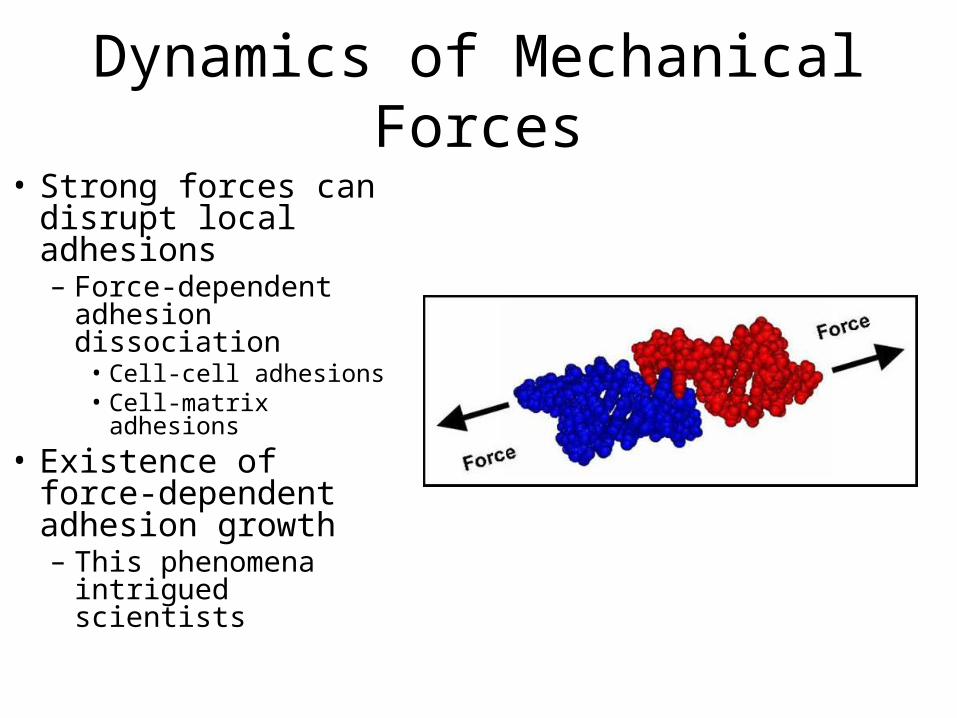
Dynamics of Mechanical Forces• Strong forces can
disrupt local adhesions– Force-dependent
adhesion dissociation• Cell-cell adhesions• Cell-matrix adhesions
• Existence of force-dependent adhesion growth– This phenomena
intrigued scientists

Focal Adhesions• Formed by different cell
types– Fibroblasts– Epithelial cells
• Large (several microns in length)
• Adhere to actin filaments at proximal end
• Formation dependent on natural matrix proteins or RGD peptides at high densities
• Interact with ECM using different integrins

Focal Adhesions (contd.)• More than 100 proteins
make up FA domain– Link integrin molecules and
actin filaments– Regulate assembly and
signaling generated by adhesive interactions
• Gives FAs ability to respond to range of signals/features– Rigidity – Mechanical perturbation– Topography

Focal Complexes (FXs)
• Type of cell-matrix adhesion which FAs evolve from
• Small (less than micron in size)
• Formed continuously from lamellipodia
• Some undergo maturation

FX-to-FA Transition• Dependent upon continually applied force– (1) Inhibition of myosin II leads to accumulation of FXs and
disappearance of FAs• Activation of myosin II induces FA assembly
– (2) Application of external force to FAs motivates growth in force direction regardless of myosin II activation
– (3 )Size of FAs and force applied generally proportional – (4) When strong enough forces unable to be generated,
large FAs not formed• E.g. on a soft matrix
– Overall trend:• FAs respond to local force by increased assembly• FAs respond to relaxation of force by disassembly

FA Mechanosensing Adhesions
• Involved in many cellular responses– Substrate stretching– Variations in substrate rigidity • Migration in direction of increasing rigidity
– Durotaxis: Directed movement of cell motility
– Fluid shear stress
Non-FA Mechanosensing Adhesions
• Integrin-mediated adhesion– Fibrillar adhesions• Associated with ECM fibrils• Evolve from FAs in force-dependent manner• Unlike FAs, do NOT disassemble when force relaxed
– Podosomes • Different substrate rigidity response than FAs
– Lifespan, not shape, depends on substrate flexibility

Non-FA Mechanosensing Adhesions
• Cadherin-mediated cell-cell adherens junctions (AJs)– Depend on myosin-II driven
contractility • Thus, the inhibition of myosin
II reduces AJ proteins at cell-cell interface
• Platelet endothelial cell adhesion molecule (PECAM)– Immunoglobulin family– Influences mechanical
response of endothelial cells to fluid shear stress

Unanswered Questions Regarding FAs
• Relationship/interaction between FA components and relative spatial organization mostly unknown
• Precise mechanisms by which cells respond to environment unanswered – Surface rigidity– Ligand density – Local/global mechanical perturbations
• Unknown which molecular interactions within FAs regulated by force

Possible Mechanosensing Signaling Mechanisms
• Modulation of phosphorylation-dependent protein-protein interactions– May explain force-
induced changes within FA components
– FAK, Src, Fyn• Protein tyrosine kinases
thought to be involved

Possible Mechanosensing Signaling Mechanisms
• Adaptor Proteins – Zyxin• Recruitment to nascent FXs accompanies maturation
into FAs• When force applied, translocates from FAs to stress
fibers following substrate stretching
• Adhesion-dependent pathways regulating activities of Rho family GTPases and Rap1
• These mechanisms have yet to be integrated into a specific molecular scheme

Physical Modeling
• Models proposed over the years to explain events in adhesion-mediated mechanosensitivity– Physical mechanisms governing FAs– Shear-stress profile along FAs– Spatial distribution of FAs– Effects of substrate elasticity on FA formation

Physical Modeling
• Three specific models of FA mechanosensing described– Stress-driven model– Strain-driven model– Thermodynamic model
• Models differ in assumptions concerning physical factors underlying mechanosensing and dynamic behavior of FAs during growth/shrinkage

Stress-driven Model• Assumed that FAs contain molecular
switches that react to application of force by changing states from inactive to active, or vice-versa– Sense stress and switch to active
conformation when stress exceeds a critical value
• Could be basis for explaining variety of mechanisms involved in mechanosensing:– Stress-induced transition of certain FA
proteins– Modulate activity of enzymes to turn on
or off phosphorylation switches• Kinases/ Phosphatases
– Regulate various adhesion molecules• Extracellular fibronectin• Adaptor proteins such as talin and
vinculin• Signaling enzymes such as Src and FAK

Stress-driven Model• Mechanosensitive protein unit
connected to actin filament moving with retrograde actin flow– Results in dragging force acting on
protein unit from filament• (a) – Passive state with weak slip
link• (b) – Active state with strong slip
link• Transition occurs when dragging
force exceeds critical value– Leads to conformational change in
mechanosensitive protein• Could explain mechanism for
maturation of FXs by ECM elasticity

Strain-driven Model• Mechanosenser switch activated
by local elastic strain – Characterized by compression or
extension• FA composed of two layers
– Lower layer contains mechanosensitive proteins and integrins and is attached to substrate
– Upper layer connected to actin filaments • Transmits force to lower layer
• Compression of top of lower layer relative to its bottom drives FA assembly– Generates strain at front edge of
FA leading to growth in pulling force direction

Energy Considerations
• Stress-driven model– Stress can promote conformational transition of
protein if thermodynamic work produced by stress decreases activation energy of the transition • Requires significant change in protein dimensions
– Example is stretch-activated ion channels• Open when 2-D membrane stress applied• Change in area of stress-sensing channel in membrane
large enough to drive transition from passive to active state
Energy Considerations• Stress-driven model (contd.)– Stress induced by actin filaments points in direction of
contractile or pulling force– Conformational transition must result in protein stretching
(ΔL)– Energy produced by stress is ΔF = -γ*L(per)* ΔL
• γ is lateral tension• L(per) is protein linear dimension measured perpendicular to the
stress– ΔL must be greater than 4 nm assuming stress-induced
force (γ*L(per)) acting on single protein is ~ 1pN and energy produced by stress greater than thermal energy
– Points out limitation in sensing stress in case of FAs

Energy Considerations• Strain-driven model– Strain sensing understood if attachment of plaque
protein to mechanosensing layer coupled to deformation of sensing layer• If sensor layer not deformed prior to binding plaque protein,
energy of required deformation is paid at expense of binding energy
• If sensor layer is deformed prior to binding, effective affinity of plaque protein increases
• Why does this matter?– Binding constant of plaque proteins can be altered by straining
mechanosensing layer» Deformation energy coupled to binding of one plaque
protein must be larger than thermal energy

Thermodynamic Model
• No protein switch• Elastic stress generated within plaque parallel to
plasma membrane by stress fibers can cause FA self-assembly and growth in pulling force direction– Reducing pulling force leads to FA disassembly
• Elastic stress decreases chemical potential within plaque, enhancing self-assembly by adding new plaque proteins
• Predicts internal treadmilling-like motion of proteins which can progress in different directions depending on organization of FA assembly/disassembly

Thermodynamic Model• (a) – FA composed of
aggregate of elastic building blocks– FA connected to substrate by
links along surface• (b) – Pulling forces results in
aggregate stretching and buildup of elastic energy
• (c) – FA assembly driven by introduction of new proteins into aggregate– Concentration of new
proteins are distributed diffusively throughout plaque
– Leads to energy relaxation

Implications of Thermodynamic Model
• FA plaque is elastic and can withstand mechanical stress
• Cannot undergo stretch-induced rupture with addition of new FA proteins– May require molecular devices similar to formin
proteins• Can maintain connection to protein complex while new
monomers are added to complex while at the same time stabilizing the growing structure
• More modeling and experimentation necessary to understand mechanism by which formins direct FA mechanosensing

Differences Between Models
• Position of mechanosensor within the FA– Stress-driven model• Stress sensors located at interface between plaque and
stress fiber– Strain-driven model• Strain sensors located in integrin layer interacting with
ECM– Thermodynamic model• Elastic stresses of plaque stimulate FA self-assembly
– Effective mechanosensors located within plaque

Differences Between Models• Proposed method of molecular exchange between FA
and cytoplasm– Stress-driven model
• Stress-mediated transition stabilizes and reinforces connection between stress fibers and plaque– Results in FX maturation into FAs
– Strain-driven model• Actin spread through the protein compresses mechanosensor
layer ahead of plaque and extends layer behind plaque– Compressed molecular switches promote binding of new plaque
proteins while extended switches promote plaque disassembly at back of FA» Results in FA treadmilling
– Thermodynamic model• Involves entire plaque volume rather than partial areas along front
and back of FA (analogous to strain-driven model)

Conclusions• Focal adhesion key example of a mechanosensitive adhesion
molecule• Three FA mechanosensitive models introduced
– Stress-driven model– Strain-driven model– Thermodynamic model
• Adhesion-mediated mechanosensitivity described and confirmed, but molecular mechanisms still not completely known– Known that force promotes directional adhesion assembly
• Regulated by Rho GTPases and transformed by protein phosphorlyation/dephosphorylation
– Unknown whether mechanosensitivity is directed by single protein or complex nor what specific proteins are involved
References
• Bershadsky A, Kozlov M, Geiger B: Adhesion-mediated mechanosensitivity: a time to experiment, and a time to theorize. Current Opinion in Cell Biology 2006, 18:472-481.

Questions


