Download - Principle of Wound Closure and Preparation

Principle of Wound Closureand Preparation
«’√™—¬ «’√ª°√≥å°¡≈ «—≤π‰°√
217
°“√¥Ÿ·≈·º≈‡ªìπ ‘Ëß ”§—≠Õ¬à“ßÀπ÷ËߢÕß·æ∑¬å ‚¥¬‡©æ“–»—≈¬·æ∑¬å‡π◊ËÕß®“°
¡’§«“¡‡°’ˬ«¢âÕß°—∫·º≈‚¥¬μ√ß»—≈¬·æ∑¬åºŸâ¥Ÿ·≈®”‡ªìπμâÕß¡’§«“¡‡¢â“„®‡°’ˬ«°—∫·º≈
∑—Èß “‡Àμÿ°“√‡°‘¥ªí®®—¬∑’Ë¡’·º≈μàÕ°“√√—°…“·≈–°“√À“¬¢Õß·º≈ √«¡∑—Èß°“√√—°…“·º≈∑’Ë
‡°‘¥¢÷Èπ °“√¡’§«“¡√Ÿâ§«“¡‡¢â“„®Õ¬à“ߥ’∑”„Àâ»—≈¬·æ∑¬å “¡“√∂„Àâ°“√¥Ÿ·≈·≈–√—°…“
·º≈∑’ˇ°‘¥¢÷Èπ‰¥âÕ¬à“ß¡’ª√– ‘∑∏‘¿“æ·≈–‡À¡“– ¡
°“√√—°…“·º≈¡’¢—ÈπμÕπÀ≈“°À≈“¬¥—ß∑’Ë®–°≈à“«μàÕ‰ª√«¡∂÷ß°“√‡μ√’¬¡°“√μ—Èß
·μà°àÕπ°“√√—°…“ √–À«à“ß°“√√—°…“·≈–¿“¬À≈—ß°“√√—°…“ ´÷Ëß„π·μà≈–¢—ÈπμÕπ¡’º≈μàÕ
°“√¥Ÿ·≈·≈–√—°…“
Principle of Wound Closure
À≈—°°“√ªî¥·º≈∫√‘‡«≥º‘«Àπ—ß®”‡ªìπ∑’Ë®–μâÕߥŸ∑—Èß à«π¢Õß·º≈·≈– ¿“æ¢Õß
ºŸâªÉ«¬ ‚¥¬®–·∫à߉¥â¥—ßπ’È
1. Patient condition ¿“æ¢ÕߺŸâªÉ«¬·≈– underlying disease ∑’Ë¡’º≈μàÕ
°“√À“¬¢Õß·º≈ ‡™àπ vascular disease, congenital condition affecting elastin
and/or wound healing ¿“«–∑“ß‚¿™π“°“√∑’ËÕ“®¡’º≈μàÕ°“√À“¬¢Õß·º≈ ‡™àπ °“√
¢“¥ “√Õ“À“√∫“ßÕ¬à“ßÀ√◊Õ«‘μ“¡‘π∫“ߪ√–‡¿∑
2. Wound condition μâÕߥŸμ—Èß·μà “‡Àμÿ°“√‡°‘¥ ‡™àπ crush injury Õ“®¡’
¿“«–°“√∫“¥‡®Á∫¢Õ߇π◊ÈÕ‡¬◊ËÕ¡“°°«à“∑’ˇÀÁπ„π§√—Èß·√°°“√μ‘¥‡™◊ÈÕ¢Õß·º≈ ¿“æ¢Õß·º≈
(¢π“¥¢Õß∫“¥·º≈, §«“¡≈÷°, ¡’Õ«—¬«– ”§—≠∫“¥‡®Á∫À√◊Õ‰¡à ‡™àπ À≈Õ¥‡≈◊Õ¥À√◊Õ‡ âπ

Principle of Wound Closure and Preparation218
ª√– “∑¡’°“√∫“¥‡®Á∫√à«¡¥â«¬, μ”·ÀπàߢÕß∫“¥·º≈, degree of contamination)
·≈–°“√¡’ ‘Ëß·ª≈°ª≈Õ¡∫√‘‡«≥·º≈
Aim of skin closure
®ÿ¥ª√– ߧå„π°“√ªî¥·º≈∫√‘‡«≥º‘«Àπ—ßÕ“®·∫àßÕÕ°‰¥â‡ªìπ 2 ®ÿ¥ª√– ߧåÀ≈—°
䴉ᡈ
1. For coverage ‡æ◊ËÕ∑”°“√·°â‰¢·º≈‡ªî¥„À⇪ìπ·º≈ªî¥‡ªìπ®ÿ¥¡ÿàßÀ¡“¬À≈—°
2. For reconstruction ‡æ◊ËÕ∑”°“√·°â‰¢·≈–‡ √‘¡ √â“ß ‚¥¬Õ“®¡’®ÿ¥¡ÿàßÀ¡“¬
„π¥â“π°“√∑”ß“π (Functional results) À√◊Õ„π¥â“𧫓¡ «¬ß“¡ (Aesthetic results)
À√◊Õ∑—Èß ÕßÕ¬à“ß√à«¡°—π
Crikelair1 ‰¥â„ÀâÀ≈—°°“√„π°“√ºà“μ—¥ªî¥·º≈∫√‘‡«≥º‘«Àπ—߉«â¥—ßπ’È
- Place incisions to follow tension lines and natural folds in the
skin
- Handle tissues gently and debride only as much as necessary
to ensure an adequately clean bed
- Ensure complete hemostasis
- Eliminate tension at the skin edges
- Use fine sutures and remove them early
- Evert wound edge
- If possible, choose patients whose age is closer to 90 than to 9
years
- Allow time for scars to mature before repeat intervention
„π°“√ªî¥·º≈∑’Ë≈÷°¡“°°«à“º‘«Àπ—ß2 ¡’À≈—°°“√¥—ßπ’È
1. to eliminate dead space
2. to provide a strong enough closure to prevent dehiscence
while wound healing is occurring
3. to precisely approximate the skin edges without tension
‚¥¬„π°“√√—°…“‰¡à®”‡ªìπ∑’Ë®–μâÕß∑”°“√ªî¥∑ÿ°™—Èπ¢Õ߇π◊ÈÕ‡¬◊ËÕ (not all layers
«’√™—¬ «’√ª°√≥å, °¡≈ «—≤π‰°√ 219
necessarily require separate closure) ·μà°“√ªî¥·º≈‰¡à§«√„À⇰‘¥™àÕß«à“ߢ÷Èπ„π·º≈
(dead space) À≈—ß°“√ªî¥º‘«Àπ—ß2
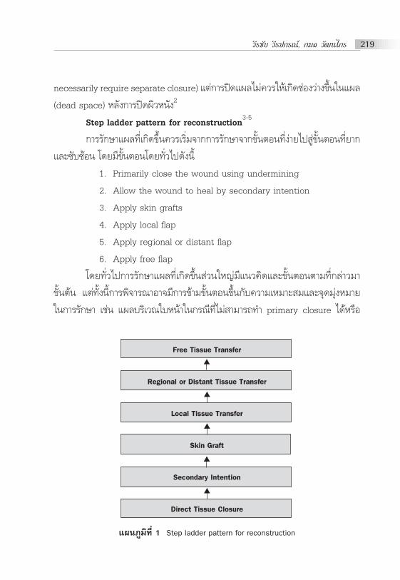
Step ladder pattern for reconstruction3-5
°“√√—°…“·º≈∑’ˇ°‘¥¢÷Èπ§«√‡√‘Ë¡®“°°“√√—°…“®“°¢—ÈπμÕπ∑’Ëßà“¬‰ª Ÿà¢—ÈπμÕπ∑’ˬ“°
·≈–´—∫´âÕπ ‚¥¬¡’¢—ÈπμÕπ‚¥¬∑—Ë«‰ª¥—ßπ’È
1. Primarily close the wound using undermining
2. Allow the wound to heal by secondary intention
3. Apply skin grafts
4. Apply local flap
5. Apply regional or distant flap
6. Apply free flap
‚¥¬∑—Ë«‰ª°“√√—°…“·º≈∑’ˇ°‘¥¢÷Èπ à«π„À≠à¡’·π«§‘¥·≈–¢—ÈπμÕπμ“¡∑’Ë°≈à“«¡“
¢—Èπμâπ ·μà∑—Èßπ’È°“√æ‘®“√≥“Õ“®¡’°“√¢â“¡¢—ÈπμÕπ¢÷Èπ°—∫§«“¡‡À¡“– ¡·≈–®ÿ¥¡ÿàßÀ¡“¬
„π°“√√—°…“ ‡™àπ ·º≈∫√‘‡«≥„∫Àπâ“„π°√≥’∑’ˉ¡à “¡“√∂∑” primary closure ‰¥âÀ√◊Õ
·ºπ¿Ÿ¡‘∑’Ë 1 Step ladder pattern for reconstruction
Free Tissue Transfer
Regional or Distant Tissue Transfer
Local Tissue Transfer
Skin Graft
Secondary Intention
Direct Tissue Closure
▲
▲
▲
▲
▲

Principle of Wound Closure and Preparation220
„π°“√∑” primary closure ·≈â«∑”„À⇰‘¥ anatomical distortion °“√√—°…“Õ“®
æ‘®“√≥“„π°“√∑”„π¢—ÈπμÕπ¢Õß skin grafting À√◊Õ„™â local flap ´÷Ëß®–„Àâº≈∑’Ë¥’°«à“
∂⓪≈àÕ¬„Àâ·º≈À“¬‡Õß (secondary intention) „π°“√√—°…“∫“¥·º≈„π∫“ßμ”·Àπàß
∂â“„Àâ¡’°“√À“¬¢Õß·º≈‡Õß (secondary intention) Õ“®∑”„À⇰‘¥¡’ªí≠À“„π¥â“𧫓¡
«¬ß“¡ (aesthetic result) À√◊Õ‡°‘¥°“√¥÷ß√—ÈߢÕ߇π◊ÈÕ‡¬◊ËÕ (scar contracture) μ“¡¡“
‰¥â ‡ªìπμâπ
Type of Skin Closure
Direct tissue closure
‡ªìπ°“√√—°…“‚¥¬°“√ªî¥·º≈∫√‘‡«≥º‘«Àπ—ß‚¥¬μ√ß „™â„π°√≥’∑’Ë·º≈¡’¢π“¥‰¡à
„À≠à¡“°·≈– contaminated πâÕ¬ „π°√≥’∑’Ë·º≈¡’§«“¡μ÷ß¡“°À≈—ß®“°‡¬Á∫ªî¥∑”„Àâ¡’
‚Õ°“ ‡°‘¥·º≈·¬° widened scar ·≈–¡’º≈μàÕ wound healing À√◊Õ„π°√≥’ inde-
terminate zone of injury ‰¡à§«√∑’Ë®–„™â°“√√—°…“‚¥¬«‘∏’π’È °“√√—°…“«‘∏’π’È¡’¢âÕ¥’„π·ßà
‡ªìπ°“√√—°…“∑’Ëßà“¬·≈–‰¡à —́∫´âÕπ °“√À“¬¢Õß·º≈¥’·≈–‰¥â anatomical alignment
∑’ˇÀ¡“– ¡ ·μà¡’¢âÕ‡ ’¬„π·ßà¡’‚Õ°“ μ‘¥‡™◊ÈÕ‚¥¬‡©æ“–·º≈∑’Ë¡’ contaminated ¡“°
·≈–Õ“®®”‡ªìπ∑’Ë®–μâÕß„™â∑—°…–∑“ß°“√ºà“μ—¥∑’Ë´—∫´âÕπ„π°√≥’∑’Ë¡’°“√„™â skin flap √à«¡
¥â«¬6
Secondary intention
‡ªìπ°“√√—°…“‚¥¬ª≈àÕ¬„Àâ¡’°“√À“¬¢Õß·º≈‡Õß ‚¥¬·æ∑¬å¡’Àπâ“∑’Ëπ” ‘Ëß·ª≈°
ª≈Õ¡·≈–‡π◊ÈÕ‡¬◊ËÕ∑’Ë쓬ÕÕ°®“°·º≈√à«¡°—∫°“√ªÑÕß°—π¿“«–μ‘¥‡™◊ÈÕ ·≈–¥Ÿ·≈·º≈„ÀâÕ¬Ÿà
„π ¿“«–∑’ˇÀ¡“– ¡ ‚¥¬°“√√—°…“«‘∏’π’È¡’¢âÕ¥’„π·ßà ‡ªìπ°“√√—°…“∑’Ëßà“¬·≈–‰¡à´—∫´âÕπ
„™â„π°√≥’∑’Ë·º≈¡’¢π“¥‡≈Á°À√◊Õ·º≈∂≈Õ° ·μà¡’¢âÕ‡ ’¬„π·ßà„™â‡«≈“„π°“√À“¬¢Õß·º≈π“π
‡ ’ˬßμàÕ°“√μ‘¥‡™◊ÈÕ Õ“®μâÕß∑”°“√‡ª≈’ˬπÕÿª°√≥åªî¥·º≈À≈“¬§√—Èß ·≈–Õ“®‡°‘¥ subop-
timal scar6
Skin grafting
Skin grafting ‡ªìπ«‘∏’¡“μ√∞“π„π°“√ªî¥·º≈„π°√≥’∑’Ë·º≈‰¡à “¡“√∂∑’Ë®–∑”
direction tissue closure À√◊Õ‡°‘¥ secondary intention ‰¥â„π‡«≈“Õ—π§«√ ‚¥¬„π

«’√™—¬ «’√ª°√≥å, °¡≈ «—≤π‰°√ 221
à«π skin graft ª√–°Õ∫¥â«¬ epidermis ·≈–∫“ß à«πÀ√◊Õ∑—ÈßÀ¡¥¢Õß dermis ¢÷Èπ
Õ¬Ÿà°—∫™π‘¥¢Õß skin graft ¢—ÈπμÕπ°“√√—°…“‚¥¬°“√π”º‘«Àπ—ß®“° à«πÀπ÷ËߢÕß√à“ß°“¬
(donor site) ‡§≈◊ËÕπ¬â“¬º‘«Àπ—ßπ—Èπ¡“«“ß„πμ”·Àπàß∑’ˇªìπμâÕß°“√ (recipient site)
º‘«Àπ—ß∑’Ëπ”¡“«“ß “¡“√∂Õ¬Ÿà‰¥â‚¥¬Õ“»—¬ “√Õ“À“√∑’Ë¡“®“°·º≈ (wound bed) ¥—ßπ—Èπ
°“√∑” skin grafting “¡“√∂𔉪„™â‰¥â„π°√≥’∑’Ë·º≈¡’ ¿“«–∑’ˇÀ¡“– ¡ (well-vas-
cularized wound bed) ‚¥¬°“√√—°…“«‘∏’π’È¡’¢âÕ¥’„π·ßà‡ªìπ°“√√—°…“∑’Ëßà“¬·≈–‡√Á«„π
°“√ªî¥·º≈‚¥¬‰¡àμâÕß„™â flap ·μà¡’¢âÕ‡ ’¬„π·ßàμâÕß¡’°“√‡μ√’¬¡ recipient wound bed
∑’ˇÀ¡“– ¡·≈–Õ“®‡°‘¥ªí≠À“¥â“π poor aesthetics, contour deformity ·≈– poor
durability ‡¡◊ËÕ‡∑’¬∫°—∫ local flap √«¡∂÷ß°“√¡’·º≈‡ªìπ∫√‘‡«≥ donor site6
Type of skin graft “¡“√∂·∫à߉¥âÀ≈“¬ª√–‡¿∑ ‡™àπ
- ·∫àßμ“¡§«“¡Àπ“¢Õß dermis ‰¥â·°à split-thickness skin graft ·≈–
full-thickness skin graft
- ·∫àßμ“¡≈—°…≥–¢Õß skin graft ‰¥â·°à meshed skin graft ·≈– sheet
skin graft
√Ÿª∑’Ë 1 Sheet skin graft·≈– Meshed skin graft

Principle of Wound Closure and Preparation222
Full-thickness skin graft and Split-thickness skin graft
- Full-thickness skin graft À≈—ß®“°∑’Ëπ”º‘«Àπ—ß∑’Ë∑” skin graft ÕÕ°¡“
®–‰¡à‡À≈◊Õ epidermal structure Õ¬Ÿà„π∫√‘‡«≥ donor site ∑”„Àâ·º≈∑’ˇ°‘¥¢÷Èπ‰¡à
“¡“√∂À“¬‰¥â‡Õß ¥—ßπ—Èπ„π°√≥’∑’Ë√—°…“‚¥¬°“√„™â Full-thickness skin graft §«√
æ‘®“√≥“π”¡“®“°∫√‘‡«≥∑’Ë “¡“√∂‡¬Á∫·º≈ªî¥‰¥âÀ√◊Õ„π°√≥’∑’ËμâÕß°“√º‘«Àπ—ߢπ“¥
„À≠à‡æ◊ËÕ®ÿ¥ª√– ߧåÕ¬à“ß„¥Õ¬à“ßÀπ÷Ëß®”‡ªìπμâÕßπ” split-thickness skin graft ¡“ªî¥
∫√‘‡«≥ donor site μ”·ÀπàߢÕß donor site ∑’Ë„™â∫àÕ¬ ‰¥â·°à postauricular area,
supraclavicular area, flexural area, thigh area, inguinal area ·≈– abdominal
area
- Split-thickness skin graft μà“ß°—∫„π°√≥’ Full-thickness skin graft
∫√‘‡«≥ donor site ®–‡À≈◊Õ adnexal remnants, pilo-sebaceous follicle À√◊Õ sweat
gland apparatus ´÷Ëß dermis ∑’ˇÀ≈◊ÕÕ¬Ÿà “¡“√∂ √â“ߺ‘«Àπ—ߢ÷Èπ¡“„À¡à‰¥â ¥—ßπ—Èπ„π
°√≥’¢Õß°“√∑” Split-thickness skin graft °“√¥Ÿ·≈∫√‘‡«≥ donor site „ÀâÕ¬Ÿà„π
¿“«–∑’ˇÀ¡“– ¡‡æ◊ËÕ√Õ„Àâ¡’°“√ √â“ߺ‘«Àπ—ߢ÷Èπ„À¡à (reepithelialization) ‚¥¬
“¡“√∂·∫àß split-thickness skin graft μ“¡§«“¡Àπ“¢Õß dermis ‰¥â¥—ßπ’È
● Thin split-thickness skin graft
● Intermediate split-thickness skin graft
● Thick split-thickness skin graft
„π∑“ߪƑ∫—μ‘ °“√·∫àßμ“¡§«“¡Àπ“¢Õß dermis Õ“®¡’ªí≠À“‡π◊ËÕß®“°§«“¡
Àπ“¢Õߺ‘«Àπ—ß„π·μà≈–™à«ßÕ“¬ÿ, ‡æ» ·≈–μ”·ÀπàߢÕߺ‘«Àπ—ß„π∫√‘‡«≥ à«πμà“ßÊ ¢Õß
√à“ß°“¬ ¡’§«“¡Àπ“¢Õß dermis ‰¡à‡∑à“°—π ‚¥¬æ∫«à“ º‘«Àπ—ߢÕ߇¥Á°·√°§≈Õ¥∫“ß
ª√–¡“≥ 3.5 ‡∑à“¢Õߺ‘«Àπ—ߺŸâ„À≠à ‚¥¬º‘«Àπ—ߢÕ߇¥Á°®–¡’§«“¡Àπ“„°≈⇧’¬ß°—∫
ºŸâ„À≠à‡¡◊ËÕÕ“¬ÿª√–¡“≥ 5 ªï „πºŸâÀ≠‘ß®–æ∫«à“¡’§«“¡Àπ“¢Õßdermis πâÕ¬°«à“„πºŸâ™“¬
·≈–º‘«Àπ—ß∫√‘‡«≥ΩÉ“¡◊Õ·≈–ΩÉ“‡∑â“Àπ“∑’Ë ÿ¥„π√à“ß°“¬·≈–º‘«Àπ—ß∫√‘‡«≥Àπ—ßμ“·≈–
À≈—ßÀŸ∫“ß∑’Ë ÿ¥„π√à“ß°“¬3,7
∫√‘‡«≥ donor site ∑’Ë¡’°“√„™â∫àÕ¬ ‰¥â·°à »’√…– μâπ·¢π μâπ¢“ ·≈–∫√‘‡«≥°âπ
°“√æ‘®“√≥“™π‘¥¢Õß split-thickness skin graft3,8 “¡“√∂æ‘®“√≥“‰¥â®“°

«’√™—¬ «’√ª°√≥å, °¡≈ «—≤π‰°√ 223
1. The setting of dermatome ‚¥¬ “¡“√∂¥Ÿ‰¥â®“°°“√μ—Èߧà“
2. The translucency of the graft ‚¥¬°“√¡Õߺà“π skin graft ‰ª¬—ß
°√–¥“…∑’Ë¡’μ—«Õ—°…√À√◊ÕÀ¡“¬‡≈¢ ·≈â«∑”°“√Õà“π ∂â“ “¡“√∂Õà“π‰¥â™—¥‡®π skin graft
∑’ˉ¥â‡ªìπ™π‘¥ thin split-thickness skin graft ·μà∂Ⓡ√“‰¡à “¡“√∂Õà“πμ—«Õ—°…√À√◊Õ
À¡“¬‡≈¢‰¥â ∂◊Õ‡ªìπ thick split-thickness skin graft
3. The pattern of bleeding of the donor site ‚¥¬Õ“»—¬°“√¥Ÿ≈—°…≥–
¢Õ߇≈◊Õ¥∑’ËÕÕ°∫√‘‡«≥ donor site
● Thin split-thickness skin graft ¡’§«“¡Àπ“·πàπ¢Õß®ÿ¥‡≈◊Õ¥ÕÕ°¡“°
·≈–≈—°…≥–®ÿ¥‡≈◊Õ¥ÕÕ°®–¡’¢π“¥‡≈Á° (a high density of tiny bleeding point)
● Thick split-thickness skin graft ¡’§«“¡Àπ“·πàπ¢Õß®ÿ¥‡≈◊Õ¥ÕÕ°
πâÕ¬√à«¡°—∫®ÿ¥‡≈◊Õ¥ÕÕ°¡’¢π“¥„À≠à‡¡◊ËÕ‡∑’¬∫°—∫ thin split-thickness skin graft
(a lower density of larger bleeding point)
The process of skin graft take “¡“√∂·∫à߉¥â‡ªìπ 2 √–¬–
- Plasmatic imbibitions ‡°‘¥¢÷Èπ∑—π∑’À≈—ß®“°∑’Ë«“ߺ‘«Àπ—ß∑’Ëπ”¡“ (skin graft)
«“ß≈ß∫π recipient bed ‚¥¬æ∫«à“¡’ fibrin network ‡°‘¥¢÷Èπ√–À«à“ß skin graft °—∫
recipient bed ∑”„Àâ skin graft ‰¡à‡§≈◊ËÕπ∑’Ë√à«¡°—∫¡’ plasma-like fluid ®“° recipi-
ent bed ºà“π‡¢â“‰ª„π skin graft „π™à«ß 48 ™—Ë«‚¡ß·√°À≈—ß∑” skin grafting æ∫«à“
skin graft ¡’πÈ”Àπ—°‡æ‘Ë¡¢÷Èπ®“°‡¥‘¡ª√–¡“≥ 30%-40%3,7 ·≈–‡√‘Ë¡æ∫ blood flow ‡°‘¥
¢÷Èπ„π skin graft
- Revascularization ‡ªìπ√–¬–∑’Ë¡’‡≈◊Õ¥°≈—∫¡“‡≈’È¬ß skin graft ‚¥¬√–¬–π’È
ª√–°Õ∫¥â«¬2 ¢∫«π°“√ ‰¥â·°à Inosculation (°“√‡™◊ËÕ¡°—π√–À«à“ßÀ≈Õ¥‡≈◊Õ¥¢Õß skin
graft °—∫ À≈Õ¥‡≈◊Õ¥¢Õß recipient bed) ·≈– Neovascularization (°“√ √â“ßÀ≈Õ¥
‡≈◊Õ¥¢÷Èπ„À¡à„π skin graft) ‚¥¬√–¬–π’ȇ°‘¥„π™à«ß 48 - 96 ™—Ë«‚¡ßÀ≈—ß°“√«“ß skin graft
≈ß∫π recipient bed3
Skin graft adherence
°“√∑’Ë skin graft ®– “¡“√∂Õ¬Ÿà√Õ¥‰¥â®”‡ªìπμâÕßμ‘¥Õ¬Ÿà°—∫ recipient bed
‚¥¬ “¡“√∂·∫à߉¥â‡ªìπ 2 √–¬– „π√–¬–·√° skin graft μ‘¥°—∫ recipient bed ‚¥¬

Principle of Wound Closure and Preparation224
Õ“»—¬ fibrin ´÷Ë߇°‘¥¢÷Èπ∑—π∑’∑’Ë«“ß skin graft √–¬–À≈—߇°‘¥¢÷Èπª√–¡“≥ 48-96 ™—Ë«‚¡ß
‚¥¬‡ªìπ√–¬–∑’Ë¡’°“√‡®√‘≠¢Õß fibrous tissue ·≈– vessels ®“° recipient bed ‡¢â“‰ª„π
skin graft2
Conditions for take
º‘«Àπ—ß∑’˪≈Ÿ°∂à“¬ “¡“√∂Õ¬Ÿà√Õ¥‰¥â¡’ªí®®—¬∑’Ë ”§—≠Õ¬Ÿà 2 Õ¬à“߉¥â·°à
- Vascular recipient bed
- Contact of the skin graft and the recipient bed
Donor site selection and Graft harvest
°“√æ‘®“√≥“ graft donor site à«π„À≠àæ‘®“√≥“®“°
1. μâÕß°“√ split-thickness skin graft À√◊Õ full-thickness skin graft
2. ≈—°…≥–¢Õß skin graft ∑’ˇÀ¡“– ¡°—∫ recipient bed ‡™àπ„π°√≥’
¢Õß ’º‘«
3. ‚Õ°“ ∑’Ë®–‡°‘¥¿“«–·∑√°´âÕπ∫√‘‡«≥ donor site
The phenomenon of bridging
„π°“√∑” skin grafting æ∫«à“„π∫“ß°√≥’∫√‘‡«≥∑’Ë∑” skin grafting ‰¡à‰¥â
‡ªìπ∫√‘‡«≥∑’Ë¡’ well-vascularized wound bed ‡ ¡Õ‰ª Õ“®¡’∫“ß∫√‘‡«≥∑’ˇªìπ
avascularized wound bed Õ¬Ÿà∫â“ß °“√∑” skin grafting æ∫«à“ skin graft “¡“√∂
∑’Ë®–Õ¬Ÿà√Õ¥‰¥â ∂â“∫√‘‡«≥ avascularized wound bed ¡’¢π“¥‡ âπºà“π»Ÿπ¬å°≈“ßπâÕ¬
°«à“ 1 ‡´π쑇¡μ√3,8
Postoperative care
°“√¥Ÿ·≈À≈—ß®“°°“√∑” skin grafting ¡’§«“¡ ”§—≠¡“° ‡æ√“–¡’º≈μàÕ‚Õ°“ ∑’Ë
graft take ·≈–¬—ß√«¡‰ª∂÷ß°“√¥Ÿ·≈∫√‘‡«≥ donor site ∂â“„π°√≥’∑’ËμâÕß°“√‡°Á∫√—°…“
skin graft ‰¡à«à“®–¥â«¬ “‡Àμÿ„¥°Áμ“¡‡æ◊ËÕπ”°≈—∫¡“„™â„πÕπ“§μ »—≈¬·æ∑¬åºŸâ∑”°“√
√—°…“®–μâÕß„À⧫“¡ ”§—≠·≈–‡¢â“„®¢—ÈπμÕπ„π°“√¥Ÿ·≈‡ªìπÕ¬à“ߥ’
- Care of skin graft ‚¥¬¡’¢âÕ§«√√–«—ß„π°“√¥Ÿ·≈·≈–·°â‰¢¥—ßπ’È
● Removal of hematomas and seromas „π°√≥’∑’Ë«“ß sheet graft ¡’
‚Õ°“ æ∫ fluid collection Õ¬Ÿà„μâ skin graft ‰¥â „π°√≥’∑’Ë ß —¬«à“¡’ fluid collection

«’√™—¬ «’√ª°√≥å, °¡≈ «—≤π‰°√ 225
·π–π”„ÀâÀ≈—ß°“√∑” skin grafting §«√∑”°“√‡ªî¥¥Ÿ„π«—π∑’Ë 2 À≈—ß∑”°“√ºà“μ—¥3 ∂â“
æ∫«à“¡’ fluid collection „μ⺑«Àπ—ß„Àâ∑”°“√·°â‰¢‚¥¬„™â No. 11 knife blade °√’¥≈ß
‰ª¬—ß skin graft ∫√‘‡«≥∑’Ë¡’ fluid collection ·≈–∑”°“√ remove ÕÕ° À≈—ß®“°π—Èπ„Àâ
∑”·º≈∑ÿ°«—π ®π°«à“®–æ∫«à“‰¡à¡’ fluid collection ‡°‘¥¢÷Èπ ·μà„π°√≥’∑’Ë·πà„®«à“‰¡à¡’ fluid
collection ·≈–°“√μ‘¥‡™◊ÈÕÀ≈—ß®“°∑” skin grafting à«π„À≠à®–∑”°“√‡ª≈’ˬπ dress-
ing ª√–¡“≥«—π∑’Ë 5-7 À≈—ß°“√∑”ºà“μ—¥ ∂⓺ŸâªÉ«¬¡’Õ“°“√ª«¥¡“°¢÷Èπ∫√‘‡«≥μ”·Àπàß∑’Ë
«“ß skin graft „Àâπ÷°∂÷ß«à“¡’°“√μ‘¥‡™◊ÈÕ‡°‘¥¢÷Èπ·≈–„π∑“ßμ√ß°—π¢â“¡ ∂â“¡’Õ“°“√ª«¥
·º≈πâÕ¬≈ß®–‡ªìπμ—«∫àß∫Õ°∂÷ß graft take3
● Care of the infected skin graft °“√μ‘¥‡™◊ÈÕ¢Õß skin graft æ∫«à“
ºŸâªÉ«¬®–¡’‰¢âμË”Ê ·º≈¡’°≈‘Ëπº‘¥ª°μ‘ ·≈–¡’Õ“°“√∫«¡·¥ß∫√‘‡«≥¢Õ∫¢Õß skin graft
à«π„À≠à¡—°‡°‘¥ª√–¡“≥«—π∑’Ë 2-4 À≈—ß°“√∑”ºà“μ—¥ °√≥’∑’Ë¡’°“√μ‘¥‡™◊ÈÕ∫√‘‡«≥ skin graft
°“√√—°…“ à«π„À≠à‡ªìπ°“√¥Ÿ·≈·º≈‡©æ“–∑’Ë (local wound care) ‚¥¬°“√π”‡π◊ÈÕ‡¬◊ËÕ
à«π∑’Ë쓬ÕÕ°, °“√„™â¬“ªØ‘™’«π–‡©æ“–∑’Ë (topical antibiotic) √à«¡°—∫°“√¥Ÿ·≈·º≈
”À√—∫ systemic antibiotic ®–„™â ”À√—∫„π°√≥’ cellulitis À√◊Õ„π°√≥’∑’Ë¡’°“√μ‘¥
‡™◊ÈÕ√ÿπ·√߇°‘¥¢÷Èπ
● Immobilization period for skin grafts °√≥’∑’Ë¡’°“√«“ß skin graft
„π∫√‘‡«≥∑’Ë∂Ⓡ°‘¥ secondary graft contraction ®–∑”„À⇰‘¥ªí≠À“μ“¡¡“‰¥â ‡™àπ∫√‘‡«≥
flexor surface ·π–π”„À⺟âªÉ«¬„ à splint ‡æ◊ËÕªÑÕß°—π‰¡à„Àâ¡’°“√À¥μ—« (contracture)
‚¥¬·π–π”„Àâ„ à‡ªìπ∫“߇«≈“¢Õß«—𠇪ìπ√–¬–‡«≈“ª√–¡“≥ 6 ‡¥◊Õπ
- Storage of skin grafts3,7,8 °“√‡°Á∫√—°…“ skin graft “¡“√∂‡°Á∫√—°…“‰¥â
¥—ßπ’È
● «“ß skin graft ∫π donor site ‡ªìπ«‘∏’°“√‡°Á∫√—°…“ split-thickness
skin graft ∑’Ë¡’ª√– ‘∑∏‘¿“æ∑’Ë ÿ¥ ·≈– “¡“√∂π”°≈—∫¡“„™â ∂â“π”¡“„™â¿“¬„π√–¬–‡«≈“
10 «—π “¡“√∂π” skin graft ÕÕ°¡“®“°∫√‘‡«≥ donor site ‰¥â‚¥¬‰¡àμâÕß„™â¬“™“·≈–
ºŸâªÉ«¬‰¡à¡’Õ“°“√‡®Á∫®“°°“√π” skin graft ÕÕ°®“°∫√‘‡«≥ donor site
● Refrigerated at 4˚C ‚¥¬π” skin graft Ààե⫬ gauze ™ÿ∫ saline
solution À√◊Õ Ringerûs solution „ à„π«— ¥ÿª√“»®“°‡™◊ÈÕ “¡“√∂‡°Á∫√—°…“ skin graft

Principle of Wound Closure and Preparation226
‰¥âπ“π∂÷ß 21 «—π
- Care of the donor site °“√¥Ÿ·≈ donor site À≈—ß®“°°“√∑” skin grafting
‡ªìπ°“√¥Ÿ·≈‡æ◊ËÕ„Àâ·º≈∑’ˇ°‘¥¢÷ÈπÕ¬Ÿà„π ¿“«–∑’ˇÀ¡“– ¡μàÕ°“√‡°‘¥ reepithelialization
·≈–ªÑÕß°—π‰¡à„À⇰‘¥¡’°“√μ‘¥‡™◊ÈÕ °“√À“¬¢Õß·º≈¢÷Èπ°—∫ª√‘¡“≥ pilosebaceous appa-
ratus, hair follicle ·≈– sweat glands ´÷Ë߇ªìπ®ÿ¥‡√‘Ë¡μâπ¢Õß°“√‡°‘¥ epithelial re-
generation ‚¥¬æ∫«à“ donor site ®“°°“√∑” thin split-thickness skin graft
“¡“√∂À“¬¿“¬„π 7-9 «—π ·≈– donor site ®“°°“√∑” thick split-thickness skin
graft „™â‡«≈“„π°“√À“¬¡“°°«à“ 14 «—π3,8 °“√¥Ÿ·≈·º≈ donor site ‚¥¬„™â semiper-
meable polyurethane membrane æ∫«à“ “¡“√∂≈¥Õ“°“√‡®Á∫·º≈‰¥â2,3,7 °“√‡ªî¥
·º≈¡—°®–√Õ„Àâ¡’ complete reepithelialization à«π„À≠àÕ¬Ÿàª√–¡“≥ 2 —ª¥“Àå „π
°√≥’∑’Ë·º≈ donor site À“¬™â“¡—°‡°‘¥®“°°“√μ‘¥‡™◊ÈÕÀ√◊Õ°“√∑” thick split-thickness
skin graft ∑’ËÀπ“¡“°‡°‘π‰ª
Characteristics of grafted skin
- Contraction of grafted skin “¡“√∂·∫à߉¥â‡ªìπ 2 √–¬–
1. Primary contraction (Graft elasticity) ‡°‘¥¢÷Èπ∑—π∑’À≈—ß®“°∑’Ë∑”°“√π”
skin graft ÕÕ°¡“®“° donor site ‚¥¬æ∫«à“ skin graft ¡’°“√À¥μ—«‡≈Á°≈ß®“°¢π“¥
‡¥‘¡‡°‘¥®“°°“√∑”ß“π¢Õß elastic fibers ∑’ËÕ¬Ÿà„π™—Èπ dermis ¢Õß skin graft ´÷Ë߉¡à‰¥â
‡ªìπº≈¡“®“° biologic process of contracture3 ‡π◊ËÕß®“°°“√À¥μ—«¢Õß skin graft
¢÷Èπ°—∫ª√‘¡“≥ elastic fibers ∑’ËÕ¬Ÿà„π™—Èπ dermis ®÷ßæ∫«à“ skin graft ∑’ËÀπ“®–æ∫°“√
À¥μ—«¡“°°«à“„π skin graft ∑’Ë∫“ß Davis ·≈– Kitlowski »÷°…“æ∫«à“ full-thickness
skin graft ¡’ primary contracture 41% ¢Õߢπ“¥æ◊Èπ∑’˺‘«‡√‘Ë¡·√° ·≈– thin split-
thickness skin graft ¡’ primary contracture ‡æ’¬ß 9% ¢Õߢπ“¥æ◊Èπ∑’˺‘«‡√‘Ë¡·√°3
2. Secondary contraction (Contraction) ‡ªìπº≈¡“®“°°“√À¥μ—«¢Õß
recipient bed ¿“¬À≈—ß®“°∑’Ë¡’°“√ªî¥·º≈¥â«¬ skin graft ‚¥¬ “¡“√∂¥Ÿ‰¥â®“°¢π“¥¢Õß
recipient bed ´÷Ë߇ªìπ¢∫«π°“√À¥μ—«Õ¬à“ß∂“«√ (true contracture) ‡ªìπº≈®“°
Myofibroblast activity √–¬–π’ȇ°‘¥¢÷Èπª√–¡“≥«—π∑’Ë 10 À≈—ß®“°°“√«“ß skin graft
·≈–¥”‡π‘π‰ª®π‡«≈“ª√–¡“≥ 6 ‡¥◊Õπ ªí®®—¬∑’Ë¡’º≈μàÕ secondary contraction3 ‰¥â·°à

«’√™—¬ «’√ª°√≥å, °¡≈ «—≤π‰°√ 227
● The thicker a skin graft
● The more rigid the recipient bed
● Complete take of a skin graft also decreases its degree of con-
tracture
”À√—∫ full-thickness skin graft ‰¡àæ∫°“√‡°‘¥ secondary contraction
- Color of grafted skin ¿“«– hyperpigmentation ¢Õß skin graft „π
μ”·Àπà߇¥’¬«°—πæ∫«à“ thin split-thickness skin graft ¡’¿“«– hyperpigmentation
¡“°°«à“„π thick split-thickness skin graft3 ‚¥¬°“√‡°‘¥¿“«– hyperpigmenta-
tion ‡°‘¥®“°ŒÕ√å‚¡π·≈– UV light ¡’º≈‰ª°√–μÿâπ melanocyte „π skin graft ‚¥¬
æ∫«à“ °“√π” skin graft ®“°∫√‘‡«≥‡¥’¬«°—π (local area) ¡“ªî¥·º≈∫√‘‡«≥‡¥’¬«°—π
®–¡’§«“¡„°≈⇧’¬ß¢Õß ’ skin graft ¡“°°«à“π”¡“®“°μ”·ÀπàßÕ◊Ëπ °“√π” skin graft
¡“ªî¥·º≈∫√‘‡«≥„∫Àπâ“ §«√π”¡“®“°∫√‘‡«≥»’√…–·≈–§Õ ∂â“π”¡“®“°∫√‘‡«≥Õ◊Ëπ®–æ∫«à“
skin graft ∑’Ëπ”¡“Õ“®„Àâ ’º‘¥ª°μ‘‰ª ‡™àπ°“√π”¡“®“°∫√‘‡«≥ ∑âÕß·≈–μâπ¢“ æ∫«à“ skin
graft ®–„Àâ ’‡¢â¡°«à“∫√‘‡«≥„∫Àπâ“ °“√ªÑÕß°—π¿“«– hyperpigmentation ∑”‰¥â‚¥¬
°“√ªÑÕß°—π‰¡à„Àâ skin graft ∂Ÿ°· ß·¥¥√à«¡°—∫°“√∑“§√’¡°—π·¥¥
- Accessory skin structures „π°“√∑” full-thickness skin graft skin
graft ¡’ hair follicles, sebaceous glands ·≈– sweat gland ¥—ßπ—Èπ À≈—ß®“°∑’Ë¡’
°“√À“¬¢Õß·º≈æ∫«à“ skin graft ¡’ hair growth, sebaceous gland function ·≈–
sweating ‡°‘¥¢÷Èπ ·≈– thick split-thickness skin graft ∫“ߧ√—ÈßÕ“®æ∫«à“¡’°“√
∑”ß“π¢Õß pilosebaceous apparatus ·≈– sweat gland ‰¥â®“°§ÿ≥ ¡∫—μ‘¥—ß°≈à“«
“¡“√∂π”¡“ª√–¬ÿ°μå„™â„π°√≥’∑’ËμâÕß°“√ª≈Ÿ°º¡À√◊Õª≈Ÿ°§‘È«
- Sensation of grafted skin „π°√≥’∑’Ëπ” skin graft ‰ª«“ß∫π sufficiently
innervated bed æ∫«à“¡’ nerve regenerate ‡¢â“‰ª¬—ß graft ‰¥â®“°∫√‘‡«≥¢Õ∫¢Õß
skin graft ·≈–®“°∫√‘‡«≥ graft bed ‚¥¬°“√ regenerate ¢Õ߇ âπª√– “∑‡ªìπ·∫∫
random pattern ‚¥¬¡’∑—Èß sensory nerve function ·≈– autonomic nerve func-
tion ‰ª¬—ß hair follicle, sweat gland ·≈– erector pili °“√°≈—∫§◊π¡“¢Õߪ√– “∑
√—∫§«“¡√Ÿâ ÷°®–‡ªìπ‰ªμ“¡§ÿ≥ ¡∫—μ‘¢Õß recipient site ‚¥¬‡√‘Ë¡æ∫ª√–¡“≥ 3

Principle of Wound Closure and Preparation228
—ª¥“ÀåÀ≈—ß®“°¡’°“√∑” skin grafting ·≈–°“√√—∫§«“¡√Ÿâ ÷°®–‡ªìπª°μ‘„π™à«ßª√–¡“≥
1-2 ªï ‚¥¬„π™à«ß·√°æ∫«à“¡’¿“«– hyperalgesia °àÕπ∑’˧«“¡√Ÿâ ÷°®–°≈—∫¡“‡ªìπª°μ‘
‚¥¬æ∫«à“ full-thickness skin graft ¡’ sensory recovery ¥’°«à“ split-thick-
ness skin graft3,7
- Durability of grafted skin æ∫«à“ full-thickness skin graft ·≈– thick
split-thickness skin graft ¡’§ÿ≥ ¡∫—μ‘∑πμàÕ°“√‡ ’¬¥ ’‰¥â¥’°«à“ thin split-thick-
ness skin graft3
- Growth of grafted skin °“√‡®√‘≠‡μ‘∫‚μ¢Õß skin graft ‡¡◊ËÕ‡∑’¬∫°—∫
°“√‡®√‘≠‡μ‘∫‚μ¢Õß√à“ß°“¬ æ∫«à“∑—Èß split-thickness skin graft ·≈– full-thickness
skin graft „π™à«ß·√°®–¡’°“√À¥μ—«¢Õß skin graft ·μà„π√–¬–‡«≈“μàÕ¡“æ∫ second-
ary phase of growth „πÕ—μ√“ à«π∑’ˇ∑à“°—π·≈–„°≈⇧’¬ß°—∫°“√‡®√‘≠‡μ‘∫‚μ¢Õß√à“ß°“¬
‚¥¬°“√‡®√‘≠‡μ‘∫‚μ¢Õß skin graft ‡°‘¥®“°°“√·√ߥ÷ß√—Èß∫√‘‡«≥¢Õ∫¢Õß skin graft ∑”„Àâ
skin graft ¡’¢π“¥„À≠à¢÷Èπ
Flaps
°“√‡≈◊Õ°„™â skin flap à«π„À≠à®–æ‘®“√≥“‡≈◊Õ°„™â„π°√≥’∑’Ë·º≈¡’ ¿“«–∑’ˉ¡à
‡À¡“– ¡„π°“√∑” skin grafting À√◊Õ„π°√≥’∑’ËμâÕß°“√®ÿ¥¡ÿàßÀ¡“¬¡“°°«à“°“√√—°…“
·º≈∑’ˇ°‘¥¢÷Èπ Õ“∑‘‡™àπ „π°√≥’∑’Ë·º≈∫√‘‡«≥„∫Àπâ“ ‡æ◊ËÕ„À≥â Aesthetic result ∑’Ë¥’
À√◊Õ„π°√≥’∑’Ë®”‡ªìπμâÕß¡’°“√√—°…“‚¥¬°“√©“¬√—ß ’ §«√æ‘®“√≥“„™â skin flap ®–
‡À¡“– ¡°«à“°“√‡≈◊Õ°„™â skin graft ‚¥¬°“√„™â skin flap ¡’¢âÕ¥’„π·ßà¡’ intravascular
circulation „πμ—«‡Õß ·≈–‰¡àμâÕßÕ“»—¬ blood supply ®“° recipient site
Uses of flaps ¡’À≈—°°“√‡≈◊Õ°„™â¥—ßπ’È
1. Closure of wounds with a poor vascular bed
2. Provide bulk, as for reconstruction of facial features
3. Provide padding over bony prominences
4. Permit operation through the flap on underlying structure
Classification of skin flaps9
ª√–‡¿∑¢Õß flap “¡“√∂·∫à߉¥â‡ªìπÀ≈“¬ª√–‡¿∑ μ“¡·μà≈—°…≥–∑’Ë°≈à“«∂÷߇™àπ

«’√™—¬ «’√ª°√≥å, °¡≈ «—≤π‰°√ 229
1. ·∫àßμ“¡ type of vascularity
a. Random pattern skin flap‚¥¬‡≈◊Õ¥∑’Ë¡“‡≈’È¬ß skin flap ¡“®“° der-
mal-subdermal plexus
b. Axial pattern skin flap ‚¥¬ skin flap ®–¡’À≈Õ¥‡≈◊Õ¥‡©æ“–¡“
‡≈’ȬßÕ¬à“ß™—¥‡®π
2. ·∫àßμ“¡ composition of tissue transposed
a. Simple flap ‡ªìπ flap ∑’˪√–°Õ∫¥â«¬‡π◊ÈÕ‡¬◊ËÕ‡æ’¬ß™π‘¥‡¥’¬« ‡™àπ skin
flap À√◊Õ muscle flap
b. Composite flap ‡ªìπ flap ∑’˪√–°Õ∫¥â«¬‡π◊ÈÕ‡¬◊ËÕÀ≈“¬™π‘¥ ‡™àπ mus-
culocutaneous flap, fasciocutaneous flap À√◊Õ osteocutaneous flap
c. Compound flap‡ªìπ°“√ª√–°Õ∫√–À«à“ß flap √à«¡°—∫ skin graft
3. ·∫àßμ“¡ proximity to the defect
a. Local flap
b. Regional flap
c. Distant flap
4. ·∫àßμ“¡ method of movement ‚¥¬‡√’¬°μ“¡°“√‡§≈◊ËÕπ∑’Ë¢Õß flap
a. Direct advancement flap
b. Transposition flap
c. Rotation flap
d. Island flap
e. Interpolation flap
f. Free flap
5. ·∫àßμ“¡ anatomic site ‚¥¬‡√’¬°™◊ËÕμ“¡μ”·ÀπàߢÕß flap donor site
‡™àπ groin flap, deltopectoral flap À√◊Õ nasolabial flap
6. ·∫àßμ“¡ method of manipulation before transfer
a. Flap delay
b. Tissue expansion

Principle of Wound Closure and Preparation230
c. Flap prefabrication
d. Flap prelamination
‚¥¬∑—Ë«‰ª „π°“√°≈à“«∂÷ß flap §«√®–μâÕß查∂÷ß
1. Method of movement
2. Tissue composition
3. Vascularity
Method of movement
√Ÿª·∫∫°“√‡§≈◊ËÕπ¬â“¬ skin flap ¡’À≈“°À≈“¬≈—°…≥– ®–¡’™◊ËÕ‡√’¬°μ“¡√Ÿª
·∫∫¢Õß°“√‡§≈◊ËÕπ¬â“¬‡π◊ÈÕ‡¬◊ËÕ‚¥¬¡’√Ÿª·∫∫°“√‡§≈◊ËÕπ¬â“¬∑’Ë„™â∫àÕ¬¥—ßπ’È
1. Advancement flap ‡ªìπ°“√‡§≈◊ËÕπ¬â“¬„π≈—°…≥–‡§≈◊ËÕπμ√߉ª¢â“ßÀπâ“
√Ÿª∑’Ë 2 Advancement flap
√Ÿª∑’Ë 3 Transposition flap

«’√™—¬ «’√ª°√≥å, °¡≈ «—≤π‰°√ 231
‡æ◊ËÕ∑”°“√ªî¥ defect ‚¥¬Õ“®¡’À≈“¬≈—°…≥– ‡™àπ direct advancement, V-Y
advancement,Y-V advancement ·≈– bipedical advancement flap
2. Transposition flap ‡ªìπ°“√‡§≈◊ËÕπ¬â“¬‡π◊ÈÕ‡¬◊ËÕ‰ª„π·π«¢â“ß ‚¥¬¡’ pivot
point (®ÿ¥À¡ÿπ) Õ¬Ÿà„πμ”·Àπàßμ√ߢⓡ°—∫ defect ‚¥¬ à«π„À≠à∫√‘‡«≥ pivot point
‡ªìπ®ÿ¥¡’·π«∑’Ë¡’·√ßμ÷ß¡“° ÿ¥ (line of greatest tension) ´÷Ëß∂â“·√ß∑’Ë¡“°√–∑”¡“°
‡°‘π‰ªÕ“®¡’º≈∑”„À⇰‘¥ flap ischemia ‰¥â „π°√≥’π’ÈÕ“®μâÕß∑”°“√·°â‰¢‚¥¬°“√∑” back
cut À√◊Õ Burowûs triangle ‡æ◊ËÕ∑”°“√≈¥§«“¡μ÷ß∫√‘‡«≥ pivot point ”À√—∫∫√‘‡«≥
flap donor site “¡“√∂∑”°“√ªî¥‰¥âÀ≈“¬«‘∏’‡™àπ skin grafting, direct closure, À√◊Õ„™â
√Ÿª∑’Ë 4 Island flap
√Ÿª∑’Ë 5 Distant flap

Principle of Wound Closure and Preparation232
secondary flap2
√Ÿª·∫∫μà“ßÊ ¢Õß transposition flap ‡™àπ Z-plasty, Dufourmental flap,
Limberg flap (Rhomboid flap) ‡ªìπμâπ
3. Rotation flap ‡ªìπ°“√‡§≈◊ËÕπ¬â“¬‡π◊ÈÕ‡¬◊ËÕ„π≈—°…≥–À¡ÿπ‡ªìπ§√÷Ëß«ß°≈¡
‚¥¬¡’ pivot point Õ¬Ÿà∫√‘‡«≥ª≈“¬ ÿ¥¢Õß skin flap ‚¥¬§”π÷ß∂÷ߧ«“¡¬◊¥À¬ÿàπ¢Õ߇π◊ÈÕ‡¬◊ËÕ
”À√—∫∫√‘‡«≥ flap donor site “¡“√∂∑”°“√ªî¥‚¥¬ skin graft À√◊Õ direct closure2
4. Interpolation flap ‡ªìπ°“√‡§≈◊ËÕπ¬â“¬‡π◊ÈÕ‡¬◊ËÕ‚¥¬«‘∏’°“√À¡ÿπ ‚¥¬μ”·Àπàß
¢Õß flap donor site ·≈– defect ‰¡à‰¥âÕ¬Ÿàμ‘¥°—π ”À√—∫ pivot point Õ¬Ÿà„π∫√‘‡«≥∑’Ë
‡ªìπ¢—È«(pedicle) ‡™àπ Littler neurovascular digital pulp flap, Deltopectoral
(Bakamjian) flap
5. Distant flap ‡ªìπ°“√‡§≈◊ËÕπ¬â“¬‡π◊ÈÕ‡¬◊ËÕ‚¥¬∑’Ë∫√‘‡«≥ flap donor site ·≈–
defect Õ¬Ÿà„πμ”·Àπàß∑’ËÀà“ß°—𠇙àπ cross leg flap, groin flap
6. Free tissue transfer ‡ªìπ°“√‡§≈◊ËÕπ¬â“¬‡π◊ÈÕ‡¬◊ËÕ‚¥¬¡’°“√μ—¥À≈Õ¥‡≈◊Õ¥
∑’Ë∑”°“√‡≈’È¬ß flap ÕÕ°®“°∫√‘‡«≥‡¥‘¡ ·≈â«∑”°“√‡§≈◊ËÕπ¬â“¬‡π◊ÈÕ‡¬◊ËÕ‰ª¬—ßμ”·Àπàß∑’Ë
μâÕß°“√√à«¡°—∫¡’°“√μàÕÀ≈Õ¥‡≈◊Õ¥¢Õß flap ‡¢â“°—∫∫√‘‡«≥ defect ‚¥¬°“√‡§≈◊ËÕπ¬â“¬
‡π◊ÈÕ‡¬◊ËÕ∫√‘‡«≥π’È®”‡ªìπμâÕß„™â‡∑§π‘§∑“ß microsurgery √à«¡¥â«¬
Maintaining the blood supply of the flap
Blood supply ∑’Ë¡“‡≈’È¬ß flap ¡’§«“¡ ”§—≠„π°“√Õ¬Ÿà√Õ¥¢Õ߇π◊ÈÕ‡¬◊ËÕ∑’Ë∑”°“√
¬â“¬‰ª‚¥¬ªí®®—¬∑’ˇ°’ˬ«¢âÕ߉¥â·°à
- Tension °“√¡’§«“¡μ÷ß∫√‘‡«≥ flap À√◊Õ pedicle ®“°°“√ºà“μ—¥‡æ◊ËÕ®—¥
«“ß flap ‚¥¬®ÿ¥∑’Ë¡’§«“¡μ÷ß¡“°∑’Ë ÿ¥Õ¬Ÿà∫√‘‡«≥∑’ˇ√’¬°«à“ çpivot pointé §«“¡μ÷ß∑’ˇ°‘¥
¢÷ÈπÕ“®¡’º≈∑”„À⇰‘¥ venous congestion ·≈– partial flap necrosis μ“¡¡“‰¥â
- Kinking “¡“√∂·°â‰¢‰¥â‚¥¬‰¡à„Àâ∫√‘‡«≥ pedicle ‡°‘¥ acute angula-
tion ‚¥¬°“√«“ß·ºπ°“√ºà“μ—¥∑’Ë¥’ °“√®—¥«“ßμ”·ÀπàߢÕß pedicle „Àâ‡À¡“– ¡·≈–∑”°“√
immobilization ‰¡à„Àâ¡’°“√‡§≈◊ËÕπ∑’Ë¢Õß pedicle „π¿“¬À≈—ß
- Pressure Õ“®‡°‘¥°“√°¥∑—∫®“°ªí≠À“¿“¬πÕ°À√◊Õ¿“¬„π flap ‚¥¬ Ex-
ternal pressure à«π„À≠à‡°‘¥®“° hematoma À√◊Õ‡°‘¥®“°°“√°¥¢ÕßÕÿª°√≥å∑”

«’√™—¬ «’√ª°√≥å, °¡≈ «—≤π‰°√ 233
·º≈´÷Ëß “¡“√∂·°â‰¢‰¥â‚¥¬ßà“¬ ”À√—∫ Internal pressure ‡°‘¥®“°°“√ flap ¡’ bulki-
ness À√◊Õ‡°‘¥°“√∫«¡¢Õß flap À≈—ß®“°ºà“μ—¥ ‚¥¬æ∫«à“ flap ®–¡’πÈ”Àπ—°‡æ‘Ë¡¢÷Èπª√–¡“≥
35% À≈—ߺà“μ—¥ºà“π‰ª 24 ™—Ë«‚¡ß3
- Hematoma “‡Àμÿ¢Õ߇°‘¥ flap necrosis à«πÀπ÷Ë߇°‘¥ external pres-
sure ®“° hematoma ·μàªí≠À“À≈—°‡°‘¥®“°°“√‡°‘¥ superoxide radicals ¢÷Èπ´÷Ëß
®–¡’º≈μàÕ flap ∑”„À⇰‘¥ flap necrosis μ“¡¡“ “¡“√∂·°â‰¢‰¥â‚¥¬°“√π” hematoma
∑’ˇ°‘¥¢÷ÈπÕÕ°
- Infection in the flap °“√μ‘¥‡™◊ÈÕ∑”„Àâ‡≈◊Õ¥∑’ˉª‡≈’È¬ß flap ≈¥≈ß√à«¡
°—∫∑”„Àâ basic metabolism „π flap ‡æ‘Ë¡¢÷Èπ¡’º≈μàÕ flap survival “¡“√∂·°â‰¢‚¥¬
°“√ proper drainage, frequent wet dressing changes ·≈–°“√„À⬓ªØ‘™’«π–
- The effects of smoking °“√ Ÿ∫∫ÿÀ√’Ë¡’º≈μàÕ flap survival ‚¥¬æ∫«à“
nicotine „π∫ÿÀ√’Ë¡’º≈μàÕ inflammatory phase, epithelialization of wound heal-
ing ·≈– peripheral vasoconstriction ‚¥¬„πºŸâªÉ«¬∑’Ë Ÿ∫∫ÿÀ√’˧«√À¬ÿ¥ Ÿ∫∫ÿÀ√’Ë
°àÕπ·≈–À≈—ߺà“μ—¥‡ªìπ‡«≈“Õ¬à“ßπâÕ¬ 2 —ª¥“Àå
- Systemic changes (hypotension or severe hypoxia)
Planning a skin flap
°“√«“ß·ºπ°“√ºà“μ—¥∑’Ë¥’ ¡’º≈∑”„Àâ°“√ºà“μ—¥ª√– ∫§«“¡ ”‡√Á® ‚¥¬°“√
ºà“μ—¥‡æ◊ËÕ∑” skin flap ®”‡ªìπ∑’Ë®–μâÕß∑√“∫∂÷ß type of vascularity, tissue composi-
tion ·≈– method of movement ‚¥¬∑’ËÀ≈—ߺà“μ—¥∫√‘‡«≥ pedicle ‰¡à§«√‡°‘¥ kinking
À√◊Õ tension ´÷Ëß®–¡’º≈μàÕ flap survival ·≈–§«√§”π÷ß„π‡√◊ËÕß pivot point √à«¡¥â«¬
Flap delay
‡ªìπ°“√‡§≈◊ËÕπ¬â“¬‡π◊ÈÕ‡¬◊ËÕ®“°∑’ËÀπ÷Ë߉ª¬—ßÕ’°∑’ËÀπ÷Ëß‚¥¬·∫àßÕÕ°‡ªìπ 2 ¢—ÈπμÕπ
‚¥¬¢—ÈπμÕπ·√°®–∑”°“√ºà“μ—¥‡æ◊ËÕ≈¥ª√‘¡“≥‡≈◊Õ¥∑’ˉª‡≈’Ȭß∫√‘‡«≥ flap ∑’ËμâÕß°“√
À≈—ß®“°∑”°“√ºà“μ—¥„π¢—ÈπμÕππ’È∫√‘‡«≥∑’ˉ¥â√—∫°“√ºà“μ—¥ ‡π◊ÈÕ‡¬◊ËÕ∫√‘‡«≥¥—ß°≈à“«®–
∑”°“√ª√—∫μ—«‡æ◊ËÕ∑’Ë®–„Àâ‡≈◊Õ¥¡“‡≈’Ȭß∫√‘‡«≥¥—ß°≈à“«‰¥â‡ªìπª°μ‘‡À¡◊Õπ°àÕπºà“μ—¥®“°
‡π◊ÈÕ‡¬◊ËÕ∑’ˬ—߇À≈◊ÕÕ¬Ÿà À≈—ß®“°π—Èπ¢—ÈπμÕπ ÿ¥∑⓬‡ªìπ°“√‡§≈◊ËÕπ¬â“¬‡π◊ÈÕ‡¬◊ËÕ‰ª¬—ß∫√‘‡«≥
∑’ËμâÕß°“√ ´÷Ëß√–¬–‡«≈“√–À«à“ߢ—ÈπμÕπ·√° ·≈–¢—ÈπμÕπ ÿ¥∑⓬®–Àà“ß°—πª√–¡“≥ 10

Principle of Wound Closure and Preparation234
«—π®π∂÷ß 3 —ª¥“Àå3
Flap delay ‡ªìπ«‘∏’∑’˙૬‡æ‘Ë¡‚Õ°“ „π°“√√Õ¥¢Õß flap ·≈– “¡“√∂‡æ‘Ë¡¢π“¥
¢Õß flap „π°“√‡§≈◊ËÕπ¬â“¬ ‚¥¬∑’Ë°≈‰≈¢Õß delay flap ¬—߉¡à “¡“√∂Õ∏‘∫“¬‰¥âÕ¬à“ß™—¥‡®π
‚¥¬¡’∑ƒ…Æ’∑’Ë°≈à“«∂÷ßÕ¬Ÿà 2 ∑ƒ…Æ’
1. Delay conditions tissue to ischemia
2. Improves or increases vascularity
°“√∑” flap delay ¡—°„™â„π°“√ºà“μ—¥‡æ◊ËÕ∑” skin flap ∑’ˇªìπ™π‘¥ Random
pattern skin flap ‡ªìπ à«π„À≠àÀ√◊Õ„™â„π°“√∑” Axial pattern skin flap ∑’ËμâÕß°“√π”
skin ¡“°°«à“ blood supply ∑’Ë¡“‡≈’Ȭßμ“¡ª°μ‘À√◊Õ skin ∫√‘‡«≥ª≈“¬ flap ‡ªìπ™π‘¥
Random pattern skin flap
Monitoring flap vascularity
§«“¡ ”‡√Á®„π°“√ºà“μ—¥ Pedicle flap À√◊Õ Free tissue transfer °“√
æ‘®“√≥“‡√◊ËÕß flap perfusion ‡ªìπ ‘Ëß®”‡ªìπ ‡æ◊ËÕ∑’Ë®–ªÑÕß°—π μ√«® Õ∫ ·≈–√—°…“¿“«–
·∑√°´âÕπ∑’ËÕ“®‡°‘¥¢÷Èπ‚¥¬‡©æ“–„π™à«ß 72 ™—Ë«‚¡ß·√°À≈—ߺà“μ—¥2
°“√æ‘®“√≥“„π‡√◊ËÕß Flap perfusion “¡“√∂∑”‰¥âÀ≈“¬«‘∏’¥—ßμ“√“ß∑’Ë 1 ·≈– 2
Skin flap ∑’Ë¥’¡’≈—°…≥– pink color ·≈– blanching on pressure „π°√≥’∑’Ë
skin flap ¡’ªè≠À“ arterial insufficiency ∑”„Àâ skin flap ¡’≈—°…≥– collapsed, empty
fails to blanche on pressure ·≈–√Ÿâ ÷°‡¬Áπ°«à“º‘«Àπ—ߪ°μ‘ ‚¥¬¡’ “‡Àμÿ®“° spasm
μ“√“ß∑’Ë 1 Clinical application for monitoring flap perfusion1
Arterial Occlusion Venous Congestion
Skin color Pale, mottled, bluish, or white Cyanotic, bluish, or dusky
Capillary refill Sluggish Brisker than normal
Tissue turgor Prune-like, turgor decreased Tense, swollen; turgor increased
Dermal bleeding Scant amount of dark blood and/or serum Rapid bleeding of dark blood
Temperature Cool Cool

«’√™—¬ «’√ª°√≥å, °¡≈ «—≤π‰°√ 235
μ“√“ß∑’Ë 2 Techniques for monitoring flap perfusion1
Number of Repeat- Time toTest Setting Uses Comments
Sifes ability perform
Color Clinical 8kin flaps, Infinite Infinite Seconds Not a reliable testI&E
Capillary Clinical Skin flaps, Infinite Infinite Seconds Not very reliable,blanching I&E needs experience to
InterpretWarmth Clinical Skin flaps, Infinite Infinite Seconds Much too subjective to
I & E be of clinical useStab wound Clinical Skin and Infinite Infinite Seconds Useful subjective
bleeding muscle measure, especiallyflaps, E in free flaps
pH Clinical and Skin and One site Infinite minutes Has beenresearch buried disappointing,
flaps, I&E although possibilitiesfor buried tissuetransfers still exist
tooO2 clinical and Skin flaps, One site Infinite 20-30 min Has beenresearch E disappointing and
most haveabandoned its use
Laser Doppler Clinical and Skin and One site Infinite Continuous Most promising testResearch buried currently avaliable
Flaps, I&E for clinical use;implantable probesrecently developed
Photopiethys- Clinical Skin flap, One site Infinite Minutes Disappointing results,mography I&E although some
modifications haveimproved its use
Surface Clinical and Skin flaps, Multiple sites, Infinite Minutes Most widely used testtemperature research E continuous for monitoring free
flaps but must beinterpreted byexperienced persons
Differential Clinical and Skin and One site Infinite Continuous Current standard forthermometry research buried buried tissue
Flaps, E transiers, althoughnot widely used

Principle of Wound Closure and Preparation236
À√◊Õ thrombosis “¡“√∂·¬°®“°°—π‚¥¬„À⇢Á¡·∑߉ª∫π skin flap ∂Ⓡªìπ arterial
spasm ®–æ∫‡≈◊Õ¥´÷¡ÕÕ°¡“ ·μà∂Ⓡªìπ arterial thrombosis ®–‰¡àæ∫‡≈◊Õ¥´÷¡ÕÕ°
¡“®“° skin flap „π°√≥’¢Õß venous congestion “‡Àμÿ‡°‘¥®“° venous thrombo-
sis À√◊Õ venous insufficiency ‚¥¬®–æ∫«à“ skin flap ®–¡’≈—°…≥–∫«¡¢÷Èπ¡“° (ex-
cessive swelling) ·≈–¡’ ’§≈È” (cyanosis)
Characteristics of skin flaps postoperatively
Color and texture ¡’≈—°…≥–‡À¡◊Õπ°—π‡π◊ÈÕ‡¬◊ËÕ∫√‘‡«≥∑’Ëπ”¡“ (donor site)
¥—ßπ—Èπ°“√„™â local flap ®–„Àâ aesthetic result ∑’Ë¥’
Bulkiness §«“¡Àπ“¢Õß flap ¢÷ÈπÕ¬Ÿà°—∫À≈“¬ªí®®—¬ ·≈–„π∫“ßμ”·ÀπàßÕ“®
μ“√“ß∑’Ë 2(μàÕ) Techniques for monitoring flap perfusion1
Number of Repeat- Time toTest Setting Uses Comments
Sifes ability perform
Fluorescain Clinical and Skin flaps, Whole flap Every 8 hr 30 min Currently the best testreseach I&E more instrumentation for
sequentilly very usefulinformation
Clearance (H, Clinical and Skin and One site, or Infinite 30-60 min Particularty useful forTc, Xe, etc.) research buried more research laboratory;
Flaps, I&E sequentily equipment may becumbersome forclinical use
Radioactive Research All flaps, Infinite 3 times Hours Golden standard formicrospheres I&E laboratory; test takes
seconds to performbut data analysistakes hours
Electromagnetic Research All island One site Infinite Continuous Technically demandingflowmetry flaps, E but gives excellent
measure of total flapflow; cannotdifferentiate nutrientfrom total flow
I = intransic failure; E = extrinsic complications.
«’√™—¬ «’√ª°√≥å, °¡≈ «—≤π‰°√ 237
¡’°“√‡ª≈’ˬπ·ª≈߉¥â‡™àπ °“√„™â abdominal flap §«“¡Àπ“ “¡“√∂‡ª≈’ˬπ·ª≈ßμ“¡
πÈ”Àπ—°μ—«
Hair growth and sebaceous secretion ¡’≈—°…≥–‡À¡◊Õπ°—π‡π◊ÈÕ‡¬◊ËÕ∫√‘‡«≥
∑’Ëπ”¡“ (donor site)
Sensation and sweating „π™à«ß·√° flap ®– Ÿ≠‡ ’¬°“√∑”ß“π¢Õߪ√– “∑
√—∫§«“¡√Ÿâ ÷° (¬°‡«âπ„π°√≥’∑’Ë𔇠âπª√– “∑¡“æ√âÕ¡°—∫ flap) ·≈–°“√∑”ß“π¢ÕßμàÕ¡
‡Àß◊ËÕ ·μà°“√∑”ß“π®–§àÕ¬Ê °≈—∫¡“„π™à«ß 6 —ª¥“Àå∂÷ß 3 ªï3
Durability ¡’≈—°…≥–‡À¡◊Õπº‘«Àπ—ߪ°μ‘
Growth “¡“√∂‚μμ“¡°“√‡®√‘≠¢Õß√à“ß°“¬ (proportional to body growth)
Principle of Wound Preparation
°“√‡μ√’¬¡·º≈„Àâ¡’ ¿“æ„Àâ‡À¡“– ¡ (wound preparation) ‡æ◊ËÕ„Àâ·º≈¡’ ¿“«–
∑’ˇÀ¡“– ¡ ”À√—∫°“√À“¬ ‚¥¬∑—Ë«‰ª¡’À≈—°¥—ßπ’È6
1. π”‡π◊ÈÕ쓬·≈–‡π◊ÈÕ∑’Ë∫“¥‡®Á∫ÕÕ°®“°·º≈ (to the removal of devital-
ized and unhealthy tissue from the wound)
2. §«∫§ÿ¡·≈–ªÑÕß°—π‰¡à„Àâ¡’¿“«–μ‘¥‡™◊ÈÕ (to control of infection and re-
duction of bacterial load)
3. §«∫§ÿ¡ ¿“«–·«¥≈âÕ¡∫√‘‡«≥·º≈„Àâ‡À¡“– ¡ (to maintainance of mois-
ture balance at the wound surface)
4. ™à«¬„Àâ¡’°“√‡®√‘≠¢Õߺ‘«Àπ—ß®“°∫√‘‡«≥¢ÕߢÕ∫·º≈ (to the epidermal
margin or advancing wound edge)
°√≥’¢Õß·º≈‡°‘¥¢÷Èπ„À¡à (Acute wound) §«√∑”°“√ªî¥·º≈„Àâ‡√Á«∑’Ë ÿ¥‡∑à“∑’Ë
“¡“√∂∑”‰¥â ‚¥¬√–¬–‡«≈“∑’ˇÀ¡“– ¡ (golden period) ¿“¬„π 6 ™—Ë«‚¡ßÀ≈—ß®“°‡°‘¥
·º≈ ‚¥¬°“√π”‡π◊ÈÕ∑’Ë∫“¥‡®Á∫·≈–‡π◊ÈÕ쓬ÕÕ° π” ‘Ëß·ª≈°ª≈Õ¡ÕÕ° ∑”§«“¡ –Õ“¥
∫√‘‡«≥∫“¥·º≈ ∑”°“√ ”√«®¥Ÿ«à“¡’Õ«—¬«–„¥∫“¥‡®Á∫√à«¡°—∫∑”°“√·°â‰¢ ·≈â«∑”°“√
ªî¥·º≈
„π°√≥’∑’Ë¡’‡π◊ÈÕ‡¬◊ËÕ∫“¥‡®Á∫√ÿπ·√ß °“√‡¬Á∫ªî¥·º≈„π∑—π∑’Õ“®¡’ªí≠À“μà“ßÊ

Principle of Wound Closure and Preparation238
μ“¡¡“‰¥â·°à °“√¡’‚Õ°“ μ‘¥‡™◊ÈÕ¡“°¢÷Èπ, ªí≠À“‡√◊ËÕ߇≈◊Õ¥‰ª‡≈’Ȭ߬—ß∫√‘‡«≥‡π◊ÈÕ‡¬◊ËÕ∑’Ë
∫“¥‡®Á∫‰¡à‡æ’¬ßæÕ, ·≈–°“√æ‘®“√≥“‡π◊ÈÕ‡¬◊ËÕ∑’Ë∫“¥‡®Á∫«à“‡ªìπ‡π◊ÈÕ‡¬◊ËÕ∑’Ë쓬À√◊Õ‰¡à ¥—ß
π—Èπ°“√¥Ÿ·≈·º≈∑’ˇ°‘¥¢÷Èπ°àÕπ∑’Ë®–∑”ªî¥·º≈„π¿“¬À≈—߇ªìπ ‘Ëß∑’Ë®”‡ªìπ ‡æ◊ËÕÀ≈’°‡≈’ˬß
¿“«–·∑√°´âÕπ¿“¬À≈—ß°“√ªî¥·º≈
°“√æ‘®“√≥“«à“¡’‡π◊ÈÕ‡¬◊ËÕ쓬À√◊Õ‰¡à ∂Ⓡπ◊ÈÕ‡¬◊ËÕ∑’Ë∫“¥‡®Á∫¡’°“√쓬®”‡ªìπμâÕß
∑”°“√μ—¥‡π◊ÈÕ‡¬◊ËÕ∑’Ë쓬ÕÕ° ‚¥¬ “¡“√∂¥Ÿ‰¥â®“°‡≈◊Õ¥∑’Ë¡“‡≈’Ȭß∫√‘‡«≥¢Õ∫·º≈ ‡æ√“–
‡π◊ÈÕ‡¬◊ËÕ∑’Ë쓬®–¢—¥¢«“ß°“√À“¬¢Õß·º≈·≈–Õ“®∑”„Àâ¡’°“√μ‘¥‡™◊ÈÕμ“¡¡“ „π°√≥’∑’ˉ¡à
·πà„®«à“‡π◊ËÕ‡¬◊ËÕ∑’Ë∫“¥‡®Á∫π—Èπ쓬À√◊Õ¬—ß Õ“®∑”‰¥â 2 °√≥’ ‰¥â·°à °“√∑”·º≈·≈â«
æ‘®“√≥“Õ’°§√—Èß„π§√—ÈßμàÕ‰ª (subsequent revisional surgery) À√◊Õ°“√π”‡π◊ÈÕ‡¬◊ËÕ∑’Ë
‰¡à·πà„®ÕÕ°·μàμâÕßæ‘®“√≥“«à“°“√π”‡π◊ÈÕ‡¬◊ËÕ¥—ß°≈à“«ÕÕ°®–‰¡à¡’º≈μàÕ°“√∑”ß“π (func-
tion) À√◊Õ√Ÿª√à“ߺ‘¥‰ª (anatomical distortion) À≈—ß„Àâ°“√√—°…“ À≈—ß®“°π—ÈπμâÕß
æ‘®“√≥“«à“¡’ ‘Ëß·ª≈°ª≈Õ¡Õ¬Ÿà„π·º≈À√◊Õ‰¡à ∂â“¡’μâÕß∑”°“√π” ‘Ëß·ª≈°ª≈Õ¡ÕÕ°‚¥¬
‡©æ“– ‘Ëß·ª≈°ª≈Õ¡∑’Ë¡’¢π“¥‡≈Á° ‡™àπ ºßÀ√◊ÕΩÿÉπ (dirt) ®”‡ªìπμâÕßπ”ÕÕ°°àÕπ∑’Ë®–¡’
°“√À“¬¢Õß·º≈ ‰¡à‡™àππ—Èπ∂â“·º≈À“¬®–‡°‘¥‡ªìπ traumatic tattoo μ“¡¡“ °“√π” ‘Ëß
·ª≈°ª≈Õ¡¢π“¥‡≈Á°ÕÕ° “¡“√∂∑”‰¥â‚¥¬„™â sharp spoon À√◊Õ wire brush ´÷Ëß®–
∑”„Àâßà“¬„π°“√π” ‘Ëß·ª≈°ª≈Õ¡¢π“¥‡≈Á°ÕÕ°8
°“√¡’‡π◊ÈÕ‡¬◊ËÕ∫“¥‡®Á∫ (traumatized tissue) §«√æ‘®“√≥“∑”°“√π”‡π◊ÈÕ‡¬◊ËÕ∑’Ë∫“¥
‡®Á∫π—ÈπÕÕ° „π°√≥’∑’Ë°“√μ—¥‡π◊ÈÕ‡¬◊ËÕÕÕ°π—Èπ‰¡à∑”„À⇰‘¥ªí≠À“„π¥â“π°“√∑”ß“πÀ√◊Õ
§«“¡ «¬ß“¡ ‡π◊ËÕß®“°°“√À“¬¢Õß·º≈Õ“®®–∑”„À⇰‘¥·º≈‡ªìπ∑’Ë¥’°«à“
°√≥’∑’Ë∑” primary intention „π·º≈∑’ˇ°‘¥®“°Õÿ∫—쑇Àμÿ §«√∑’Ë®–‡¬Á∫ªî¥·º≈
„À⇢â“∑’ˇ¥‘¡‚¥¬‡©æ“–„π°√≥’∑’Ë∫“¥·º≈¡’≈—°…≥–©’°¢“¥√ÿàß√‘Ëß §«√æ‘®“√≥“®“° landmark
μà“ßÊ ‡À¡◊Õπ°“√μàÕ jigsaw ‡æ◊ËÕ„Àâ‡π◊ÈÕ‡¬◊ËÕ°≈—∫‰ªÕ¬Ÿà ≥ ∑’ˇ¥‘¡°àÕπ∑’Ë®–‰¥â√—∫°“√∫“¥
‡®Á∫ ¬°‡«âπ„π∫“ß°√≥’ ‡™àπ √Õ¬·º≈Õ¬Ÿà„π≈—°…≥–∑’Ë®–°àÕ„À⇰‘¥·º≈‡ªìπ∑’ˇÀÁπ™—¥ (no-
ticeable scar) Õ“®æ‘®“√≥“∑”°“√·°â‰¢‚¥¬°“√„™â‡∑§π‘§μà“ßÊ √à«¡¥â«¬´÷Ëß®–μâÕß
æ‘®“√≥“‡ªìπ√“¬Ê ‰ª
„π°√≥’·º≈‡√◊ÈÕ√—ß ®”‡ªìπμâÕßÀ“ “‡ÀμÿÀ√◊Õªí®®—¬∑’Ë¡’º≈μàÕ°“√À“¬¢Õß·º≈ ·≈–
∑”°“√·°â‰¢ “‡Àμÿ·≈–ªí®®—¬∑’Ë¡’º≈μàÕ°“√À“¬¢Õß·º≈°àÕπ„π°√≥’∑’Ë “¡“√∂∑”‰¥â

«’√™—¬ «’√ª°√≥å, °¡≈ «—≤π‰°√ 239
Preparation of open wound
„π°√≥’¢Õß·º≈‡ªî¥ ‘Ëß ”§—≠∑’ËμâÕßæ‘®“√≥“‰¥â·°à blood supply ·≈– quan-
titative bacteriology ªí®®ÿ∫—π¡’«‘∏’°“√∑’Ë°â“«Àπâ“„π°“√ªî¥∫“¥·º≈¥â«¬«‘∏’μà“ßÊ
πÕ°‡Àπ◊Õ®“°∑’Ë°≈à“«¡“ ‰¥â·°à Skin substitute, tissue expander, negative-pres-
sure wound therapy, ·≈– wound dressing ™π‘¥μà“ßÊ1,5
Errors in wound management
à«π„À≠৫“¡º‘¥æ≈“¥„π°“√¥Ÿ·≈·º≈4 ‡°‘¥®“°
1. failure to remove all dirt from the wound
2. creation of a scar with gross suture marks
3. failure to suture the various wound edges in the precise posi-
tion
Postoperative care
°“√¥Ÿ·≈·º≈À≈—ß°“√ªî¥·º≈ ¡’®ÿ¥¡ÿàßÀ¡“¬ (The aim of good postopera-
tive treatment)4 ‡æ◊ËÕ
1. prevent haematoma
2. provide rest for healing
3. prevent suture marks
°“√¥Ÿ·≈À≈—ß°“√ºà“μ—¥
√Ÿª∑’Ë 6 Avulsion wound of ear
Principle of Wound Closure and Preparation240
The dressing
°“√„™âÕÿª°√≥å∑”·º≈·≈–°“√∑”·º≈‡ªìπ à«π™à«¬„Àâ·º≈Õ¬Ÿà„π ¿“«–·«¥≈âÕ¡∑’Ë
‡À¡“– ¡μàÕ°“√À“¬¢Õß·º≈√«¡∑—È߬—ߙ૬ªÑÕß°—π·º≈®“°°“√μ‘¥‡™◊ÈÕ·≈–°“√∫“¥‡®Á∫
¿“¬À≈—ß Õÿª°√≥å∑”·º≈∫“ߪ√–‡¿∑Õ“®¡’ à«πº ¡¢Õ߬“ªØ‘™’«π–„π°“√™à«¬ªÑÕß°—π
°“√μ‘¥‡™◊ÈÕ√à«¡¥â«¬ „π∫“ß°√≥’Õÿª°√≥å„π°“√∑”·º≈Õ“®‡ªìπ à«π™à«¬¥“¡À√◊ÕªÑÕß°—π
°“√‡§≈◊ËÕπ‰À«¢Õß·º≈À√◊Õ°√–¥Ÿ°∑’ËÀ—° „π∫“ß°√≥’Õ“®„™â„π·ßà pressure dressing
‡æ◊ËÕªÑÕß°—π‰¡à„Àâ¡’‡≈◊Õ¥ÕÕ°
Suture removal
°“√π” suture material ÕÕ°®“°·º≈§«√π”ÕÕ°„Àâ‡√Á«∑’Ë ÿ¥‡∑à“∑’Ë®–∑”‰¥â‚¥¬
‰¡à¡’º≈°√–∑∫μàÕ°“√À“¬¢Õß·º≈ ‡æ◊ËÕªÑÕß°—π°“√‡°‘¥ suture marks ∑—Èßπ’È¢÷Èπ°—∫
ªí®®—¬μà“ßÊ ‡™àπ degree of tension, μ”·ÀπàߢÕß·º≈, line of wound ‡ªìπμâπ ‚¥¬
§”π÷ß«à“¢≥–∑’Ë removed ·º≈¬—ß¡’ tensile strength ‰¡à¡“°·≈–Õ“®‡°‘¥·º≈·¬° (wound
dehiscence) μ“¡¡“‰¥â„π°√≥’∑’Ë¡’·√ß¡“°√–∑”‡æ’¬ß‡≈Á°πâÕ¬
Subsequent support of the wound
„π¢≥–∑’Ëπ” suture material ÕÕ°®“°·º≈ æ∫«à“∫“¥·º≈¬—ß¡’ tensile
strength ‰¡à¡“°¥â«¬‡Àμÿπ’È °“√ªÑÕß°—π‰¡à„Àâ¡’°“√·¬°¢Õß·º≈μ“¡¡“‡ªìπ ‘Ëß ”§—≠
°“√ªÑÕß°—πÀ√◊Õ°“√„™âÕÿª°√≥å∑”·º≈™à«¬ªÑÕß°—π„π™à«ß·√°®–‡ªìπ°“√™à«¬‰¡à„Àâ·º≈
‡°‘¥°“√·¬°μ“¡¡“
‡Õ° “√Õâ“ßÕ‘ß
1. Thornton JF, Gosman AA. Skin grafts and skin substitutes and principles of flaps.Selected Readings in Plastic Surgery 2004;10(1).
2. Thorne CH. Techniques and principles in plastic surgery. In: Thorne CH, BeasleyRW, Aston SJ, editors. Grabb and Smithûs Plastic Surgery. 6th ed. Philadelphia:Lippincott William and Wilkins; 2007. p. 3-14.
3. Jankauskas S, Cohen IK, Grabb WC. Basic technique of plastic surgery. In: SmithJW, Aston SJ, editors. Grabb and Smithûs Plastic Surgery. 4th ed. Philadelphia:Lippincott-Raven; 1991. p. 3-90.
4. Tschoi M, Hoy EA, Granick MS. Skin flaps. Surg Clin N Am 2009;89:643-59.

«’√™—¬ «’√ª°√≥å, °¡≈ «—≤π‰°√ 241
5. Janis JE, Kwon RK, Attinger CE. The new reconstructive ladder: modifications tothe traditional model. Plast Reconstr Surg 2011;127(suppl):205s-212s.
6. Lee CK, Hansen SL. Management of acute wounds. Clin Plast Surg. 2007;34:685-96.7. Paletta CE, Pokorny JJ, Rumbolo P. Skin grafts. In: Mathes SJ, editor. Plastic surgery.
vol 1. 2nd ed.. Philadelphia: Saunders Elsevier; 2006. p. 293-316.8. McGregor AD, McGregor IA. Fundamental techniques of plastic surgery and their
surgical applications. 10th ed. London: Churchill Livingstone; 2000.9. Smith JD, Pribaz JJ. Flaps. In: Achauer BM, Eriksson E, Guyuron B, Colemann III JJ,
Russell RC, Vanderkolk CA, editors. Plastic Surgery: Indications, Operations, andOutcomes. Vol 1. St Louis: Mosby; 2000. p. 261-90.
10. Knox KR, Datiashvili RO, Granick MS. Surgical wound bed preparation of chronicand acute wounds. Clin Plast Surg 2007;34:633-41.


