THE JOURNAL OF I NVESTIGATIVE 0EHMATOI.OGY
Copyright © 1972 hy The WilliamR & Wilkins Co.
Vol. 58. No.3 Printed in U.S.A.
THE HISTOPATHOLOGY OF EXPERIMENTAL PINTA IN THE CHIMPANZEE *
FRANCIS W. CHANDLER, Jn. , D.V.M., ARNOLD F. KAUFMANN, D. v :M. i· AND U. S. G. KUHN III, D.V.M.
ABSTRACT
Pinta les ions in various stages of development from experimentally infected chimpa nzees wer e subjected to histopathologic examination. Both early lesions and lesions of long duration were strikingly similar to the corresponding lesions in man. However, observed differences in t he chimpanzee lesions included t he demonstration of Treponema carateum in the upper dermis, a location in which t hey are rarely found in m a n . The endstage of chimpanzee pinta was hy perpigmentation or norma l pigmentation of t he affected a rea as opposed to irreversible hypop igmentation more commonly seen in ma n.
Recent preliminary reports by Kuhn, et al. (1 , 2) described t he successful tra nsfer of Treponema carateum , the causative agen t of pinta, from patients in southern Mexico a nd Venezuela to the chimpanzee (Pan troglodytes). Although pinta has been transmitted experimental ly to ma n (3, 4), this was the first report of t he experimenta l transfer a nd establi shmen t of t he d isease in a ny species of laboratory animal.
We wish to report our observations on t he histologic a lterat ions of experim e ntal pinta lesions at various stages of develupment in fi ve chimpanzees . A m ore deta iled repor t on clinical and serologic changes in the affected anima ls will fo llow.
MATERIALS AND METHODS
Skin lesion biopsy sites included the cheek, thoracic back, lumbar back and lateral thigh.
Specimens for biopsy were taken with a 3 or 4 mm punch while the chimpanzees were sedated wi th phencyclidine hydro-chloride. 1
• 2 Lesions to be biopsied were
not prepared aseptically in any way in order to prevent detachm ent of superficial scales. ln most biopsies, the lesion border with norma l t issue was included. Addi tional specimens for biopsy included normal sk in taken adjacent to lesions in vary ing stages of deve lopment.
A ll t issues were routin ely fixed in 10 per cent neutral buffered forma lin, embedded in paraffin, sectioned at 6p., and stained with hematoxylin and eosin . A modificat ion of the Krajian silver impregnat ion technique (5) was used for the demonstration of treponemes in selected specimens. Sections of rabbit testicular tissue infected with Treponema. pa.llidum were included to serve as a positi ve sta ining control. Selected biopsies
Received Septem ber 15, 1971; accepted for publicat ion September 17, 1971.
* From the Venereal Disease Research· Laboratory Venereal Disease Program, State and Community Serv: ices Division, Center for Disease Control, Health Services and Mental Hea lth Ad ministrat ion, Pub li c Hea lth Service, U.S. Department of Health, Education, and Welfare, Atlanta, Georgia 30333.
t Chief, Bacteria l Zoonoses Section, Bacterial Diseases Branch, Epidemiology P rogra m, CDC, HSMHA, PHS, HEW.
1 Sernylan®, Bio-Ceutics Laboratories, Inc., St. Joseph, Mo. 64502, U. S. A.
2 Trade names are used for ident ification only and do not represent an endorsement by the Public Hea lth Service or the U.S. Dept. of HEW.
103
were prepared for fl uorescent ant ibody t issue staining (6) by embedding in O.C.T. compound 3
, cryostat sect ioning at 4J.L, and applyi ng fluoresce in -tagged anti-T. pa.llidum rabbi t globulin (7) produced in this laboratory (Lot. No. 712227R2) that had common ant ibody removed by adsorp t ion with Reiter treponemes (8, 9). Posit ive (tissue sections contain ing T. pa.llidum) and negative (smears of Reiter t reponemes) controls were prepared with each run of s lides.
RESULTS
Fourteen biops ied Pinta lesio ns of 15 days to 20 m onths durat ion fr o m five chimpa nzees were examined . The biopsies represented single lesions with two excep t ions-one seri es of three biopsies and a nother of two made consecut ively from two different les ions at 4 to 6 m onth in terva ls. The biopsy specimens included four differen t primary lesions a nd seven les ions r esu! t ing from reinoculation of previous ly in fected a nimals.
Grossly, the expe rim e n tal pinta les ion in the chimpanzee is the typical sca ling erythe matosqu a mous plaq ue as d escribed in huma n pinta .
Figures 1- 4 are represe ntative of norma l and of a ltered skin of chimpanzees infected with pinta. T he lesions were morphologically similar , varying s lightly wit h th e relative activity or duration of t he biopsy site. It was n ot possible, on t he basis of morphology, to differen t iate primary lesions from t hose due to reinoculation of infective material. Histopathologic a lterations were restri cted for the most pa rt to t he epidermis. The a lterations a re summa ri zed by les ion in t he Table.
The youngest les ion , 15 days duration , was characterized by mod erate parakeratosis and mild acanthosis. T he pa rakeratotic layer con ta in ed clefts filled wit h ede ma fluid . A mild to moderate spongiosis which was m ost prominent in t he interpapill ary pegs was presen t in the lower layers of the epidermis. Many of th e m ore basa l cell s were vacuolated, lea ding to th e apparen t dissolu ti on of some. The upper dermis was edematous. The dermal edema was most marked adjacent to zones of spongiotic ep ith elium wh ich tended to obscure the norma l clear-cut divis ion between these two layers. A mild inflammatory in filtrate
3 Ames Co., division of Mil es Laboratories, Inc., Elkhart, Indiana. U. S. A.
104 THE JOURNAL OF INVESTIGATIV E DERMATOLOGY
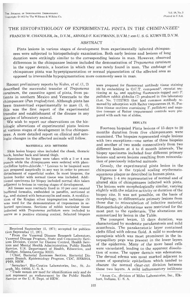
FIG. 1. Photom icrograph of normal chim panzee skin taken from the right latera l th igh. Hematoxylin and eosin, X 400.
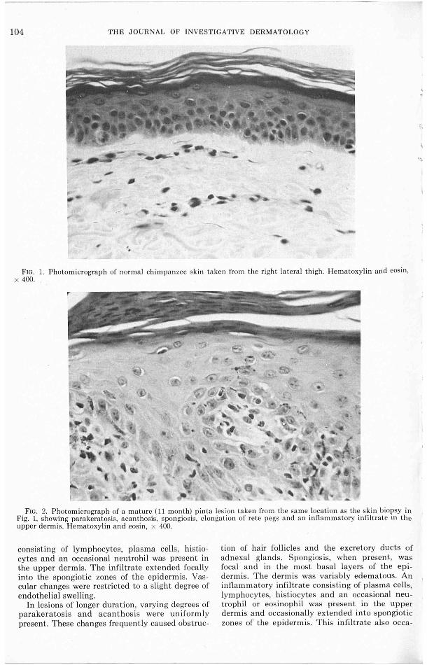
FIG. 2. Photomicrograph of a mature (11 mont h) pin ta les ion taken from the sa me locat ion as the skin biopsy in Fig. 1, showing parakeratosis, acan thosis, spongiosis, elongation of rete pegs a nd a n in!la mmatory infil t rate in the upper dermis. H ematoxylin a nd eosin , x 400.
consisting of lymp hocytes, plasma cells, histiocytes and an occas ional neut rohil was present in the upper dermis. T he infil trate extended foca lly into the spon giotic zones of the epidermis. Vascu lar changes were restricted to a s li ght degree of endothelia l swell ing.
In les ions of longe r duration, varying degrees of parakeratosis and aca nt hosis were uniform ly present. These changes freq uent ly caused obstruc -
tion of hair follicles and the excretory ducts of adnexa l glands. Spongios is, when present, was focal and in the most basa l layers of t he epidermis. T he derm is was variab ly edematous. An inflamm atory infiltrate consisting of plasma cells, lymphocytes, histiocytes and an occasional neutrophil or eos inophil was present in the upper dermis and occasiona lly extended in to spongiotic zones of t he epidermis. This infil trate a lso occa-
PINTA IN THE CHIMPANZEE 105
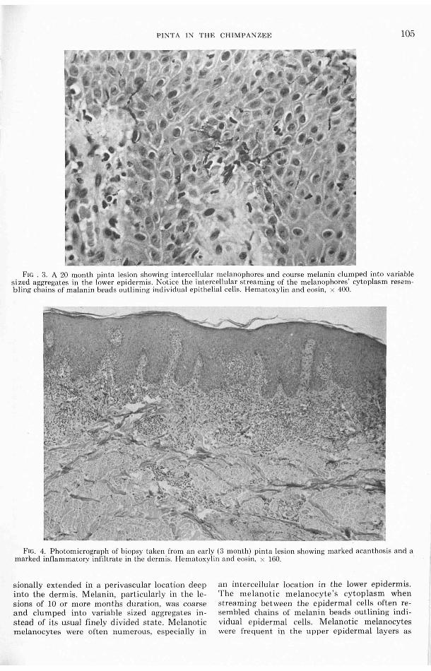
FtG . 3. A 20 month pinta lesion showi ng intercellular melanophores and course melanin clumped in to variab le sized aggregates in the lower epidermis. Notice the intercellular stream ing of the melanophores ' cytoplasm resembling chains of ma lanin beads out lining indi vidua l ep ithelial ce lls. Hematoxylin a nd eosin, x 400.
Ftc. 4. Photomicrograph of biopsy taken from an early (3 month) pin ta les ion showing marked acanthosis a nd a marked infla mmatory infi ltrate in the dermis. Hematoxylin a nd eos in, x 160. ·
siona lly extended in a perivascular location dee p into the dermis. Melanin, particularly in the lesions of 10 or more mont hs duration, was coarse and clumped into variable sized aggregates in stead of its usual finely divided state. Melanotic melanocytes were often numerous, es pec ia lly in
an in tercellula r location in the lower epidermis. The m e la not ic m elanocyte's cytop lasm when streaming between the epidermal cells often resembled cha ins of melanin beads outlining indi vidual epidermal cells . Melanotic melanocytes were fre quent in t he upper epiderma l layers as
106 THE JOURNAL OF INV ESTIGATIVE DERMATOLOGY
TABLE
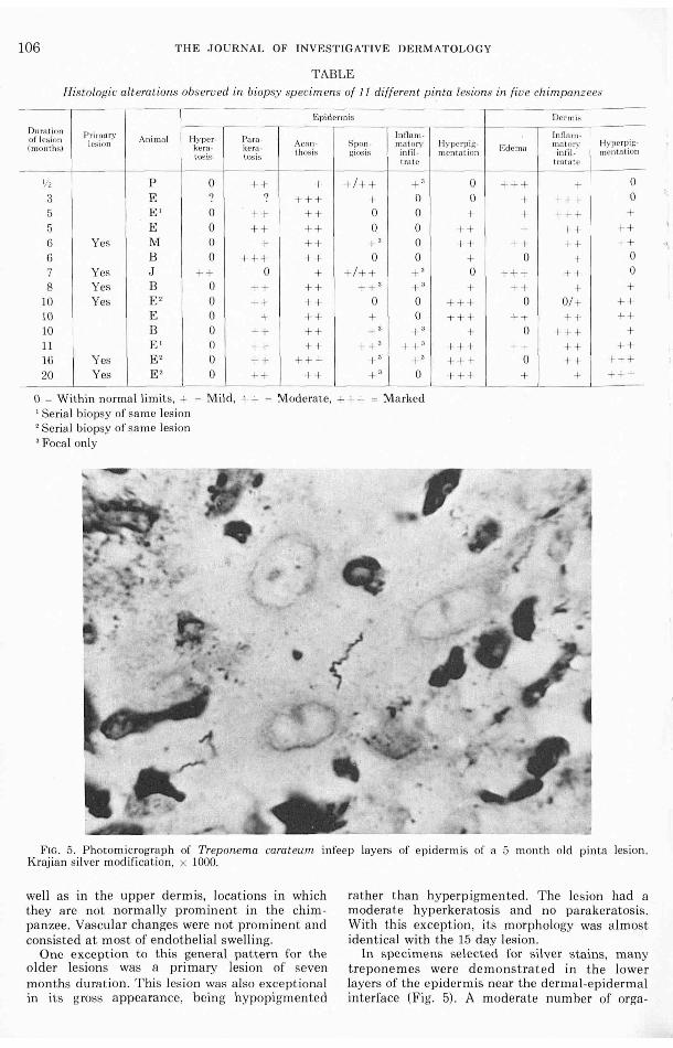
Histologic alterations observed in biopsy sp ecimens of 1 I different pinta lesions in fiv e chimpanzees
Epid ermis Dcrrni :-;
Duration PrimtlrY lnfln m- lnfla m-of lesion lc:; ion Animal Hyper- Para - Acn n- Spc11~ - mu tory Hy·pc rpig- mrttor;.• Hy pe n~ i g-
( months) kera - kera - Edema tos is tosis thosis j.! IOSIS infil - mental ion in fit - menwt 1011
I ra te t rat a te
'!' p 0 + + +I++ + ' 0 +++ + 0
3 E ? ? +++ + 0 0 + +++ 0
5 E' 0 ++ ++ 0 0 + + +++ 5 E 0 ++ ++ 0 0 ++ + ++ ++ 6 Yes M 0 + + + + ' 0 ++ ++ ++ ++ 6 B 0 + ++ ++ 0 0 + 0 + 0 7 Yes J ++ 0 + +I+ + + ' 0 +++ + + 0
8 Yes B 0 + + + ++ ' + " + ++ + + 10 Yes E' 0 ++ ++ 0 0 +++ 0 0/ -t- ++ 10 E 0 + + + + 0 +++ ++ ++ ++ 10 B 0 ++ ++ +' + ' + 0 +++ + 11 E' 0 ++ ++ ++ ' ++ ' +++ ++ ++ ++ 16 Yes E ' 0 + + + ++ + ' + ' +++ 0 ++ +++ 20 Yes E' 0 ++ ++ + ' 0 +++ + + +++
0 = Within norma l limits, + = Mild, ++ = Moderate, +++ = Marked ' Seria l biopsy of same les ion ' Serial bi opsy of sa me lesion 3 Focal only
FIG. 5. Photomicrograph of Treponema carateum infeep layers of epidermis of a 5 month old pinta lesion. Krajian silver modification , x 1000.
well as in the upper dermis, locations in which they are not n ormally promin ent in the chirppanzee. Vascular changes were not promine nt and consisted at most of endothelial swell ing.
One exception to this genera l pattern for the o ld er les ions was a primary lesion of seven months duration. This lesion was a lso exceptiona l in its gross a ppeara nce, being hypopigm ented
rather than h yperpigmented. The lesion had a moderate hyperkeratosis a nd no parakeratosis. With this exception , its morphology was a lmost identical with the 15 day lesion .
In spe cime ns selected for s ilver s tains, many treponemes were dem onstrated in the lower layers of the epidermis near the derma l-ep id erma l interface (Fig. 5). A moderate number of orga-
PINTA IN THE CHIMPANZEE 107
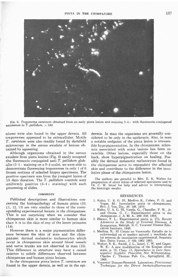
FIG. 6. Treponema caroteum obtained from an early pinta lesion and staining 3- 4+ with fluorescein-conjugated antiserum to T. pallidum , x 540.
nisms were also found in the upper dermis. All t reponemes appeared to be extracellula r. Motil e T . carateum were a lso readily found by clarkfield microscopy in the serous exudate of lesions obtained by squeezing.
Although organisms obta in ed in t he serous exudate from pinta lesions (Fig. 6) eas ily accepted the fluorescein conjugated anti- T . pallidum globulin (3- 4+ staining on a 0- 4 sca le), we were able to demonstrate f1uorescing treponemes in only 1 of 6 frozen sections of se lected biopsy specimens. The positive s pecimen was from the youngest lesion of 15 days duration . The T. pallidum controls were uniformly pos it ive (3-4 + sta ining) with each processing of slides.
COMMENTS
Published descriptions and illustrations concerning the his topathology of huma n pinta (10, 11, 12, 13) a re very similar to t hose of the corres ponding experimenta l les ions in t he chimpanzee. T his is not surpris ing when we consider that chimpanzee skin is more similar to human skin t han it is to t he skin of a ny of the lesser primates ( 14) .
However there is a major pigmentation differ ence between· t he skin of man and t he chim panzee: d erma l melanotic melanocytes which occur in chimpanzee skin around blood vessels and nerve trunks are not observed in man (15). T his difference in structure may have a causa l relation to subt le differences observed between chimpan zee a nd human pinta les ions.
In the chimpanzee pinta les ion T . carateum are found in the upper dermis, as well as in the epi-
dermis. In ma n t he organ isms are generally considered to be only in the epidermis, Also, in man a notable endpoint of the pinta lesion is irreversible hypo pigmentation. In the chim panzee, achromia · assoc iated with some lesions has been reversible. Other lesions, es pec ia lly t hose on t he back, show hyperpigmen tation on healing. Poss ibly the dermal melanotic melanocytes found in t he chimpanzee serve to repopulate the affected skin and contribute to t he difference in t he involu t ive phase of t he chimpanzee lesion .
The authors are grateful to Mrs. E. K. Walter for preparat ion of silver stains of selected specimens and to Dr. C. W. Israel for help and adv ice in interpret ing the histologic results.
REFEREN CES 1. Kuhn, U. S. G. Ill , Medina R. , Cohen, P. G. and
Vegas, M.: Inoculation pin ta in chimpanzees. Br it . J. Ven. Dis. , 46: 198, 1970.
2. Kuhn, U. S. G. III, Varela, G., Chandler, F. W., Jr. and Osuna, G. C.: Experimenta l pinta in the chim panzee. J. A. M. A., 206: 839, 1968.
3. Blanco, F. L.: Experimental pinta, p. 275, Recent Advances in the Study of Venereal Diseases, A Symposium. Raleigh, N. C. Venereal Disease Education Institute, 1948.
4. Med ina, R.: El Carate en Venezuela: Estudio de ~s · enfermedad en el med ico natural y resultado de los ensayos de inoculacion experimenta l. Separata Rev. Derm Venez., 3: 160, 1962- 1963.
5. Walter, E. K., Smith, J. L. , Israel, C. W. and Gager, W. E.: A new modification of the Krajian silver stain , p. 70, Chap. 13, Spirochetes in Late Seronegative Sy philis, Penicillin N ot Withstanding. Charles C. Thomas Pub. Co., Springfield, Ill. , 1969.
6. Venerea l Disease Research Laboratory: Provisional T echnique for the Direc t Immunoflu orescent

108 THE JOURNAL OF INVESTIGATIVE DERMATOLOGY
Identifi cation of Treponema pallidum in Body Fluids and Tissue Sections in Current Use at the Venereal Disease Research Laboratory. Venereal Disease Research Laboratory, Center for Disease Control, Atlanta, Georgia 30333, June 15, 1967.
7. Venereal Disease Research Laboratory: Properties and Instructions for Use of Lyophilized Fluorescein-Conjugated Anti-Treponema pallidum Globulin (Rabbit Origin). Venereal Disease Research Laboratory, Center for Disease Control, Atlanta, Georgia 30333, June 15, 1967.
8. Deacon, W. E. and Hunter, E. F.: Treponema! antigens as related to identification and syphilis serology. Proc. Soc. Exp. Bioi., 110: 352, 1962.
9. Meyer, P . E. and Hunter, E. F.: Antige nic relat ion ships of 14 treponemes demonstrated by immunoflu orescence. J. Bact. , 93: 783, 1967.
10. Blanco, F. L.: Histologia patologica de las lesiones cutaneas y de los glanglios tinfacticos en el mal del pinto. Rev. Kuba Med . Trop. , 5: 329, 1940.
11. Blanco, F. L. and De Laosa, 0.: The primary lesion of pinta. Amer. J. Syph., 31: 600, 1947.
12. Hasse lma nn, C. M.: Comparat ive studies on the histopathology of syphi lis, yaws and pinta . Brit. J . Vener. Dis., 33: 5, 1957.
13. Pardo-Castello, V. and Ferrer, I.: Pinta: ma l del pinto: carate.: Arch. Derm. Syph. , 45: 843, 1942.
14. Ford, D. M. and Perk ins, E . M.: The sk in of the chimpanzee, pp. 82-119, The Chimpanzee, Vol. 3. Ed., Bourne G. H., Karger Publishing Co., Basei/Muchen/New York, 1970.
15. Schu ltz, A. H.: Va ria bility in man and other primates. Amer. J . Phys. Anthrop., 5: 1, 1947.


