dr. andrás palkó - u-szeged.hu€¦ · dr. andrás palkó. department of radiology, university of...
TRANSCRIPT

Department of Radiology, University of Szeged
Imaging of the suprarenal glands
Dr. András Palkó

Department of Radiology, University of Szeged
Methods of examination:
• plain x-ray (radiography, fluoroscopy, tomography)
• x-ray with contrast material
• ultrasound (b-mode, Doppler, color, duplex)
• computed tomography
• magnetic resonance imaging
• x-ray-, US-, CT-guided intervention
• isotope

Department of Radiology, University of Szeged
Space occupying lesion I. (benign)
• hyperplasia (F/N)
• pseudotumor (N)
• bleeding (N): acute - > 40 HU, old - fluid-fluid level
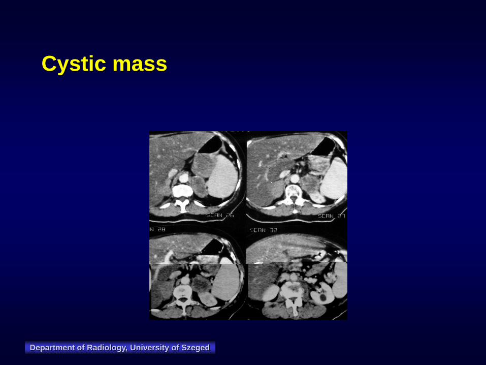
• cyst, pseudocyst (N): 15 % calcified
• adenoma (F/N): 1-5 cm, <0 / 10 / <10 HU,
moderate enhancement, rarely calcified
• myelolipoma (N): fat density, moderate enhancement,
20 % calcified
F/N: function y/n

Department of Radiology, University of Szeged
Space occupying lesion II. (malignant):
• Medullary:
– pheochromocytoma
(paraganglioma) (F):
enhancement,
calcification, avoid
biopsy (?!)
– multiple endocrine
neoplasia (MEN) (F)
– neuroblastoma (N): 90
% calcified, > 5 cm
• Cortical:
– Adrenocortical
carcinoma (F/N): > 5
cm, heterogeneous
enhancement,
necrosis, bleeding, 50
% calcified
– metastasis (N):
heterogeneous
enhancement, bilateral,
irregular
– lymphoma (N)
F/N: function y/n
Department of Radiology, University of Szeged
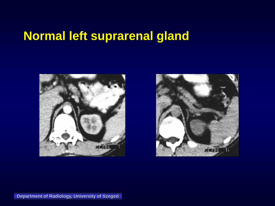
Normal left suprarenal gland

Department of Radiology, University of Szeged
Normal right suprarenal gland

Department of Radiology, University of Szeged
Adrenal hemorrhage
Department of Radiology, University of Szeged
Cystic mass

Department of Radiology, University of Szeged
Hyperplasia

Department of Radiology, University of Szeged
Hypoperfusion shock

Department of Radiology, University of Szeged
Adenoma

Department of Radiology, University of Szeged
Adenoma

Department of Radiology, University of Szeged
Adenocarcinoma

Department of Radiology, University of Szeged
Pheochromocytoma

Department of Radiology, University of Szeged
Phaeochromocytoma

Department of Radiology, University of Szeged
Phaeochromocytoma

Department of Radiology, University of Szeged
Adrenocortical carcinoma

Department of Radiology, University of Szeged
Neuroblastoma

Department of Radiology, University of Szeged
Metastasis

Department of Radiology, University of Szeged
Biopsy
Department of Radiology, University of Szeged
Imaging of other retroperitoneal
organs

Department of Radiology, University of Szeged
Department of Radiology, University of Szeged

Department of Radiology, University of Szeged

Department of Radiology, University of Szeged
Retroperitoneal lymphadenomegaly:
• malignant lymphoma
• lymphogenic metastasis
• inflammation, infection
• sarcoidosis
• amyloidosis
• Castleman disease
• Whipple disease
• HIV adenopathy
Department of Radiology, University of Szeged
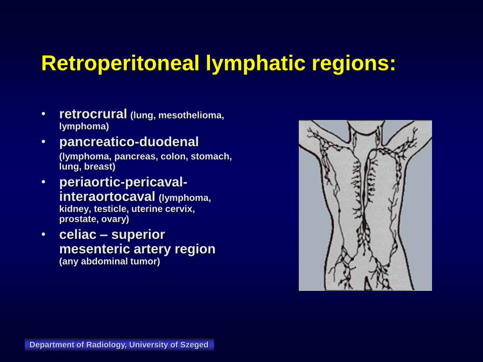
Retroperitoneal lymphatic regions:
• retrocrural (lung, mesothelioma, lymphoma)
• pancreatico-duodenal (lymphoma, pancreas, colon, stomach,
lung, breast)
• periaortic-pericaval-interaortocaval (lymphoma, kidney, testicle, uterine cervix, prostate, ovary)
• celiac – superior mesenteric artery region (any abdominal tumor)

Department of Radiology, University of Szeged

Department of Radiology, University of Szeged
Threshold value of largest axial lymph
node diameter
• retrocrural: > 6 mm
• pancreatico-duodenal: > 10 mm
• periaortic-pericaval-interaortocaval: > 10 mm
• celiac – superiopr mesenteric artery region: > 9 mm

Department of Radiology, University of Szeged
Limitation of detectability:
• US may be impeded by bowel gas and large body diameter
• CT & MR may be hampered by the lack of retroperitoneal fat
• US/CT/MR are not able to recognize pathologic changes in a normal
size lymph node
• most of lymphatic regions are unavailable for lymphography

Department of Radiology, University of Szeged
Limitations of characterisation:
• lymph nodes of normal size may harbor pathologic foci
• lymph node enlargement may be of benign or malignant
origin
• it may be difficult to differentiate enlarged lymph nodes
from cross-section of vessels/bowels by US/CT/MR
• filling defect detected by lymphography may be of
benign nature (fibrosis, fat)
Department of Radiology, University of Szeged
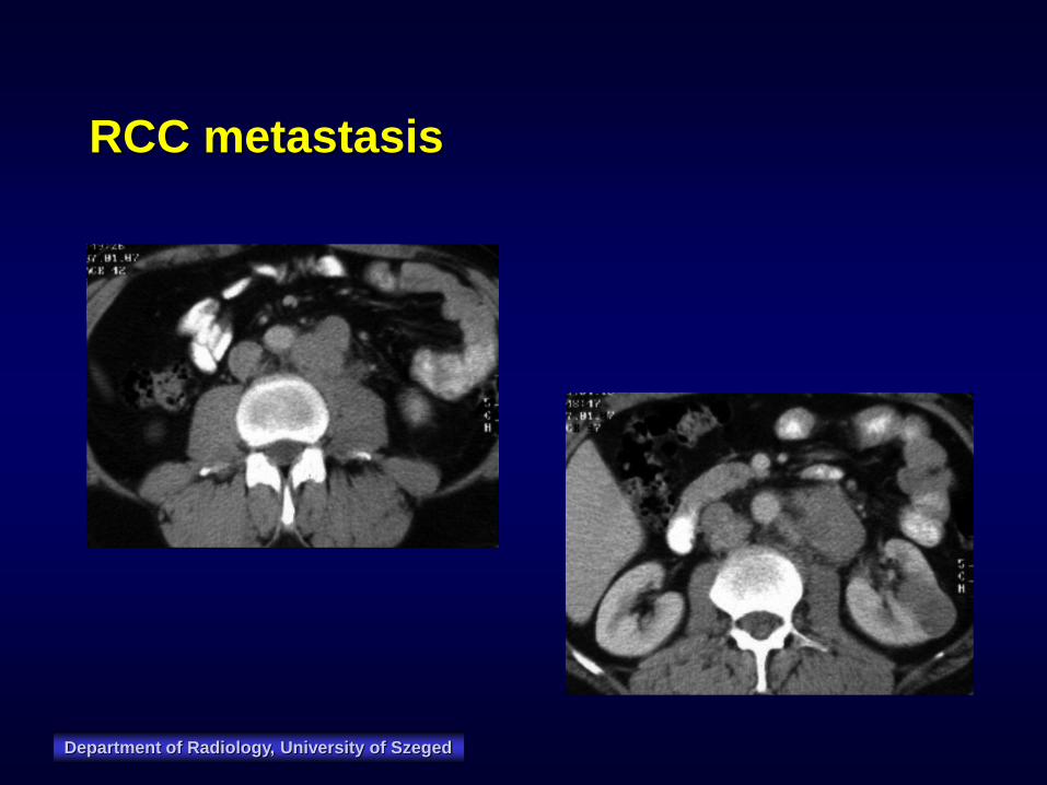
RCC metastasis

Department of Radiology, University of Szeged
NHL

Department of Radiology, University of Szeged
Diseases of the aorta
• atherosclerosis
• aneurysm (sclerosis, infection, injury)
– thrombosis
– dissection
– bleeding (chronic)
– bleeding (rupture)
• postoperative condition

Department of Radiology, University of Szeged
AAA rupture
3
4
Department of Radiology, University of
Szeged, Hungary

Department of Radiology, University of Szeged
AAA contained rupture

Department of Radiology, University of Szeged
AA dissection
3
6

Department of Radiology, University of Szeged
AA dissection
3
7
Department of Radiology, University of Szeged
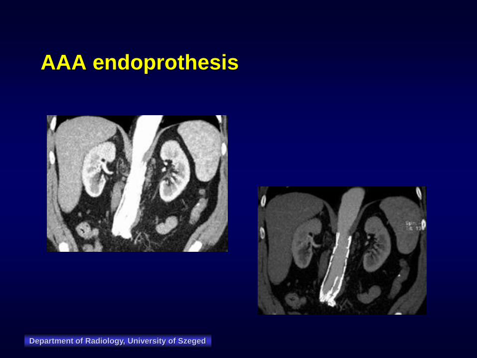
AAA endoprothesis

Department of Radiology, University of Szeged
Endoleak

Department of Radiology, University of Szeged
Diseases of the IVC
• congenital anomalies
• thrombosis
• tumor thrombus
• external compression
• cava filter

Department of Radiology, University of Szeged
VCI tumor thrombus

Department of Radiology, University of Szeged
Retroperitoneal tumors:
Malignant
• fibrosarcoma
• malignant teratoma
• liposarcoma
• leiomyosarcoma
• rhabdomyosarcoma
• fibrous histiocytoma
• tumor of the kidney,
suprarenal gland,
pancreas
Benign
• haemangiopericytoma
• lymphangioma
• neurinoma
• teratoma
• Schwannoma
• fibrosus histiocytoma
• neurilemmoma
• lipoma

Department of Radiology, University of Szeged
Liposarcoma

Department of Radiology, University of Szeged
Rhabdomyosarcoma

Department of Radiology, University of Szeged

Department of Radiology, University of Szeged
Other retroperitoneal changes
• retroperitoneal fibrosis
• gas (perforation, emph. pyelonephr.)
• pseudocyst
• urinoma
• echinococcus cyst
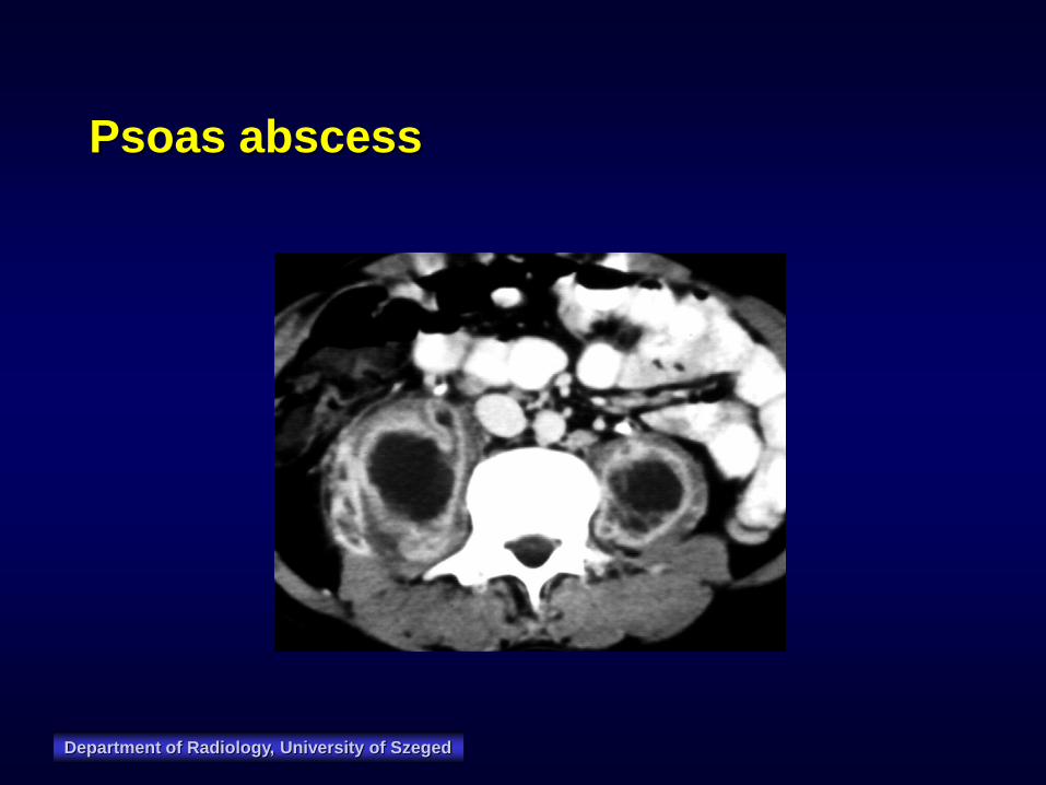
• abscess (psoas!)
• haematoma
• lymphocele
• meningocele
• retrocaval ureter

Department of Radiology, University of Szeged
Haematoma

Department of Radiology, University of Szeged
Retroperitoneal fibrosis

Department of Radiology, University of Szeged
Abscedating pyelonephritis
Department of Radiology, University of Szeged
Psoas abscess

Department of Radiology, University of Szeged
Retroperitoneal abscess

Department of Radiology, University of Szeged
Rectum perforation

Department of Radiology, University of Szeged
Celiac ganglion blockade