dr. harvey richman, od, faao, fcovd diplomate...
TRANSCRIPT

Dr. Harvey Richman, OD, FAAO, FCOVD
Diplomate American Board of Optometry
Executive Committee AOA Third Party Center
Founder Ask the AOA Coding Experts

92000 Codes Special Ophthalmological Services
Describe Services in which a special Evaluation
of part of the visual system is made, which goes
beyond the services, or in which special treatment
is given.
Special ophthalmological services may be reported
in addition to the general ophthalmological services
or evaluation and management services
2

Disclaimers 1. Medicare policy changes frequently so links to
the source documents have been provided for your reference.
2. This presentation is prepared as a tool to assist providers and is not intended to grant rights or impose obligations.
3. Every reasonable effort has been made to assure the accuracy of the information.
4. Ultimate responsibility for the correct submission of claims and response to any remittance advice lies with the provider of services.

Disclaimers 6. This presentation is general summary that explains certain
aspects of the Medicare program, but is not a legal document. The official Medicare program provisions are contained in the relevant laws, regulations, and rulings.
7. The Medicare Learning Network (MLN) is the brand name for official CMS educational products and information for Medicare fee-for-service providers. For additional information visit the Medicare Learning Network’s web page at www.cms.hhs.gov/MLNGenInfo on the CMS website.
8. Current Procedural Terminology (CPT) is copyright by the American Medical Association. All Rights Reserved. No fee schedules, basic units, relative values, or related listings are included in CPT. The AMA assumes no liability for the data contained herein. Applicable FARS/DFARS restrictions apply to government use.
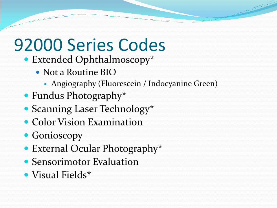
92000 Series Codes Extended Ophthalmoscopy*
Not a Routine BIO Angiography (Fluorescein / Indocyanine Green)
Fundus Photography*
Scanning Laser Technology*
Color Vision Examination
Gonioscopy
External Ocular Photography*
Sensorimotor Evaluation
Visual Fields*

Documentation Requirements: Reason for performing the examination
Technique used
Image or Drawing of the ocular site showing anatomy seen including the pathology
Legible narrative report of the findings
Documentation supporting medical necessity must be submitted

Effect of Lenses
Without Lenses
With Lenses

Refraction-92015 Determination of refractive state
Statutorily not covered by Medicare
RVU $38.09
Consider Modifiers

Refraction 92015 By CMS Statute a Non-Covered Service
Patient Responsibility
ABN Not Required but Useful
GY Modifier
Multilevel Refraction Codes 92015?
Phoropter
Trial Frame
Telescope

Modifiers 21-Prolonged E&M Services (NO LONGER EXISTS)
When the face to face service is prolonged or otherwise greater than that usually required for the highest E & M service within a given category. A report may be appropriate.
22- Increased Procedural Services When the work required to provide a service is
substantially greater than what is typically required. Documentation must support the substantial additional work and the reason for the additional work. (Time, difficulty of procedure, severity of patient condition)
Not to be used with E & M

Gonioscopy 92020
Gonioscopic exam to diagnose injury or disease in the anterior chamber of the eye, performed under local anesthetic due to necessity of placing specialized lens directly on the eye to obtain a clear image
Bilateral Procedure Code
LCD Utilization

Topography 92025
Computerized corneal topography, unilateral or bilateral with interpretation and report
Detection of subtle corneal surface irregularity and astigmatism
Report one time only
12

Indications & Limitations of Coverage:
Post penetrating keratoplasty Post kerato-refractive complications Post op irregular astigmatism Corneal dystrophy, bullous keratopathy Complications of transplanted cornea, Keratoconus

Sensorimotor Exam 92060
Sensorimotor examination (i.e. of the movement of the eye), conducted by taking measurements as the eyes focus on different locations or through one or more prisms. Searches for deviations in normal eye movements, which may result from injury or disease. Includes interpretation and report.

New Codes!!!! 92071 Fitting of a contact lens for treatment of ocular surface
disease
Report materials in addition to this code, using either 99070 or the appropriate HCPCS Level II material code.
This is the appropriate code to use for fitting a bandage contact lens.

New Codes!!!! 92072 Fitting of a contact lens for management of
keratoconus, initial fitting.
For subsequent fittings, please use either the 9921X or 9201X codes.
Report materials in addition to this code, using either 99070 or the appropriate HCPCS Level II material code.
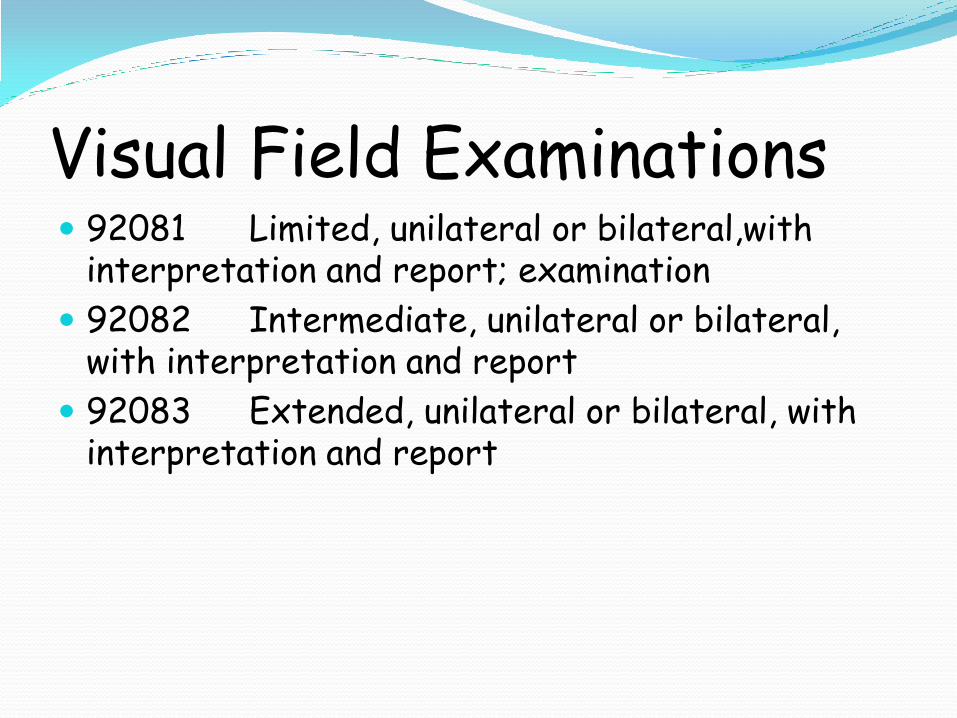
Visual Field Examinations 92081 Limited, unilateral or bilateral,with
interpretation and report; examination
92082 Intermediate, unilateral or bilateral, with interpretation and report
92083 Extended, unilateral or bilateral, with interpretation and report

Indications & Limitations of Coverage
Necessary to establish a diagnosis
Monitor a course for treatment
Determine if a change in therapeutic plan
Indications & Limitations of Coverage
92081-92082 medically necessary to diagnose and follow retinal disorders
92083 diagnosis or follow-up of glaucoma

Coding Guidelines All services are considered bilateral
-50 modifier is not appropriate
-52 modifier if only doing one eye

Blepharoplasty Guidelines Visual field examinations to determine the need
for blepharoplasty are sometimes performed twice, once with the eye(s) taped and immediately repeated without the eye(s) taped. In this situation, the repeated service should be submitted with CPT modifier 76 on a separate detail line.
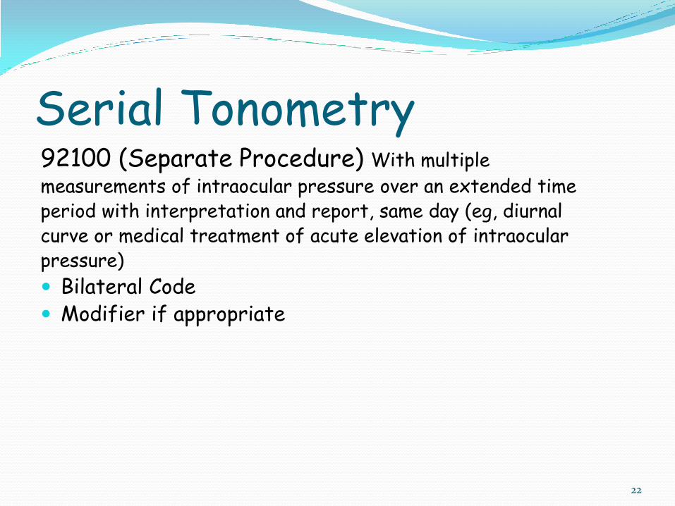
Serial Tonometry 92100 (Separate Procedure) With multiple
measurements of intraocular pressure over an extended time period with interpretation and report, same day (eg, diurnal curve or medical treatment of acute elevation of intraocular pressure)
Bilateral Code Modifier if appropriate
22

Scanning Laser Tests Confocal laser scanning ophthalmoscopy
(topography)
Optical Coherence tomography

Coding guidelines 92132-3-4 Scanning computerized ophthalmic
diagnostic imaging (e.g., scanning laser) with interpretation and report, unilateral
Using either a 52- LT or RT modifier if reduced
CPT codes not covered with SLT:
92225, 92226, 76512, 92250
59 modifier usage
GA modifier usage with ABN

92132-SCANNING COMPUTERIZED OPHTHALMIC DIAGNOSTIC IMAGING,
ANTERIOR SEGMENT, WITH INTERPRETATION AND REPORT, UNILATERAL OR BILATERAL
Narrow angle, suspected narrow angle, and mixed narrow and open angle glaucoma
Determining the proper intraocular lens for a patient who has had prior refractive surgery and now requires cataract extraction
Iris tumor Presence of corneal edema or opacity that precludes
visualization or study of the anterior chamber Calculation of lens power for cataract patients who have
undergone prior refractive surgery. Payment will only be made for the cataract codes as long as additional documentation is available in the patient record of their prior refractive procedure. Payment will not be made in addition to A-scan or IOL master.
Certain exceptions that must be determined on a case-by-case basis with the appropriate documentation.
92133Glaucoma Indications
SCANNING COMPUTERIZED OPHTHALMIC DIAGNOSTIC IMAGING, POSTERIOR SEGMENT, WITH INTERPRETATION AND REPORT, UNILATERAL OR BILATERAL; OPTIC NERVE
Technological improvements have rendered SCODI as a valuable diagnostic tool in the diagnosis and treatment of glaucoma. These improvements enable discernment of changes of the nerve fiber even in advanced cases of glaucoma.
It is expected that only two exams/eye/year would be required to manage the patient who has glaucoma or is suspected of having glaucoma.

MILD visual field abnormality (inner circle = 10 degrees, outer circle = 20 degrees) ICD 9 365.71

MODERATE visual field abnormality (inner circle = 10 degrees, outer circle = 20 degrees) ICD 9 365.72

SEVERE visual field abnormality (inner circle = 10 degrees, outer circle = 20 degrees) ICD9 365.73

Glaucoma Severity Level Scanning Laser Frequency
The current frequency limitations for Scanning Laser for most regions are:
Mild or Suspect Glaucoma 1 Time per year
Moderate Glaucoma 2 Times per year
Advanced or Severe Glaucoma NO Scanning laser. Up to 4 Visual Fields per year

Utilization Guidelines-GLC Although CMS guidelines state
Only two exams/eye/year are allowed for the patient who has or is suspected of having glaucoma
Most LCD state once per year to follow pre-glaucoma patients or those with “mild” damage
One or two tests per year for patients with “moderate damage,” followed with SLT or visual fields if both SLT and visual fields are used, only one of each
tests
“Advanced damage,” field testing preferred by Medicare guidance

92134-SCANNING COMPUTERIZED OPHTHALMIC DIAGNOSTIC IMAGING, POSTERIOR
SEGMENT, WITH INTERPRETATION AND REPORT, UNILATERAL OR BILATERAL; RETINA
Retinal disorders are the most common causes of severe and permanent vision loss. These technologies are valuable tools for the evaluation and treatment of patients with retinal disease, especially macular abnormalities.
These imaging techniques are useful tools to measure the effectiveness of therapy, and in determining the need for ongoing therapy, or the safety of cessation of therapy.
It is expected that only one exam/eye/2 months would be required to manage the patient whose primary ophthalmological condition is related to a retinal disease. However, for those patients who are undergoing active treatment for macular degeneration or diabetic retinopathy one exam/eye/month may be appropriate for the management of their disease.
The use of fluorescein angiography, indocyanine green angiography and SCODI to study the patient’s same eye per clinical encounter will NOT be authorized. However, SCODI and fluorescein angiography may be obtained on the patient’s same eye per clinical encounter if the medical record substantiates the need for both studies.

Utilization Guidelines-AMD/DR Only one exam/eye/2 months is allowed for the patient
whose primary ophthalmological diagnosis is related to a retinal disease
One exam/eye/month is allowed for the patient who is undergoing active treatment for macular degeneration or diabetic retinopathy

Extended Ophthalmoloscopy
92225 - Ophthalmoscopy, extended, retinal drawing with interpretation & report; initial
92226 - ... Subsequent

Extended Ophthalmoscopy
Reserved for the meticulous evaluation of a severe ophthalmologic problem
Always include indirect ophthalmoscopy & one other method viewing detail
Retinal drawing with detail a must.

Coding Guidelines: unilateral procedure
Do not report codes with modifier –50
Service on both eyes, use LT or RT (uncommon)

Documentation Guidelines??? Drawing has to be a certain size (no)
Observation with two or more lenses (maybe)
Scleral Indentation must be done
Colored Drawings with colored pencils (match international recommendations).
Must have interpretation and report as well as orders!

92250:Fundus Photography Fundus photography with interpretation and report
Bilateral Code
38

Photography Document abnormalities
Check carrier’s medical policy for limitations or restrictions of coverage
Obtain filing requirements from carrier for bilateral or multiple procedures
39

92250 Utilization Guidelines Fundus photography. Generally, it is not medically
necessary to repeat fundus photography more often than every 2 years for follow-up of stable glaucoma. Repeat photographs for retinopathy are rarely necessary.

• Color vision examination, extended, eg, anomaloscope or
equivalent
(Color vision testing with pseudoisochromatic plates [such as HRR
or Ishihara] is not reported separately. It is included in the
appropriate general or ophthalmological service, or 99172)
92283 Color Vision

92286
Endothelial Microscopy
• Special anterior segment
photography with
interpretation and report;
with specular endothelial
microscopy and cell count
• Unilateral Code

Anterior Segment Photography
92285
External ocular photography with interpretation and report for documentation of medical progress (eg, close-up photography, slit lamp photography, gonio-photography, stereo-photography)
Medicare Fees National Non-Facility Fee $43.58

92310 Prescription of optical and physical characteristics of
and fitting of contact lens, with medical supervision of adaptation; corneal lens, both eyes, except for aphakia:
Fitting of one eye, append -52 modifier Non Covered Service for Medicare Non-Facility Fee $91.81
HCPCS: V codes
44
92311-92313
Prescription of optical and physical characteristics of and fitting of contact lens, with medical supervision of adaptation; corneal lens for aphakia, …..
99311-one eye 99312-two eyes 99313-corneoscleral lens
45
92314 Prescription of optical and physical characteristics of
and fitting of contact lens, with medical supervision of adaptation and direction of fitting by independent technician; corneal lens, both eyes, except for aphakia:
Fitting of one eye, append -52 modifier Non Covered Service for Medicare Non-Facility Fee $72.64 HCPCS: Vcodes
46

92315-92317
Prescription of optical and physical characteristics of and fitting of contact lens, with medical supervision of adaptation and direction of fitting by independent technician; corneal lens for aphakia, …..
92315-one eye 92316-two eyes 92317-corneoscleral lens
47

Contact Lens Evaluations 92325
Modification of contact lens (separate procedure), with medical supervision of adaptation
Lay description-Modification of contact lens, typically by grinder or polisher, to provide a better fit
Non-Facility Fee $29.13
Contact Lens Evaluations 92326
Replacement of contact lens under current prescription (due to damage, loss, etc.).
Non-Facility Fee $34.66

Thanks for your time, now……