draft - university of toronto t-space · and centrifuged (ca. 30 times) at 4500 rpm, until the...
TRANSCRIPT

Draft
Dye rejection membranes prepared from oxidized graphite
particles
Journal: Canadian Journal of Chemistry
Manuscript ID cjc-2016-0628.R1
Manuscript Type: Article
Date Submitted by the Author: 02-Feb-2017
Complete List of Authors: Colomba, Anastasia; The University of Western Ontario, Department of Chemistry Biesinger, Mark; The University of Western Ontario, Surface Science Western Divigalpitiya, Ranjith; 3M Canada Company Brandys, Frank; 3M Canada Company
Gilroy, Joe; The University of Western Ontario, Department of Chemistry
Keyword: Materials Characterization, Chemically Modified Graphite, Dye Rejection Membranes, Carbon-Based Materials, Wastewater Purification
https://mc06.manuscriptcentral.com/cjc-pubs
Canadian Journal of Chemistry
Draft
1
Dye rejection membranes prepared from oxidized graphite particles
Anastasia Colombaa, Mark C. Biesinger
b, Ranjith Divigalpitiya
c, Frank A. Brandys
c,
and Joe B. Gilroya*
aDepartment of Chemistry and the Centre for Advanced Materials and Biomaterials Research
(CAMBR), The University of Western Ontario, London, Ontario, Canada, N6A 5B7.
bSurface Science Western, The University of Western Ontario, London, Ontario, Canada,
N6A 5B7.
c3M Canada Company, 1840 Oxford Street East, London, Ontario, Canada, N5V 3R6.
*Corresponding author. E-mail: [email protected] (Joe B. Gilroy)
Page 1 of 23
https://mc06.manuscriptcentral.com/cjc-pubs
Canadian Journal of Chemistry
Draft
2
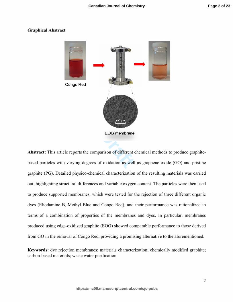
Graphical Abstract
Abstract: This article reports the comparison of different chemical methods to produce graphite-
based particles with varying degrees of oxidation as well as graphene oxide (GO) and pristine
graphite (PG). Detailed physico-chemical characterization of the resulting materials was carried
out, highlighting structural differences and variable oxygen content. The particles were then used
to produce supported membranes, which were tested for the rejection of three different organic
dyes (Rhodamine B, Methyl Blue and Congo Red), and their performance was rationalized in
terms of a combination of properties of the membranes and dyes. In particular, membranes
produced using edge-oxidized graphite (EOG) showed comparable performance to those derived
from GO in the removal of Congo Red, providing a promising alternative to the aforementioned.
Keywords: dye rejection membranes; materials characterization; chemically modified graphite;
carbon-based materials; waste water purification
Page 2 of 23
https://mc06.manuscriptcentral.com/cjc-pubs
Canadian Journal of Chemistry
Draft
3
Introduction
Water purification is one of the fastest growing markets: Boston Consulting Group reported a
demand of $47.7 billion for products related to waste water treatment in the top 40 national
markets in 2012, with an estimated compound annual growth rate of 10.2% for the period
between 2014 and 2019.1 Recent advances in nanotechnology offer opportunities to develop the
next-generation of water supply systems, with applications ranging from adsorption to
photocatalysis and membrane filtration systems.2 Since the discovery of graphene, there have
been numerous studies related to its applications across the physical sciences, health sciences,
and engineering. Graphene has been widely studied for its electrical properties, transparency and
flexibility for energy storage, sensors, electronic devices, polymer composites and many other
applications,3-9 but it is graphene oxide (GO) that has mainly been studied for filtration
applications.10-11 GO is hydrophilic, easy to disperse in water and other solvents and readily
undergoes chemical functionalization.12 It has been shown that GO or functionalized GO sheets
can be stacked to produce high-flux membranes that can effectively be used for pervaporation,13
nanofiltration,14 ultrafiltration,15 microfiltration,16 reverse osmosis17 and photocatalytic
degradation of contaminants.18
GO is often produced via the harsh oxidation of graphite through Hummers’ method,19
with the main drawbacks of introducing severe disruption to the structure of the graphene sheets
and the use of high concentrations of hazardous chemicals. More recently, different groups have
reported relatively mild conditions for the functionalization of graphite to produce edge-oxidized
graphite (EOG), that could then be intercalated or exfoliated into graphene nano-sheets.20-22
In this study, we aimed to produce particles derived from graphite with different degrees
of oxidation, ranging from mild oxidation to heavily oxidized GO, and compare their physico-
Page 3 of 23
https://mc06.manuscriptcentral.com/cjc-pubs
Canadian Journal of Chemistry
Draft
4
chemical properties. The particles were then used for the production of supported membranes on
a modified nylon substrate by means of a simple vacuum-assisted self-assembly technique.
Finally, the rejection of three different organic dyes (Rhodamine B, Methyl Blue, Congo Red)
from water in stirred cell experiments was investigated.
Experimental
Materials
TIMREX® HSAG300 (PG: 6 µm diameter, 300 m2/g surface area) was purchased from
TIMCAL and used as a starting material. NaNO3 (Anachemica), KMnO4 (Sigma Aldrich),
concentrated H2SO4, 70% HNO3, and H2O2 (Caledon) were used as reagents without further
purification. Methyl Blue, Congo Red (Sigma Aldrich) and Rhodamine B (Alfa Aesar) were
used as dyes for the rejection studies. Modified nylon membranes (EF004, average pore diameter
0.04 µm) were obtained from 3M Canada.
Sample preparation
Nitric acid oxidized (NAO) particles were prepared following a modified version of the
procedure reported by Dalai and co-workers.23 5.0 g of HSAG300 were added to 32 mL of 70%
HNO3. This mixture was refluxed at 35 °C for 2.5 h using a reflux condenser connected to a
recirculating chiller. A magnetic stirrer was used for mixing during the reflux. The sample was
washed with H2O to remove residual acid and centrifuged repeatedly (ca. 15 times) at 4500 rpm,
until the supernatant pH reached neutrality. The resulting product was dried overnight at 80 °C to
yield 4.8 g of black powder.
Edge-oxidized graphite was prepared according to the method previously reported by
Wu, Zou and co-workers.21 1.0 g of graphite was added to 23 mL of concentrated H2SO4 and 0.1
g NaNO3 in an ice bath, and stirred for 10 min. 0.8 g of KMnO4 were then slowly added to the
Page 4 of 23
https://mc06.manuscriptcentral.com/cjc-pubs
Canadian Journal of Chemistry
Draft
5
solution to maintain the temperature below 20 °C. The mixture was then brought to 35 °C and
stirred for an additional 30 min, followed by the slow addition of 45 mL H2O. After 15 min, 140
mL H2O and 7.5 mL of 30% H2O2 were added. The solution was then washed with H2O and
centrifuged repeatedly (ca. 15 times) at 4500 rpm, until the supernatant pH reached neutrality.
The resulting product was dried overnight at 80 °C to yield 1.1 g of dark grey powder.
Graphene oxide (GO) was produced according to a modified version of Hummers'
method.22 1.0 g of graphite was added to 100 mL concentrated H2SO4 and soaked for 12 h, after
which 0.74 g NaNO3 were added. The mixture was then cooled down in an ice bath and 3.4 g of
KMnO4 were added, followed by stirring for 2 h in an ice bath and room temperature for 5 days.
5 mL of H2O were then slowly added to the mixture, and after 5 min another addition of 4 mL
and 3 mL of 30% H2O2 was performed. 500 mL of H2O were then added to reduce the viscosity
of the mixture. The obtained solution (brown in colour) was then repeatedly washed with H2O
and centrifuged (ca. 30 times) at 4500 rpm, until the supernatant pH reached neutrality. The
resulting product was dried overnight at 80 °C to yield 0.9 g of black powder.
Powder characterization methods
Thermogravimetric analysis (TGA) profiles were obtained using a TA Instrument Q50 TGA and
Pt sample pans under a flow of N2. Prior to the run, the temperature was equilibrated at 25 °C for
1 min followed by an increase to 1000 °C at a rate of 10 °C/min.
Powder X-ray diffraction (PXRD) analyses were conducted from 5−120° (2θ) using an
Inel CPS Powder Diffractometer with Cu X-ray radiation, equipped with an Inel XRG 3000
generator and Inel CPS 120 detector on an aluminum sample holder.
Page 5 of 23
https://mc06.manuscriptcentral.com/cjc-pubs
Canadian Journal of Chemistry
Draft
6
Raman spectroscopy was carried out using a Renishaw Invia Raman microscope
equipped with a 20× magnification objective and a 633 nm excitation laser.
Energy-dispersive X-ray (EDX) spectroscopy measurements were carried out using a
Hitachi TM-3000 SEM equipped with a Bruker Quantax 70 EDX detector at a 30× magnification
and 15 kV.
X-ray photoelectron spectroscopy (XPS) was performed on a Kratos AXIS Nova
Spectrometer located at Surface Science Western (The University of Western Ontario). All
samples were mounted by pressing a thick layer of the sample onto double sided adhesive tape.
The Kratos charge neutralizer system was used on all specimens. Survey scan analyses were
carried out with an analysis area of 300 × 700 microns and a pass energy of 160 eV. High
resolution analyses were carried out with an analysis area of 300 × 700 microns and a pass
energy of 20 eV. Spectra have been charge corrected to the C=C line of the carbon 1s spectrum
(graphitic carbon) set to 284.5 eV. Spectra were analysed using CasaXPS software (version
2.3.14).
Membrane preparation and characterization
Membranes were prepared as follows: a 400 ppm solution of the different samples was prepared
and sonicated for 2 h. After this, 100 mL of the solution were deposited on a 14.6 cm2 disk of
porous substrate (modified nylon, average pore diameter 0.04 µm) by vacuum filtration. The
membranes were then air dried overnight in a fume hood.
The membranes thicknesses were measured with two different methods: a micrometer
(MITUTOYO IP 65 MICROMETER) and by SEM (NanoFabrication Facility, The University of
Page 6 of 23
https://mc06.manuscriptcentral.com/cjc-pubs
Canadian Journal of Chemistry

Draft
7
Western Ontario). The SEM measurements were carried out on a cross section of the membrane
using a LEO (Zeiss) 1540XB FIB/SEM at a 200x magnification and 5 kV EHT voltage level.
Dye rejection studies
Rejection experiments were carried out in a 316 stainless steel stirred cell (Steriltech HP4750) at
an operating pressure of 60 psi. Three solutions were prepared: Rhodamine B (50 mg/L), Methyl
Blue (10 mg/L) and Congo Red (50 mg/L). The experiments were carried out by running seven
consecutive filtration cycles and collecting 30 mL aliquots of each dye solution for each
membrane; their concentration was monitored using a Shimadzu UV-1800 UV-vis
spectrophotometer at wavelengths of 554, 594 and 498 nm, respectively. The data reported in
Fig. 10 were taken from the seventh filtration cycle, chosen as the % rejection values changed by
less than 5% compared to the sixth filtration cycle, to ensure that the adsorption sites on the
surface of the carbon layer were saturated. The data for each of the seven filtration cycles can be
found in Fig. S8−S10. The time required to collect the 30 mL sample was recorded and used to
calculate the flux normalized with respect to the surface and pressure as:
���
���=
��� (�)
�� �(��) ∙ ��� (ℎ) ∙ �� ���� (���)
Results and discussion
Powder characterization
Details of the preparation of oxidized graphitic particles are outlined in the experimental section.
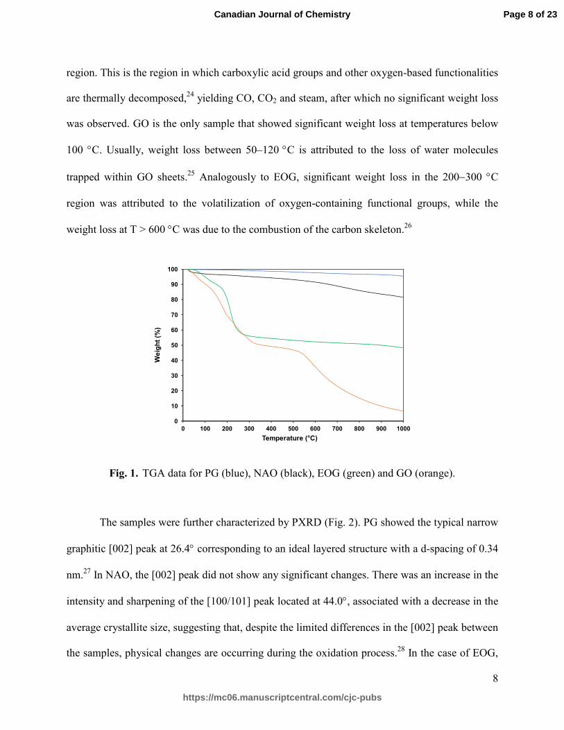
Fig. 1 shows thermogravimetric analysis (TGA) data for the four different powder samples.
Pristine graphite (PG), used as a control, showed very limited weight loss (about 5%) in the
temperature window studied (25→1000 °C), while NAO had a total weight loss of about 19%,
mostly at temperatures above 600 °C. EOG showed significant weight loss in the 200−300 °C
Page 7 of 23
https://mc06.manuscriptcentral.com/cjc-pubs
Canadian Journal of Chemistry
Draft
8
region. This is the region in which carboxylic acid groups and other oxygen-based functionalities
are thermally decomposed,24 yielding CO, CO2 and steam, after which no significant weight loss
was observed. GO is the only sample that showed significant weight loss at temperatures below
100 °C. Usually, weight loss between 50–120 °C is attributed to the loss of water molecules
trapped within GO sheets.25 Analogously to EOG, significant weight loss in the 200−300 °C
region was attributed to the volatilization of oxygen-containing functional groups, while the
weight loss at T > 600 °C was due to the combustion of the carbon skeleton.26
Fig. 1. TGA data for PG (blue), NAO (black), EOG (green) and GO (orange).
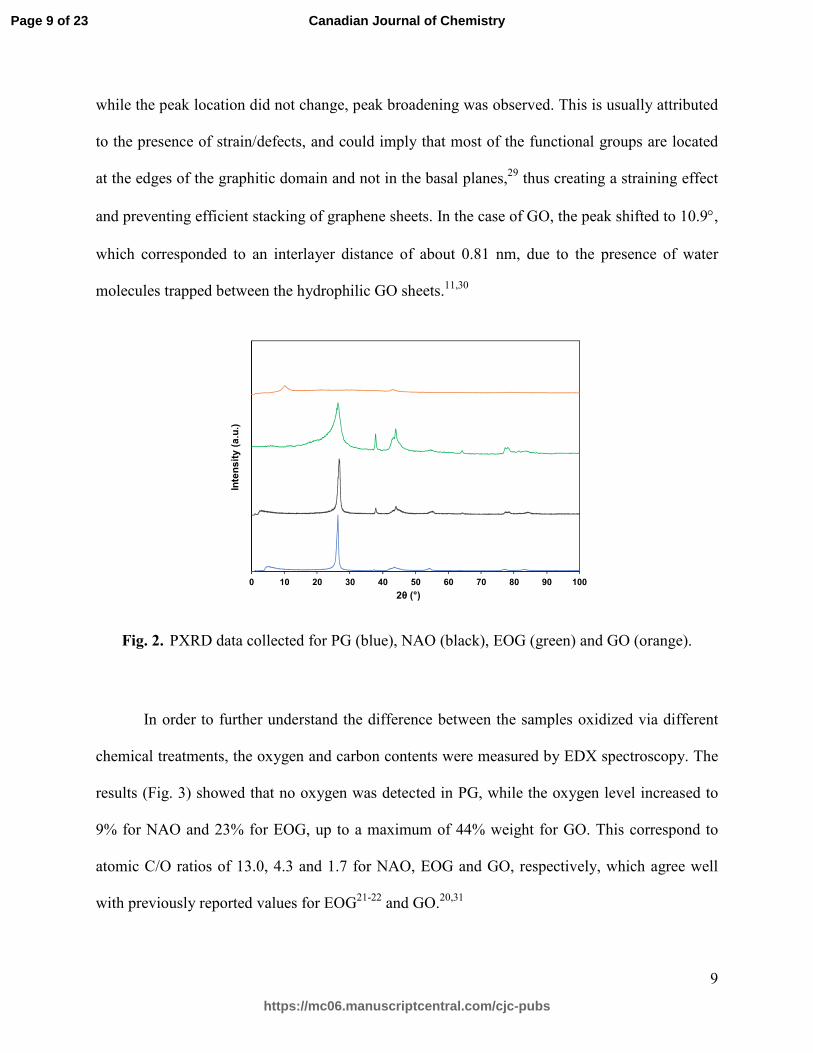
The samples were further characterized by PXRD (Fig. 2). PG showed the typical narrow
graphitic [002] peak at 26.4° corresponding to an ideal layered structure with a d-spacing of 0.34
nm.27 In NAO, the [002] peak did not show any significant changes. There was an increase in the
intensity and sharpening of the [100/101] peak located at 44.0°, associated with a decrease in the
average crystallite size, suggesting that, despite the limited differences in the [002] peak between
the samples, physical changes are occurring during the oxidation process.28 In the case of EOG,
0
10
20
30
40
50
60
70
80
90
100
0 100 200 300 400 500 600 700 800 900 1000
We
igh
t (%
)
Temperature (°C)
Page 8 of 23
https://mc06.manuscriptcentral.com/cjc-pubs
Canadian Journal of Chemistry
Draft
9
while the peak location did not change, peak broadening was observed. This is usually attributed
to the presence of strain/defects, and could imply that most of the functional groups are located
at the edges of the graphitic domain and not in the basal planes,29 thus creating a straining effect
and preventing efficient stacking of graphene sheets. In the case of GO, the peak shifted to 10.9°,
which corresponded to an interlayer distance of about 0.81 nm, due to the presence of water
molecules trapped between the hydrophilic GO sheets.11,30
Fig. 2. PXRD data collected for PG (blue), NAO (black), EOG (green) and GO (orange).
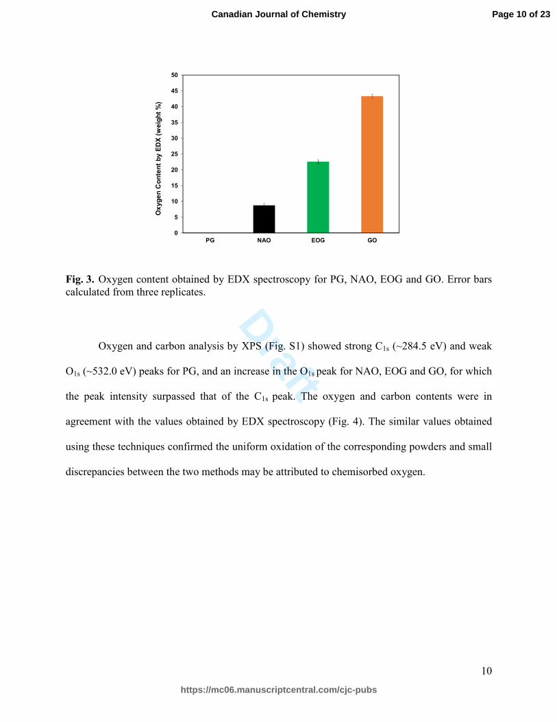
In order to further understand the difference between the samples oxidized via different
chemical treatments, the oxygen and carbon contents were measured by EDX spectroscopy. The
results (Fig. 3) showed that no oxygen was detected in PG, while the oxygen level increased to
9% for NAO and 23% for EOG, up to a maximum of 44% weight for GO. This correspond to
atomic C/O ratios of 13.0, 4.3 and 1.7 for NAO, EOG and GO, respectively, which agree well
with previously reported values for EOG21-22 and GO.20,31
0
0.5
1
1.5
2
2.5
3
3.5
4
0 10 20 30 40 50 60 70 80 90 100
Inte
ns
ity (
a.u
.)
2θ (°)
Page 9 of 23
https://mc06.manuscriptcentral.com/cjc-pubs
Canadian Journal of Chemistry
Draft
10
Fig. 3. Oxygen content obtained by EDX spectroscopy for PG, NAO, EOG and GO. Error bars calculated from three replicates.
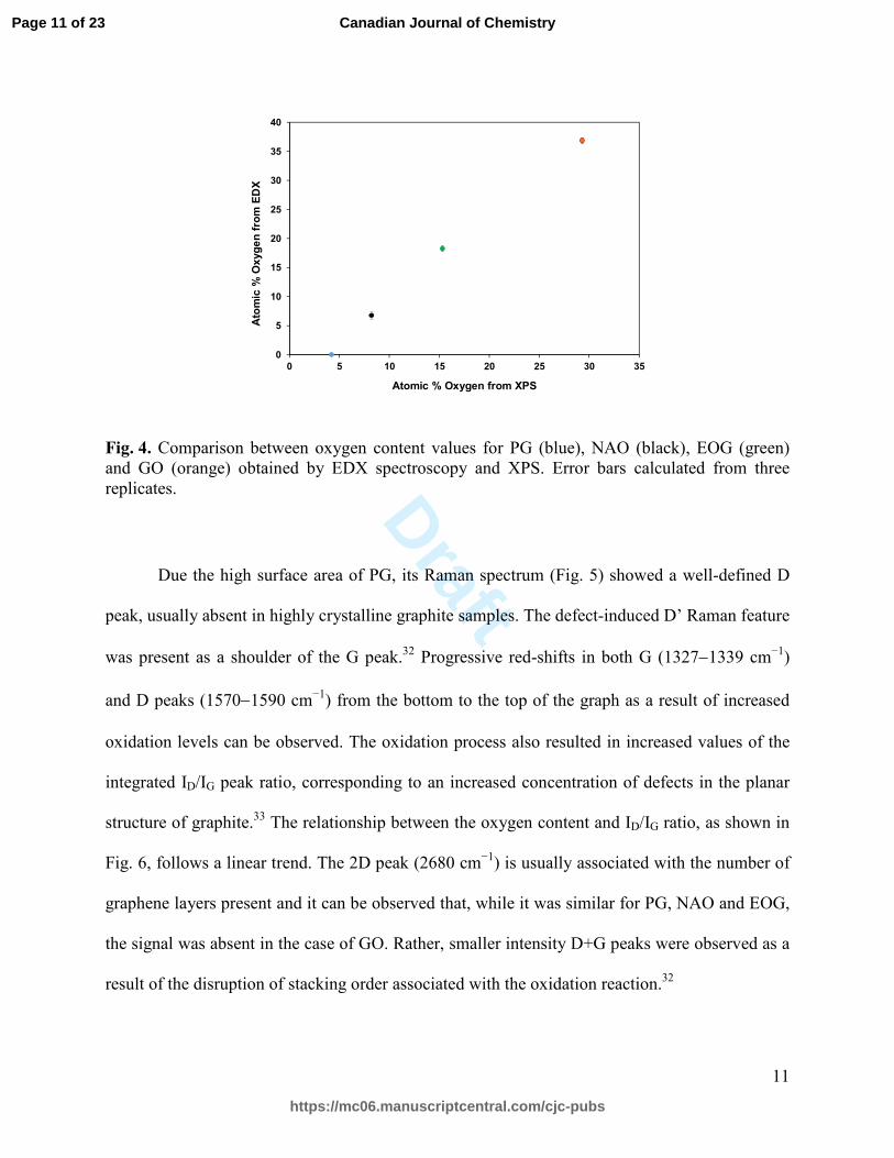
Oxygen and carbon analysis by XPS (Fig. S1) showed strong C1s (~284.5 eV) and weak
O1s (~532.0 eV) peaks for PG, and an increase in the O1s peak for NAO, EOG and GO, for which
the peak intensity surpassed that of the C1s peak. The oxygen and carbon contents were in
agreement with the values obtained by EDX spectroscopy (Fig. 4). The similar values obtained
using these techniques confirmed the uniform oxidation of the corresponding powders and small
discrepancies between the two methods may be attributed to chemisorbed oxygen.
0
5
10
15
20
25
30
35
40
45
50
PG NAO EOG GO
Ox
yg
en
Co
nte
nt
by E
DX
(w
eig
ht
%)
Page 10 of 23
https://mc06.manuscriptcentral.com/cjc-pubs
Canadian Journal of Chemistry
Draft
11
Fig. 4. Comparison between oxygen content values for PG (blue), NAO (black), EOG (green) and GO (orange) obtained by EDX spectroscopy and XPS. Error bars calculated from three replicates.
Due the high surface area of PG, its Raman spectrum (Fig. 5) showed a well-defined D
peak, usually absent in highly crystalline graphite samples. The defect-induced D’ Raman feature
was present as a shoulder of the G peak.32 Progressive red-shifts in both G (1327−1339 cm−1)
and D peaks (1570−1590 cm−1) from the bottom to the top of the graph as a result of increased
oxidation levels can be observed. The oxidation process also resulted in increased values of the
integrated ID/IG peak ratio, corresponding to an increased concentration of defects in the planar
structure of graphite.33 The relationship between the oxygen content and ID/IG ratio, as shown in
Fig. 6, follows a linear trend. The 2D peak (2680 cm−1) is usually associated with the number of
graphene layers present and it can be observed that, while it was similar for PG, NAO and EOG,
the signal was absent in the case of GO. Rather, smaller intensity D+G peaks were observed as a
result of the disruption of stacking order associated with the oxidation reaction.32
0
5
10
15
20
25
30
35
40
0 5 10 15 20 25 30 35
Ato
mic
% O
xyg
en
fro
m E
DX
Atomic % Oxygen from XPS
Page 11 of 23
https://mc06.manuscriptcentral.com/cjc-pubs
Canadian Journal of Chemistry

Draft
12
Fig. 5. Raman spectra of PG (blue), NAO (black), EOG (green) and GO (orange).
Fig. 6. Relationship between the ID/IG ratios and oxygen content from XPS measurements for PG (blue), NAO (black), EOG (green) and GO (orange).
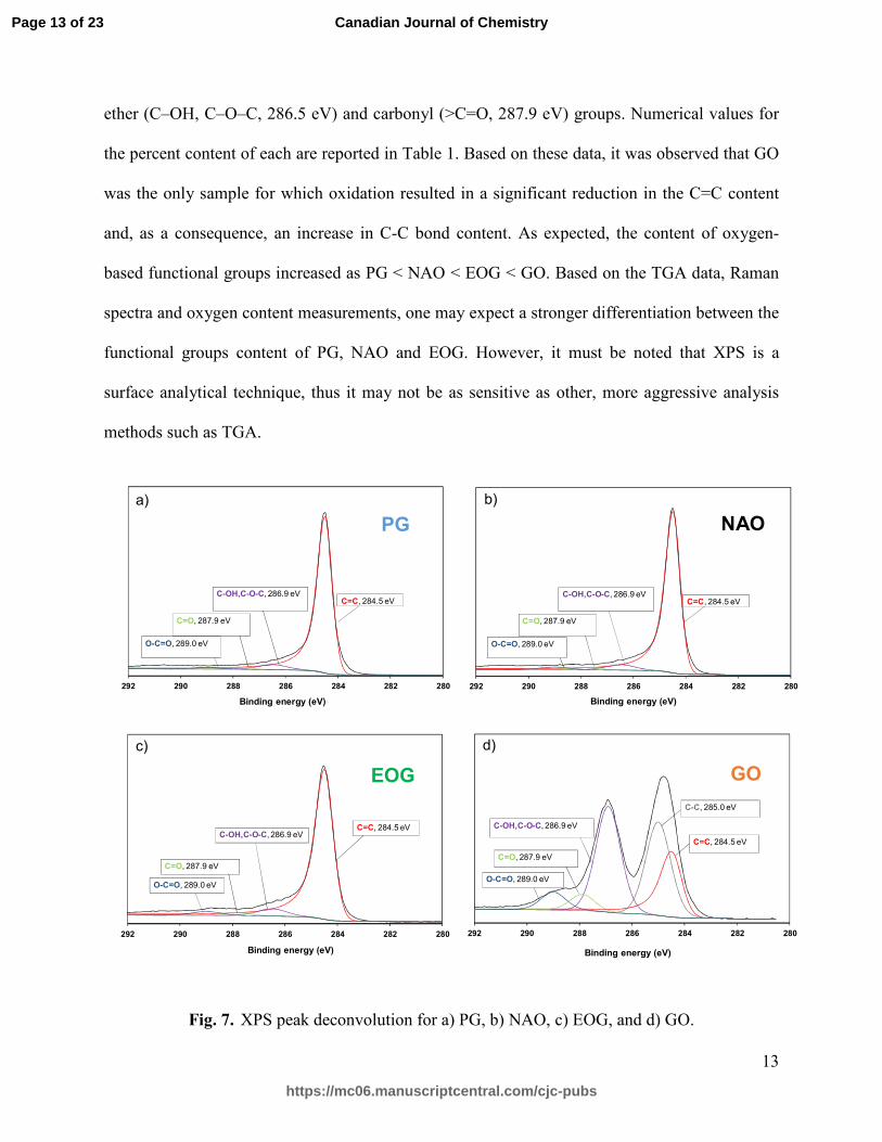
XPS data (Fig. 7) was further analyzed in order to reveal the differences between the
powder samples in terms of five different chemical functionalities observed at 284.5, 285.0,
286.5, 287.9 and 289.2 eV, respectively. These signals can be assigned to sp2 carbon atoms in
aromatic rings (284.5 eV), C-C and C-H bonds (285.0 eV), C atoms bonded to hydroxyl and
0.0
0.5
1.0
1.5
2.0
2.5
3.0
3.5
4.0
800 1000 1200 1400 1600 1800 2000 2200 2400 2600 2800
Inte
ns
ity (
a.u
.)
Raman shift (cm-1)
0
0.2
0.4
0.6
0.8
1
1.2
1.4
0 5 10 15 20 25 30 35
I D/I
G
Oxygen content (atomic %)
Page 12 of 23
https://mc06.manuscriptcentral.com/cjc-pubs
Canadian Journal of Chemistry
Draft
13
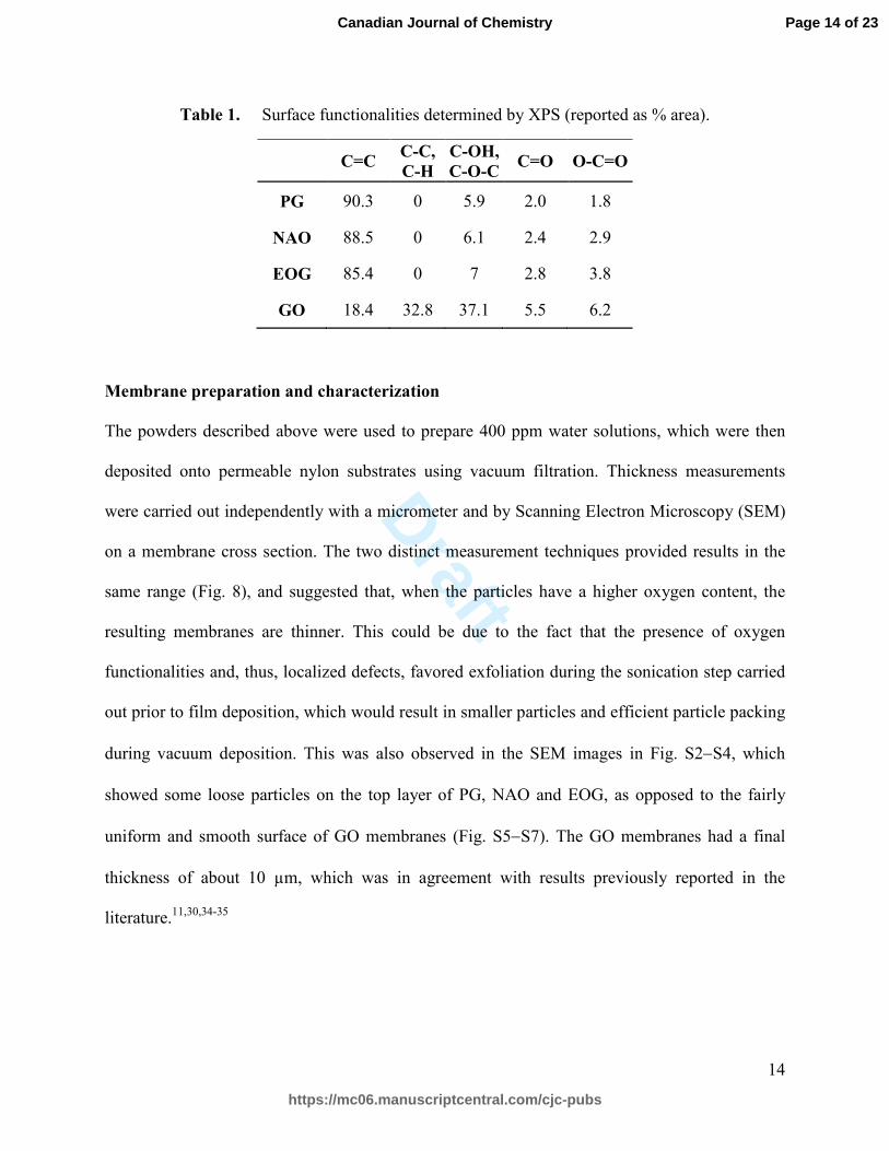
ether (C–OH, C–O–C, 286.5 eV) and carbonyl (>C=O, 287.9 eV) groups. Numerical values for
the percent content of each are reported in Table 1. Based on these data, it was observed that GO
was the only sample for which oxidation resulted in a significant reduction in the C=C content
and, as a consequence, an increase in C-C bond content. As expected, the content of oxygen-
based functional groups increased as PG < NAO < EOG < GO. Based on the TGA data, Raman
spectra and oxygen content measurements, one may expect a stronger differentiation between the
functional groups content of PG, NAO and EOG. However, it must be noted that XPS is a
surface analytical technique, thus it may not be as sensitive as other, more aggressive analysis
methods such as TGA.
Fig. 7. XPS peak deconvolution for a) PG, b) NAO, c) EOG, and d) GO.
280282284286288290292
Binding energy (eV)
C=O, 287.9 eV
O-C=O, 289.0 eV
C=C, 284.5 eV
C-OH,C-O-C, 286.9 eV
C-C, 285.0 eV
280282284286288290292
Binding energy (eV)
C=O, 287.9 eV
O-C=O, 289.0 eV
C=C, 284.5 eVC-OH,C-O-C, 286.9 eV
280282284286288290292
Binding energy (eV)
C=O, 287.9 eV
O-C=O, 289.0 eV
C=C, 284.5 eVC-OH,C-O-C, 286.9 eV
280282284286288290292
Binding energy (eV)
C=O, 287.9 eV
O-C=O, 289.0 eV
C=C, 284.5 eVC-OH,C-O-C, 286.9 eV
a) b)
c) d)
PG NAO
EOG GO
Page 13 of 23
https://mc06.manuscriptcentral.com/cjc-pubs
Canadian Journal of Chemistry
Draft
14
Table 1. Surface functionalities determined by XPS (reported as % area).
C=C C-C,
C-H
C-OH,
C-O-C C=O O-C=O
PG 90.3 0 5.9 2.0 1.8
NAO 88.5 0 6.1 2.4 2.9
EOG 85.4 0 7 2.8 3.8
GO 18.4 32.8 37.1 5.5 6.2
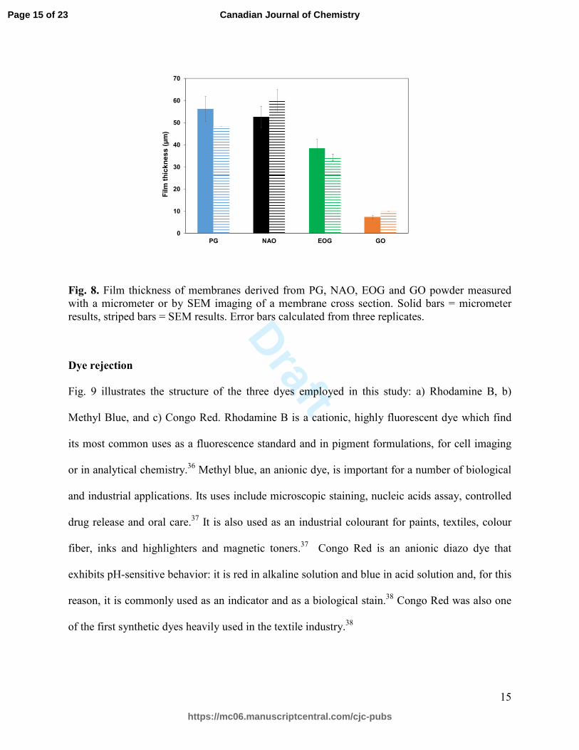
Membrane preparation and characterization
The powders described above were used to prepare 400 ppm water solutions, which were then
deposited onto permeable nylon substrates using vacuum filtration. Thickness measurements
were carried out independently with a micrometer and by Scanning Electron Microscopy (SEM)
on a membrane cross section. The two distinct measurement techniques provided results in the
same range (Fig. 8), and suggested that, when the particles have a higher oxygen content, the
resulting membranes are thinner. This could be due to the fact that the presence of oxygen
functionalities and, thus, localized defects, favored exfoliation during the sonication step carried
out prior to film deposition, which would result in smaller particles and efficient particle packing
during vacuum deposition. This was also observed in the SEM images in Fig. S2−S4, which
showed some loose particles on the top layer of PG, NAO and EOG, as opposed to the fairly
uniform and smooth surface of GO membranes (Fig. S5−S7). The GO membranes had a final
thickness of about 10 µm, which was in agreement with results previously reported in the
literature.11,30,34-35
Page 14 of 23
https://mc06.manuscriptcentral.com/cjc-pubs
Canadian Journal of Chemistry
Draft
15
Fig. 8. Film thickness of membranes derived from PG, NAO, EOG and GO powder measured with a micrometer or by SEM imaging of a membrane cross section. Solid bars = micrometer results, striped bars = SEM results. Error bars calculated from three replicates.
Dye rejection
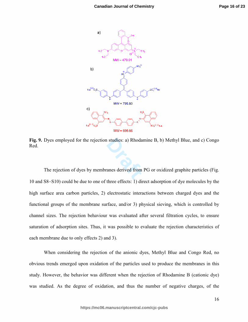
Fig. 9 illustrates the structure of the three dyes employed in this study: a) Rhodamine B, b)
Methyl Blue, and c) Congo Red. Rhodamine B is a cationic, highly fluorescent dye which find
its most common uses as a fluorescence standard and in pigment formulations, for cell imaging
or in analytical chemistry.36 Methyl blue, an anionic dye, is important for a number of biological
and industrial applications. Its uses include microscopic staining, nucleic acids assay, controlled
drug release and oral care.37 It is also used as an industrial colourant for paints, textiles, colour
fiber, inks and highlighters and magnetic toners.37 Congo Red is an anionic diazo dye that
exhibits pH-sensitive behavior: it is red in alkaline solution and blue in acid solution and, for this
reason, it is commonly used as an indicator and as a biological stain.38 Congo Red was also one
of the first synthetic dyes heavily used in the textile industry.38
0
10
20
30
40
50
60
70
PG NAO EOG GO
Film
th
ick
ne
ss
(µ
m)
Page 15 of 23
https://mc06.manuscriptcentral.com/cjc-pubs
Canadian Journal of Chemistry
Draft
16
Fig. 9. Dyes employed for the rejection studies: a) Rhodamine B, b) Methyl Blue, and c) Congo Red.
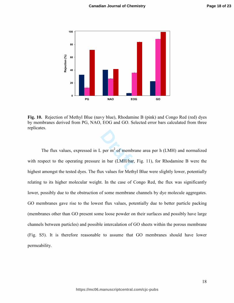
The rejection of dyes by membranes derived from PG or oxidized graphite particles (Fig.
10 and S8−S10) could be due to one of three effects: 1) direct adsorption of dye molecules by the
high surface area carbon particles, 2) electrostatic interactions between charged dyes and the
functional groups of the membrane surface, and/or 3) physical sieving, which is controlled by
channel sizes. The rejection behaviour was evaluated after several filtration cycles, to ensure
saturation of adsorption sites. Thus, it was possible to evaluate the rejection characteristics of
each membrane due to only effects 2) and 3).
When considering the rejection of the anionic dyes, Methyl Blue and Congo Red, no
obvious trends emerged upon oxidation of the particles used to produce the membranes in this
study. However, the behavior was different when the rejection of Rhodamine B (cationic dye)
was studied. As the degree of oxidation, and thus the number of negative charges, of the
Page 16 of 23
https://mc06.manuscriptcentral.com/cjc-pubs
Canadian Journal of Chemistry
Draft
17
membranes was increased their ability to reject Rhodamine B was also enhanced. This
observation provides critical insight into future membrane design, whereby membrane oxidation
may be used to tune the rejection of cationic molecules. Understanding dye rejection in the
context of physical sieving was slightly more difficult. When examining the rejection data for the
three dyes, no obvious correlation between the molecular weights of the dyes and their rejection
were observed. For each membrane type, Congo Red rejection was most efficient. We attribute
this behavior to Congo Red’s propensity to aggregate in aqueous and organic solutions via π-π
stacking interactions.39,40 Thus, we postulate that Congo Red was rejected more efficiently due to
the larger relative size of these aggregates in solution.
Despite the fact that GO-based membranes showed the highest rejection for Rhodamine
B and Congo Red, EOG-based membranes showed a rejection of 87% for Congo Red. Thus, by
combining the information regarding the physico-chemical properties (charge, aggregation, etc)
of the dye or target molecule to be rejected and the properties of the membranes employed,
partially oxidized graphite-based membranes may provide a valid replacement for those based on
GO. Crucially, the use of such membranes would also circumvent the need for prolonged, harsh
chemical oxidation methods (i.e., Hummers' method).
Page 17 of 23
https://mc06.manuscriptcentral.com/cjc-pubs
Canadian Journal of Chemistry
Draft
18
Fig. 10. Rejection of Methyl Blue (navy blue), Rhodamine B (pink) and Congo Red (red) dyes by membranes derived from PG, NAO, EOG and GO. Selected error bars calculated from three replicates.
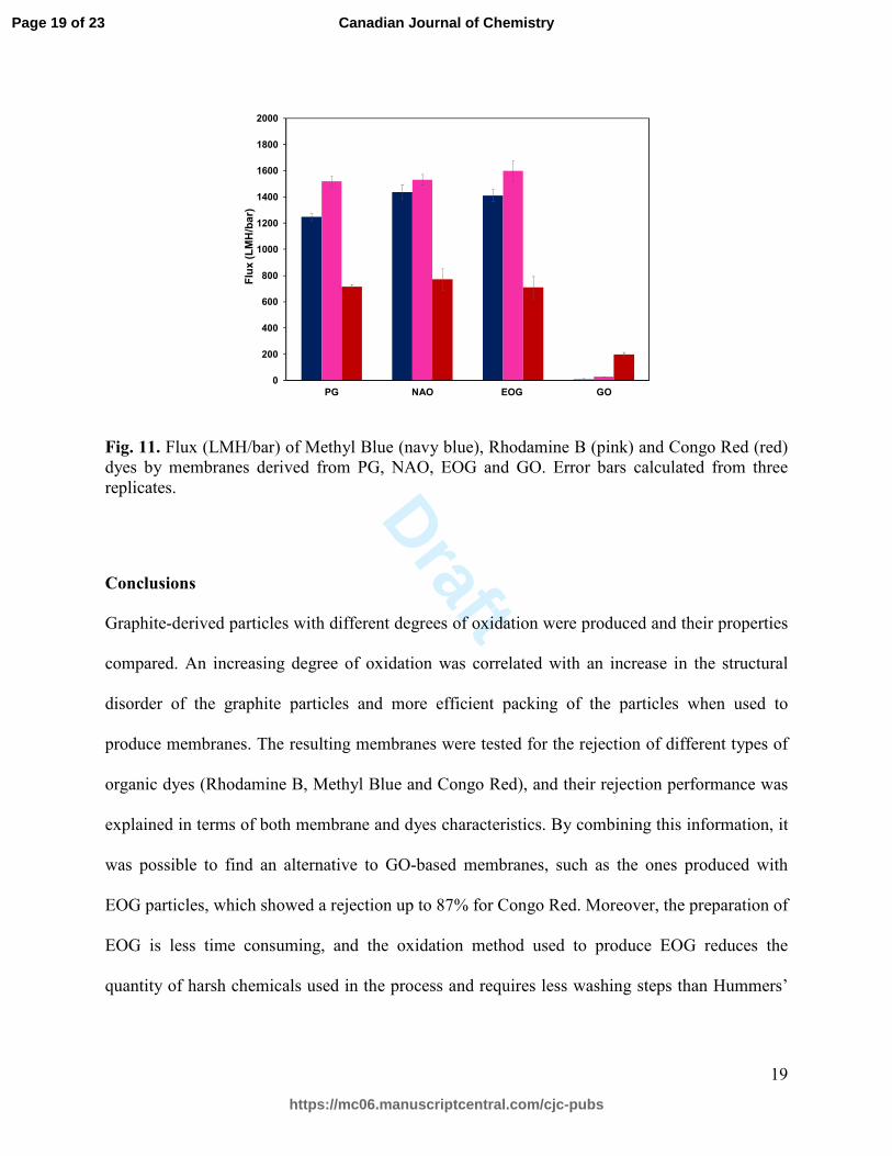
The flux values, expressed in L per m2 of membrane area per h (LMH) and normalized
with respect to the operating pressure in bar (LMH/bar, Fig. 11), for Rhodamine B were the
highest amongst the tested dyes. The flux values for Methyl Blue were slightly lower, potentially
relating to its higher molecular weight. In the case of Congo Red, the flux was significantly
lower, possibly due to the obstruction of some membrane channels by dye molecule aggregates.
GO membranes gave rise to the lowest flux values, potentially due to better particle packing
(membranes other than GO present some loose powder on their surfaces and possibly have large
channels between particles) and possible intercalation of GO sheets within the porous membrane
(Fig. S5). It is therefore reasonable to assume that GO membranes should have lower
permeability.
0
20
40
60
80
100
PG NAO EOG GO
Re
jec
tio
n (%
)
Page 18 of 23
https://mc06.manuscriptcentral.com/cjc-pubs
Canadian Journal of Chemistry
Draft
19
Fig. 11. Flux (LMH/bar) of Methyl Blue (navy blue), Rhodamine B (pink) and Congo Red (red) dyes by membranes derived from PG, NAO, EOG and GO. Error bars calculated from three replicates.
Conclusions
Graphite-derived particles with different degrees of oxidation were produced and their properties
compared. An increasing degree of oxidation was correlated with an increase in the structural
disorder of the graphite particles and more efficient packing of the particles when used to
produce membranes. The resulting membranes were tested for the rejection of different types of
organic dyes (Rhodamine B, Methyl Blue and Congo Red), and their rejection performance was
explained in terms of both membrane and dyes characteristics. By combining this information, it
was possible to find an alternative to GO-based membranes, such as the ones produced with
EOG particles, which showed a rejection up to 87% for Congo Red. Moreover, the preparation of
EOG is less time consuming, and the oxidation method used to produce EOG reduces the
quantity of harsh chemicals used in the process and requires less washing steps than Hummers’
0
200
400
600
800
1000
1200
1400
1600
1800
2000
PG NAO EOG GO
Flu
x (
LM
H/b
ar)
Page 19 of 23
https://mc06.manuscriptcentral.com/cjc-pubs
Canadian Journal of Chemistry
Draft
20
method, presenting obvious manufacturing advantages and reduced environmental impact when
compared to GO production.
Acknowledgements
The authors would like to acknowledge the Natural Science and Engineering Research Council
(NSERC) of Canada (Engage, EGP 487122-15), Mitacs Accelerate (IT0622), the Canada
Foundation for Innovation (JELF, 33977) and 3M Canada for funding this work. We thank Dr.
Todd Simpson and Tim Goldhawk from the Western Nanofabrication Facility for assistance with
SEM imaging, Dr. Paul Boyle and Aneta Borecki for assistance with PXRD measurements, and
Catherine Bardeau and Jennifer Johnson from 3M Canada for assistance with EDX
measurements.
Supplementary material
Supplementary data are available with the article through the journal Web site at ########.
References
(1) B. C. Group Water and Wastewater Treatment Technologies: Global Markets, 2013.
(2) Qu, X.; Alvarez, P. J. J.; Li, Q. Water Res. 2013, 47, 3931. doi:
10.1016/j.watres.2012.09.058.
(3) Park, S.; Ruoff, R. S. Nat. Nanotechnol. 2009, 4, 217. doi: 10.1038/nnano.2009.58.
(4) Liu, C.; Yu, Z.; Neff, D.; Zhamu, A.; Jang, B. Z. Nano Lett. 2010, 10, 4863. doi:
10.1021/nl102661q.
(5) Chung, K.; Lee, C.-H.; Yi, G.-C. Science 2010, 330, 655. doi: 10.1126/science.1195403.
(6) Shao, Y.; Wang, J.; Wu, H.; Liu, J.; Aksay, I. A.; Lin, Y. Electroanal. 2010, 22, 1027. doi:
10.1002/elan.200900571.
Page 20 of 23
https://mc06.manuscriptcentral.com/cjc-pubs
Canadian Journal of Chemistry
Draft
21
(7) Wang, G.; Shen, X.; Yao, J.; Park, J. Carbon 2009, 47, 2049. doi:
10.1016/j.carbon.2009.03.053.
(8) Zhang, L.; Shi, G. J. Phys. Chem. C 2011, 115, 17206. doi: 10.1021/jp204036a.
(9) Kumar, M.; Singh, K.; Dhawan, S. K.; Tharanikkarasu, K.; Chung, J. S.; Kong, B.-S.; Kim,
E. J.; Hur, S. H. Chem. Eng. J. 2013, 231, 397. doi: 10.1016/j.cej.2013.07.043.
(10) Gao, W.; Majumder, M.; Alemany, L. B.; Narayanan, T. N.; Ibarra, M. A.; Pradhan, B. K.;
Ajayan, P. M. ACS Appl. Mater. Interfaces 2011, 3, 1821. doi: 10.1021/am200300u.
(11) Joshi, R. K.; Carbone, P.; Wang, F. C.; Kravets, V. G.; Su, Y.; Grigorieva, I. V.; Wu, H. A.;
Geim, A. K.; Nair, R. R. Science 2014, 343, 752. doi: 10.1126/science.1245711.
(12) Chen, D.; Feng, H.; Li, J. Chem. Rev. 2012, 112, 6027. doi: 10.1021/cr300115g.
(13) Hung, W.-S.; Tsou, C.-H.; De Guzman, M.; An, Q.-F.; Liu, Y.-L.; Zhang, Y.-M.; Hu, C.-
C.; Lee, K.-R.; Lai, J.-Y. Chem. Mater. 2014, 26, 2983. doi: 10.1021/cm5007873.
(14) Han, Y.; Jiang, Y.; Gao, C. ACS Appl. Mater. Interfaces 2015, 7, 8147. doi:
10.1021/acsami.5b00986.
(15) Zhang, J.; Xu, Z.; Shan, M.; Zhou, B.; Li, Y.; Li, B.; Niu, J.; Qian, X. J. Membr. Sci. 2013,
448, 81. doi: 10.1016/j.memsci.2013.07.064.
(16) Zhao, C.; Xu, X.; Chen, J.; Yang, F. Desalination 2014, 334, 17. doi:
10.1016/j.desal.2013.07.011.
(17) Kim, S. G.; Hyeon, D. H.; Chun, J. H.; Chun, B.-H.; Kim, S. H. Desalin. Water Treat. 2013,
51, 6338. doi: 10.1080/19443994.2013.780994.
(18) Gao, Y.; Hu, M.; Mi, B. J. Membr. Sci. 2014, 455, 349. doi: 10.1016/j.memsci.2014.01.011.
(19) Hummers, W. S.; Offeman, R. E. J. Am. Chem. Soc. 1958, 80, 1339. doi:
10.1021/ja01539a017.
Page 21 of 23
https://mc06.manuscriptcentral.com/cjc-pubs
Canadian Journal of Chemistry
Draft
22
(20) Jeon, I.-Y.; Shin, Y.-R.; Sohn, G.-J.; Choi, H.-J.; Bae, S.-Y.; Mahmood, J.; Jung, S.-M.;
Seo, J.-M.; Kim, M.-J.; Wook Chang, D.; Dai, L.; Baek, J.-B. Proc. Natl. Acad. Sci. U.S.A. 2012,
109, 5588. doi: 10.1073/pnas.1116897109.
(21) Bai, M.; Chen, J.; Wu, W.; Zeng, X.; Wang, J.; Zou, H. Colloids Surf. A Physicochem. Eng.
Asp. 2016, 490, 59. doi: 10.1016/j.colsurfa.2015.11.033.
(22) Wei, L.; Wu, F.; Shi, D.; Hu, C.; Li, X.; Yuan, W.; Wang, J.; Zhao, J.; Geng, H.; Wei, H.;
Wang, Y.; Hu, N.; Zhang, Y. Sci. Rep. 2013, 3, 2636. doi: 10.1038/srep02636.
(23) Rambabu, N.; Azargohar, R.; Dalai, A. K.; Adjaye, J. Fuel Process. Technol. 2013, 106,
501. doi: 10.1016/j.fuproc.2012.09.019.
(24) Zacharia, R. Doctoral dissertation, Freie Universität Berlin, 2004.
(25) Abdolhosseinzadeh, S.; Asgharzadeh, H.; Seop Kim, H. Sci. Rep. 2015, 5, 10160. doi:
10.1038/srep10160.
(26) Song, J.; Wang, X.; Chang, C.-T. J. Nanomater. 2014, 2014, 6. doi: 10.1155/2014/276143.
(27) Spyrou, K.; Rudolf, P. In Functionalization of Graphene; Wiley-VCH Verlag GmbH & Co.
KGaA: Weinham, 2014.
(28) Girgis, B. S.; Temerk, Y. M.; Gadelrab, M. M.; Abdullah, I. D. Carbon Lett. 2007, 8, 95.
doi: 10.5714/CL.2007.8.2.095.
(29) Kosynkin, D. V.; Higginbotham, A. L.; Sinitskii, A.; Lomeda, J. R.; Dimiev, A.; Price, B.
K.; Tour, J. M. Nature 2009, 458, 872. doi: 10.1038/nature07872.
(30) Nair, R. R.; Wu, H. A.; Jayaram, P. N.; Grigorieva, I. V.; Geim, A. K. Science 2012, 335,
442. doi: 10.1126/science.1211694.
(31) Pham, V. H.; Cuong, T. V.; Hur, S. H.; Oh, E.; Kim, E. J.; Shin, E. W.; Chung, J. S. J.
Mater. Chem. 2011, 21, 3371. doi: 10.1039/C0JM02790A.
Page 22 of 23
https://mc06.manuscriptcentral.com/cjc-pubs
Canadian Journal of Chemistry
Draft
23
(32) Pimenta, M.; Dresselhaus, G.; Dresselhaus, M. S.; Cancado, L.; Jorio, A.; Saito, R. Phys.
Chem. Chem. Phys. 2007, 9, 1276. doi: 10.1039/B613962K.
(33) Zhao, J.; Liu, L.; Li, F., Ed. Graphene Oxide: Physics and Applications; Springer-Verlag:
Berlin, 2014.
(34) Coleman, M.; Tang, X. Nano Res. 2015, 8, 1128. doi: 10.1007/s12274-014-0593-x.
(35) Hung, W.-S.; An, Q.-F.; De Guzman, M.; Lin, H.-Y.; Huang, S.-H.; Liu, W.-R.; Hu, C.-C.;
Lee, K.-R.; Lai, J.-Y. Carbon 2014, 68, 670. doi: 10.1016/j.carbon.2013.11.048.
(36) Beija, M.; Afonso, C. A. M.; Martinho, J. M. G. Chem. Soc. Rev. 2009, 38, 2410. doi:
10.1039/B901612K.
(37) Sharma, P.; Hussain, N.; Borah, D. J.; Das, M. R. J. Chem. Eng. Data 2013, 58, 3477. doi:
10.1021/je400743r.
(38) Steensma, D. P. Arch. Path. Lab. Med. 2001, 125, 250. doi: 10.1043/0003-
9985(2001)125<0250:CR>2.0.CO;2.
(39) Frid, P.; Anisimov, S. V.; Popovic, N. Brain Res. Rev. 2007, 53, 135. doi:
10.1016/j.brainresrev.2006.08.001.
(40) Stopa, B.; Jagusiak, A.; Konieczny, L.; Piekarska, B.; Rybarska, J.; Zemanek, G.; Król, M.;
Piwowar, P.; Roterman, I. J. Mol. Model. 2013, 19, 4731. doi: 10.1007/s00894-012-1744-1.
Page 23 of 23
https://mc06.manuscriptcentral.com/cjc-pubs
Canadian Journal of Chemistry