drinking mechanisms in the zebra finch and the bengalese … · zebra finch is able to drink by a...
TRANSCRIPT
THE CONDOR A JOURNAL OF AVIAN BIOLOGY
Volume 92 Number 1
I_\!
February 1990
MAR Oh WI0 The Condor 92: 1-28 Q The Cooper Ornithological Society 1990
WVERSITY OF ~[4br-fJ- DRINKING MECHANISMS IN THE ZEBRA FINCH AND
THE BENGALESE FINCH’
J. HEIDWEILLER AND G. A. ZWEERS~ Department of Neurobehavioral Morphology, Zoological Laboratory,
University of Leiden, The Netherlands
Abstract. Two kinds of drinking behavior were studied by film and radiogram analysis of tip down drinking Zebra Finches (Poephila guttata) and tip up drinking Bengalese Finches (Lonchura striata) which use similar scooping tongue motions to carry water into the mouth. Water transport through the pharynx differs: the Zebra Finch uses a scooping motion of the larynx that reoccurs in every motion cycle, while the beak is kept down. The Bengalese Finch elevates the head allowing water to flow downward due to gravity and pharyngeal properistalsis. Extensive analyses show the anatomy of the species to be highly similar. The Zebra Finch is able to drink by a double scoop mechanism, because-unlike the Bengalese Finch-reflexes for glottis closure and esophageal peristalsis are used. Integration of these reflexes and a shift in timing of the larynx-scoop has modified tip up into tip down drinking. Thus, tip down is more complex than tip up drinking, since here actions from different cycles and patterns are integrated in one motion cycle. Increased kinematic complexity is, apart from any historical scenario, an argument that tip down is derived from tip up drinking in Estrildidae. An evolutionary scenario is presented in which developments of scooping anatomical elements are seen as preadaptations. These developed by selection on elements serving the highly specialized kind of feeding on seeds of Gramineae under high predator pressure in open fields, and allowed a wide secondary extension of the feeding area.
Key words: Drinking; oropharyngeal anatomy: Estrildidae; Zebra Finch; Bengalese Finch.
INTRODUCTION
Estrildid finches exhibit two kinds of drinking: tip up drinking and suction drinking (Poulsen 1953; Wickler 1961; Immelmann 1962, 1965). To understand how divergence in the evolution of these two different kinds of behavior for one role may have developed, we need to know the physical mechanisms used in such drinking.
These two kinds of drinking can easily be clas- sified into the two main categories of avian drinking since estrildid drinking patterns are characterized by the same names (Wickler 196 1): beak tip up drinking, and suction drinking in which the beak tips are kept down. This classi-
’ Received 23 May 1989. Final acceptance 2 October 1989.
2 Address reprint requests to Dr. G. A. Zweers.
fication, however, is not sufficient to understand which drinking mechanism really operates in a given species because several kinds of mecha- nisms are found within each of the two main categories of drinking. The same is true for both of the two successive steps in drinking behavior: intake of water into the mouth, and transport of the water from the mouth through the pharynx and into the esophagus.
For example, water is taken into the mouth in the Cacatuinae by scooping the water with the lower beak (Homberger 1980); in Gallus, the mouth opens and closes several times, taking some water each time (McLelland 1979); in Anus, a complex interplay of capillary action and pres- sure changes in the different areas of the mouth serves to transport the water into and through the mouth (Kooloos and Zweers 1989). A similar feature is found in the category labeled “suction
111

2 J. HEIDWEILLER AND G. A. ZWEERS
drinking.” For example, Psittacinae ladle water with the tip of the tongue, and Lorriculinae drink with a suctioning action (Homberger 1980) whereas for Columba a double suction mecha- nism has been proposed, in which lower air pres- sure develops first in the mouth and immediately thereafter in the pharynx (Zweers 1982a).
Several conclusions may be drawn from the reviews of Schonholzer (1959) Homberger (1980) and Kooloos and Zweers (1989). The main difference found between the two categories of avian drinking is in the transport of the water from the mouth through the pharynx into the esophagus. In tip up drinking behavior, this part of the water transport occurs while the beaks are kept in a “tipped up” position, whereas in suc- tion drinking behavior, the same transport oc- curs while the head is “tipped down.” Further, tip up swallowing occurs in a series of drinking bouts which resemble poly draught behavior, whereas tip down swallowing comprises one drinking bout which has a mono draught ap- pearance. Therefore, we propose to apply the fol- lowing descriptive terms to the main categories ofdrinking: tip up swallowing and tip down swal- lowing. Both categories are certainly present in the Estrildidae.
Immelmann (1962) takes estrildid tip up drinking as a self evident feature, while for suc- tion drinking he proposes that peristalsis of the cranial part of the esophagus pumps in water. In the literature, the drinking mechanisms of the eight suction drinking estrildid species are thought to be identical with the drinking mechanism in pigeons (Poulsen 1953). Wickler (196 1) also con- cluded that the drinking behaviors of Geopelia cuniata (Columbidae) and Poephila acuticauda (Estrildidae) were identical. Immelmann (1965) stated that Zebra Finches (Poephila guttata) drink in a pigeon-like manner. Immelmann and Im- melmann (1967) assumed, as did Lorentz (1939) for pigeons, that sucking is caused by peristaltic movements of the anterior part of the esophagus. Fisher et al. (1972) suggested that Zebra Finches drink like honey eaters. These authors provide no further evidence for the proposed models. However, Zweers (1982a) has shown that Co- lumba livia does not drink by peristalsis in the esophagus, so that either the mechanisms must be different or suction drinking Estrildidae must drink by the same double suction mechanism that has been proposed for C. livia. The first aim of this paper is to analyze mechanisms for drink-
ing. Therefore the Zebra Finch and the Bengalese Finch (Lonchura striata) have been selected for analysis. The Zebra Finch is one of the eight species of Australian Estrildidae that show tip down swallowing. The Bengalese Finch which originally lived in Southeast Asia is a tip up, poly draught swallower of water.
The morphology of the interior of the mouth and pharynx at first looks similar in the different species of Estrildidae. Therefore differences in drinking behavior may either be correlated with minor differences in the epidermal structure, or may be identified as “different applications of similar epidermal elements.” In the latter case, the muscle-bone apparatus, the modal action patterns for drinking, or both may be different. A second aim of this research was to test whether the assumption holds that different kinematic patterns may serve in highly similar morpholo- gies to elicit different drinking mechanisms. Therefore the anatomy of the epidermal struc- tures and the lingual muscle-bone elements must be compared. Since we have found no anatom- ical descriptions in the literature that are suffi- ciently accurate, these must be included here. Description of the lingual muscle-bone appara- tus does, however, not serve for functional ex- planation of that apparatus, but rather for gath- ering anatomical evidence that lingual and pharyngeal integumental elements are equipped such that they may carry out similar motion pat- terns.
The development in highly similar morphol- ogies of different behavioral patterns for the same role may have a specific cause. Immelmann and Immelmann (1967) consider tip down swallow- ing of water to be a mechanism with a selective advantage for those Estrildidae that must rely upon desert water holes for their water needs. Their argument is that suction drinking allows much faster intake of water than tip up drinking, so that predator pressure decreases when the fast- er mechanism develops. Their view includes the assumption that suction drinking is derived from tip up drinking.
As a second cause of the development of suc- tion drinking, Immelmann and Immelmann (1967) assume that in semiarid climates small dewdrops are the only water source available, and that these drops can only be ingested via the suction mechanism they propose. These two as- sumptions need further testing. A third aim here is thus to measure the different kinds of perfor-

DRINKING MECHANISMS IN ESTRILDIDAE 3
mance of both mechanisms and to apply the re- sults to a discussion of selection pressure.
It is not only selection pressure, however, which determines change in a system that is as complex as the oropharyngeal system. For example, to elicit appropriate drinking behavior, over 30 muscles in the mouth and pharynx must act in an orderly fashion. Internal constraints and physical boundary conditions also determine the direction of development. These may or may not have created a discontinuity between the two mechanisms. A comparison is needed to see which factors are important here. We start from a hypothetical ancestral tip up, poly draught drinking mechanism for Estrildidae that is rel- atively simple and close to the drinking behavior seen in Gallus: water is scooped with the lower beak, then the head is tipped up; water runs down through the pharynx due mainly to gravity plus some pharyngeal properistaltic actions (Mc- Lelland 1979). Whether the two drinking mech- anisms may be deduced from this mechanism in one series or in two diverging directions is then discussed to see if the mechanisms themselves suggest boundaries or constraints which, in ad- dition to an evolutionary scenario, may have de- termined how development has occurred.
METHODS AND MATERIALS
ANATOMY
Fresh and preserved specimens of the Zebra Finch (Poephila guttata castanotis (Gould 1873)) and the Bengalese Finch (Lonchura striata (L.) var. domestica) were used for dissection of the mus- cle-bone apparatus of beak and tongue. The pre- served specimens were perfused via the left ven- tricle with 4% formalin. For each species, a total head was embedded in Epon C and cut in trans- verse sections of 10 p to allow microscopic com- parisons. Sections were stained according to a combined Mallory-hematoxylin staining proce- dure (Zweers et al. 1977). Beaks of embedded specimens were closed, and tongue and larynx were brought into similar positions. SEM pho- tographs were made to locate orifices of glands and to enable the description of epidermal struc- tures. The anatomical terminologies of Baumel (1979) and McLelland (1979) adapted according to Zweers (1982b), were used.
KINEMATIC ANALYSIS
Cinematography and radiography. The drinking behavior of both species was filmed with a Pail-
\ CAUDAL
ROSTRAL
FIGURE 1. The points at which measurements were made in analysis of drinking are illustrated for the Ze- bra Finch. The reference line of the neurocranium is indicated by two markers (1 and 2). The upper beak line runs from the beak tip to a marker at the cranio- facial joint (3). The lower beak line connects the beak tip with a marker at the quadrato-mandibularjoint (4). Three markers represent the rostral, intermediate, and caudal mouth floor (5, 6, 7). Additional points were the very tips of the beak and tongue.
lard Bolex 16-mm camera, using Kodak Plus X reversal film at 67 fps, as well as with a Teledyne highspeed camera using Eastman Ektachrome Video Newsfilm at 250 fps. The head was marked with a series of triangular white markers as shown in Figure 1. The reflexes of tongue and larynx, which occur when water drops fall upon the low- er beak tip and the lingual base, were filmed at 67 fps, while the birds were slightly anesthetized with equithesin and were fixed in a stereotactic apparatus (Zweers 1971) with their beaks wide open.
About 100 radiograms were taken laterally of both species drinking a suspension of barium sulfate in water. The X-ray generator (Siemens- Reiniger; Gigantos) was adjusted at 120 KV, 450 mA, and 0.003-see exposure time. Radiofilms were made with a camera (Arriflex) connected to the X-ray apparatus, which was adjusted at 110 KV, 25 mA, and 0.0 1-set exposure time.

4 J. HEIDWEILLER AND G. A. ZWEERS
The radiograms were put in an order representing a general drinking bout by comparison with films taken simultaneously.
Analysis offilms. Films were digitized frame by frame with a graphic tablet (Tectronic 4049) connected to a Vax computer (Digital Inc.). The following parameters were measured (Fig. 1): (1) The distance between the beak tips (gape). (2) The angle between the neurocranium reference line and the upper beak line. (3) The angle be- tween the neurocranium reference line and the lower beak line. (4) Height of the water level between the beaks relative to the beak tip. (5) The distance between the rostra1 mouth floor and the reference line on the neurocranium. (6) The distance between the intermediate mouth floor and the reference line on the neurocranium. (7) The distance between the caudal mouth floor and a reference line on the neurocranium. (8) The distance between the tip of the tongue and a line connecting the quadrato-mandibularjoint mark- er with the lower beak tip (dorsoventral motion). (9) The distance between the tip of the tongue and a line connecting the beak tips (rostrocaudal motion).
The terminology used in the movement anal- ysis is as follows: a bout of drinking is called a scene. A scene is composed of phases, each in- cluding one or more cycles of typical motion patterns. Each motion cycle may be subdivided into stages. A scene runs from the fixation of the head a few centimeters above the water to the point at which the head is again in rest position and the water has entered the esophagus. Tip up behavior comprises the phases of head approach, beak immersion, head upstroke, beak tip up, and return to rest position (cf. Kooloos and Zweers 1989). Tip down behavior comprises a head fix- ation, a head approach, and a beak immersion, after which the head returns to a rest position (cf. Zweers 1982a).
DRINKING PERFORMANCES
The birds were kept in pairs in cages to which small bay windows were attached for filming pur- poses. The birds were trained to drink in an ex- perimental setup designed to determine water intake speed (Fig. 2). A pipette (filled with water and closed on top) was placed in a small (16 x 6 x 8 mm) water box. The pipette was sawn obliquely open at the lower end so that the level of the meniscus of the water box did not change while a bird was drinking. Water intake was mea-
sured with an accuracy of 0.01 ml. Further, a cathode was placed in the water box, and the stick in front of the water box was made of con- ductive material to form an anode. When a bird put its bill in the water box, contact was made. The current was recorded by an oscillomink, thus determining water intake speed (ml/set). The time the bill is in the water is only a minor part of the drinking behavior in L. striata. To determine drinking speed, the total time spent drinking was measured from the films. Lonchura striata often interrupts swallowing actions to inspect the en- vironment. This inspection time was subtracted from total swallowing time.
The ability to drink small drops of water was tested as follows: for five successive days, the only water available to the birds was in small drops on a perspex sheet, offered for 3 min, five to 10 times a day. The drops were made smaller each day, but each time the total quantity of water was 4 ml. On the first day, the birds were offered four drops of 0.1 ml per session. The drops were reduced to 0.067,0.05, and 0.03 ml; on the last day, 40 drops of 0.01 ml were sup- plied. These experiments were recorded on film and videotape to determine drinking speed and possible changes in drinking patterns. Birds were assumed to be performing drinking actions when movements of the throat and floor of the mouth were visible. The birds had access to food ad libitum during all experiments.
RESULTS
ANATOMY Integument
Epidermal structures. In this paper the oral cav- ity is defined as the area running from the beak tips to the transverse plane connecting the Plica lingualis (i.e., the caudal edge of the lingual wings) and the caudal edge of the transversely oriented Corpus cavemosum maxillare. The pharynx ex- tends from this plane caudally to the plane con- necting the tips of the dorsal and ventral pha- ryngeal scrapers (cf. Zweers 1982b). The main epidermal elements are shown in Figures 3-7.
The roof of the mouth in P. guttata consists of a layer of keratin with a system of pronounced hard ridges (Figs. 3a, 6B). These longitudinal rugae play an important role in seed husking (Zis- wiler 1965, 1967, 1979). During closure of the beak, the tomial edge of the mandible is placed between the sharp Ruga palatina lateralis and

DRINKING MECHANISMS IN ESTRILDIDAE 5
FIGURE 2. Experimental arrangement used to mea- sure drinking speed. Markers for film analysis are shown as used for the Bengalese Finch. The water-filled pipette keeps the level in the water box constant. The length of contact between the beak tips and water box is mea- sured via an electric circuit (A, K).
intermedialis. Caudal to the rugae lies the Corpus cavernosum maxillare. That cushion consists of a relatively thick epidermis, loosely packed con- nective tissue, and many venous sinuses (Figs. 4d; 6B, C, D). The shape varies from an H to a horseshoe, tending more toward the former. This epidermal element has smooth, curved walls that are perpendicular to the surface of the upper beak. The roof of the mouth in L. striata has the same elements seen in P. guttata. The cavernous body is equally variable, but tends more toward a horseshoe shape.
In P. guttata, the roof of the pharynx carries over 20 small, caudally directed, and rather flex- ible spines. The secondary choana lies in the me- dian caudal to the cavernous body (Figs. 4d, 7A) and diverges somewhat caudad (Fig. 7B). The edges carry about 10 somewhat enlarged spines oriented mediocaudad (Fig. 4d). Caudal to the secondary choana lies an infundibulum. The me- dian ridge in the secondary choana gradually ap- proaches and merges caudally into the roof of the pharynx. The gape of the internal choana is actively changeable. The dorsal pharyngeal scrapers (syn. Papillae pharyngeales dorsales) lie caudal to this area. These are bilateral flaps, each carrying a series of some 10 flexible papillae with a spine projecting ventrocaudad (Figs. 3a, 4a). The roof of the pharynx in L. striata has a similar appearance except that the edges ofthe secondary choana carry somewhat larger spines (Fig. 4e).
The floor of the mouth of P. guttata is a deep gutter between the high walls of the mandibles,
SCu TMn CCL OPG AL BL ’ dl La 6G VPF I: I
‘I
FIGURE 3. Main anatomical elements in mouth and pharynx of the Zebra Finch. a. Ventral view of the roof of the mouth and pharynx. The position of the three major salivary glands is indicated by broken lines and solid dots (orifices). The fat dot rostra1 to the Corpus cavemosum maxillare indicates the orifice of the an- terior palatine gland. b. Dorsal view of the floor of the mouth and pharynx. (See the list of abbreviations for explanation.)
the dorsal edges of which tend to curve caudally to medial. Most of this gutter is filled with the lingual mass while a slit-like area, the oropharyn- geal groove, lies between the mandibular walls and the lingual mass (Figs. 3b; 6C, D). The tongue carries a seed cup at its rostra1 end. The hairs that form the seed cup develop from the ventral side of the tongue (Fig. 4b). Caudal to the seed cup lies a bulging area that forms the mid-part of the tongue. This area contains a large structure consisting of venous sinuses separated by loosely packed reticular and collagen fibers and sur- rounded by a network of connective tissue, form- ing the Corpus cavemosum linguale (Fig. 6C). The thick dermis of this area has many papillae piercing into the epidermis, which contain nu- merous corpuscles of Herbst and Merkel (Krulis 1978). Caudal to this bulging lingual area lie the erectable bilateral Alae linguales (Figs. 3b, 4c, 7A). At their caudal edge, these carry about three large and several smaller flexible spines, pointing caudally. The cartilaginous laterocaudal pro- cesses of the paraglossal point into the most lat- eral spines (Fig. 7A). The floor of the mouth in

6 J. HEIDWEILLER AND G. A. ZWEERS
FIGURE 4. SEM photographs of the main anatomical elements in mouth and pharynx. a. Bilateral dorsal pharyngeal flaps with orifices of the gll. sphenopterygoidei in the Bengalese Finch (top is rostral). b. Seec kup at the lingual tip in the Bengalese Finch. c. Floor of the pharynx in the Bengalese Finch. From left to ril mt (i.e. from rostra1 to caudal): lingual alae, lingual base and transversal folds of the integument, larynx and glottis, bilateral ventral pharyngeal flaps, rostra1 end of esophagus. d. Roof of the mouth and pharynx in the Be ngalese Finch. From left to right (i.e. from rostra1 to caudal): Corpus cavemosum maxillare, secondary choana. palate

DRINKING MECHANISMS IN ESTRILDIDAE I
L. striata has the same elements as that of P. guttata except that the gutter of the lower beak is somewhat deeper. The slightly wider lingual wings carry a few more, but smaller, spines.
At its most rostra1 point, the floor of the phar- ynx in P. guttata has a widely extendable, smooth lingual base (Figs. 3b; 4c; 7A, B). From this area, deep but narrow lateral pharyngeal grooves run caudolaterad along the laryngeal area. In the me- dian lies the large larynx, covered with many small spines (Fig. 7C). These spines are larger along the glottal edges. Dorsocaudal upon the larynx lie the ventral pharyngeal scrapers (syn. Papillae pharyngeales ventrales). These carry some 10 large flexible spines pointing caudally (Figs. 3b, 4~). The cartilaginous Processus dor- socaudalis arytenoideus extends into the most medial spine. The larynx as a whole can be el- evated and the scraper area can also be actively erected by the elevation of the procricoid area (Zweers and Berkhoudt 1987). The floor of the pharynx in L. striata has the same elements as in P. guttata although there are larger and more numerous spines on the dorsocaudal procricoid area of the larynx.
Glands. Granivorous birds like the Estrildidae have well-developed glands due to their diet of dry food (McLelland 1979). The glands are ar- ranged in the pattern typical for granivorous song birds, and are relatively large (Figs. 3a, 5). The terminologies of Anthony (1920) and Baumel (1979) adapted according to Zweers (1982b), were used.
The two main groups of the Gil. palatinae and the Gil. sphenopterygoideae (Figs. 3a; 7A, B) are found in the roof of the pharynx. The internal and external portions of the Gil. palatinae an- teriores form one rather large volume of glands, with a length of 6.5 mm and a width of 2 mm, that run along the secondary choana. The left and right efferent tubes extend rostrad and merge into each other. Their joint orifice lies rostra1 to the Corpus cavernosum maxillare in the median. In and lateral to the infundibulum lie the Gil. palatinae posteriores which have 1 O-20 orifices. In the dorsal pharyngeal flaps lies a field of nu- merous orifices of the Gil. sphenopterygoideae (Fig. 4e).
LI-LSA LSP MP MA CA
FIGURE 5. Scheme illustrating the position and size of the salivary glands and their orifices (solid dots) in the floor of mouth and pharynx. The upper half refers to the Bengalese Finch and the lower half to the Zebra Finch. (See the list of abbreviations for explanation.)
At approximately 1 mm and 2.5 mm caudal to the mandibular symphysis, the floor of the mouth shows the two pairs of orifices of the large Gl. mandibularis anterior (there are three pairs present in L. striata). The gland runs along the whole floor of the pharynx medial to the jaw adductor muscles and dorsal to the geniohyoid muscle. Rostrally, the efferent ducts merge more and more, until two pairs of ducts remain (Figs. 5; 6C, D; 7A, B). These extend rostrad in the floor of the mouth. The tongue contains a few Gil. linguales inferiores and Gil. linguales super- iores anteriores, which, in P. guttata, lie ventral to the Corpus cavernosum linguale (Fig. 6D) and in L. striata, dorsal to that structure. A few ef- ferent ducts run dorsocaudad and open at the base of the lingual alae.
The floor of the pharynx contains the Gil. mandibulare posteriores, Gil. linguales super- iores posteriores (Fig. 4f) and the Gil. cricoary- tenoideae (Fig. 7). The posterior mandibular gland is laterally flattened and lies medial to the Gl. mandibularis anterior from the cricoid to the rictus level. The four efferent ducts open in the deep groove lateral to the lingual base. The Gil. linguales superiores posteriores extend over the whole lingual base and a field of orifices are pres- ent (Figs. 5, 7). These are surrounded by taste buds (Fig. 4f). Caudally, this field of glands changes into that of the Gil. cricoarytaenoideae,
e and infundibulum. e. Secondary choana with spines along the edges in the Bengalese Finch (top is rostral). f. An orifice of the gll. linguales superiores posteriores with orifices of two taste buds at the lingual base in the Zebra Finch. The bars indicate 1 mm, except the bar in f which is 0.1 mm long.

8 J. HEIDWEILLER AND G. A. ZWEERS
A 6B6C 6D 7A7B7C
Mx
RPM RPI RPL
scu
Mn
Mx
PIE CCM
Mn
CCL
PAL
PIE
CCM
GH
w --/- . MP
FIGURE 6. Microscopic cross sections of the mouth of the Zebra Finch. The position of the sections is indicated in A. (See the list of abbreviations and text for explanation.)

DRINKING MECHANISMS IN ESTRILDIDAE 9
PAL
SC
PIE
’ ’ \ \ StH
HT
4 CG
‘/- IM HVL
MA
GP
MP
HVM BH
LSP CrH
HT CB MP
GHC
FIGURE 7. Microscopic cross sections of the pharynx of the Zebra Finch. The position of the indicated in Figure 6A. (See the list of abbreviations and the text for explanation.)
sections is

10 J. HEIDWEILLER ANLI G. A. ZWEERS
which run along the dorsal side of the larynx and in the ventral pharyngeal flaps. Only slight dif- ferences are found between P. guttata and L. striata. All glands in L. striata are somewhat larger and run somewhat more to caudal than in P. guttata (Fig. 5).
Osteology and myology
Jaw apparatus. The functional morphology of feeding upon seeds in Estrildidae has been ex- plained by Ziswiler (1965). He distinguished two main kinds of seed husking: husk cutting (“schneiden”) and husk crushing (“quetschen”). Most Estrildidae feed upon seeds of Gramineae and apply the husk-crushing mechanism (Zis- wiler 1979). The cutting mechanism requires a highly complex pattern of jaw motion, in which protraction, retraction, laterad motion, and el- evation and depression of the lower jaw occurs. The crushing mechanism shows only elevation and depression of the mandibles. For this reason we do not expect qualitative differences in the jaw apparatuses of P. guttata and L. striata. We refer to Sims (1955) for the descriptions of the jaw apparatus that are accurate enough for the present goal.
Tongue apparatus. The tongue apparatus shows much variation in birds (McLelland 1979). These variations are often closely related to feeding spe- cializations (e.g., Homberger 1980). George and Berger (1966) reviewed descriptions of tongue musculature in various bird species. Their de- scription for Passeriformes was based on the work of Engels (1938). Engels described the tongue musculature of 19 species in seven passerine families.
Ziswiler (1979) explains that in estrildids the tasks of orienting and locating a seed for proper crushing, expulsion of the shell parts, and seed transport, are functions of the lingual apparatus. In those passeriform families in which the tongue plays the roles just mentioned during seed eating, each family examined has a different kind of stiff- ening of the external tongue portion. Ziswiler argues that in these families several elements of the lingual apparatus have adapted to these roles, independently. In the estrildids these adapta- tions are found in the shape of the paraglossals, the development of the Corpus cavernosum lin- guale, and a neomorph bony element, the “OS hypentoglossum.” Further he assumes that the high mobility of the paraglosso-basihyal articu- lation is accomplished by modifications in M.
hyoglossus obliquus and M. ceratoglossus. En- gels (1938) did not describe these adaptations and hence a number of differences in lingual myology in the Zebra Finch and Bengalese Finch relative to other families was observed. There- fore, Engels’ description is not sufficiently de- tailed for our goal. This goal is, as said earlier, to gather sufficient anatomical evidence to test the assumption that the integumental elements of tongue and pharynx floor are equipped with a lingual muscle-bone apparatus that may carry out similar motion patterns in both species. Therefore a summary of the anatomy is in order. (Upon request an extensive anatomical descrip- tion of the tongue apparatus is available in a Dutch laboratory report.)
Lingual osteology. The lingual bony elements have the general avian composition (Figs. 6, 7, 8a). The paraglossal is a bilateral S-shaped ele- ment. Left and right paraglossals fuse at their rostra1 tips. At their midpoints they have a me- dial horizontal extension, the Ala medialis par- aglossalis, which articulates with the basihyal by a heterocoelous, saddle-shaped joint. This allows elevation and depression as well as rotation of the paraglossals along their longitudinal axis. In the median between the rostra1 part of the par- aglossals lies the oval-shaped hypoglossal (Fig. 8a; OS hypentoglossum, in Ziswiler 1979) a neo- morph that sustains the Corpus cavemosum lin- guale. The basihyal and urohyal are fused. They form a heavy rod that is flattened laterad at the rostra1 part and flattened dorsoventrad at the caudal end. The caudal end bears a horizontally flattened cartilaginous disc, the Cartilago urohy- alis, that lies ventral to the cricoid. This element sustains the larynx and guides the protraction and retraction of the larynx relative to the lingual apparatus. The basihyal has, at its midpoint, a lateral process that articulates with the cerato- branchial. The ceratobranchials are relatively long (7.7 mm) and heavy elements. The epibranchials and pharyngobranchials lie more caudally and they are much shorter (4.5 and 0.7 mm, respec- tively); the latter element is cartilaginous and its caudal tip points to dorsal.
Lingual myology. (Figs. 6,7,8) There are three lingual muscle groups: intrinsic, extrinsic and ex- ternal lingual muscles. The terminology applied here is according to Vanden Berge (1979) as adapted by Zweers (1982b). There are two in- trinsic lingual muscles, which connect lingual bones: M. hyoglossus obliquus and M. cerato-

DRINKING MECHANISMS IN ESTRILDIDAE 11
glossus; there is no M. hyoglossus anterior. M. hyoglossus obliquus originates along the ventro- lateral side of the basihyal, from its cranial tip up to and including the capsule of the basihyal- ceratobranchial joint. This muscle inserts on the ventral and medial sides of the paraglossal, cau- da1 to the joint between paraglossals and basi- hyal. M. hyoglossus obliquus is trapezium-shaped in lateral view and V-shaped in cross section. It runs perpendicular to the axial length of the hyoid apparatus. The muscle is relatively large and pro- vides the apparatus with a rather long working arm; its contraction causes elevation of the tip of the tongue. The antagonist, M. ceratoglossus, originates along the full length of the cerato- branchial and inserts via a strong aponeurosis on the caudoventral side of the paraglossal, rostra1 to the basihyal-paraglossal joint.
Five extrinsic lingual muscles are present, con- necting the lingual bones to the jaws, the larynx and trachea or other elements (Fig. 8~). M. gen- iohyoideus is a large protractor muscle compris- ing two separate portions. The rostra1 part orig- inates along the ventromedial side at the midpoint of the mandible. The caudal part originates from the ventrolateral side of the mandible at the level of the foramen mandibularis. Both slips insert along the epibranchials and pharyngobranchials. M. stylohoideus is a large, flat, triangular-shaped retractor muscle that originates ventrocaudola- teral at the angular part of the mandible and inserts dorsal to the rostra1 part of the basihyal- ceratobranchial joint. M. ceratohyoideus retracts the strand of connective tissue that proceeds cra- nially in the median raphe of the intermandibu- lar muscle. The muscle connects the medial side of the caudal end of the ceratobranchial with the median strand mentioned, and may elevate the floor of the pharynx.
M. hyovalvularis (cf. Zweers 1982b), pars me- dialis, connects the caudal part of the area dorsal to the basihyal-ceratobranchial joint to the der- mis of the Papillae pharyngeales ventrales. Pars lateralis connects the dorsomedial side of the ros- tral end of the ceratobranchial to the dermis of the ventral pharyngeal flaps. The two parts are only separated near their origin by M. hyotra- chealis. M. hyovalvularis runs dorsolateral to M. hyotrachealis (contrary to the position as drawn in Fig. 8b) and medial to the ventral pharyngeal groove. M. hyovalvularis may elevate the flaps and/or procricoid area of the larynx (Zweers and Berkhoudt 1987). M. cricohyoideus runs dor-
Mn TMn HG PG BH CB UH EB PB
I I I I III I I I HT CrH Cr TLD CIV VPF
HVL CG UH TLV CIC TR
GPGHRGHC GH UH Mn SH
MR PG IM iiH StH CH CB ES &
FIGURE 8. Osteology and myology of lower jaw and tongue apparatus in the Zebra Finch. a. Osteology. The lowerjaw has an extensive, strong symphysis and sharp tomial edges; at the rictus level the dorsal edges slant mediad. The hypoglossal is a relatively small element that lies dorsomediad to the rostra1 tips of the para- glossals. b. Myology. Intrinsic lingual muscles and the muscles connecting lingual and laryngeal elements are shown. c. Myology. Ventral view ofmuscles connecting the lingual and jaw apparatus are drawn. The jaws are rotated slightly laterad so that the lingual apparatus is visible. All muscles are present bilaterally, but they are drawn on only one side. (See the list of abbreviations for explanation.)
somedial to the latter muscle; it originates caudal to the origin of M. hyovalvularis and runs to the rostra1 edge of the cricoid. M. hyotrachealis con- nects the area dorsorostral to the basihyo-cera- tobranchial joint to the lateral side of the first

12 J. HEIDWEILLER AND G. A. ZWEERS
tracheal ring. The last three muscles may protract the laryngeal area relative to the tongue.
External lingual muscles indirectly determine the position of the lingual apparatus (Fig. 8b, c). There are four muscles that may retract or de- press the pharyngeal floor area. M. claviculocri- coideus and M. claviculovalvularis originate jointly from the rostrodorsal edge of the clavicle. They run dorsorostrad along and interwoven with the dermis of the ventral neck, then split and cross the subcutaneal area ventral to the larynx, attaching separately to the cricoid and to the der- mis of the ventral pharyngeal flaps, respectively. The very long M. trachealis lateralis runs along and closely connected to the trachea, separate from the previous muscles. The dorsal and ven- tral portions originate at the tracheal ring dorsal to the syrinx and run craniad along the trachea, inserting into the dorsal side of the first complete tracheal ring and into the cricoid, respectively.
Three muscles may elevate the depressed oro- pharyngeal floor. A relatively very heavy M. in- termandibularis attaches to the medial side of the dorsal edge of the mandible, from the sym- physis to the level of the corner of the mouth. Left and right muscles run ventrad and turn me- diad at the level of the ventral edge of the man- dible, meeting each other at a median raphe; in cross section, they form a U shape. They may elevate the gutter they form for the tongue. A flat and strong M. serpihyoideus originates ventral to the auditory meati from the exoccipital; the muscle runs ventrorostrad, turns mediorostrad at the ventral edge of the mandible and attaches to the same median raphe as, and ventral to, M. ceratohyoideus. A broad, very thin, M. cutaneus colli, closely connected to the dermis, of the neck may increase the elevation of the pharyngeal floor.
Anatomical differences between the Zebra Finch and the Bengalese Finch
The morphologies of the two species are very similar. There are a few minor differences in the integument. In L. striata: (1) the Corpus caver- nosum maxillare tends to be shaped more like a horseshoe than like an H; (2) the papillae and spines at the palate and along the edges of the secondary choana are larger and extend further caudad; (3) the mandibular gutter is slightly deeper; (4) the lingual wings are wider and carry more but smaller papillae; and (5) there are more and larger spines on the procricoid area. Lon- chura striata also exhibits a few differences in the
glands: (1) The Gl. mandibularis anterior has three rather than two pairs of efferent ducts. (2) The Gil. linguales inferiores lie dorsal, not ven- tral, to the Corpus cavemosum lingualis. (3) The Gl. mandibularis anterior and posterior lie more lateral. (4) The caudal part of the lingual base contains fewer glands. Finally, the muscle-bone apparatus in L. striata shows two more small differences: (1) M. geniopharyngealis has an extra slip that connects the mandibular symphysis and the paraglossal, just caudal to the paraglossal- basihyal joint. (2) M. ceratohyoideus is less ex- tended to rostral.
DRINKING IN THE ZEBRA FINCH
Kinematics of a drinking bout. Drinking bouts of Zebra Finches are very similar. They all include almost identical phases, each composed of one or more cyclic motion patterns. Only slight dif- ferences between drinking bouts occur. These represent the flexibility of the behavior, since they are related to motivation and to circum- stances such as how the water is offered and whether the birds feel comfortable or not. The differences are found in the amplitudes and in the number of repetitions of a cyclic motion pat- tern. Therefore we have developed a generalized picture of a drinking scene that will be illustrated by describing a representative drinking scene (Fig. 9).
The head is kept fixed about 2 cm above the water for O-20 msec, with the eyes focused binocularly upon the offered water. A fast head approach towards the water follows, lasting 120- 160 msec, during which time the beak tips open to a gape of about 1 mm. The beaks are im- mersed, but not further than one-third of their length. Water intake now occurs during the im- mersion phase. As many as 50 cyclic repetitions of protraction and retraction of the tongue occur with a frequency of 18-20 Hz, so that one tongue cycle takes about 50 msec. The gape increases to a maximum of 3 mm. The average duration of the immersion phases filmed is 1,120 msec, but the phase may last a few seconds. During the upstroke phase, the head is once again elevated. The beak is kept wide open during the 100 msec that this phase may last. As soon as the head reaches a horizontal position the beak is closed. During the next phase, the tip up phase, the head is kept in a tipped up position for about 420 msec while several cyclic throat motions occur; the head then returns to rest.

DRINKING MECHANISMS IN ESTRILDIDAE 13
(many motion cycled I I I 1300
Ob
1400 1800
eQq MSEC.
FIGURE 9. Schematic representation of a drinking scene in the Zebra Finch (upper part) and in the Bengalese Finch (lower part). The sequence of the successive phases has been indicated along the abscissa. The immersion phase of the Zebra Finch is made up of many motion cycles. For the Bengalese Finch, two drinking scenes from a bout including several more are shown.
Kinematics of an immersion cycle. The pre- vious description of a drinking bout still did not allow the formulation of models for water intake and transport mechanisms. Therefore, we made a quantitative frame-by-frame analysis of 20 cycles representative of the behavior in the mid- dle of the immersion phase (Fig. lo), and we developed a generalized immersion cycle (Fig. 11). An immersion cycle is defined as beginning when the tongue starts protracting.
The upper and lower beaks rotate simultan- ously in the same direction over very small an- gles. The elevation of the upper beak is in most immersion phases somewhat larger than that of the lower beak so that the gape ofthe beak changes slightly. During elevation of the beak the gape increases, but less than 0.4 mm. Often the gape does not change at all, but in the course of the whole immersion phase the elevation of the up- per beak increases, resulting in a slight overall increase in the gape. Every gape cycle occurs si- multaneously with a cycle of lingual motions.
These are partly visible on the films: depression of the rostra1 and midpoints on the floor of the mouth (throat) and elevation of the caudal point indicate that protraction of the lingual apparatus occurs.
The immersion cycle may be subdivided into five stages, as follows (Figs. 10, 11): the tongue begins to protract at the start of stage 1 while the beaks are already depressing and the gape is in- creasing slightly. Stage 2 starts when the beak motions reverse: elevation begins and the gape starts to decrease. At the same time, lingual pro- traction continues, and a rapid elevation of the lingual tip occurs. The lingual protraction is con- firmed by the motions of the mouth floor mark- ers. The rostra1 point continues to depress, and the midpoint depresses slightly more or does not move because the lingual apparatus has already passed that point. The caudal marker continues to elevate for the same reason. Stage 3 starts with a reversal of lingual motions: the tongue retracts very fast, the lingual tip depresses very fast, and

14 J. HEIDWEILLER AND G. A. ZWEERS
CAUDAL 9.0.
MOUTH 8.5. ‘\
FLOOR 80. / ,‘LE”ATmN
0 40 80 120 160 200 240 280 320 MSEC.
FIGURE 10. Representative kinetogram of six mo- tion cycles from the middle section of a drinking bout of the Zebra Finch. All measurements are in milli- meters, except for the angles of the beaks with the neurocranium, which are in degrees. For the frames in which the tongue tip is not visible, the positions mea- sured are connected by dashed lines.
STAGE NO.
UPPER BILL
LOWER BILL
GAPE
MANDIBLE
12345
IMMFRlnN
123 4 123 4 5
E
I RETRACTION
I m I I II DEPRESSION
1 . I . . . . . _ , . _ . _ . . . , . . . . . _ , . _ . _ . . _ .
TAENIOPYGIA LONCHURA
the beaks and gape continue to elevate and de- crease, respectively. The lingual motions are again confirmed by the motions of the mouth floor markers: the rostra1 point elevates, the midpoint is stable, and the caudal point depresses.
A sudden second reversal of the lingual motion pattern marks the start of stage 4: a very fast second elevation of the lingual tip occurs, while lingual retraction comes to a halt or even reverses into protraction for a moment. The small shoul- der in the curve which describes the elevating rostra1 mouth floor marker confirms these sud- den lingual motions. The gape starts to increase while the beaks continue to elevate. Stage 5 be- gins with the reversal of the lingual elevation, which once again rapidly depresses; lingual re- traction is continued. In this stage the beaks can show either elevation followed by a fast depres- sion, or a rather slow depression.
To allow the formulation of a model for the mechanism of water intake, this description of a cycle in the immersion phase requires additional information from X-ray analysis about motions ofwater and larynx. Six radiograms were selected that represent the most typical water and larynx positions during the immersion cycle (Fig. 12).
FIGURE 11. Generalized kinetograms of drinking. The left column represents the immersion cycle of the Zebra Finch; the middle and right columns show the immersion cycle and tip up cycle of the Bengalese Finch, respectively. Protraction and retraction of the man- dible could be included for the Bengalese Finch in the right column. Dashed lines indicate kinetograms found occasionally. (See text for explanation.)
In the radiograms, the water is black due to the barium sulfate, and the larynx appears as a light grey area at the end of the trachea. For clarity, a few preliminary remarks about the water trans- port mechanism are given in the following para- graph. These indicate the relationship of the ra- diograms to the model, and will be explained in the next section.
Radiogram (a) represents the start of a drink- ing bout: the beak is open, the larynx and lingual apparatus are retracted and depressed. This ra- diogram demonstrates the rise of the water level along the beaks (by adhesion). Radiogram (b) is from the start of stage 1: the larynx is retracted and in dorsal position. The tongue is retracted and elevated, the lingual alae are positioned against the Corpus cavernosum maxillare. The narrow space between tongue and upper bill is

DRINKING MECHANISMS IN ESTRILDIDAE 15
a b
FIGURE 12. explanation.)
A selection of radiograms of the Zebra Finch drinking a barium sulfate suspension. (See text for

16 J. HEIDWEILLER AND G. A. ZWEERS
FIGURE 13. A schematic representation of the double scoop drinking mechanism of the Zebra Finch. The first scoop is a lingual scoop occurring at the end of stage 2 (c), whereas the second scoop is a larynx scoop, performed in stages 3 and 4. (See text for explanation.)
filled with water (by capillary action). Some water from the previous scoop is present, still on the lingual base. Radiogram (c) is from the start of stage 2: the tongue is protracted and still de- pressed; the larynx is also protracted and ele- vated against the Corpus cavernosum maxillare. (From the latter position, the tongue scoops the water caudad while the larynx is rapidly de- pressed. This is shown in radiogram (d).) A large dose of water is present on top of the tongue. The preceding water dose lies in the esophagus, caudal to the pharyngeal haps. Radiogram (e) represents the start of stage 4. The larynx is pro- tracted as far as possible and its rostra1 part is elevated against the Corpus cavemosum max- illare (therefore it now scoops a dose of water). The water dose is on the caudal part of the larynx and caudal to it. From this position, the elevated larynx is retracted (radiogram (f), mid-stage 5) (pushing the water dose into the esophagus). Fur- ther, the radiograms demonstrate that some water
enters the choana (Figs. 12c, d, t), that densely packed separate water doses are visible in the esophagus (Figs. 12c, d, e) (indicating that peri- stalsis occurs), and that variation in beak posi- tions may occur relative to the more stable mo- tion pattern of the tongue.
Tip down drinking by a double scoop mecha- nism. The mechanism for water intake and water transport can now be formulated as a double scoop mechanism. This mechanism is effected first by the lingual tip and second by the rostra1 tip of the larynx. One dose of water is taken into the mouth and transported through the pharynx during each immersion cycle (Figs. 11, 13).
In stage 1 the tongue runs rostrad along the mouth floor; some water remains in the opening mouth by adhesion to the beaks. In stage 2, the tongue protracts still further and the lingual tip is elevated very rapidly (first scoop) while the mouth once again closes. Some increase of pres- sure in the rostra1 mouth, together with the

DRINKING MECHANISMS IN ESTRILDIDAE 17
scooping action of the lingual seedcup, forces the water dose to run over the tongue between the lingual wings to the lingual base. The depression of this area causes a pressure decrease. In stage 3 the rostra1 tip of the larynx is protracted and elevated very fast relative to the tongue (second scoop). This action ofthe larynx pushes the tissue of the lingual base against the lingual wings so that the lingual tip is again depressed very fast.The somewhat folded tissue mass of the rostra1 tongue base and the lingual wings are pressed against the palate at the end of stage 3. That mass of tissue keeps the water dose, which during this stage is held above the lingual base, from running back into the oral cavity. The protracting and elevating larynx brakes the retraction of the tongue so that the lingual retraction comes to a halt for a moment in stage 4. Then the elevated larynx retracts along the palate. As a result, the water dose is pushed caudad and the folded lin- gual base once again unfolds. In stage 5, the water dose is pushed into the esophagus by the retract- ing larynx, the lingual retraction slows down, and the tip of the larynx and all lingual elements are depressed so that the mouth and pharynx are prepared for the next intake of a water dose. In the esophagus, water is transported by peristalsis.
Two remarks must be added: an extra dose of water may be moved to the pharynx in stage 3. The very rapidly depressing tongue may press the water, that is present due to capillary action, in the lateral oropharyngeal grooves between the tongue and mandible, caudad. Further, a mouth- ful ofwater is moved caudad in the mouth during the upstroke by a slight increase in the gape. This feature may be compared to a drop of water run- ning away from the tips of a pair of forceps as they are slowly opened. The water is swallowed in a way similar to that used during immersion.
Additional evidence for the presence of a dou- ble scoop mechanism comes from reflex studies. It might be expected that, if the water is trans- ported directly over the larynx and along the choana, these wide openings would show a clos- ing reflex when touched by water arriving ros- trally on the lingual base. This assumption was tested. Water was dropped at several points in the oropharynx and the reflex reactions of the choana and larynx were filmed while the head was fixed in a stereotactic apparatus. The closing reflex of the glottis occurred and the choana nar- rowed, but it did not completely close when water
was dripped on the lingual base. Further, in this situation, the tongue as well as the larynx showed clearly the same scooping motions as derived from film and radiogram analyses.
DRINKING IN THE ZEBRA FINCH
Kinematics of a drinking bout. A generalized drinking bout was formulated under the same conditions as for P. guttata; a representative scene will be described (Fig. 9). The scene is made up of the same phases as described for P. guttata; thus only differences will be mentioned. The ap- proach phase lasts about 250 msec and the im- mersion phase does not take more than 350 msec. During this time only three to maximally eight cycles of beak and lingual motions occur. The gape does not exceed 1.5 mm; by the end of the immersion phase, the beak is almost closed. The upstroke phase lasts about 100 msec during which the head is elevated and positioned horizontally. The tip up phase shows a series of clear cyclic throat and beak motions. From this description, it may be expected that water intake and the collection of several water doses in the oropha- ryngeal cavity will occur during the immersion phase, and that transport of water to the esoph- agus occurs during the tip up phase.
Kinematics of the immersion cycles. Four or five similarly composed immersion cycles are usually found, during which the amplitudes of the motions involved gradually change (Figs. 11, 14). The start of an immersion cycle is defined as the point at which the beak begins to open. The cycle is subdivided into four stages. In stage 1, the beak opens and the upper beak elevates while the lower beak depresses. The mouth floor at the rictus level depresses while the cranial, mid-, and caudal markers upon the throat also depress. At the end of the cycle, the caudal mark- er stops depressing. The latter motions indicate that the tongue is protracting along the floor of the mouth. In stage 2 all motions continue except that of the mid-throat marker, which reverses into elevation but does not elevate to its original level. In stage 3 all motions continue, with the exception of the rostra1 throat marker which now elevates. In stage 4 the mouth closes again, the upper beak depresses, and the lower beak ele- vates. Closure occurs faster than does opening. The mouth floor motions continue except at the rictus level where elevation again occurs. The mouth floor markers indicate lingual protraction

18 J. HEIDWEILLER AND G. A. ZWEERS
STAGE NO. 123 4
MID 15.5- / Ft%Z 15.0-
14.5.
CAUDAL 14.0.
k%~
r----I,+/
13.0. 1 ELEVATION
0 40 80 120160200 MSEC.
FIGURE 14. Representative kinetograms of the im- mersion phase of the Bengalese Finch. All measure- ments are in millimeters, except for the angles of the beaks relative to the neurocranium, which are in de- grees.
and retraction. The lingual tip is only visible in a protracted position when the tip is seen to be elevated.
In the course of the phase some changes do occur. The gape increases while the position of the beak tips rotates ventrad. Depressions are larger than elevations for all of the markers along the throat. This means there must be a gradual increase in the space available at the level of the rostra1 part of the pharyngeal cavity.
Figure 15 shows four radiograms (a-d) that are representative for the immersion cycle of drink- ing Bengalese Finches. Stages l-4 refer to the water intake mechanism and are explained in a later section. Radiogram (a) shows the start of the immersion phase. The tongue and larynx are both in a depressed position and are already partly protracted (stage 1). The area above the tongue is still empty. In radiogram (a) the rise of the water level along the beaks (due to adhesion) is clearly visible. Radiograms (b), (c), and (d) dem- onstrate the process of water intake in L. striata. Radiogram (b) shows the larynx in an elevated and protracted position. The tongue is in a de- pressed and retracted position (start of stage 2). From this position the tongue moves rostrad.
(Due to this tongue movement, water flows cau- dad on top of the tongue. When the tongue is in a fully protracted position, the lingual tip is el- evated [lingual scoop] and the tongue retracts.) The onset of this motion is shown in radiogram (c) (stage 3) which shows a more retracted lar- ynx, still elevated up to the palate, and a larger amount of water on top of the lingual base. In radiogram (d) (stage 4) the larynx is in its most caudal position, keeping the entrance of the esophagus closed. There is still some water in the rostra1 part of the esophagus. The small black line is either some barium sulfate left lateral to the larynx or a little water running back since it is possible that not all the water from the pre- vious dose will have run into the crop. The lin- gual base is fully depressed and the pharyngeal area above the lingual base is full of water. The tongue is retracted, the lingual alae are elevated and the tip depressed. Radiogram (d) also indi- cates that the esophagus of L. striata does not show peristaltic contractions.
Kinematics of the tip up cycles. Usually about five to seven cycles occur. These cycles are re- markable for two reasons (Figs. 11, 16): strong and increasing protractions and retractions of the mandible occur, as do increasing and strong la- ryngeal motions. The start of a tip up cycle is defined as the start of the opening of the beak; that moment coincides with the start of the man- dibular protraction. Five stages may be de- scribed. In stage 1 the beak opens, the mandible protracts, and the throat markers depress. In stage 2 the same motions occur, except that the caudal throat marker elevates very fast. In stage 3 all motions are continued except that the caudal throat marker is again depressed very rapidly and the rostra1 marker is now elevated. At the end of stage 4 the beak begins to close and the motion of the caudal throat marker is reversed into el- evation while the rostra1 marker continues to elevate. In stage 5, beak motions continue while the throat markers again reverse from elevation to depression.
The remarkable motion of the caudal throat marker is explained as follows. In the first cycle drawn in Figure 16, the extra dip in the curve, illustrating the elevation and depression of the marker which separates stages 2 and 3, is absent. However, this dip develops in the second cycle and increases in subsequent cycles. The larynx lies dorsal to this marker so that this extra dip in the curve must be caused by an extra up-down

DRINKING MECHANISMS IN ESTRILDIDAE 19
FIGURE 15. A selection of radiograms of the Bengalese Finch drinking a barium sulfate suspension. a-d. These radiograms represent stages during the immersion phase. *h. These radiograms illustrate stages during the tip up phase. (See text for explanation.)

20 J. HEIDWEILLER AND G. A. ZWEERS
80 240 320 MSEC.
FIGURE 16. Representative kinetograms of the tip up phase of the Bengalese Finch. All measurements are in millimeters, except for the angles of the beaks rel- ative to the neurocranium, which are in degrees.
motion of the larynx, superimposed upon the cyclic protraction and retraction of the linguo- laryngeal apparatuses. This motion may be in- terpreted as a scooping motion.
Figure 15 (e-h) shows four radiograms that are representative for a cycle of the tip up phase. In the following description, a few remarks are added about the swallowing mechanism. They are ex- plained in the next section. Radiogram (e) shows the buccal cavity completely filled with water. Some water has also entered the secondary choana. Water that has adhered to the outside of the lower bill may run down along the throat as a big drop. The bill tips are slightly separated. In radiogram (e) (stage 1) the tongue is in a re- tracted and elevated position while the larynx is in a protracted and depressed position, so that the rostra1 part of the larynx is below a fold of the lingual base. Radiogram (0 is from stage 3. Tongue and larynx have the same position rel- ative to each other as in radiogram (e) except that the rostra1 part of the lingual base and the larynx are depressed even more. Now water runs from the oral cavity through the bilateral oro- pharyngeal grooves to the rostra1 part of the pha- ryngeal cavity. This water is added to the water mass collected at the lingual base during the im- mersion phase. Radiogram (g) is from the end of stage 3. The tongue is still retracted and elevated. In this stage the caudal part of the larynx is de- pressed while the rostra1 part and the lingual base
are elevated, which decreases the volume avail- able for the water mass on top and along the lingual base and larynx. (This motion squeezes water along the larynx into the esophagus.) In radiogram (h), tongue and larynx are both de- pressed (stage 5) in order to squeeze the last water from the lateral pharyngeal grooves into the space above the lingual base. This radiogram demon- strates again that peristalsis does not occur in the Bengalese Finch.
Separate mechanisms for water intake and water transport. Two separate mechanisms op- erate to transport water into the esophagus. The first mechanism occurs during the immersion phase, transporting water to the rostra1 part of the pharynx. The second occurs during the tip up phase, transporting water into the esophagus. The intake of water is a delicate interplay of several mechanisms: adhesion, capillarity, and changing pressure (Fig. 17). The mechanism of water intake during immersion is as follows: in stage 1, water runs between the beak tips as a result of adhesion and capillary action. During stages 2 and 3, the slow opening of the beak causes water to run further into the mouth as a result of capillary action, again comparable to a water drop running back along a pair of spreading forceps. In stages 2 and 3 the lingual base de- presses, although the larynx keeps its elevated position. That enlarges the rostra1 part of the pharyngeal cavity, which causes a decrease in air pressure. As a result this area is prepared to re- ceive water. Further, the protracting tongue builds up extra pressure in the rostra1 part of the mouth. During the protraction of the tongue in stages 2 and 3, the lingual tip is depressed and the lingual alae elevated. That tongue position prevents water already collected on the lingual base from retro- grade flow. In stage 4, water is scooped by the elevating lingual tip and transported into the pharynx by the retracting tongue. The closing mouth and elevating tongue cause increasing pressure in the mouth so that the scooped water as well as the water present by capillary action in the narrow oropharyngeal grooves is pressed caudad into the rostra1 part of the pharyngeal cavity. As soon as the tips are dipped in water, these actions transport doses of water from the water mass running between the beak tips due to adhesion and capillary action. Eventually, rep- etition of these actions moves doses of water to the caudal oral and rostra1 pharyngeal area. The
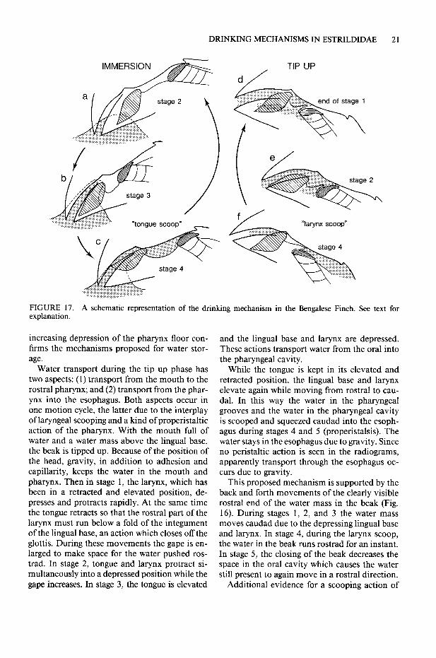
DRINKING MECHANISMS IN ESTRILDIDAE 2 1
TIP UP
FIGURE 17. A schematic reoresentation of the drinking mechanism in the Bengalese Finch. See text for explanation.
increasing depression of the pharynx floor con- firms the mechanisms proposed for water stor- age.
Water transport during the tip up phase has two aspects: (1) transport from the mouth to the rostra1 pharynx; and (2) transport from the phar- ynx into the esophagus. Both aspects occur in one motion cycle, the latter due to the interplay of laryngeal scooping and a kind of properistaltic action of the pharynx. With the mouth full of water and a water mass above the lingual base, the beak is tipped up. Because of the position of the head, gravity, in addition to adhesion and capillarity, keeps the water in the mouth and pharynx. Then in stage 1, the larynx, which has been in a retracted and elevated position, de- presses and protracts rapidly. At the same time the tongue retracts so that the rostra1 part of the larynx must run below a fold of the integument of the lingual base, an action which closes off the glottis. During these movements the gape is en- larged to make space for the water pushed ros- trad. In stage 2, tongue and larynx protract si- multaneously into a depressed position while the gape increases. In stage 3, the tongue is elevated
and the lingual base and larynx are depressed. These actions transport water from the oral into the pharyngeal cavity.
While the tongue is kept in its elevated and retracted position, the lingual base and larynx elevate again while moving from rostra1 to cau- dal. In this way the water in the pharyngeal grooves and the water in the pharyngeal cavity is scooped and squeezed caudad into the esoph- agus during stages 4 and 5 (properistalsis). The water stays in the esophagus due to gravity. Since no peristaltic action is seen in the radiograms, apparently transport through the esophagus oc- curs due to gravity.
This proposed mechanism is supported by the back and forth movements of the clearly visible rostra1 end of the water mass in the beak (Fig. 16). During stages 1, 2, and 3 the water mass moves caudad due to the depressing lingual base and larynx. In stage 4, during the larynx scoop, the water in the beak runs rostrad for an instant. In stage 5, the closing of the beak decreases the space in the oral cavity which causes the water still present to again move in a rostra1 direction.
Additional evidence for a scooping action of

22 J. HEIDWEILLER AND G. A. ZWEERS
TABLE 1. Drinking performances. n = number of observations, SD = standard deviation.
Poephila guffata Lonchura striata
SD n SD n
Mean daily water intake 1.58 ml 0.61 28 1.59 ml 0.87 14 Mean water intake speed 0.50 ml/set 0.16 24 0.27 ml/set 0.06 27 Mean drinking speed 0.44 ml/set 0.20 12 0.04 ml/set 0.01 10
the larynx comes from films of reflex studies of birds fixed in a stereotactic apparatus while drops of water were applied to several points in the mouth and pharynx. These films show that the choana partly closed and the glottis did not close at all when water was applied at the lingual base. But two other reactions were clear: first, the lar- ynx was pulled rostrad underneath a fold of in- tegument of the lingual base; second, strong pro- tractions and retractions moved the drops caudad. The glottis also did not close if the protracted tongue was kept fixed, and so much water was dropped upon the lingual base that it began to run into the trachea. This water was blown out by powerful exhaling.
DRINKING PERFORMANCES
The average water dose taken per immersion cycle was calculated by dividing the measured volume of water ingested per bout by the number of im- mersion cycles that was seen on the films. The average was determined from 22 filmed bouts. The water dose taken per cycle was somewhat larger in the Zebra Finch (0.024 ml, SD = 0.005, n = 12) than in the Bengalese Finch (0.021 ml, SD = 0.005, IZ = lo), but the difference is not significant (P > 0.05 ANOVA model II Sokal and Rohlf 198 1). The average daily water intake, the average water intake speed, and the average drinking speed are listed in Table 1. The two species drink about the same amount per day in our study. The average water intake speed of the Zebra Finch is about two times higher than that of the Bengalese Finch. Because the Zebra Finch is able to swallow the water during intake, its average drinking speed is 10 times higher.
Average drinking speeds for drops of varying volumes are illustrated in Figure 18. This figure clearly shows that the drinking speed of the Zebra Finch increases when drops increase in size, while the drinking speed of the Bengalese Finch is rath- er constant at a low level. Finally, these data show that both species are quite able to drink small drops.
DISCUSSION
EARLIER MODELS FOR ESTRILDID DRINKING ”
The double scoop model proposed in this study for P. guttata does not support the drinking mechanism proposed by Immelmann and Im- melmann (1967). These authors assumed water intake was caused by esophageal suction result- ing from peristaltic movements. Although peri- stalsis indeed occurs, such a mechanism is im- probable for two reasons. First, there are no muscles to enlarge the esophagus. Second, P. gut- tata drinks with a wide open beak which is not completely immersed. Thus even if an area of lower air pressure develops due to peristalsis of the esophagus, air will be swallowed instead of water.
A second generally accepted opinion about es- trildid drinking is also not confirmed by the pres- ent study: e.g., Wickler (196 1) and Immelmann (1965) suggest that Zebra Finches drink by suck- ing in a pigeon-like manner. This requires further discussion. It has been proposed that the drink- ing mechanism of the pigeon (Columba livia)
ml/s .1O-2 8-
n (u 6. 4 0 _ P ‘2 4 P .E G - c7
22 P
sl 4 P 7 I I I I I I I I, 1
.02 .04 .06 .08 0.1
volume of water drops ml
FIGURE 18. Differences in drinking performance of the Zebra Finch and the Bengalese Finch. The graph illustrates the relation between drinking speed (ml/set) and drop volume for the Zebra Finch (circles) and the Bengalese Finch (triangles).

DRINKING MECHANISMS IN ESTRILDIDAE 23
operates like a double suction pump. Zweers (1982a) summarizes this mechanism as follows (p. 3 13): “As a result of the retraction of the tongue in the mouth (acting as a piston in a cyl- inder) low air pressure develops in the buccal cavity and water is sucked into the mouth. Sec- ondly, lower air pressure develops in the pharynx as a result of a depression of its floor, so that the water in the mouth is given a momentum cau- dad, by which it is forced over the larynx into the esophagus. Neither peristaltic action, nor an alternative lower air pressure area is recorded in the esophagus. The collection of the swallowed water at the lowest place occurs by gravity.” This study indicates that the pigeon’s drinking mech- anism is effective only if the gape is very small. In contrast, the beak of the Zebra Finch must be wide open to allow the tongue to make the scoop- ing movements needed for fast drinking. Further, anatomical differences between P. guttata and C. livia make it rather improbable that these birds drink by similar mechanisms.
Whether Zebra Finches drink like honeyeaters, as suggested by Fisher et al. (1972) is still un- known since the drinking mechanism of these birds has not been analyzed on a functional-an- atomical level. The drinking mechanism of Lon- chura seen in the present study, involving two separate sequences of two different kinds of scoops, has not previously been described in birds. A few studies described below specify tip up drinking, but these descriptions point to mech- anisms other than those proposed here for L. striata.
According to White (1970) the chicken (Gal- lus gallus) collects water on the lingual base by quick rostro-caudad movements. In the swallow- ing phase the water runs laterally along the larynx into the esophagus by gravity. Further, in con- trast to the observations for L. striata, Schon- holzer (1959) states that the Black Swan (Cygnus atratus) stretches its neck and points its bill into the air, which probably causes water to run into the esophagus by gravity without tongue or lar- ynx movements. A model for drinking in the Mallard (Anasplatyrhynchos) has been proposed by Kooloos and Zweers (1989). These authors conclude from a quantitative study that water transport through the mouth is accomplished by capillarity and pressure differentials in two small tubes and two chambers, respectively. Delicate lingual motions change the Mallard’s mouth, which is built to expel water, into a mechanism
for water intake. Thus far, no clear parallels can be seen that make it possible to define tip up drinking as based upon one general behavioral mechanism, other than that this is a two-step mechanism-one for water intake during im- mersion and one for swallowing water during tip up-each accomplished by different mecha- nisms.
DIFFERENCES BETWEEN THE TWO SPECIES
The major differences in drinking behavior be- tween the tip down, mono draught drinking P. guttata and the tip up, poly draught drinking L. striata have been described in the literature (e.g., Immelmann and Immelmann 1967). These are confirmed at the level of scene analysis. How- ever, one level of organization lower, the differ- ences are much smaller. The basic behavioral elements of tongue and larynx movements that make up drinking behavior are quite similar in both species. For example, the lingual scooping motion that causes the transport of water to the lingual base is basically the same in both species. The difference is that P. guttata protracts its tongue without elevating the lingual alae. Clear- ly, there is no need for such an elevation: there is no water on the lingual base that might run back into the water box, because each water dose is immediately transported further into the esophagus. For the same reason, P. guttata is able to drink with a much wider gape than L. striata. A further striking similarity occurs when the two species drink very small drops. When P. guttata drinks small drops, it opens its beak, quite like L. striata, but over only a small distance, and the water transport to the lingual base takes place via the same sort of lingual scooping as in L. striata. This similarity explains two features. First, intake speeds in the two species are equal when they drink small drops. Second, L. striata is, de- spite being a tip up drinker, quite able to “suck in” small water volumes by applying the same lingual scooping actions found in P. guttata.
The small differences in the anatomy of the two species may relate to small advantages in- herent in each of the two kinds of drinking. The anatomical differences and their assumed ad- vantages are: L. striata has a somewhat smaller mass of glands in the lingual base so that L. striata may easily pull the larynx underneath the integument to close the glottis. This motion is not needed in P. guttata since the arytenoids close the glottis as soon as a water dose arrives. In L.

24 J. HEIDWEILLER AND G. A. ZWEERS
striata, this reflex pattern is missing, but a large mass of water can nevertheless collect at the lin- gual base. Thus the alternative way of glottis clo- sure, via an integumental fold, also serves in L. striata to enlarge the space for collecting water. Further, the many large palatal papillae of L. striata may be an advantage because they in- crease surface area and keep water on the lingual base by adhesion. In contrast to L. striata, P. guttata has only a few small palatal papillae. This enables the larynx to scoop water along the palate easily.
HISTORICAL POLARITY IN AVIAN DRINKING MECHANISMS
To allow integration of the proposed mecha- nisms into an evolutionary context, ancestral drinking mechanisms need to be known. Unfor- tunately, we know of no relevant studies. There- fore the following approach appears reasonable. Cracraft (1988) argues in a review that it is gen- erally accepted that the galliforms are one lineage of the earliest dichotomy in the monophyletic Neognathae (e.g., Sibley and Ahlquist 1986). Cracraft also shows a summarizing dendrogram in which all avian orders-including the Passer- iformes-radiate from the next point of bifur- cation, except possibly the Anseriformes (but see Olson 1985). If cladograms reflect genealogy, as cladists suggest, Galliformes and Anseriformes are ancestral to Passeriformes and hence to Es- trildidae. Bock (pers. comm.) concludes that es- trildid drinking mechanisms may have devel- oped from galliform-like drinking because of the relative rarity of the estrildid mechanisms. If the rarity and genealogy assumptions hold the con- clusions point in the same direction. In order to make progress in the explanation of the devel- opment of a drinking mechanism, the only op- tion is to assume that the evolution of drinking behavior in all ordines, e.g., Passeriformes, be- gan from a mechanism close to either galliform or anseriform drinking.
Anseriformes have a highly specialized feeding apparatus for straining. Although a tip up drink- ing mechanism is used, it is very complex. A delicate lingual motion pattern modifies the mouth-built to expel water-into a mouth that keeps water in (Kooloos and Zweers 1989). How- ever, an ancestral estrildid drinking behavior may not have been derived from the anseriform drinking mechanism, because of its highly com- promised and specialized design. So far, there is
no reason to consider galliform drinking to be a specialized or highly compromised mechanism. Therefore the main lines of McLelland’s (1979) description-based on White’s (1970) analysis- may preliminarily serve as a starting point for an ancestral mechanism for passeriform, e.g., es- trildid, drinking.
Although further functional-anatomical anal- ysis is needed, White’s description envisions lin- gual retraction as the main cause for the transport of water into the pharynx, while laryngeal re- traction-comparable to what has been called “properistalsis” in this study-serves for trans- port into the esophagus. There is no indication of any lingual or laryngeal scooping. However, it is clear that once a scoop-like element has been developed at the lingual tip, this element is a clear preadaptation for the lingual scoop that op- erates in both estrildid drinking mechanisms ex- amined here.
HISTORICAL POLARITY IN ESTRILDID DRINKING MECHANISMS
The difference between the two kinds of drinking reported here may be explained as a difference in behavioral complexity. The two mechanisms are basically similar in their two main behavioral elements: lingual scooping and laryngeal scoop- ing. The main difference is that in tip up behavior these elements occur in two separate sequences, involving different kinds of motion cycles, whereas in tip down behavior both behavioral elements are incorporated in one single type of motion cycle, which is repeated many times. In L. striata a series of lingual scooping cycles trans- ports water to the rostra1 pharynx during the im- mersion phase, and next a series of laryngeal scooping plus properistaltic motion cycles while the beak is tip up squeezes the water into the esophagus. Neither glottis closure nor esophageal peristalsis is involved. In P. guttutu a lingual scoop moves a water dose into the rostra1 esoph- agus in each motion cycle; it is immediately car- ried into the rostra1 esophagus by a laryngeal scoop, while the glottis closes, the secondary choana narrows and esophageal peristalsis trans- ports the water dose directly into the crop.
It is apparent that the main behavioral ele- ments, which occur separately in L. striata, are closely connected in P. guttata. Further, behav- ioral elements from quite different patterns, such as glottis closure and esophageal peristalsis-part of respiration/vocalization and ingestion, re-

DRINKING MECHANISMS IN ESTRILDIDAE 25
spectively-are found in separate phases in L. striata, but, in P. guttata are seen to be integrated into one motion cycle. Generally speaking, such an increase in complexity may occur as a result of decoupling and recoupling of behavioral ele- ments. First different kinds of behavioral ele- ments occur successively in a chain of subse- quent motion cycles (e.g., L. striata), and then these behavioral elements are found recoupled and functionally present in one highly integrated pattern ofjust one motion cycle (e.g., P. guttata). Thus the tip down mechanism is of a much higher level of complexity (i.e., of integration of func- tionally meaningful behavioral elements per unit of time) than the tip up mechanism. Such an increase in complexity is in itself an argument that indicates historical polarity if the anatomical elements do not differ. An example of decoupling and recoupling of behavioral elements is offered here (see Roth and Wake 1989). Therefore, it is concluded that the tip down mechanism de- scribed here is derived from/advanced in com- parison to the proposed tip up mechanism.
EVOLUTIONARY SCENARIO FOR ESTRILDID DRINKING
Based on the analyses presented, the following scenario is proposed (Fig. 19) and open to further testing (cf. methods in Bock 1985). The ancestral estrildids are assumed to have lived in humid woodlands, some invading nearby open grass- lands bordered by semiarid and arid areas (Im- melmann 1965). This assumption is supported by the fact that all recent Estrildidae feed on grass seeds (Ziswiler 1979). The ancestral estrildids are assumed to have fed upon rather small seeds by pecking, and to have drunk by tip up behavior comparable to the galliform-like mechanisms in- dicated above as the ancestral ones. Selection favoring anatomical elements that enable fast seed eating must certainly have occurred, due to pred- ator pressure on ancestral estrildids feeding in relatively open fields (Immelmann 1965, Im- melmann and Immelmann 1967): proper diges- tion of the small seeds of Gramineae requires that they be husked before being swallowed. This assumption is supported by findings showing that almost all recent Estrildidae husk grass seeds (cf. Ziswiler 1967, 1979), and that husking these seeds is a very time consuming act (Morris 1955). A first requirement for seed husking is that the seeds
be manipulated into a position for being either crushed or cut open (Ziswiler 1965). Only the lingual tip can perform this action. Development of a seedcup composed of numerous keratin hairs at the lingual tip without doubt creates a very useful element for seed manipulation. Previous sections have shown that the development of such a lingual seedcup may serve as an anatom- ical preadaptation for drinking. (Homberger [ 19801 arrived at a similar conclusion for the development of drinking mechanisms in par- rots.) There is apparently no real need for drink- ing by lingual scooping. But since both the scoop element and the required musculature for scoop- ing are available, the behavioral ability to scoop water lingually may have developed neutrally. Once present, small advantages in terms of im- proved water intake may have fixed the lingual scoop mechanism, and moreover, extended the niche: the lingual scoop may have improved the ability to drink the small drops ofwater on blades of grass after rain, or alternatively, to ingest dew- drops. This capability may have generated an energy gain by decreasing travel costs to water, thus increasing time available for feeding. This narrative may serve as an example of the theme of evolutionary change called “possessing the be- havioral capacity to utilize a new ecological op- portunity” (Lauder et al. 1989).
The next step in the scenario is the develop- ment of tip down drinking. Unfortunately nei- ther functional anatomical nor performance analyses are available regarding transport of seeds through the pharynx. But clearly the develop- ment of any anatomical element or motion pat- tern that speeds up the transport of husked seeds through the pharynx is subject to strong predator pressure selection because of the long day of feed- ing in rather open fields. For example, devel- opment of a mechanism that scoops a husked seed from the palatal cavernous body and scrapes it off onto the caudal palatal papillae is a first option. One obvious element that could serve this goal is the rostra1 cricoid edge. Once such a mechanism has developed, it too is of course a clear preadaptation. This preadaptation later makes the laryngeal scoop available for use in double scoop drinking, which allows water to be swallowed 10 times faster than in tip up drinking. The behavioral pattern for double scoop drink- ing could have developed neutrally. But, once available, tip down drinking would have strongly supported the extension of the feeding area for

26 J. HEIDWEILLER AND G. A. ZWEERS
Extension and Selective change Preadaptations from change of habitat in Feeding selection on
mechanisms Feeding mechanisms
Semi-desert
1
Primarily neutral change in Drinking mechanisms
4 Double scoop
(Tip down) Rostra1 cricoid
edge
I Super transport to esophagus
4 Single scoop
(TiD UD) Grasslands
4 /L- Lingual seed cup
/
Husking seeds in mouth
f Woodlands Galliform-like pecking Galliform-like tip up
f / - _ drinking
Ancestral estrildidae
FIGURE 19. Diagram of a tentative evolutionary scenario for estrildid drinking mechanisms in relation to the development of feeding mechanisms under selection from economizing and predator pressure. Increased behavioral complexity occurred primarily neutral by de- and recoupling of behavioral elements. Functionally meaningful patterns may secondarily have been fixed by selection on advantages in extended and subsequently changed habitats such as drinking small drops, drinking upside down, economizing water intake, decrease of traveling costs to water sources, decrease of predator pressure at waterholes.
three reasons: first, drinking places where a bird must drink upside down now became accessible (as observed in Poephila species drinking from cattle troughs; Immelmann 1965). Second, due to the higher rate of drinking for larger dewdrops, that source may meet their water needs; less trav- el from feeding area to water would, again, reduce energy losses. A third advantage of the double scoop mechanism is the fact that predator pres- sure at water holes in semiarid areas would be diminished strongly. However, it should be re- marked that Zebra Finches need additional water because they spend much time bathing. This does not falsify the scenario proposed, because bath- ing may decrease if water becomes scarce. Fur- ther ecomorphological and function morpholog- ical analyses are clearly needed to test these as- sumptions. These analyses should include the Estrildidae living in the dry parts of Africa, to test whether their lack of tip down drinking is due to their more insectivorous feeding habits, as suggested by Immelmann and Immelmann (1967). Again this narrative scenario is felt to be an example of behavioral capacity to use new ecological opportunities (Lauder et al. 1989).
To sum up, the evidence suggests the follow- ing: tip up drinking and tip down drinking are different behavioral patterns comprising highly
similar basic behavioral elements. However, the tip down mechanism is much more complex. Here, all behavioral elements plus some extra elements from different patterns are integrated in one motion cycle, whereas in the tip up mech- anism these elements occur in separate phases and patterns. Increase in behavioral complexity is in itself an argument for historical polarity, if the morphologies are similar. Our anatomical analyses strongly support this argument. Thus, tip down is taken to be derived from tip up drink- ing in Estrildidae. The literature provides argu- ments that estrildid tip up drinking is derived from chicken-like ancestral tip up drinking. An evolutionary scenario is developed here, based upon strong selection on anatomical elements which serve to speed up specialized feeding on seeds of Gramineae, due to predator pressure in open fields. It has been shown that some of these elements are clear preadaptations for specialized drinking mechanisms. The behavioral patterns involved may have developed neutrally by de- coupling and a different but functionally ade- quate recoupling of behavioral elements. But the mechanisms, once available, allowed a strong secondary extension of the feeding area. Selec- tion may then have fixed the increased behav- ioral complexity.

DRINKING MECHANISMS IN ESTRILDIDAE 27
ACKNOWLEDGMENTS MCLELLAND. J. 1979. Digestive svstem. D. 69-181.
The authors would like to thank R. Gunn for correcting In A. S.‘King and J. McLelland [eds.],‘Form and
the text, J. Simons for photographic assistance, and function in birds. Academic Press, London.
M. Brittijn for drawing the figures. MORRIS, K. R. 1955. The seed preferences of certain
finches under controlled conditions. Avic. Msg. 61:271-287.
LITERATURE CITED Orson, S. L. 1985. The fossil record of birds, p.79-
238. In D. S. Farner, J. R. King, and K. C. Parkes ANTHONY, M. 1920. fiber die speicheldriisen der Vo- [eds.], Avian biology. Vol. 8. Academic Press, New
York. gel. Zool. Jahrb. Abt. Syst. Gekol. Geogr. Tiere 4: 545-660.
BAUMEL. J. J. 1979. Osteoloaia. D. 53-121. In .I. J. Baumel, A. S. King, A. My Lucas, J. E. Breazillk, and H. E. Evans [eds.], Nomina anatomica Av- ium. Academic Press, London.
BOCK, W. J. 1985. The arboreal theory for the origin of birds. In M. K. Hecht, J. H. Ostrom, G. Viohl, and P. Wepenhofer [eds.], The beginning of birds. Proc. Int. Archeopteryx Conf. Eichstatt (1984): 199- 207.
CRACRA~, J. 1988. The major clades of birds, p. 339-36 1. In M. Benton [ed.], The phylogeny and classification of tetrapods. Vol. I: Amphibians, reptiles, birds. Clarendon Press, Oxford.
ENGELS, W. L. 1938. Tongue musculature of passer- ine birds. Auk 55:642-650.
FISHER, C. D., E. LINDGREN, AND W. R. DAWSON. 1972. Drinking patterns and behavior of Australian des- ert birds in relation to their ecology and abun- dance. Condor 74: 111-136.
GEORGE, J. C., AND A. J. BERGER. 1966. Avian myol- ogy. Academic Press, New York.
HOMBERGER, D. 1980. Funktionell-morphologische Untersuchungen zur Radiation der Emahrungs- und Trinkmethoden der Papageien (Psittaci). Bonn. Zool. Monogr. 13: l-92.
IMMELMANN, K. 1962. Beitrage zu einer vergleigen- den Biologie australischer Prachtfinken (Spermes- tidae). Zool. Jahrb. Abt. Syst. Oekol. Geogr. Tiere 90: l-l 96.
IMMELMANN, K. 1965. Australian finches in bush and aviary. Angus and Robertson, Sydney.
IMMELMANN, K., AND G. IMMELMANN. 1967. Ver- haltensoekologischen Studien an Afrikanischen und Australischen Estrildiden. Zool. Jahrb. Abt. Syst. Oekol. Geogr. Tiere 94:609-686.
Kootoos, J.G.M., AND G. A. ZWEERS. 1989. Me- chanics of drinking in the Mallard (Anus plutv- rhynchos; Anatidae). J. Morphol. 199327-347:
K~ULIS. V. 1978. Struktur und Verteilune von Tast- rezkptoren im Schnabel-Zungenbereiih von Si- ngvijgeln, im besonderen der Fringillidae. Rev. Suisse Zool. 851385-447.
LAUDER, G. V., A. W. CROMFTON, C. GANS, J. HANKEN, K. F. LIEM, W. 0. MAIER, A. MEYER, R. PRESLEY, 0. C. RIEPPEL, G. ROTH, D. SCHLUTER, AND G. A. ZWEERS. 1989. How are feeding systems inte- grated and how have evolutionary innovations been introduced?. D. 97-l 15. In D. B. Wake and G. Roth [eds.],’ Complex organismal functions: In- tegration and evolution in vertebrates. J. Wiley and Sons, Chichester. England.
POULSEN, H. 1953. A study of incubation responses and some other behavior patterns in birds. Vi- densk. Medd. Dan. Naturhist. Foren. 115: l-l 3 1.
ROTH, G., AND D. B. WAKE. 1989. Conservatism and innovation in the evolution of feeding in Verte- brates. D. 7-21. In D. B. Wake and G. Roth Ieds.1. Complex organismal functions: Integration and evolution in vertebrates. J. Wiley and Sons, Chic- hester, England.
SCH~~NHOUER, L. 1959. Beobachtungen iiber das trinkverhalten bei Zoo-tieren. Zool. Garten (NF) 241345-434.
SIBLEY, C. G., AND J. E. AHLQUIST. 1988. Phylogeny of non-passeriform birds, based on DNA com- parisons. XIX Int. Omithol. Congr. (1986). Pos- ter.
SIMS, R. W. 1955. The morphology of the head of the hawtinch (Coccothraustes coccothraustes). Bull. Br. Mus. Nat: Hist. 13:371-393.
SOKAL, R. R., AND F. J. ROHLF. 1981. Biometry. 2nd ed. W. H. Freeman and Co., San Francisco.
VANDEN BERGE, J. 1979. Myologia, p. 175-219. In J. J. Baumel, A. S. King, A. M. Lucas, J. E. Brea- zille, and H. E. Evans [eds.], Nomina anatomica avium. Academic Press, London.
WHITE, S. S. 1970. The larynx of Gallus domesticus. Ph.D. diss. Univ. of Liverpool, England.
WICKLER, W. 196 1. Uber die Stammesgeschichte und den taxonomischen Wert einiger Verhaltenswei- sen der Vogel. Z. Tierpsychol. 18:320-342.
ZISWILER, V. 1965. Zur Kenntnis des Samenoffnens und der Struktur des homemen Gaumens bei kor- nerfressenden Oscines. J. Omithol. 106: l-48.
ZISWILER, V. 1967. Vergleichend morphologische Untersuchungen am Verdauungstrakt komfres- sender Singvogel zur Abklarung ihrer systema- tischen Stellung. Zool. Jahrb. Abt. Syst. Oekol. Geogr. Tiere 94:427-520.
ZISWILER, V. 1979. Zungenfunktionen und Zungen- versteifung bei granivoren Singvogeln. Rev. Suisse Zool. 86:823-83 1.
ZWEERS, G. A. 197 1. A stereotactic atlas of the brain- stem of the Mallard (Anasplutyrhynchos L.). Stud. Neuro-Anat. 10: l-l 50.
ZWEERS, G. A. 1982a. Drinking of the pigeon (Co- lumba livia L.). Behaviour 80:274-3 17.
ZWEERS, G. A. 1982b. The feeding system of the pi- geon (Columba livia L.). Adv. Anat. Embryol. Cell Biol. 73: l-l 11.
ZWEERS, G. A., AND H. BERKHOUDT. 1987. Larynx and pharynx of crows (Corvus corone L. and C. monedula L.. Passeriformes: Corvidae). Neth. J.
LORENTZ, K. Z. 1939. Vergleichende Verhaltens- Zool. 37:365-393. forschung. Zool. Anz. Suppl. 12:69-102. ZWEERS, G. A., A. F. CH. GERRITSEN, AND P. J. VAN
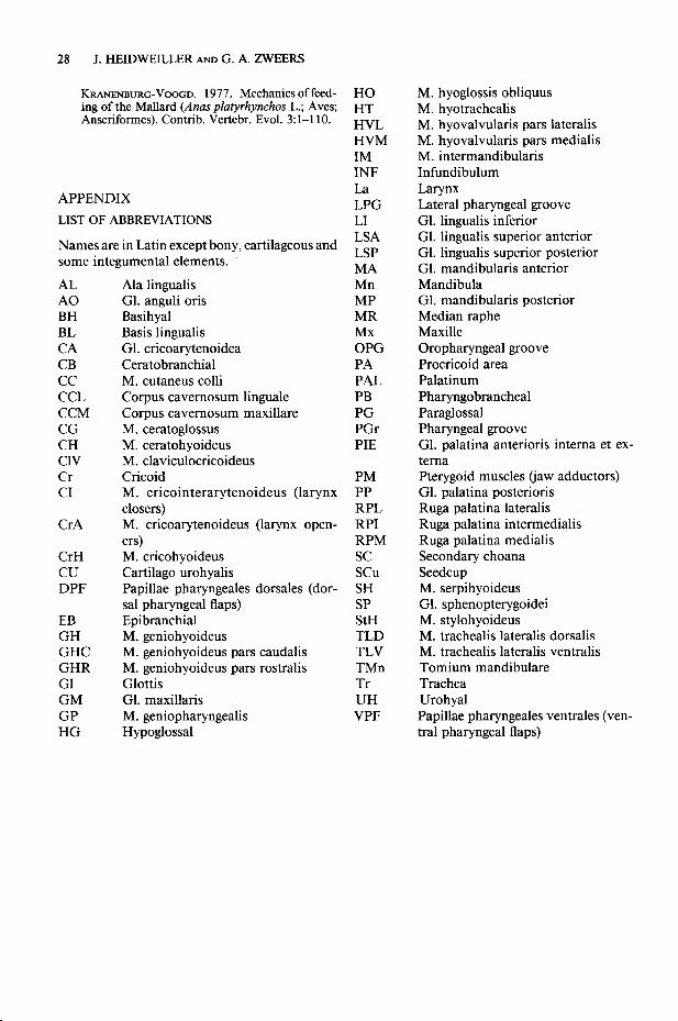
28 J. HEIDWEILLER AND G. A. ZWEERS
KFCANENBURG-VOOGD. 1977. Mechanics of feed- HO ing of the Mallard (Anus platyrhynchos L.; Aves; H-I- Anseriformes). Contrib. Vertebr. Evol. 3:1-l 10. HVL
HVM IM INF
APPENDIX La LPG
LIST OF ABBREVIATIONS LI
Names are in Latin except bony, cartilageous and LSA
some integumental elements. LSp
MA AL A0 BH BL CA CB cc CCL CCM CG CH ClV Cr CI
Ala lingualis Gl. anguli oris Basihyal Basis lingualis Gl. cricoarytenoidea Ceratobranchial M. cutaneus colli Corpus cavernosum linguale Corpus cavemosum maxillare M. ceratoglossus M. ceratohyoideus M. claviculocricoideus Cricoid M. cricointerarytenoideus (larynx closers) M. cricoarytenoideus (larynx open- ers) M. cricohyoideus Cartilago urohyalis Papillae pharyngeales dorsales (dor- sal pharyngeal flaps) Epibranchial M. geniohyoideus M. geniohyoideus pars caudalis M. geniohyoideus pars rostralis Glottis Gl. maxillaris M. geniopharyngealis Hypoglossal
Mn MP MR Mx OPG PA PAL PB PG PGr PIE
CrA
CrH cu DPF
EB GH GHC GHR Gl GM GP HG
PM PP RPL RPI RPM SC scu SH SP StH TLD TLV TMn Tr UH VPF
M. hyoglossis obliquus M. hyotrachealis M. hyovalvularis pars lateralis M. hyovalvularis pars medialis M. intermandibularis Infundibulum Larynx Lateral pharyngeal groove Gl. lingualis inferior Gl. lingualis superior anterior Gl. lingualis superior posterior Gl. mandibularis anterior Mandibula Gl. mandibularis posterior Median raphe Maxille Oropharyngeal groove Procricoid area Palatinum Pharyngobrancheal Paraglossal Pharyngeal groove Gl. palatina anterioris intema et ex- tema Pterygoid muscles (jaw adductors) Gl. palatina posterioris Ruga palatina lateralis Ruga palatina intermedialis Ruga palatina medialis Secondary choana Seedcup M. serpihyoideus Gl. sphenopterygoidei M. stylohyoideus M. trachealis lateralis dorsalis M. trachealis lateralis ventralis Tomium mandibulare Trachea Urohyal Papillae pharyngeales ventrales (ven- tral pharyngeal flaps)