drug coated balloon vs drug eluting stent in compex sfa...
TRANSCRIPT

Drug coated balloon vs drug eluting stent in compex SFA lesions
Yes, DCB are definitelysuperior
Frank Vermassen

Disclosure
Speaker name: Frank Vermassen
I have the following potential conflicts of interest to report:
Consulting: Medtronic, Abbott Vascular, Terumo, Boston
Scientific, Spectranetics,
Employment in industry
Shareholder in a healthcare company
Owner of a healthcare company
Other(s)

Primary vs selective stenting in the SFASurvival free of vascular events
Becquemin (J Vasc Surg 2003)
PTA
Stent

Early recoil,
dissection
Negative vessel
remodelling
Neo-intimal
hyperplasia
Reasons for restenosis
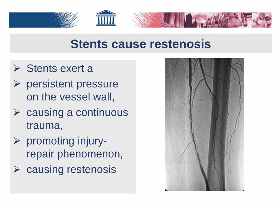
Stents exert a
persistent pressure
on the vessel wall,
causing a continuous
trauma,
promoting injury-
repair phenomenon,
causing restenosis
Stents cause restenosis
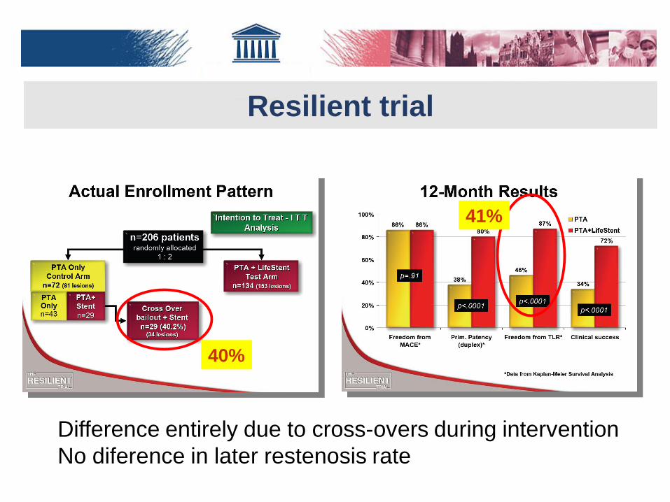
Resilient trial
40%
41%
Difference entirely due to cross-overs during intervention
No diference in later restenosis rate

Drug-elution to inhibit SMC proliferation and intimal hyperplasia
Cascade of events leading to wound
healing also leads to restenosis
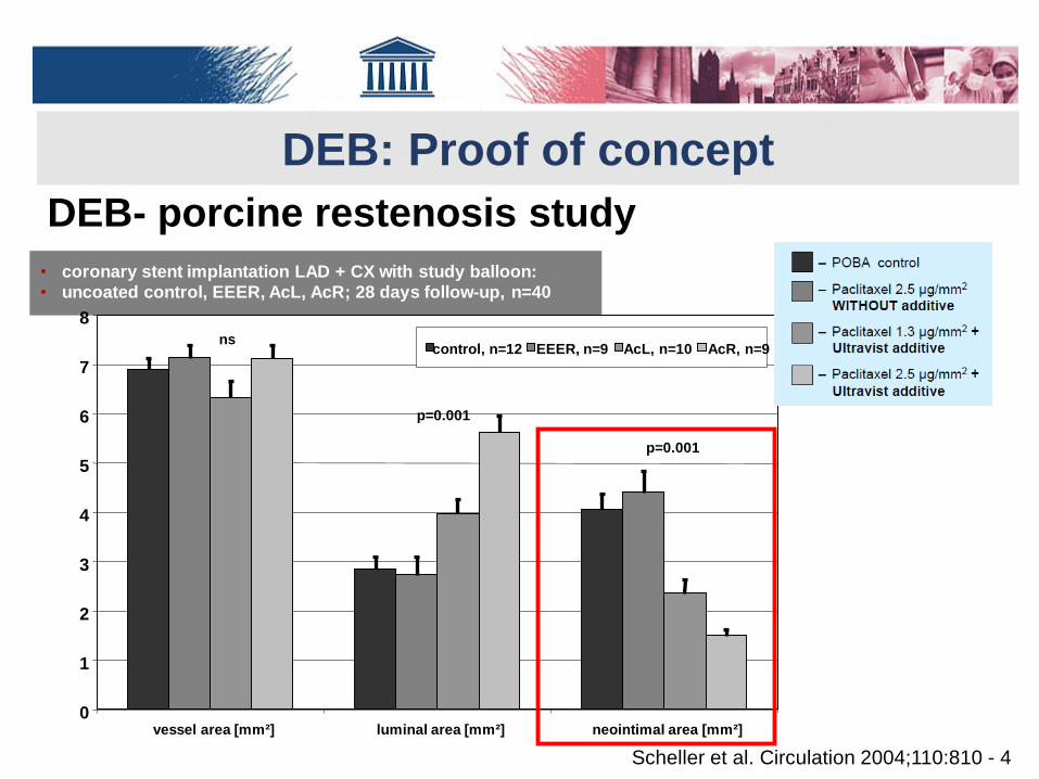
DEB: Proof of concept
0
1
2
3
4
5
6
7
8
vessel area [mm²] luminal area [mm²] neointimal area [mm²]
control, n=12 EEER, n=9 AcL, n=10 AcR, n=9ns
p=0.001
p=0.001
Scheller et al. Circulation 2004;110:810 - 4
• coronary stent implantation LAD + CX with study balloon:
• uncoated control, EEER, AcL, AcR; 28 days follow-up, n=40
DEB- porcine restenosis study

6 DEB Technologies / 7 Trials (6-month LLL Primary Endpoint)
[1] G.Tepe et al. - NEJM 2008; [2] M.Werk et al. - Circulation 2008; [3] D.Scheinert - TCT 2012 oral presentation; [4] M.Werk et al. - Circulation CI
2012; [5] D.Scheinert – EuroPCR 2012 oral presentation; [6] D.Scheinert – LINC 2013 oral presentation; [7] P.Peeters – LINC 2013 oral presentation
Short term results


LEVANT II – 1 yr
1 yr
1 yr
62.5%
82.7%
92.3%
52.6%
• Lutonix DEB vs POBA
• 476 patients randomized 2:1
• Rutherford cat: 2-4
• Single de novo lesions > 70%
• < 15 cm length
• SFA or prox. PA
• Mean lesion length: 6.3 cm

IN.PACT SFA – 1 yr
(p<0.001 by log-rank test)
(p<0.001 by log-rank test)
• IN.PACT admiral vs POBA
• 331 patients randomized 2:1
• Rutherford cat: 2-4
• Single de novo lesions > 70%
• 4-18 cm length (occlusions < 10 cm)
• SFA or prox. PA
• Mean lesion length: 8.9 cm
Freedom from binary restenosis
Freedom from CD-TLR

Ranger (Boston Scientific) Illumenate (Spectranetics)
Preliminary results with other DCB
First in men study
50 DCB – 1 yrRCT: DCB vs POBA 2:1
105 Patients
Freedom from TLR Primary patency
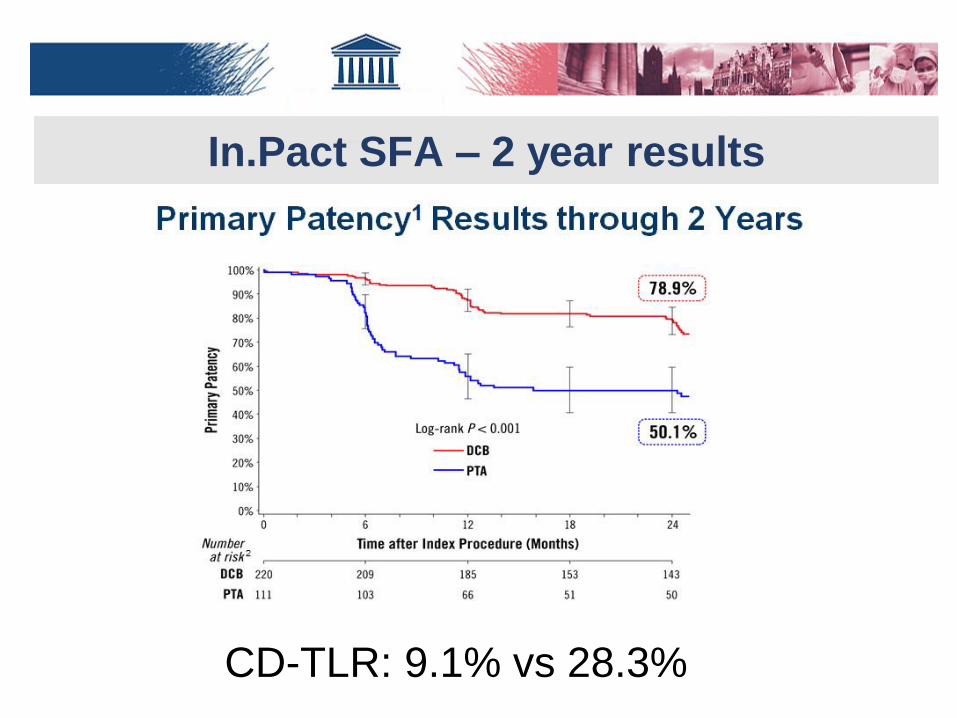
In.Pact SFA – 2 year results
CD-TLR: 9.1% vs 28.3%
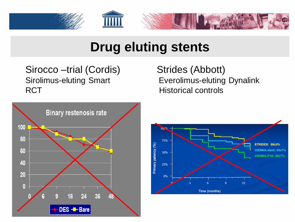
Drug eluting stents
Sirocco –trial (Cordis)Sirolimus-eluting Smart
RCT
Strides (Abbott)Everolimus-eluting Dynalink
Historical controls

Drug-eluting stents
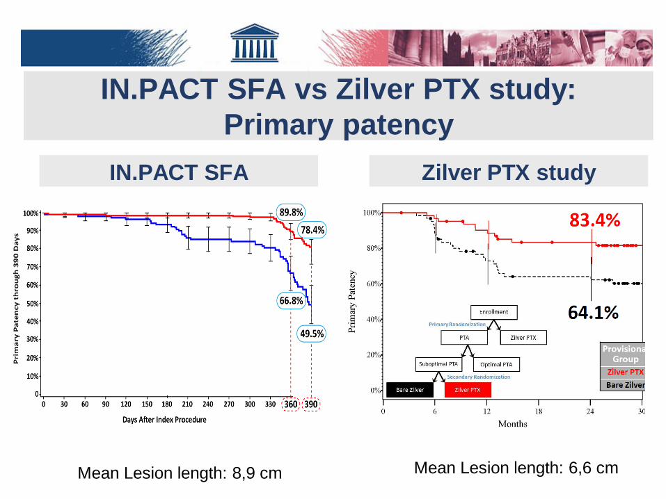
IN.PACT SFA vs Zilver PTX study:
Primary patency
IN.PACT SFA Zilver PTX study
Mean Lesion length: 8,9 cm Mean Lesion length: 6,6 cm

IN.PACT SFA vs Zilver PTX study:
Freedom from CD-TLR
IN.PACT SFA Zilver PTX study
Mean Lesion length: 8,9 cm Mean Lesion length: 6,6 cm

1-year SFA results (%)
Katsanos K, et al. Bayesian meta-analysis in the femoropopliteal artery. JVS 2014
Baseline risk adjusted random effects mixed treatment comparison
Freedom from Restenosis at 1
YearFreedom from TLR at 1 Year

Long-term: Probability best
Katsanos K, et al. Bayesian meta-analysis in the femoropopliteal artery. JVS 2014
Baseline risk adjusted random effects mixed treatment comparison
Freedom from Restenosis Freedom from TLR

In.Pact SFA subgroups
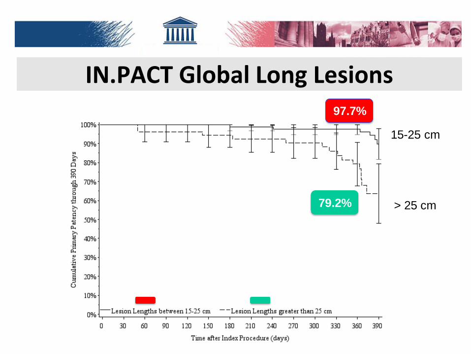
IN.PACT Global Long Lesions97.7%
79.2%
15-25 cm
> 25 cm
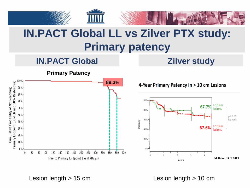
IN.PACT Global LL vs Zilver PTX study:
Primary patency
89.3%
Primary Patency
IN.PACT Global Zilver study
Lesion length > 15 cm Lesion length > 10 cm

DEB vs. DES in long SFA lesions
228-Patients retrospective, propensity score analysis
(Zeller T. et al. JEVT 2014: 21: 39-368)

IN.PACT® Global CTO Imaging Cohort
Procedure Success 100%
(125/125)
Clinical Success 99.2%
(124/125)
Pre-dilatation94.4%
(119/126)
Post-dilatation50%
(62/126)
Provisional Stent 46.8%(59/126)
Lesions (N) 128
Lesion type- de novo- restenosis- ISR
92.2% (118/128)
7.8% (10/128)
0%
Lesion Length (mean ±SD) 22.90± 9.75 cm
Occluded Lesion Length 11.97± 8.11
Calcification 71.2%% (89/125)
RVD (mm ±SD) 5.056 ± 0.657
Diameter Stenosis (% ±SD) 100%
Dissections: None 32.8% (42/128)
A-C 43.8% (56/128)
D-F 23.4% (30/128)
Dierk Scheinert, MD Presented at Veith Symposium 2016
Primary patency rate at 12 Mo= 84.4% (95 cases)

DEB vs DES for In stent restenosis
• Freedom from TLR superior with DCB over DES
Soukas LINC 2015

• Independent, prospective, multicentre single arm study
• 105 pts
• Lesion length 251.71 ±78.89 mm.
– De novo 94.6%
– CTO 49.5%
– Provisional stenting 10.5%
89.3%
77.2%
Primary Patency
• Primary patency at 360 days
89.3%
• Freedom from CD-TLR 96%
• MAE composite at 12mo 6.9%
• Thrombosis: 1% (1 event)
Micari A et al. JACC 2016; 9: 950-6
TASC C & D - SFA- Long Study at 1 Yr
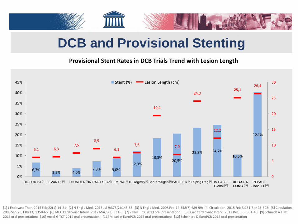
DCB and Provisional Stenting
[1] J Endovasc Ther. 2015 Feb;22(1):14-21; [2] N Engl J Med. 2015 Jul 9;373(2):145-53; [3] N Engl J Med. 2008 Feb 14;358(7):689-99; [4] Circulation. 2015 Feb 3;131(5):495-502; [5] Circulation. 2008 Sep 23;118(13):1358-65; [6] JACC Cardiovasc Interv. 2012 Mar;5(3):331-8; [7] Zeller T CX 2013 oral presentation; [8]. Circ Cardiovasc Interv. 2012 Dec;5(6):831-40; [9] Schmidt A LINC 2013 oral presentation; [10] Ansel G TCT 2014 oral presentation; [11] Micari A EuroPCR 2015 oral presentation; [12] Scheinert D EuroPCR 2015 oral presentation
6,7%2,5% 4,0%
7,3% 9,0%
12,3%
18,3%20,5%
23,3% 24,7%
10,5%
40,4%
6,1 6,37,5
8,9
6,17,6
19,4
7,0
24,0
12,2
25,126,4
0
5
10
15
20
25
30
0%
5%
10%
15%
20%
25%
30%
35%
40%
45%
BIOLUX P-I LEVANT 2 THUNDER IN.PACT SFA FEMPAC IT Registry Bad Krozingen PACIFIER Leipzig Reg. IN.PACTGlobal
DEB-SFA-Long IN.PACTGlobal LL
Provisional Stent Rates in DCB Trials Trend with Lesion Length
Stent (%) Lesion Length (cm)
LEVANT 2[2] THUNDER [3]IN.PACT SFA[4]FEMPAC [5] IT Registry [6] Bad Krozigen [7] PACIFIER [8] Leipzig Reg.[9]BIOLUX P-I [1] DEB-SFA
LONG [11]
IN.PACT
Global LL[12]
IN.PACT
Global [10]

DEB and STENTS: DEBATE SFA
• DEB + stent vs PTA + stent
• Single centre RCT (Liistro F.)
• 110 patients randomized 1:1
• Rutherford cat: 3-6
• SFA or prox. PA
• Concomitant PTA BTK > 50%
• Mean lesion length: 9.5 cm
12-month Restenosis and TLR (per lesion)
DEB+Stent PTA+Stent
Restenosis per lesion length Restenosis per Revasc Technique
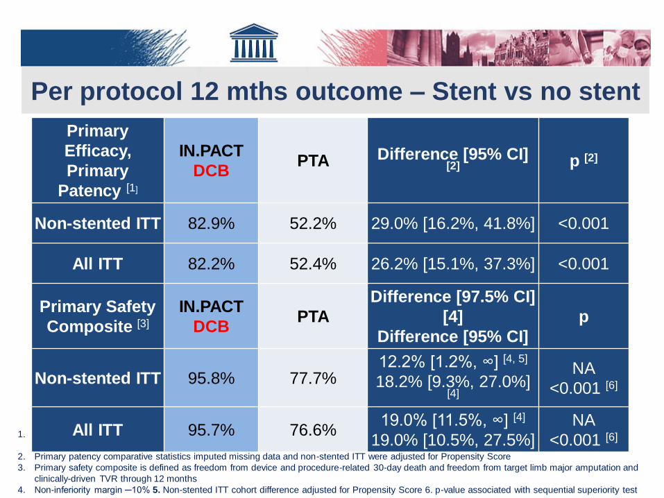
1. Primary patency is defined as freedom from clinically-driven TLR and freedom from restenosis as determined by duplex ultrasound (DUS) Peak
Systolic Velocity Ratio (PSVR) ≤ 2.4
2. Primary patency comparative statistics imputed missing data and non-stented ITT were adjusted for Propensity Score
3. Primary safety composite is defined as freedom from device and procedure-related 30-day death and freedom from target limb major amputation and
clinically-driven TVR through 12 months
4. Non-inferiority margin ─10% 5. Non-stented ITT cohort difference adjusted for Propensity Score 6. p-value associated with sequential superiority test
Primary
Efficacy,
Primary
Patency [1]
IN.PACT
DCBPTA Difference [95% CI]
[2] p [2]
Non-stented ITT 82.9% 52.2% 29.0% [16.2%, 41.8%] <0.001
All ITT 82.2% 52.4% 26.2% [15.1%, 37.3%] <0.001
Primary Safety
Composite [3]
IN.PACT
DCBPTA
Difference [97.5% CI]
[4]
Difference [95% CI]
p
Non-stented ITT 95.8% 77.7%12.2% [1.2%, ∞] [4, 5]
18.2% [9.3%, 27.0%] [4]
NA
<0.001 [6]
All ITT 95.7% 76.6%19.0% [11.5%, ∞] [4]
19.0% [10.5%, 27.5%]
NA
<0.001 [6]
Per protocol 12 mths outcome – Stent vs no stent

Algorythm for treatment of SFA-lesions
standard PTA
YESde-novo, short
(<4 cm), no-CTO?
Restenosis Pre-Dilatation for
CTO / sub-occl. / Ca++
Flow-limit Dissection or
residual stenosis >50%?
Post-Dilatation:
Success?
YES
YES
NO
Stent
NO
DCB
NO

Conclusions
• DCB results are at least equivalent to DES results,
even in complex lesions
• DCB does not leave a metallic implant, causing
continuous harm to the vessel wall, and
hampering later treatment
• If needed DCB can be combined with a bare metal
stent without influencing the results
• DCB with provisonal stenting is more cost-
effective than routine DES implantation
DCB always wins