drug discov ther 2008; 2(3):140-155. review
TRANSCRIPT
www.ddtjournal.com
Drug Discov Ther 2008; 2(3):140-155. 140
Microfabrication-derived DDS: From batch to individual production
Kanji Takada*
Department of Pharmacokinetics, Kyoto Pharmaceutical University, Kyoto, Japan.
*Correspondence to: Dr. Kanji Takada, Department of Pharmaceutics, Kyoto Pharmaceutical University, Yamashina-ku, Kyoto 607-8412, Japan; e-mail: [email protected]
ABSTRACT: As a result of recent advances in microfabrication technology (MFT), microparticles including microcapsules and microspheres can be prepared individually and the disadvantages of the conventional microparticles produced by batch production, i.e. (i) low loading efficiency, (ii) large size variation, and (iii) initial burst release, have been remedied. In addition, all conventional microparticles have the same structure, a spherical shape, so they have only one function, sustained release. Three-layer microcapsules (TLMCs) have been designed to address these issues. TLMCs consist of a surface layer, a drug carrying layer, and a basement layer. TLMCs have sustained release as well as adhesiveness and targeting functions. TLMCs are prepared using ink-jet printer nozzle technology. The obtained TLMCs are used for the oral delivery of peptide/protein drugs and long-term sustained-release injection preparation. In addition, self-dissolving micropiles (SDMPs) can be individually produced by MFT as a percutaneous preparation. MFT allows biopharmaceutical drugs like insulin, erythropoietin, and growth hormone to be absorbed through the skin. Thus, advances in MFT have accelerated the development of pharmaceutical technology.
Keywords: Microfabrication, DDS, Three-layer microcapsules, Micropiles, Individual production
1. Introduction
Many scientists are interested in nanotechnology, and governments are supporting scientific research on nanotechnologies. In the field of pharmaceutics, nanotechnology is an attractive technology, and research on nanocarr iers l ike l iposomes and nanospheres has been widely performed (1-3).
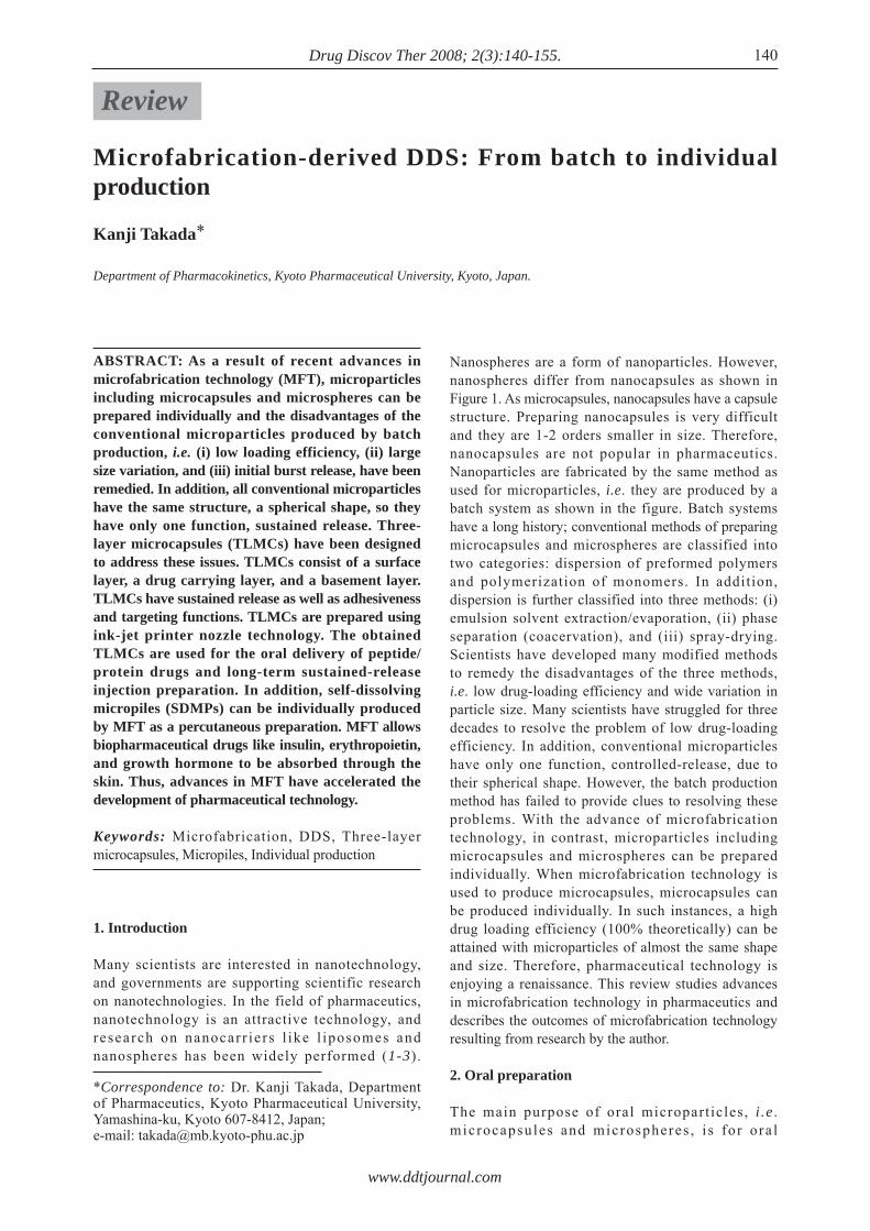
Nanospheres are a form of nanoparticles. However, nanospheres differ from nanocapsules as shown in Figure 1. As microcapsules, nanocapsules have a capsule structure. Preparing nanocapsules is very difficult and they are 1-2 orders smaller in size. Therefore, nanocapsules are not popular in pharmaceutics. Nanoparticles are fabricated by the same method as used for microparticles, i.e. they are produced by a batch system as shown in the figure. Batch systems have a long history; conventional methods of preparing microcapsules and microspheres are classified into two categories: dispersion of preformed polymers and polymerization of monomers. In addition, dispersion is further classified into three methods: (i) emulsion solvent extraction/evaporation, (ii) phase separation (coacervation), and (iii) spray-drying. Scientists have developed many modified methods to remedy the disadvantages of the three methods, i.e. low drug-loading efficiency and wide variation in particle size. Many scientists have struggled for three decades to resolve the problem of low drug-loading efficiency. In addition, conventional microparticles have only one function, controlled-release, due to their spherical shape. However, the batch production method has failed to provide clues to resolving these problems. With the advance of microfabrication technology, in contrast, microparticles including microcapsules and microspheres can be prepared individually. When microfabrication technology is used to produce microcapsules, microcapsules can be produced individually. In such instances, a high drug loading efficiency (100% theoretically) can be attained with microparticles of almost the same shape and size. Therefore, pharmaceutical technology is enjoying a renaissance. This review studies advances in microfabrication technology in pharmaceutics and describes the outcomes of microfabrication technology resulting from research by the author.
2. Oral preparation
The main purpose of oral micropart icles, i .e . microcapsules and microspheres , i s for ora l
Review
www.ddtjournal.com
Drug Discov Ther 2008; 2(3):140-155. 141
sustained-release preparat ion. Both chemical and natural polymers are used as a wall-forming material. Cellulose and cellulose derivatives such as hydroxypropylmethyl cellulose (HPMC), ethyl cellulose (EC), cellulose acetate (CA), cellulose acetate trimellitate (CAT), cellulose acetate butyrate (CAB), cellulose acetate phthalate, cellulose acetate propionate, hydroxypropylmethyl cellulose phthalate (HPMCP), hydroxypropylmethyl cellulose acetate succinate (HPMCAS), carboxymethyl cellulose (CMC), methyl cellulose (MC), sodium cellulose sulfate, and sodium carboxymethyl cellulose are used as chemical materials. Natural polymers that are used as a wall-forming material include chitosan, gelatin, and alginate. The polymerization method has not been used for oral pharmaceutical preparations because of the safety problem of the polymers obtained. In pharmaceuticals, the safety problem is crucial. When the polymerization method is used, polymerized substances with different degrees of polymerization are formed. If safety studies are performed with each polymerized substance, the
production cost of the microparticles will increase tremendously. Therefore, the dispersion method is generally used. Using these polymers as wall-forming materials, microcapsules and microspheres were prepared by both of the methods mentioned above. However, each method has both advantages and disadvantages. The coacervation method can be performed under room temperature. However, a coacervating agent is needed. Therefore, the coacervation method often suffers from residual solvents and coacervating agents detected in the final microcapsules. In addition, each polymer requires its own coacervating agent, so there is no universal rule for the combination of a wall-forming polymer material and coacervating agent. The solvent extraction/evaporation method has been widely used in pharmaceutics to prepare microcapsules. This requires the evaporation of the solvent by increasing the temperature during the preparation process. Therefore, the possibility of degradation increases when drugs that are susceptible to heating,
Figure 1. Conventional micro- and nano-particles prepared by batch production method.
Microspheres
Nanocapsules
Nanrospheres
Nanoparticles
Microcapsules
Microparticles
Batch productionI. polymerization of monomersII. dispersion of preformed polymers
1. Emulsion solvent extraction/evaporation method
2. Phase separation (coacervation) method
3. Spray-drying method
103-105 particles/ batch
・ShapeSpherical
・Function Monofunction(Sustained-
release)
www.ddtjournal.com
Drug Discov Ther 2008; 2(3):140-155. 142
In the last two decades, research focused on the wall-forming material, and natural polymers like sodium alginate were introduced in microcapsules and microspheres. Reports mentioned, for example, dic lofenac sodium microspheres prepared by emuls i f icat ion (46 ) , L- lacta te dehydrogenase microparticles prepared by spray-drying (47), and indomethacin microspheres prepared by precipitation (48 ) . Egg a lbumin mic rospheres con ta in ing nitrofurantoin were prepared by phase separation (49). In addition, chitosan was used as a wall-forming material and ketoprofen was used as the core drug for preparation by emulsification/solvent evaporation (50). In addition, melatonin was loaded onto chitosan microcapsules by ionotropic gelation (51). Chitosan microspheres and nanoparticles were applied to insulin (52,53). The review by Kas provides useful information on microparticles made of chitosan (54). The purpose of those microparticles was to provide oral sustained-release preparations. On the other hand, mucoadhesive chitosan microspheres were prepared by spray-drying, and the interaction between the obtained microspheres and rat small intestinal mucosal tissue was investigated (55 ) . In addi t ion, chi tosan microspheres and
like biopharmaceuticals including peptide/protein drugs, are used as the active pharmaceutical ingredients (API). In addition, the microcapsules obtained have a substantial variation in size. The review by Freitas et al. provides useful information on microencapsulation by the solvent evaporation/extraction method (4). T h e s p r a y - d r y i n g m e t h o d i s s i m p l e a n d microspheres are easily obtained. This method cannot produce authentic microcapsules. However, microspheres can be converted to microcapsules by modifying the surface of the microspheres during the formation process. The disadvantage of this method is the difficulty in limiting the size of microspheres. Li et al. adequately described the conventional large-scale production of these microparticles by spray-drying method (5). Table 1 summarizes the research on microparticles, indicating the core substance, API, wall-forming mate r i a l , and method o f p repara t ion . These microparticles were prepared with either of the aforementioned methods or a modified form of one of those methods. In all cases, the obtained microparticles are spherical and have only one function, sustained-release of the formulated API.
Table 1. Microcapsules and microspheres as oral sustained-release preparation
CA, cellulose acetate; CAB, cellulose acetate butyrate; CAP, cellulose acetate phthalate; CAT, cellulose acetate trimellitate; CMC, carboxymethylcellulose; CMEC, carboxymethylethylcellulose; EC, ethylcellulose; HPC, hydroxypropylcellulose; HPMC, hydroxypropylmethylcellulose; HPMCP, hydroxypropylmethylcellulose phthalate; MC, microcapsules; MP, microparticles; MS, microspheres; NS, nanospheres; PHBV, poly(3-hydroxybutyrate-co-3-hydroxyvalerate); PMMA, polymethyl methacrylate; PVA, poly(vinyl alcohol).
Drug Wall-forming material Method Reference
Acetylsalicylic acid Eudragit RS MS solvent evaporation (6,7) CMEC MC neutralization reaction (8) Eudragit RS MS solvent partition (9)Allopurinol EC MC solvent evaporation (10)Bacampicillin Eudragit E MS solvent evaporation (11)Bitolterol EC MC phase separation (12)Chlorothiazide whey protein MC cross-link (13)Dexamethasone Eudragit S100 NS spray-dry (14)Diclofenac Na CAB, PVA MS solvent evaporation (15) CMC MS crosslink (16)Fenoterol EC MS solvent evaporation (17)5-fl uorouracil EC MS solvent evaporation (18)Furosemide EC MS spherical crystallization (19)Ibuprofen EC MS solvent evaporation (20) CAB MS solvent evaporation (21) PHBV MC solvent evaporation (22) Eudragit RS MS emulsion solvent diffusion (23)Indomethacin EC, Eudragit RL MS solvent evaporation (24,25) Polyesters NS spray-dry (26)Isosorbide dinitrate EC/HPC MC oil-in-oil emulsion evaporation (27)Ketoprofen CAT, EC MS spray dry (28) CAB, HPMCP MS spray-dry (29) Eudragit RS MS coacervation/spray-dry (30)Nifedipine cetearyl alcohol/poloxamer MP hot air coating (31)Nitrofurantoin CMC MC coacervation (32,33)Pantoprazole Eudragit S100/HPMC MP spray-dry (34)Piroxicam Hyaluronate MS spray-dry (35)Propranolol HCl CAB MC emulsion non-solvent addition method (36)Quercetin PMMA MC solvent evaporation (37)Sulfamethoxazole CAP MC spray-dry (38)Theophylline HPMC MO spray-drying (39,40) CMC-Na, HPMC MC spray-drying (41) EC MC emulsion non-solvent addition (42) Polyglycerol esters of fatty acids MS spray-chilling (43) Eudragit RL, Eudragit RS MC phase separation (44)Verapamil HCl CA, cellulose acetate propionate, CAB MS emulsion-solvent evaporation (45)
www.ddtjournal.com
Drug Discov Ther 2008; 2(3):140-155.
nanoparticles were prepared for the colon delivery of prednisolone (56) and oral delivery of protein (57). Pargaonkar et al. (58) used a new method, electrostatic layer-by-layer (LbL) self-assembling, to make core-shell structures for encapsulation of dexamethasone microcrystals with a polyelectrolyte shell. The LbL self-assembly process was used to encapsulate dexamethasone particles with up to five double layers formed by alternating the adsorption of positively charged poly(dimethyldiallyl ammonium chloride), negatively charged sodium poly(styrenesulfonate), and, depending on the pH, positively or negatively charged gelatins of type A (acid pretreated/porcine gelatin) or type B (alkali processed/bovine gelatin) onto the surface of the negatively charged dexamethasone particles. Direct surface modification of dexamethasone microcrys ta l s v ia the LbL process produced monodispersed suspensions with diffusion-controlled sustained drug release via the polyelectrolyte multilayer shell. Although many studies have been performed with oral microparticle preparations, a high drug loading efficiency independent on the method of preparation was not attained. All of the methods of preparing these microparticles are batch production methods.
Since the primary goal of oral microparticles, i.e., providing an oral sustained-release preparation, has been attained, scientists are now working to develop an oral delivery system for peptide/protein drugs with microparticle technology. Peptide/protein drugs undergo hydrolysis before being absorbed by the gastrointestinal (GI) tract. Microparticles are a solid preparation and can protect peptide/protein drugs from attack by hydrolytic enzymes. Cui et al. prepared insulin loaded copoly(lactic/glycolic) acid (PLGA)-hydroxypropylmethyl cellulose (HP55) nanoparticles as an oral DDS (59). The nanoparticles were prepared by diffusion of a modified emulsion solvent in water, and their physicochemical characteristics, drug release in vitro, and hypoglycemic effects in diabetic rats were evaluated. The particle sizes of the PLGA nanoparticles (PNP) and PLGA-HP55 nanoparticles (PHNP) were 150-169 nm, and the drug loading rates were 50.3-65.4%. The initial burst release of insulin from the nanoparticles in simulated gastric fluid over 1 h was 50.5-19.8%. In diabetic rats, the relative bioavailability (BA) of insulin from PNP and PHNP was, in comparison to subcutaneous (s.c.) injection (1.0 IU/kg) of insulin, 3.68-6.27%. Ye et al. (60) prepared chitosan and sodium alginate microcapsules containing insulin by LbL self-assembly.
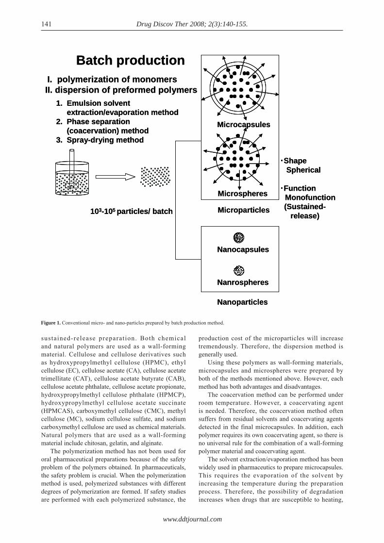
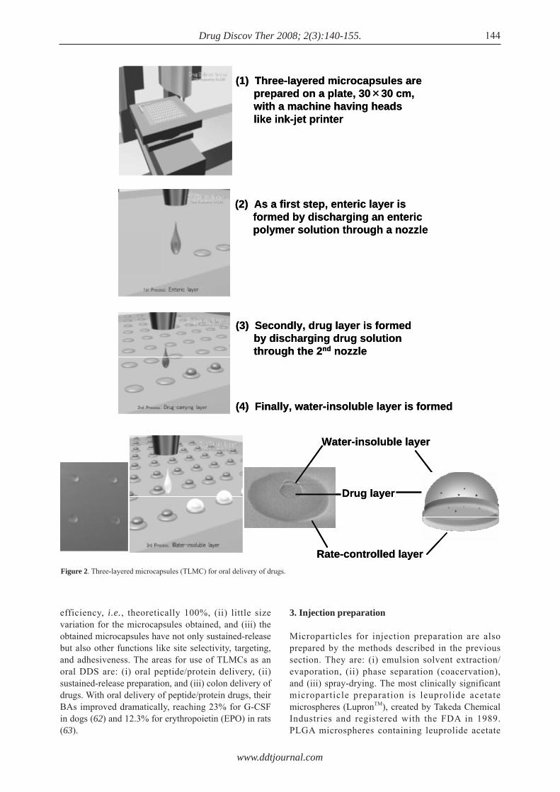
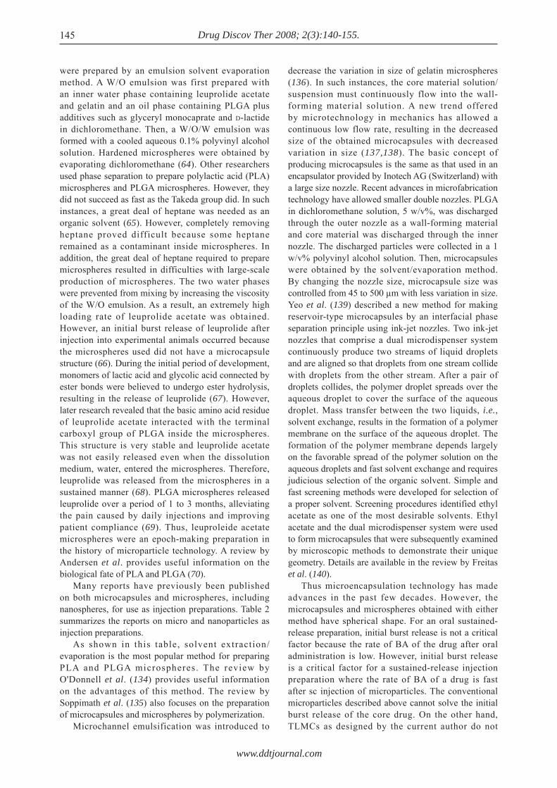
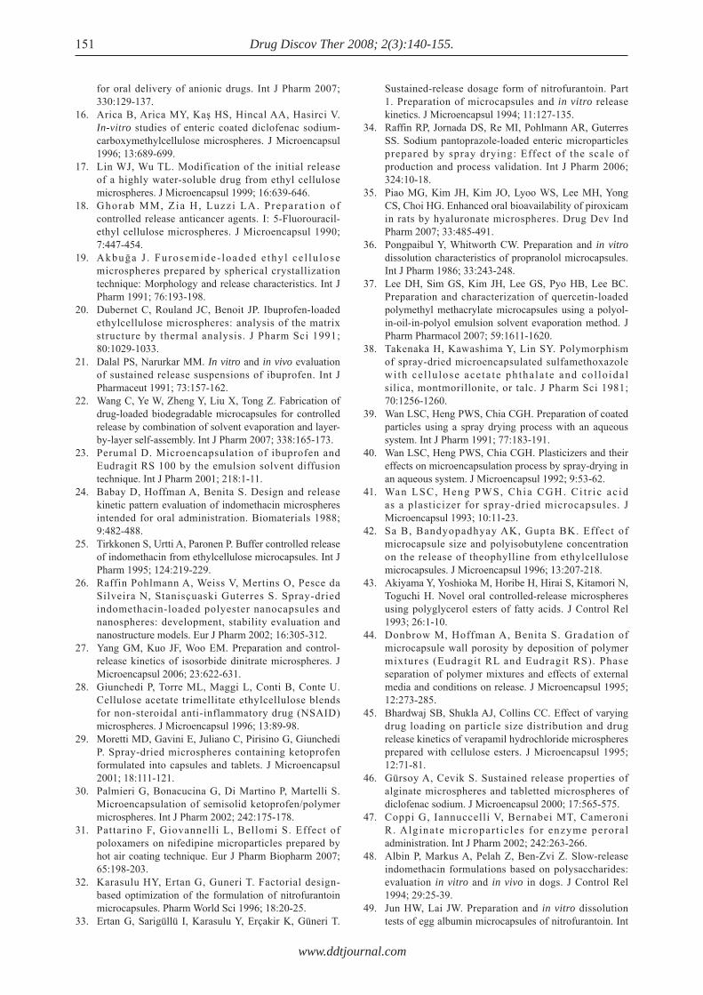
In contrast, the current author designed three-layer microcapsules (TLMCs). TLMCs were prepared individually. Figure 2 shows the basic method of preparing TLMCs by discharge as is widely used in printing technology, where it is known as the ink-jet method. As each TLMC is prepared individually, the obtained microcapsules have far less variation in
shape and size than conventional microcapsules. In addition, TLMCs do not have only one function, i.e. controlled release but other functions like targeting and adhesiveness. TLMCs were used in an oral preparation as a gastrointestinal (GI) mucoadhesive patch system known as GI-MAPS. GI-MAPS was designed to surmount the two hurdles for oral peptide/protein preparations, i.e. hydrolytic degradation by digestive enzymes and poor membrane permeability of peptide/protein drugs due to their 3D structures. As many conventional oral drug delivery systems (DDS) including absorption enhancers, emulsions, liposomes, and micro- and nano-capsules, protein unfolding technology, protein conjugates, and colon delivery technology have been examined to develop oral peptide/protein drugs. However, trials of all of these drugs have all faced the hurdle of a low BA because the dilution and spread of an absorption enhancer in the GI tract reduces the effectiveness of the absorption enhancer on peptide/protein drugs. GI-MAPS is designed to solve these problems. GI-MAPS consists of three layers: (i) a water-insoluble basement membrane, (ii) a drug-carrying layer, and (iii) a pH sensitive bioadhesive surface membrane. After oral administration, the surface layer dissolves at the targeted intestinal site and adheres to the small intestinal wall, where a closed space is created at the target site of the GI mucosa by adherence to the mucosal membrane. As a result, both drug and absorption enhancer coexist in the closed space and a high drug concentration gradient is formed between the system and enterocytes, contributing to the enhanced absorption of peptide/protein drugs because peptide/protein drugs are absorbed by a passive diffusion mechanism. As a result, the absorption enhancer is used to full advantage.
Microfabrication technology has been developed to prepare micron-sized GI-MAPS with a diameter of 500-1,000 μm. Figure 2 shows a manufacturing process using this method; a large-scale GI-MAPS-producing machine was developed in 2008. This machine has three to four nozzles that are modified to discharge a polymer solution prepared with an organic solvent. Three kinds of solutions are discharged in series; for example, an enteric polymer solution is first discharged onto the surface of a glass plate and then a drug solution with an absorption enhancer and adhesive polymer is discharged onto the dried enteric polymer layer with a smaller diameter of the drug layer than that of the first enteric layer. Finally, a water-insoluble polymer solution is discharged onto the drug layer with a diameter larger than that of drug layer. Figure 2 also shows the GI-MAPS obtained by this method. TLMCs are made individually via this method. A previous review by the current author provides useful information on the biopharmaceutical evaluation of GI-MAPS (61).
The advantages of TLMCs are: (i) high drug loading
143
www.ddtjournal.com
Drug Discov Ther 2008; 2(3):140-155.
efficiency, i.e., theoretically 100%, (ii) little size variation for the microcapsules obtained, and (iii) the obtained microcapsules have not only sustained-release but also other functions like site selectivity, targeting, and adhesiveness. The areas for use of TLMCs as an oral DDS are: (i) oral peptide/protein delivery, (ii) sustained-release preparation, and (iii) colon delivery of drugs. With oral delivery of peptide/protein drugs, their BAs improved dramatically, reaching 23% for G-CSF in dogs (62) and 12.3% for erythropoietin (EPO) in rats (63).
3. Injection preparation
Microparticles for injection preparation are also prepared by the methods described in the previous section. They are: (i) emulsion solvent extraction/evaporation, (ii) phase separation (coacervation), and (iii) spray-drying. The most clinically significant microparticle preparation is leuprolide acetate microspheres (LupronTM), created by Takeda Chemical Industries and registered with the FDA in 1989. PLGA microspheres containing leuprolide acetate
144
Water-insoluble layer
Rate-controlled layer
Drug layer
(1) Three-layered microcapsules areprepared on a plate, 30×30 cm, with a machine having heads like ink-jet printer
(2) As a first step, enteric layer is formed by discharging an enteric polymer solution through a nozzle
(3) Secondly, drug layer is formed by discharging drug solution through the 2nd nozzle
(4) Finally, water-insoluble layer is formed
Figure 2. Three-layered microcapsules (TLMC) for oral delivery of drugs.
www.ddtjournal.com
Drug Discov Ther 2008; 2(3):140-155.
were prepared by an emulsion solvent evaporation method. A W/O emulsion was first prepared with an inner water phase containing leuprolide acetate and gelatin and an oil phase containing PLGA plus additives such as glyceryl monocaprate and D-lactide in dichloromethane. Then, a W/O/W emulsion was formed with a cooled aqueous 0.1% polyvinyl alcohol solution. Hardened microspheres were obtained by evaporating dichloromethane (64). Other researchers used phase separation to prepare polylactic acid (PLA) microspheres and PLGA microspheres. However, they did not succeed as fast as the Takeda group did. In such instances, a great deal of heptane was needed as an organic solvent (65). However, completely removing heptane proved difficult because some heptane remained as a contaminant inside microspheres. In addition, the great deal of heptane required to prepare microspheres resulted in difficulties with large-scale production of microspheres. The two water phases were prevented from mixing by increasing the viscosity of the W/O emulsion. As a result, an extremely high loading rate of leuprolide acetate was obtained. However, an initial burst release of leuprolide after injection into experimental animals occurred because the microspheres used did not have a microcapsule structure (66). During the initial period of development, monomers of lactic acid and glycolic acid connected by ester bonds were believed to undergo ester hydrolysis, resulting in the release of leuprolide (67). However, later research revealed that the basic amino acid residue of leuprolide acetate interacted with the terminal carboxyl group of PLGA inside the microspheres. This structure is very stable and leuprolide acetate was not easily released even when the dissolution medium, water, entered the microspheres. Therefore, leuprolide was released from the microspheres in a sustained manner (68). PLGA microspheres released leuprolide over a period of 1 to 3 months, alleviating the pain caused by daily injections and improving patient compliance (69). Thus, leuproleide acetate microspheres were an epoch-making preparation in the history of microparticle technology. A review by Andersen et al. provides useful information on the biological fate of PLA and PLGA (70).
Many reports have previously been published on both microcapsules and microspheres, including nanospheres, for use as injection preparations. Table 2 summarizes the reports on micro and nanoparticles as injection preparations.
As shown in this table, solvent extraction/evaporation is the most popular method for preparing PLA and PLGA microspheres. The review by O'Donnell et al. (134) provides useful information on the advantages of this method. The review by Soppimath et al. (135) also focuses on the preparation of microcapsules and microspheres by polymerization.
Microchannel emulsification was introduced to
decrease the variation in size of gelatin microspheres (136). In such instances, the core material solution/suspension must continuously flow into the wall-forming material solution. A new trend offered by microtechnology in mechanics has allowed a continuous low flow rate, resulting in the decreased size of the obtained microcapsules with decreased variation in size (137,138). The basic concept of producing microcapsules is the same as that used in an encapsulator provided by Inotech AG (Switzerland) with a large size nozzle. Recent advances in microfabrication technology have allowed smaller double nozzles. PLGA in dichloromethane solution, 5 w/v%, was discharged through the outer nozzle as a wall-forming material and core material was discharged through the inner nozzle. The discharged particles were collected in a 1 w/v% polyvinyl alcohol solution. Then, microcapsules were obtained by the solvent/evaporation method. By changing the nozzle size, microcapsule size was controlled from 45 to 500 μm with less variation in size. Yeo et al. (139) described a new method for making reservoir-type microcapsules by an interfacial phase separation principle using ink-jet nozzles. Two ink-jet nozzles that comprise a dual microdispenser system continuously produce two streams of liquid droplets and are aligned so that droplets from one stream collide with droplets from the other stream. After a pair of droplets collides, the polymer droplet spreads over the aqueous droplet to cover the surface of the aqueous droplet. Mass transfer between the two liquids, i.e., solvent exchange, results in the formation of a polymer membrane on the surface of the aqueous droplet. The formation of the polymer membrane depends largely on the favorable spread of the polymer solution on the aqueous droplets and fast solvent exchange and requires judicious selection of the organic solvent. Simple and fast screening methods were developed for selection of a proper solvent. Screening procedures identified ethyl acetate as one of the most desirable solvents. Ethyl acetate and the dual microdispenser system were used to form microcapsules that were subsequently examined by microscopic methods to demonstrate their unique geometry. Details are available in the review by Freitas et al. (140).
Thus microencapsulation technology has made advances in the past few decades. However, the microcapsules and microspheres obtained with either method have spherical shape. For an oral sustained-release preparation, initial burst release is not a critical factor because the rate of BA of the drug after oral administration is low. However, initial burst release is a critical factor for a sustained-release injection preparation where the rate of BA of a drug is fast after sc injection of microparticles. The conventional microparticles described above cannot solve the initial burst release of the core drug. On the other hand, TLMCs as designed by the current author do not
145
www.ddtjournal.com
Drug Discov Ther 2008; 2(3):140-155.
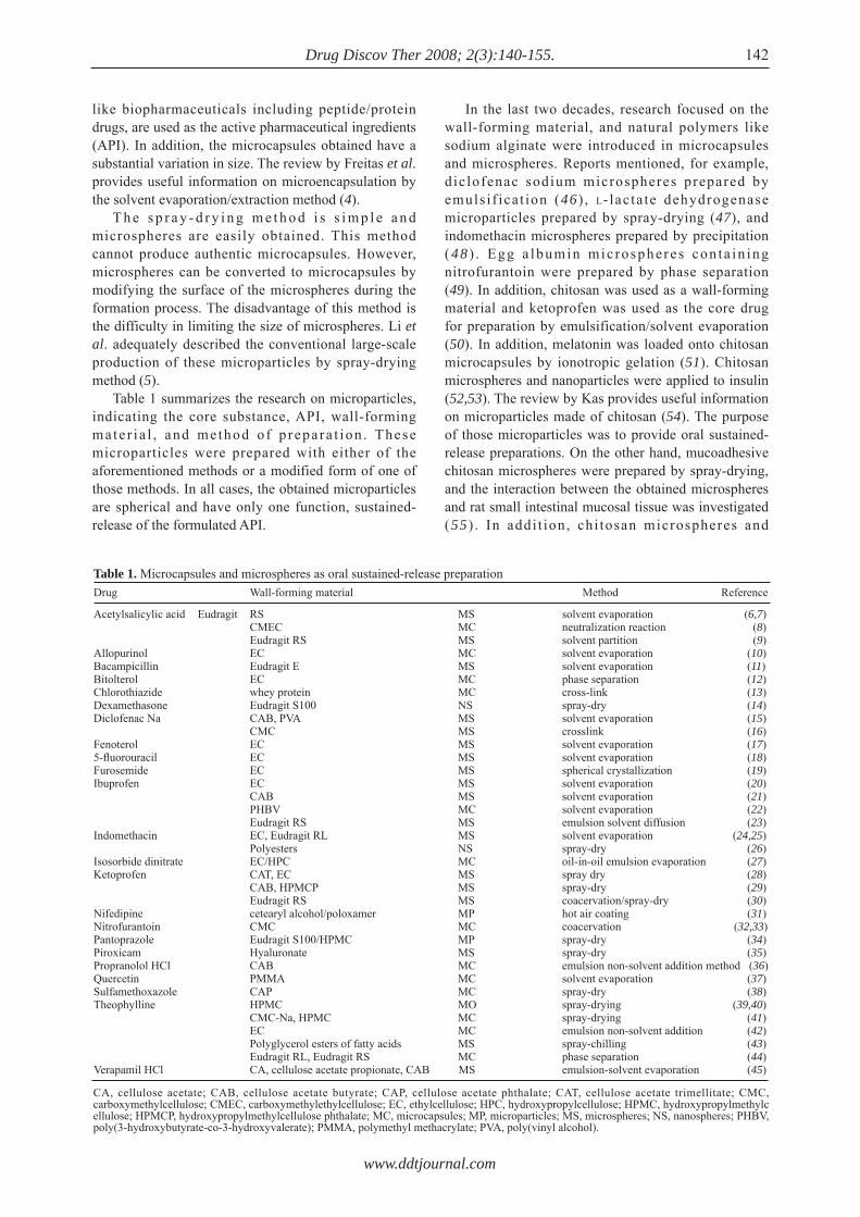
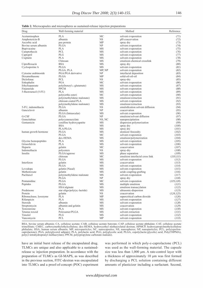
have an initial burst release of the encapsulated drug. TLMCs are unique and also applicable to a sustained-release sc injection preparation. In accordance with the preparation of TLMCs as GI-MAPS, as was described in the previous section, FITC-dextran was encapsulated into TLMCs and a proof-of-concept (POC) experiment
was performed in which poly-ε-caprolactone (PCL) was used as the wall-forming material. The capsule size was less than 1,000 μm. A rate-control layer with a thickness of approximately 10 μm was first formed by discharging a PCL solution containing different amounts of plasticizer including a surfactant. Second,
146
Drug Wall-forming material Method Reference
Acetaminophen PLA MC solvent evaporation (71)Amphotericin-B albumin NS pH-coacervation (72)Ascorbic acid pea protein MP spray dry (73)Bovine serum albumin PLGA NP solvent evaporation (74)Bupivacaine PLA MS solvent evaporation (75)Camptothecin PCL MS solvent evaporation (76)Captopril PLG MS solvent evaporation (77)Cisplatin PLA MS solvent evaporation (78) Chitosan MS emulsion-chemical crosslink (79)Ciprofl oxacin BSA MS spray dry (80)Cyclosporine A PLGA MS solvent evaporation (81) PLGA, PLA MP, NP solvent evaporation (82)Cytosine arabinoside PGA/PGA derivative NP interfacial deposition (83)Dexamethasone PLGA MP solid-oil-oil-oil (84)Diclofenac PCL NS spray-dry (85)Enkephalin PGA MC solvent evaporation (86)Ethyliopanoate poly(benzyl L-glutamate) MS solvent evaporation (87)Finasteride PPCM MS solvent evaporation (88)5-fl uorouracil (5-FU) PLA MS solvent evaporation (89) poly(ortho ester) MC solvent evaporation (90) poly(methylidene malonate) MS emulsion/extraction (91) chitosan coated PLA MS solvent evaporation (92) poly(methylidene malonate) MS emulsion/extraction (93)5-FU, indomethacin PLGA NS emulsifi cation solvent diffusion (94)Ganciclovir albumin NP coacervation (95) PLGA (Intraocular) MS solvent evaporation (96)G-CSF PLGA NP emulsion/solvent diffusion (97)Gemcitabine polycyanoacrylate NS, NC nanoprecipitation (98)Gentamicin coralline hydroxyapatite MS dispersion polymerization (99) BSA MS spray dry (100) PLA/PLGA MS spray dry (101)human growth hormone PLGA MS atomizer freezedry (102) PLGA MC solvent evaporation (103) dex-HEMA MS emulsion/polymerization (104)Glycine homopeptides PLA MS solvent evaporation (105)Griseofulvin PLA MS solvent evaporation (106)Heparin gelatin MC coacervation (107)Indomethacin polyesters NS spray-dry (108)Insulin PLGA/agarose MS phase separation (109) chitosan MC emulsion interfacial cross link (110,111) PLGA MS solvent evaporation (112)Interferon gelatin MS coacervation (113) PLGA MS solvent evaporation (114)Levodopa gelatin (Nasal) MS solvent evaporation (115)Methotrexate gelatin MS azide coupling-grafting (116)Paclitaxel poly(methylidene malonate MS solvent evaporation (117) PLGA NP spray dry (118)Pentamidine PLGA MC solvent evaporation (119,120)Peptides PLGA MS multiple emulsion (121) HSA/alginate MS emulsion transacylation (122)Prednisone star oligo/poly(DL-lactide) MS ultrasonic-dispersion (123)Protein gelatin NS coacervation (124,125)Ribonuclease, lysozyme PLA MP supercritical carbon dioxide (126)Rifampicin PLA MS solvent evaporation (127)Steroids albumin MS solvent evaporation (128)Streptomycin albumin and gelatin MS coacervation (129)Testosterone PLA MS solvent evaporation (130)Tetanus toxoid Poloxamer/PLGA MS solvent extraction (131)Timolol PLG MS solvent evaporation (132)Vancomycin PCL MP solvent evaporation (133)
Table 2. Microcapsules and microspheres as sustained-release injection preparations
BSA, bovine serum albumin; CA, cellulose acetate; CAB, cellulose acetate butyrate; CAP, cellulose acetate phthalate; CAT, cellulose acetate trimellitate; CMEC, carboxymethylethylcellulose; dex-HEMA, hydroxyethyl methacrylated dextran; HPMCP, hydroxypropylmethylcellulose phthalate; HSA, human serum albumin; MP, microparticles; NC, nanocapsules; NS, nanospheres; NP, nanoparticles; PCL, poly(epsilon-caprolactone); PGA, poly(glycerol adipate); PLA, polylactic acid; PLG, polyglycolic acid; PLGA, copoly(lactic/glycolic) acid; Poly(THPMA), poly(2-tetrahydropranyl methacrylate); PPCM, poly(propylene carbonate maleate).
www.ddtjournal.com
Drug Discov Ther 2008; 2(3):140-155.
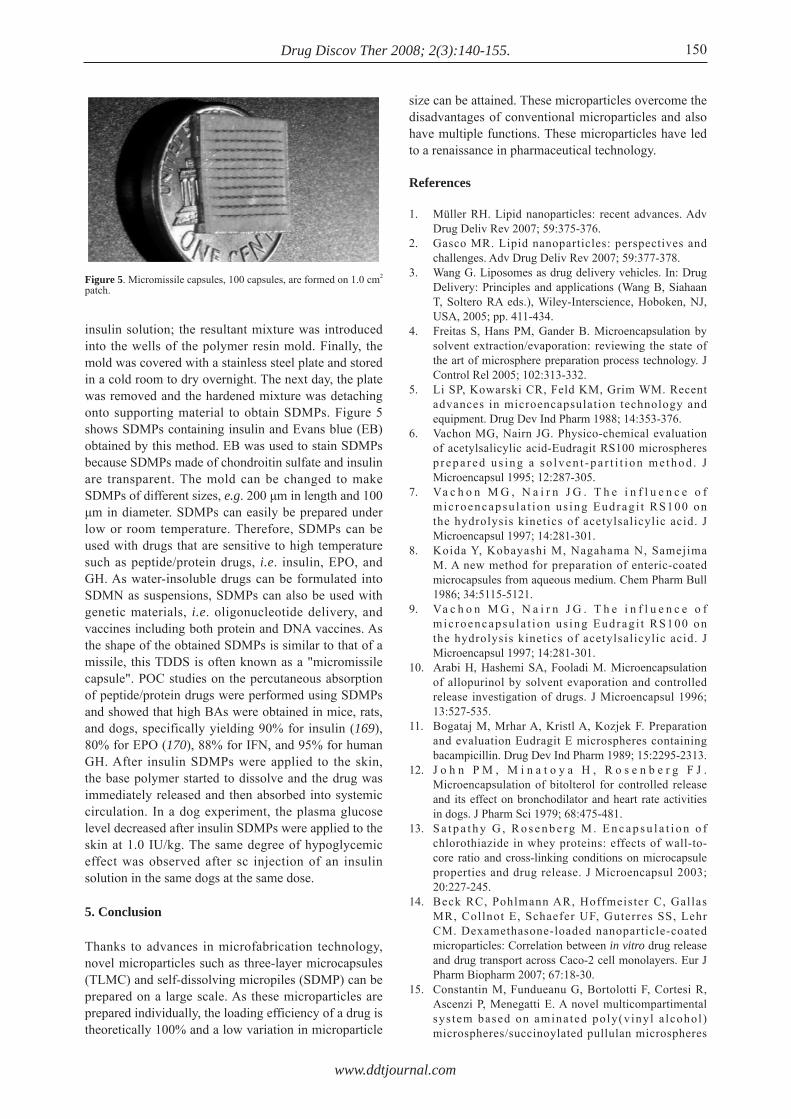
FITC-dextran was discharged. Finally, the PCL solution was discharged and a water-insoluble basement layer was formed. The in vitro release experiment showed long term sustained-release characteristics as shown in Figure 3, and an initial burst release of FITC-dextran was not observed. TLMCs are also applicable to a wide variety of peptide/protein drugs. Therefore, TLMCs containing leuprolide acetate were prepared and sustained-release characteristics were ascertained from the serum leuprolide concentration vs. time profile for more than 10 days after sc administration of the TLMC preparation in rats. The advantages of TLMCs are: (i) a high drug loading efficiency, theoretically 100%, (ii) no initial burst release, and (iii) little variation in particle size. A GI-MAPS-producing machine can also be used to prepare TLMCs, although its nozzle size must be decreased.
4. Percutaneous preparation
Thanks to advances in biotechnology, several important biopharmaceuticals such as insulin, erythropoietin (EPO), granulocyte colony stimulating factor (G-CSF), growth hormone (GH), and interferon (IFN) have been developed (141). The demand for the delivery of macromolecular biopharmaceuticals like peptide/protein drugs is increasing (142). Although the most preferable form of dosage is an oral preparation, the BAs of these drugs are almost 10-23% even when strong absorption enhancers are formulated into GI-MAPS. As a result, no oral preparation of these drugs has entered the pharmaceutical market. Even today, these drugs are administered by iv and/or sc injections. Percutaneous administration is an attractive alternative for the delivery of these drugs because of its many advantages: (i) no or less degradation by hydrolytic enzymes than in the GI tract, (ii) no first-pass effects of the liver associated with oral delivery, (iii) no or less pain than sc injection, (iv) convenience
of administration over iv injection, (v) a better and continuously controlled-delivery rate than oral and sc sustained-release preparations, and (vi) easy removal when side-effects appear. Despite their many potential advantages, transdermal drug delivery systems (TDDS) are severely limited by the poor permeability of drugs through the human skin, i.e. most drugs do not permeate through the skin at therapeutically relevant rates. Many DDSs have been examined in order to increase the rate of drug permeation through the skin, including chemical enhancers and physical methods. Among them, chemical enhancers have contributed most to the development of TDDS. Table 3 shows TDDS products launched on the American market prior to 2007. The permeability of small molecules through the skin can be enhanced by chemical enhancers (143). However, their use is limited because they trigger skin irritation or cause other safety concerns. Iontophoresis, electroporation, and ultrasound have been studied as methods of enhancing physical absorption (144). Iontophoresis uses an electric field to drive ionized molecules across the skin by electrophoresis and nonionized molecules by electroosmosis. Despite concerns about skin irritation, iontophoresis may be useful in delivering some peptides and small proteins (145). As shown in Table 3, a TDDS with lidocaine by iontophoresis was launched on the American market in 2004. Electroporation and ultrasound also provided temporary enhancement of skin permeability of both small drugs and macromolecules (146,147).
However, recent advances in microfabrication technology have allowed preparation of microneedles, which may represent a novel TDDS. Since their first description by Henry et al. in 1998 (148), microfabrication techniques for the production of silicon, metal, glass, and polymer microneedle arrays with micrometer dimensions have been reported (149-152). The microneedles are either solid or hollow and posses a geometrical shape. A microneedle TDDS is roughly defined by a micron-sized needle preparation through and by which a drug is percutaneously administered. Microneedle TDDSs are classified as follows: (i) extremely small needles through which a drug solution can be injected into the skin, (ii) metallic
147
4 12 16 2400
10
20
30
40
50
60
70
Time (days)
Cum
ulat
ive
rele
ase
of F
ITC
-Dex
tran
(%)
8 204 12 16 2400
10
20
30
40
50
60
70
Time (days)
Cum
ulat
ive
rele
ase
of F
ITC
-Dex
tran
(%)
8 20
Figure 3. In vitro release profiles of FITC-Dextran from three-layered microcapsules (TLMC) made of poly-ε-caprolactone. Release rate was controlled by the addition of plasticizer, PEG 400, (●) 30%, (♦) 25%, (■) 20% and (▲) 5%. Each point shows the mean ± SE of 3-4 experiments.
1980 scopolamine patch1981 nitroglycerin patch1983 clonidine patch1985 estradiol patch1991 fentanyl patch1992 estradiol/norethindrone patch1993 nicotine patch1994 testosterone patch1999 lidocine patch2002 norelgestromin/ethynyl estradiol patch2003 oxybutynin patch2004 lidocaine iontophoresis2005 selegilene patch2006 methylphenidate patch2007 rotigitine patch, rivastigmine patch
Table 3. Transdermal DDS products in USA
www.ddtjournal.com
Drug Discov Ther 2008; 2(3):140-155.
and/or silastic microneedles onto which a surface drug is coated, and (iii) metallic and/or silastic microneedles by which conduits known as micropores are made on the skin and a drug solution is applied once the microneedles are removed. The physiology of the skin must be understood in order to fully appreciate the function of microneedles.
Human skin consists of three layers, i.e. stratum corneum (SC), epidermis, and dermis. The SC is the outer layer of the skin with a thickness of 10-15 μm and is dead tissue. The SC is a strong primary barrier against exogenous compounds including drugs. The second barrier is the viable epidermis (50-100 μm), which contains tissue-like living cells. However, there are no blood vessels in the epidermis. Deeper still, there are blood capillaries in the dermis, which accounts for the bulk of skin volume and contains living cells in the form of nerves. When microneedle arrays are inserted into the skin, conduits are created for the penetration of a drug across the SC. Once a drug penetrates the SC, it can diffuse rapidly through the deeper tissue and permeate the underlying capillaries for systemic absorption. As microneedles do not penetrate to the dermis, where the nerve system exists, pain does not result. Based on this understanding of the skin anatomy, microneedles were designed to penetrate the SC without stimulating the pain receptors found in deeper tissue (153).
Silicon microneedles are produced with a standard microelectromechanical system, i.e. microfabrication techniques. Chabri et al. (154) prepared arrays of microconduits for direct and controlled access of molecules across the SC; when inserted into the skin, the arrays enabled drugs to diffuse into the underlying viable epidermis and dermis. Although microneedle arrays originally utilized solid projections for delivery of materials, microfabricated microneedle arrays combined with fluidic microchannels for transdermal extraction of extracellular fluid and blood have also been investigated. Chabri et al. prepared microneedles using a modified form of the BOSCH deep reactive ion etching process, which consists of a combination of an isotropic etch and BOSCH reaction. The microfabrication of microneedles involves the use of tools developed by the microelectronic industry to make integrated circuits. Although these tools offer the potential for mass production of microneedles, production is often highly specialized and includes complex multi-step processes (155,156). For example, 450-μm-thick silicon wafers were spun-coated with photoresist and baked pre-exposure. The wafers were then exposed with the test mask and developed. The wafers were baked postexposure; the thickness of the resist obtained was approximately 8 μm. A standard lithographic mask bearing the appropriate dot array pattern was used during UV exposure to produce a photoresist etch mask. The surface was subsequently
etched using a reactive blend of fluorinated and oxygen gases, with those regions directly underlying the photoresist mask being resistant to the etching process. The waters were loaded and subjected to an SF6 etch to provide an isotropic etch profile. Subsequently, ASETM etch was used to define the length of the microneedle. Finally, the resist was removed in oxygen plasma. Thus, the method of Chabri et al. falls under technology used in the field of semiconductors.
In addition to silicon-based microneedles, metallic microneedles were also proposed. They are classified into two categories, hollow microneedles (149,151) and a microneedle array made of stainless steel (157) and titanium (158). Silastic and metallic microarrays are used in two ways. One is the application of a drug solution to the skin after physical conduits are made by inserting a metallic and/or silastic microarray. The second way is to use a microarray with the drug coating its surface. After the insertion of the microarray into the skin, the drug is dissolved and absorbed into the skin. Hollow microneedles have also been developed in which a drug solution is injected into the skin through hollow microneedles. As is readily apparent, these hollow microneedles are quite distinct from pharmaceuticals. Furthermore, silicon microneedle arrays are fragile, the use of silicon is relatively expensive, and silicon has yet to be proven to be a biocompatible material. Therefore, these microneedles fall under the category of medical devices.
After Prausnitz et al. showed that the absorption of a protein antigen, ovalbumin, was extensively increased by microneedle technology (158), the absorption-enhancing effects of microneedle arrays on the following drugs have been reported: (i) small compounds with a MW of less than 1 kDa like diclofenac (159), methyl nicotinate (160), and bischloroethyl nitrosourea (161), (ii) intermediate compounds (MW between 1 and 10 kDa) like FITC-Dextran (162), desmopressin (163), and insulin (149,151,157,159), and (iii) macromolecules (MW larger than 10 kDa) like FITC-Dextran (162), bovine serum albumin (164), ovalbumin (158), antisense oligonucleotides (165), plasmid DNA (166), and nanospheres (167).
Another area of study has been self-dissolving micropiles (SDMPs). Miyano et al. (168) proposed SDMPs made of maltose for the percutaneous application of dye for tattoos and cosmetics. In their system, maltose was used as a base to make SDMPs. To make maltose SDMPs, maltose was melted by heating it to its melting point, 103°C, and SDMPs were made by introducing maltose into a metallic mold. As a high temperature is needed to make SDMPs, insulin may easily degrade and lose its pharmacological activity. In addition, maltose is a disaccharide, so it causes difficulties in obtaining SDMPs with a hard, steep top because under high humidity in particular it absorbs
148
www.ddtjournal.com
Drug Discov Ther 2008; 2(3):140-155.
water from the air; the top of micropile then bends, resulting in difficulty inserting the micropile into the skin.
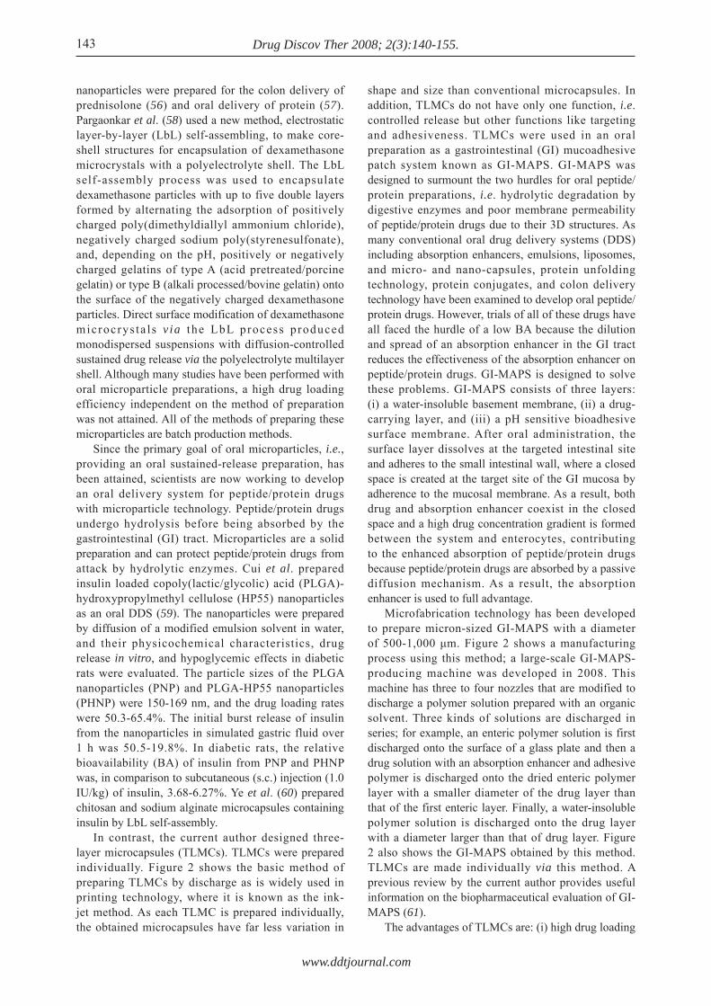
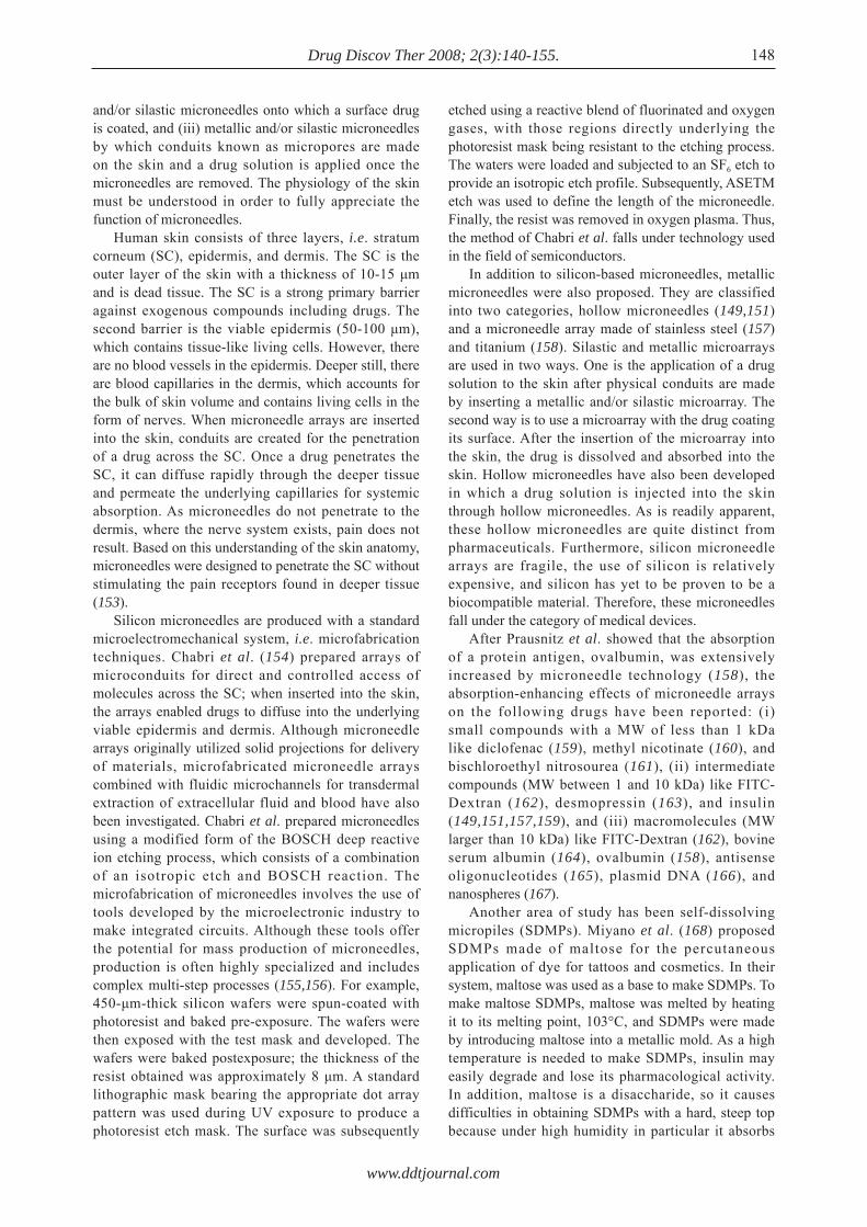
To overcome these pitfalls, SDMPs made of water-soluble thread-forming polymer were developed. Polysaccharides like chondroitin sulfate, dextran, and hyaluronic acid, proteins like albumin, and synthetic polymers like polyvinyl alcohol were used as a water-soluble thread-forming polymer. A drug solution or drug powder was added to a dense solution of a polymer or combination of polymers. SDMPs were originally formed by withdrawing the top of the micropile tip after the drug and polymer were mixed under low or room temperature. However, as shown in Figure 4, microfabrication technology allows SDMPs to be made individually by means of a mold with micron-sized pores, for example, 500 μm in length and 300 μm in diameter, in the opening base. Their size can be
changed from 500 μm to 100 μm in length and from 500 μm to 100 μm in diameter. The method of preparing SDMPs is simple in comparison to preparation by microelectromechanical technology. Namely, a mixture of polymer and drug solution is dispensed into a mold made of polymer resin and dried under pressure. After they fully dry, SDMPs are removed from the mold. A pressing system is useful in accelerating the polymer and drug mixture's insertion into the mold and drying. Metallic microneedles can be formed with MEMS, for example, to make a polymer resin mold. In research by the current author, 100 microneedles with a length of 500 μm and base diameter of 300 μm were formed in a 1.0-cm2 area on a base plate. A polymer resin mold with 100 microwells was obtained wth these master micropiles. A mixture of polymer and drug was obtained by kneading water-soluble thread-forming polymer, chondroitin sulfate, and a small amount of
149
Figure 4. Fabrication process of self-dissolving micropile (SDMP) array.
www.ddtjournal.com
Drug Discov Ther 2008; 2(3):140-155.
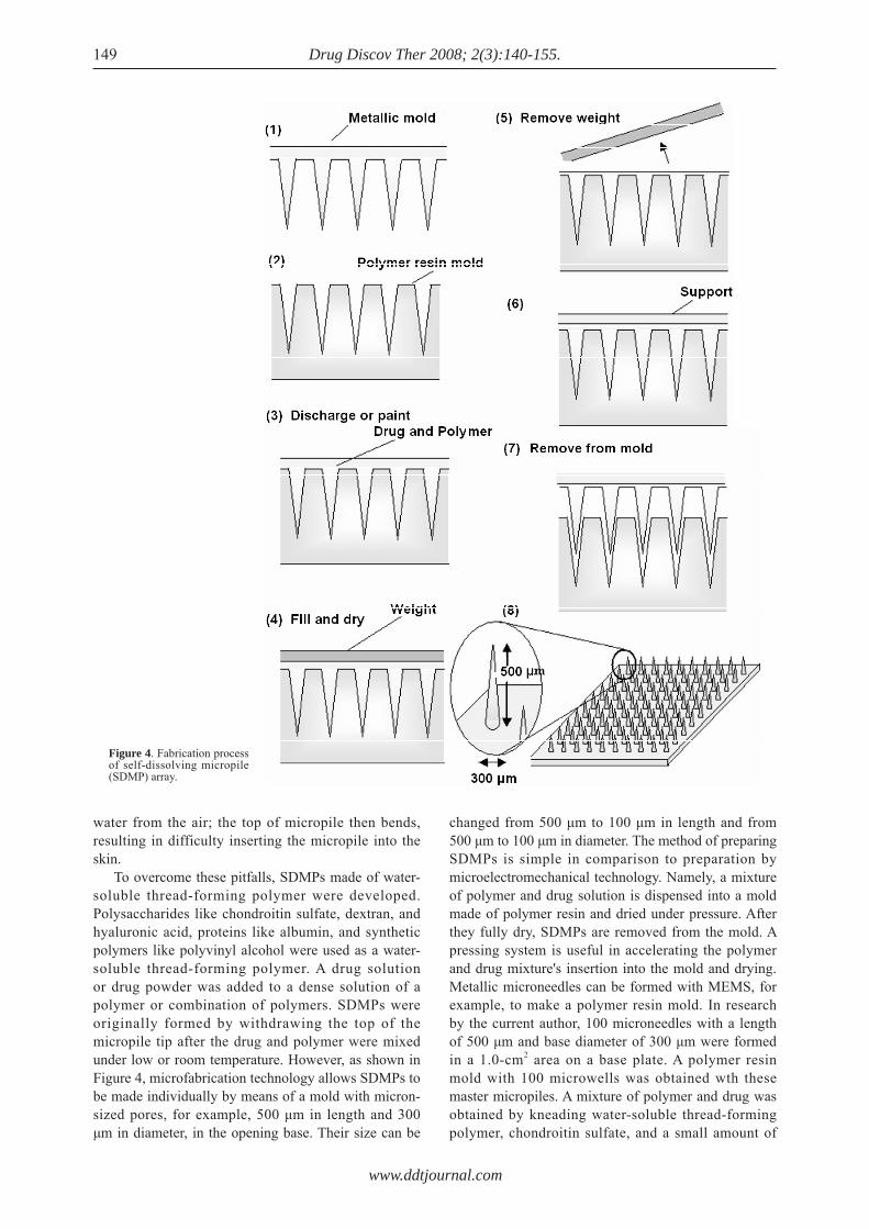
insulin solution; the resultant mixture was introduced into the wells of the polymer resin mold. Finally, the mold was covered with a stainless steel plate and stored in a cold room to dry overnight. The next day, the plate was removed and the hardened mixture was detaching onto supporting material to obtain SDMPs. Figure 5 shows SDMPs containing insulin and Evans blue (EB) obtained by this method. EB was used to stain SDMPs because SDMPs made of chondroitin sulfate and insulin are transparent. The mold can be changed to make SDMPs of different sizes, e.g. 200 μm in length and 100 μm in diameter. SDMPs can easily be prepared under low or room temperature. Therefore, SDMPs can be used with drugs that are sensitive to high temperature such as peptide/protein drugs, i.e. insulin, EPO, and GH. As water-insoluble drugs can be formulated into SDMN as suspensions, SDMPs can also be used with genetic materials, i.e. oligonucleotide delivery, and vaccines including both protein and DNA vaccines. As the shape of the obtained SDMPs is similar to that of a missile, this TDDS is often known as a "micromissile capsule". POC studies on the percutaneous absorption of peptide/protein drugs were performed using SDMPs and showed that high BAs were obtained in mice, rats, and dogs, specifically yielding 90% for insulin (169), 80% for EPO (170), 88% for IFN, and 95% for human GH. After insulin SDMPs were applied to the skin, the base polymer started to dissolve and the drug was immediately released and then absorbed into systemic circulation. In a dog experiment, the plasma glucose level decreased after insulin SDMPs were applied to the skin at 1.0 IU/kg. The same degree of hypoglycemic effect was observed after sc injection of an insulin solution in the same dogs at the same dose.
5. Conclusion
Thanks to advances in microfabrication technology, novel microparticles such as three-layer microcapsules (TLMC) and self-dissolving micropiles (SDMP) can be prepared on a large scale. As these microparticles are prepared individually, the loading efficiency of a drug is theoretically 100% and a low variation in microparticle
size can be attained. These microparticles overcome the disadvantages of conventional microparticles and also have multiple functions. These microparticles have led to a renaissance in pharmaceutical technology.
References
1. Müller RH. Lipid nanoparticles: recent advances. Adv Drug Deliv Rev 2007; 59:375-376.
2. Gasco MR. Lipid nanoparticles: perspectives and challenges. Adv Drug Deliv Rev 2007; 59:377-378.
3. Wang G. Liposomes as drug delivery vehicles. In: Drug Delivery: Principles and applications (Wang B, Siahaan T, Soltero RA eds.), Wiley-Interscience, Hoboken, NJ, USA, 2005; pp. 411-434.
4. Freitas S, Hans PM, Gander B. Microencapsulation by solvent extraction/evaporation: reviewing the state of the art of microsphere preparation process technology. J Control Rel 2005; 102:313-332.
5. Li SP, Kowarski CR, Feld KM, Grim WM. Recent advances in microencapsulation technology and equipment. Drug Dev Ind Pharm 1988; 14:353-376.
6. Vachon MG, Nairn JG. Physico-chemical evaluation of acetylsalicylic acid-Eudragit RS100 microspheres p r epa red u s ing a so lven t -pa r t i t i on me thod . J Microencapsul 1995; 12:287-305.
7. Va c h o n M G , N a i r n J G . T h e i n f l u e n c e o f mic roencapsu la t ion us ing Eudrag i t RS100 on the hydrolysis kinetics of acetylsalicylic acid. J Microencapsul 1997; 14:281-301.
8. Koida Y, Kobayashi M, Nagahama N, Samejima M. A new method for preparation of enteric-coated microcapsules from aqueous medium. Chem Pharm Bull 1986; 34:5115-5121.
9. Va c h o n M G , N a i r n J G . T h e i n f l u e n c e o f mic roencapsu la t ion us ing Eudrag i t RS100 on the hydrolysis kinetics of acetylsalicylic acid. J Microencapsul 1997; 14:281-301.
10. Arabi H, Hashemi SA, Fooladi M. Microencapsulation of allopurinol by solvent evaporation and controlled release investigation of drugs. J Microencapsul 1996; 13:527-535.
11. Bogataj M, Mrhar A, Kristl A, Kozjek F. Preparation and evaluation Eudragit E microspheres containing bacampicillin. Drug Dev Ind Pharm 1989; 15:2295-2313.
12. J o h n P M , M i n a t o y a H , R o s e n b e r g F J . Microencapsulation of bitolterol for controlled release and its effect on bronchodilator and heart rate activities in dogs. J Pharm Sci 1979; 68:475-481.
13. S a t p a t h y G , R o s e n b e rg M . E n c a p s u l a t i o n o f chlorothiazide in whey proteins: effects of wall-to-core ratio and cross-linking conditions on microcapsule properties and drug release. J Microencapsul 2003; 20:227-245.
14. Beck RC, Pohlmann AR, Hoffmeister C, Gallas MR, Collnot E, Schaefer UF, Guterres SS, Lehr CM. Dexamethasone-loaded nanoparticle-coated microparticles: Correlation between in vitro drug release and drug transport across Caco-2 cell monolayers. Eur J Pharm Biopharm 2007; 67:18-30.
15. Constantin M, Fundueanu G, Bortolotti F, Cortesi R, Ascenzi P, Menegatti E. A novel multicompartimental sys tem based on aminated poly(vinyl a lcohol) microspheres/succinoylated pullulan microspheres
150
Figure 5. Micromissile capsules, 100 capsules, are formed on 1.0 cm2 patch.
www.ddtjournal.com
Drug Discov Ther 2008; 2(3):140-155.
for oral delivery of anionic drugs. Int J Pharm 2007; 330:129-137.
16. Arica B, Arica MY, Kaş HS, Hincal AA, Hasirci V. In-vitro studies of enteric coated diclofenac sodium-carboxymethylcellulose microspheres. J Microencapsul 1996; 13:689-699.
17. Lin WJ, Wu TL. Modification of the initial release of a highly water-soluble drug from ethyl cellulose microspheres. J Microencapsul 1999; 16:639-646.
18. Ghorab MM, Zia H, Luzzi LA. Prepara t ion of controlled release anticancer agents. I: 5-Fluorouracil-ethyl cellulose microspheres. J Microencapsul 1990; 7:447-454.
19. Akbuğa J . Furosemide- loaded e thy l ce l lu lose microspheres prepared by spherical crystallization technique: Morphology and release characteristics. Int J Pharm 1991; 76:193-198.
20. Dubernet C, Rouland JC, Benoit JP. Ibuprofen-loaded ethylcellulose microspheres: analysis of the matrix structure by thermal analysis. J Pharm Sci 1991; 80:1029-1033.
21. Dalal PS, Narurkar MM. In vitro and in vivo evaluation of sustained release suspensions of ibuprofen. Int J Pharmaceut 1991; 73:157-162.
22. Wang C, Ye W, Zheng Y, Liu X, Tong Z. Fabrication of drug-loaded biodegradable microcapsules for controlled release by combination of solvent evaporation and layer-by-layer self-assembly. Int J Pharm 2007; 338:165-173.
23. Perumal D. Microencapsulation of ibuprofen and Eudragit RS 100 by the emulsion solvent diffusion technique. Int J Pharm 2001; 218:1-11.
24. Babay D, Hoffman A, Benita S. Design and release kinetic pattern evaluation of indomethacin microspheres intended for oral administration. Biomaterials 1988; 9:482-488.
25. Tirkkonen S, Urtti A, Paronen P. Buffer controlled release of indomethacin from ethylcellulose microcapsules. Int J Pharm 1995; 124:219-229.
26. Raffin Pohlmann A, Weiss V, Mertins O, Pesce da Silveira N, Stanisçuaski Guterres S. Spray-dried indomethacin-loaded polyester nanocapsules and nanospheres: development, stability evaluation and nanostructure models. Eur J Pharm 2002; 16:305-312.
27. Yang GM, Kuo JF, Woo EM. Preparation and control-release kinetics of isosorbide dinitrate microspheres. J Microencapsul 2006; 23:622-631.
28. Giunchedi P, Torre ML, Maggi L, Conti B, Conte U. Cellulose acetate trimellitate ethylcellulose blends for non-steroidal anti-inflammatory drug (NSAID) microspheres. J Microencapsul 1996; 13:89-98.
29. Moretti MD, Gavini E, Juliano C, Pirisino G, Giunchedi P. Spray-dried microspheres containing ketoprofen formulated into capsules and tablets. J Microencapsul 2001; 18:111-121.
30. Palmieri G, Bonacucina G, Di Martino P, Martelli S. Microencapsulation of semisolid ketoprofen/polymer microspheres. Int J Pharm 2002; 242:175-178.
31. Pattarino F, Giovannelli L, Bellomi S. Effect of poloxamers on nifedipine microparticles prepared by hot air coating technique. Eur J Pharm Biopharm 2007; 65:198-203.
32. Karasulu HY, Ertan G, Guneri T. Factorial design-based optimization of the formulation of nitrofurantoin microcapsules. Pharm World Sci 1996; 18:20-25.
33. Ertan G, Sarigüllü I, Karasulu Y, Erçakir K, Güneri T.
Sustained-release dosage form of nitrofurantoin. Part 1. Preparation of microcapsules and in vitro release kinetics. J Microencapsul 1994; 11:127-135.
34. Raffin RP, Jornada DS, Re MI, Pohlmann AR, Guterres SS. Sodium pantoprazole-loaded enteric microparticles prepared by spray drying: Effect of the scale of production and process validation. Int J Pharm 2006; 324:10-18.
35. Piao MG, Kim JH, Kim JO, Lyoo WS, Lee MH, Yong CS, Choi HG. Enhanced oral bioavailability of piroxicam in rats by hyaluronate microspheres. Drug Dev Ind Pharm 2007; 33:485-491.
36. Pongpaibul Y, Whitworth CW. Preparation and in vitro dissolution characteristics of propranolol microcapsules. Int J Pharm 1986; 33:243-248.
37. Lee DH, Sim GS, Kim JH, Lee GS, Pyo HB, Lee BC. Preparation and characterization of quercetin-loaded polymethyl methacrylate microcapsules using a polyol-in-oil-in-polyol emulsion solvent evaporation method. J Pharm Pharmacol 2007; 59:1611-1620.
38. Takenaka H, Kawashima Y, Lin SY. Polymorphism of spray-dried microencapsulated sulfamethoxazole wi th ce l lu lose ace ta te phtha la te and col lo ida l silica, montmorillonite, or talc. J Pharm Sci 1981; 70:1256-1260.
39. Wan LSC, Heng PWS, Chia CGH. Preparation of coated particles using a spray drying process with an aqueous system. Int J Pharm 1991; 77:183-191.
40. Wan LSC, Heng PWS, Chia CGH. Plasticizers and their effects on microencapsulation process by spray-drying in an aqueous system. J Microencapsul 1992; 9:53-62.
41. Wan LSC, Heng PWS, Ch ia CGH. Ci t r i c ac id as a plasticizer for spray-dried microcapsules. J Microencapsul 1993; 10:11-23.
42. Sa B, Bandyopadhyay AK, Gupta BK. Effect of microcapsule size and polyisobutylene concentration on the release of theophylline from ethylcellulose microcapsules. J Microencapsul 1996; 13:207-218.
43. Akiyama Y, Yoshioka M, Horibe H, Hirai S, Kitamori N, Toguchi H. Novel oral controlled-release microspheres using polyglycerol esters of fatty acids. J Control Rel 1993; 26:1-10.
44. Donbrow M, Hoffman A, Benita S. Gradation of microcapsule wall porosity by deposition of polymer mixtures (Eudragit RL and Eudragit RS). Phase separation of polymer mixtures and effects of external media and conditions on release. J Microencapsul 1995; 12:273-285.
45. Bhardwaj SB, Shukla AJ, Collins CC. Effect of varying drug loading on particle size distribution and drug release kinetics of verapamil hydrochloride microspheres prepared with cellulose esters. J Microencapsul 1995; 12:71-81.
46. Gürsoy A, Cevik S. Sustained release properties of alginate microspheres and tabletted microspheres of diclofenac sodium. J Microencapsul 2000; 17:565-575.
47. Coppi G, Iannuccelli V, Bernabei MT, Cameroni R. Alginate micropar t ic les for enzyme peroral administration. Int J Pharm 2002; 242:263-266.
48. Albin P, Markus A, Pelah Z, Ben-Zvi Z. Slow-release indomethacin formulations based on polysaccharides: evaluation in vitro and in vivo in dogs. J Control Rel 1994; 29:25-39.
49. Jun HW, Lai JW. Preparation and in vitro dissolution tests of egg albumin microcapsules of nitrofurantoin. Int
151
www.ddtjournal.com
Drug Discov Ther 2008; 2(3):140-155.
J Pharm 1983; 16:65-77.50. Genta I, Perugini P, Conti B, Pavanetto F. A multiple
emulsion method to entrap a lipophilic compound into chitosan microspheres. Int J Pharm 1997; 152:237-246.
51. El-Gibaly I, Meki AMA, Abdel-Ghaffar SK. Novel B melatonin-loaded chitosan microcapsules: in vitro characterization and antiapoptosis efficacy for aflatoxin B1-induced apoptosis in rat liver. Int J Pharm 2003; 260:5-22.
52. Sarmento B, Ribeiro A, Veiga F, Sampaio P, Neufeld R, Ferreira D. Alginate/chitosan nanoparticles are effective for oral insulin delivery. Pharm Res 2007; 24:2198-2206.
53. Ubaidulla U, Khar RK, Ahmed FJ, Sultana Y, Panda AK. Development and characterization of chitosan succinate microspheres for the improved bioavailability of insulin. J Pharm Sci 2007; 96:3010-3023.
54. Kas HS, Chitosan: properties, preparations and application to microparticulate systems. J Microencapsul 1997; 14:689-711.
55. He P, Davis Stanley S, Illum L. In vitro evaluation of the mucoadhesive properties of chitosan microspheres. Int J Pharm 1998; 166:75-88.
56. Onishi H, Oosegi T, Machida Y, McGinity JW. Eudragit coating of chitosan-prednisolone conjugate microspheres and in vitro evaluation of coated microspheres. Drug Dev Ind Pharm 2007; 33:848-854.
57. Li T, Shi XW, Du YM, Tang YF. Quaternized chitosan/alginate nanoparticles for protein delivery. J Biomed Mater Res 2007; 83A:383-390.
58. Pargaonkar N, Lvov YM, Li N, Steenekamp JH, de Villiers MM. Controlled release of dexamethasone from microcapsules produced by polyelectrolyte layer-by-layer nanoassembly. Pharm Res 2005; 22:826-835.
59. Cui F, Tao A, Cun D, Zhang L, Shi K. Preparation of insulin loaded PLGA-HP55 nanoparticles for oral delivery. J Pharm Sci 2007; 96:421-427.
60. Ye S, Wang C, Liu X, Tong Z, Ren B, Zeng F. New loading process and release properties of insulin from polysaccharide microcapsules fabricated through layer-by-layer assembly. J Control Rel 2006; 112:79-87.
61. Takada K. Oral delivery of haematopoietic factors: Progress with gastrointestinal mucoadhesive patches, microdevices and other microfabrication technologies. Am J Drug Deliv 2006; 4:65-77.
62. Eiamtrakarn S, Itoh Y, Kishimoto J, Yoshikawa Y, Shibata N, Murakami M, Takada K. Gastrointestinal mucoadhesive patch system (GI-MAPS) for oral administration of G-CSF, a model protein. Biomaterial 2002; 23:145-152.
63. Venkatesan N, Uchino K, Amagase K, Ito Y, Shibata N, Takada K. Gastro-intestinal patch system for the delivery of erythropoietin. J Control Rel 2006; 111:19-26.
64. O g a w a Y, Ya m a m o t o M , Ta k a d a S , O k a d a H , Shimamoto T. Controlled-release of leuprolide acetate from polylactic acid or copoly(lactic/glycolic)acid microcapsules: influence of molecular weight and copolymer ratio of polymer. Chem Pharm Bull 1988; 36:1502-1507.
65. Toguchi H, Ogawa Y, Okada H, Yamamoto M. Once-a-month injectable microcapsules of leuprorelin acetate. Yakugaku Zassi 1991; 111:397-409.
66. Okada H, Inoue Y, Heya T, Ueno H, Ogawa Y, Toguchi H. Pharmacokinetics of once-a month injectable microspheres of leuprolide acetate. Pharm Res 1991; 8:787-791.
67. Cohen S, Yoshioka T, Lucarelli M, Hwang LH, Langer R. Controlled delivery systems for proteins based on poly(lactic/glycolic acid) microspheres. Pharm Res 1991; 8:713-720.
68. Ogawa Y. Development of long-acting leuprolide acetate preparation. Chemistry Today 2005; 406:63-66.
69. Cox MC, Scripture CD, Figg WD. Leuprolide acetate given by a subcutaneous extended-release injection: Less of a pain? Expert Rev Anticancer Ther 2005; 5:605-611.
70. Anderson JM, Sh ive MS. Biodegrada t ion and biocompatibility of PLA and PLGA microspheres. Adv Drug Deliv Rev 1997; 28:5-24.
71. Lai MK, Tsiang RC. Encapsulating acetaminophen into poly(L-lactide) microcapsules by solvent-evaporation technique in an O/W emulsion. J Microencapsul 2004; 21:307-316.
72. Santhi K, Dhanaraj SA, Rajendran SD, Raja K, Ponnusankar S, Suresh B. Nonliposomal approach-A study of preparation of egg albumin nanospheres containing amphotericin-B. Drug Dev Ind Pharm 1999; 25:547-551.
73. Pierucci AP, Andrade LR, Baptista EB, Volpato NM, Rocha-Leão MH. New microencapsulation system for ascorbic acid using pea protein concentrate as coat protector. J Microencapsul 2006; 23:654-662.
74. S o n g C X , L a b h a s e t w a r V, M u r p h y H , Q u X , Humphrey WR, Shebuski RJ, Levy RJ. Formulation and characterization of biodegradable nanoparticles for intravascular local drug delivery. J Cont Rel 1997; 43:197-212.
75. Le Corre P, Estèbe JP, Chevanne F, Mallédant Y, Le Verge R. Spinal controlled delivery of bupivacaine from DL-lactic acid oligomer microspheres. J Pham Sci 1995; 84:75-78.
76. Dora CL, Alvarez-Silva M, Trentin AG, de Faria TJ, Fernandes D, da Costa R, Stimamiglio M, Lemos-Senna E. Evaluation of antimetastatic activity and systemic toxicity of camptothecin-loaded microspheres in mice injected with B16-F10 melanoma cells. J Pharm Sci 2006; 9:22-31.
77. Schartel B, Volland C, Li YX, Wendorff JW, Kissel T. Dielectr ic and thermodynamic propert ies of biodegradable poly(D,L-lactide-co-glycolide) and the effect on the micro-encapsulation and release of captopril. J Microencapsul 1997; 14:475-488.
78. Yoshida M, Uemura Y, Yoshizawa H, Kawano Y, Natsugoe S, Aikou T, Hatate Y. Application of microsphere for cancer treatment. Pharm Tech Jpn 2000; 16:85-91.
79. Wang YM, Sato H, Adachi I, Horikoshi I. Optimization of the formulation design of chitosan microspheres containing cisplatin. J Pham Sci 1996; 85:1204-1210.
80. Li FQ, Hu JH, Lu B, Yao H, Zhang WG. Ciprofloxacin-loaded bovine serum albumin microspheres: preparation and drug-release in vitro. J Microencapsul 2001; 18:825-829.
81. Malaekeh-Nikouei B, Sajadi Tabassi SA, Jaafari MR. The effect of different grades of PLGA on characteristics of microspheres encapsulated with cyclosporine A. Curr Drug Deliv 2006; 3:343-349.
82. Lee WK, Park JY, Yang EH, Suh H, Kim SH, Chung DS, Choi K, Yang CW, Park JS. Investigation of the factors influencing the release rates of cyclosporin A-loaded micro- and nanoparticles prepared by high-pressure homogenizer. J Control Rel 2002; 84:115-123.
152
www.ddtjournal.com
Drug Discov Ther 2008; 2(3):140-155.
83. Puri S, Kallinteri P, Higgins S, Hutcheon GA, Garnett MC. Drug incorporation and release of water soluble drugs from novel functionalised poly(glycerol adipate) nanoparticles. J Control Rel 2008; 125:59-67.
84. Thote AJ, Gupta RB. Formation of nanoparticles of a hydrophilic drug using supercritical carbon dioxide and microencapsulation for sustained release. Nanomedicine 2005; 1:85-90.
85. Müller CR, Schaffazick SR, Pohlmann AR, de Lucca Freitas L, Pesce da Silveira N, Dalla Costa T, Guterres SS. Spray-dried diclofenac-loaded poly(epsilon-caprolactone) nanocapsules and nanospheres. Preparation and physicochemical characterization. Pharmazie 2001; 56:864-867.
86. Graves RA, Freeman T, Pamajula S, Praetorius N, Moiseyev R, Mandal TK. Effect of co-solvents on the characteristics of enkephalin microcapsules. J Biomater Sci Polym Ed 2006; 17:709-720.
87. Li C, Yang DJ, Kuang LR, Wallace S. Polyamino acid microspheres: Preparation, characterization and distribution after intravenous injection in rats. Int J Pharm 1993; 94:143-152.
88. Peng D, Huang K, Liu Y, Liu S. Preparation of novel polymeric microspheres for controlled release of finasteride. Int J Pharm 2007; 342:82-86.
89. Ciftci K, Hincal AA, Kas HS, Ercan MT, Ruacan S. Microspheres of 5-fluorouracil using poly(dl-lactic acid): in vitro release properties and distribution in mice after i.v. administration. Eur J Pharm Sci 1994; 1:249-258.
90. Lin YH, Vasavada RC. Studies on microencapsulation of 5-fluorouracil with poly(ortho ester) polymers. J Microencapsul 2000; 17:1-11.
91. Fournier E, Passirani C, Colin N, Breton P, Sagodira S, Benoit JP. Development of novel 5-FU-loaded poly(methylidene malonate 2.1.2)-based microspheres for the treatment of brain cancers. Eur J Pharm Biopharm 2004; 57:189-197.
92. Chandy T, Das GS, Rao GH. 5-Fluorouracil-loaded chitosan coated polylactic acid microspheres as biodegradable drug carriers for cerebral tumours. J Microencapsul 2000; 17:625-638.
93. Fournier E, Passirani C, Colin N, Breton P, Sagodira S, Benoit JP. Development of novel 5-FU-loaded poly(methylidene malonate 2.1.2)-based microspheres for the treatment of brain cancers. Eur J Pharm Biopharm 2004; 57:189-197.
94. Niwa T, Takeuchi H, Hino T, Kunou N, Kawashima Y. Preparations of biodegradable nanospheres of water-soluble and insoluble drugs with D,L-lactide/glycolide copolymer by a novel spontaneous emulsification solvent diffusion method and the drug release behavior. J Control Rel 1993; 25:89-98.
95. Merodio M, Ruiz J, Bustos M, Galan FM, Campanero MA, Irache JM. Encapsulation of ganciclovir in albumin nanoparticles enhances the thymidine kinase suicide gene therapy. J Drug Del Scl Tech 2005; 15:121-127.
96. Herrero-Vanrell R, Ramirez L, Fernandez-Carballido A, Refojo MF. Biodegradable PLGA microspheres loaded with ganciclovir for intraocular administration. Encapsulation technique, in vitro release profiles, and sterilization process. Pharm Res 2000; 17:1323-1328.
97. Choi SH, Park TG. G-CSF loaded biodegradable PLGA nanoparticles prepared by a single oil-in-water emulsion method. Int J Pharm 2006; 311:223-228.
98. Stella B, Arpicco S, Rocco F, Marsaud V, Renoir JM,
153
Cattel L, Couvreur P. Encapsulation of gemcitabine lipophilic derivatives into polycyanoacrylate nanospheres and nanocapsules. Int J Pharm 2007; 344:71-77.
99. Sivakumar M, Rao KP. Preparation, characterization, and in vitro release of gentamicin from coralline hydroxyapatite-alginate composite microspheres. J Biomed Mater Res 2003; 65:222-228.
100. Haswani DK, Nettey H, Oettinger C, D'Souza MJ. Formulation, characterization and pharmacokinetic evaluation of gentamicin sulphate loaded albumin microspheres. J Microencapsul 2006; 23:875-886.
101. Prior S, Gamazo C, Irache JM, Merkle HP, Gander B. Gentamicin encapsulation in PLA/PLGA microspheres in view of treating Brucella infections. Int J Pharm 2000; 196:115-125.
102. Johnson OL, Jaworowicz W, Cleland JL, Bailey L, Charnis M, Duenas E, Wu C, Shepard D, Magil S, Last T, Jones AJ, Putney SD. The stabilization and encapsulation of human growth hormone in to b iodegradable microspheres. Pharm Res 1997; 14:730-735.
103. Takada S, Yamagata Y, Misaki M, Taira K, Kurokawa T. Sustained release of human growth hormone from microcapsules prepared by a solvent evaporation technique. J Control Rel 2003; 88:229-242.
104. Vlugt-Wensink KDF, de Vrueh R, Gresnigt MG, Hoogerbrugge CM, van Buul-Offers SC, de Leede LGJ, Sterkman LGW, Crommelin DJA, Hennink WE, Verrijk R. Preclinical and clinical in vitro in vivo correlation of an hGH dextran microsphere formulation. Pharm Res 2007; 24:2239-2248.
105. Pradhan RS, Vasavada RC. Formulation and in vitro release study on poly(DL-lactide)microspheres containing hydrophilic compounds: glycine homopeptides. J Control Rel 1994; 30:143-154.
106. Vudathala GK, Rogers JA. Microencapsulation of solid dispersions: Release of griseofulvin from griseofulvin: Phospholipid coprecipitates in microspheres. Pharm Res 1992; 9:759-763.
107. Tsung M, Burgess DJ. Preparation and stabilization of heparin/gelatin complex coacervate microcapsules J Pharm Sci 1997; 86:603-607.
108. Raffin Pohlmann A, Weiss V, Mertins O, Pesce da Silveira N, Stanisçuaski Guterres S. Spray-dried indomethacin-loaded polyester nanocapsules and nanospheres: Development, stability evaluation and nanostructure models. Eur J Pharm Sci 2002; 16:305-312.
109. Wang N, Wu XS. A novel approach to stabilization o f p r o t e i n d r u g s i n p o l y ( l a c t i c - c o - g l y c o l i c acid)microspheres using agarose hydrogel. Int J Pharm 1998; 166:1-14.
110. Aiedeh K, Gianasi E, Orienti I, Zecchi V. Chitosan microcapsules as controlled release systems for insulin. J Microencapsul 1997; 14:567-576.
111. Bugamelli F, Raggi MA, Orienti I, Zecchi V. Controlled insulin release from chitosan microparticles. Arch Pharm Pharm Med Chem 1998; 331:133-138.
112. Igartua M, Hernandez RM, Esquisabel A, Gascon AR, Calvo MB, Pedraz JL. Influence of formulation variables on the in-vitro release of albumin from biodegradable microparticulate systems. J Microencapsul 1997; 14:349-356.
113. Tabata Y, Uno K, Muramatsu S, Ikada Y. In vivo effects of recombinant interferon alpha A/D incorporated in gelatin microspheres on murine tumor cell growth. Jpn J
www.ddtjournal.com
Drug Discov Ther 2008; 2(3):140-155.
Cancer Res 1989; 80:387-393.114. Yang J, Cleland JL. Factors affecting the in vitro release
of recombinant human interferon-γ (rhIFN-γ) from PLGA microspheres. J Pharm Sci 1997; 86:908-914.
115. Brime B, Ballesteros MP, Frutos P. Preparation and in vitro characterization of gelatin microspheres containing levodopa for nasal administration. J Microencapsul 2000; 17:777-784.
116. Narayani R, Rao KP. Solid tumor chemotherapy using injectable gelatin microspheres containing free methotrexate and conjugated methotrexate. Int J Pharm 1996; 142:25-32.
117. Le Visage C, Rioux-Leclercq N, Haller M, Breton P, Malavaud B, Leong K. Efficacy of paclitaxel released from bio-adhesive polymer microspheres on model superficial bladder cancer. J Urol 2004; 171:1324-1329.
118. Wang J, Ng CW, Win KY, Shoemakers P, Lee TK, Feng SS, Wang CH. Release of paclitaxel from polylactide-co-glycolide (PLGA) microparticles and discs under irradiation. J Microencapsul 2003; 20:317-327.
119. Graves RA, Pamujula S, Moiseyev R, Freeman T, Bostanian LA, Mandal TK. Effect of different ratios of high and low molecular weight PLGA blend on the characteristics of pentamidine microcapsules. Int J Pharm 2004; 270:251-262.
120. Mandal TK, Bostanian LA, Graves RA, Chapman SR, Idodo TU. Porous biodegradable microparticles for delivery of pentamidine. Eur J Pharm Biopharm 2001; 52:91-96.
121. Couvreur P. Blanco-Prieto MJ, Puisieux F, Roques B, Fattal E. Multiple emulsion technology for the design of microspheres containing peptides and oligopeptides. Adv Drug Deliv Rev 1997; 28:85-96.
122. Hurteaux R, Edwards-Lévy F, Laurent-Maquin D, Lévy MC. Coating alginate microspheres with a serum albumin-alginate membrane: application to the encapsulation of a peptide. Eur J Pharm Sci 2005; 24:187-197.
123. Zou T, Li SL, Cheng SX, Zhang XZ, Zhuo RX. Fabrication and in vitro drug release of drug-loaded star oligo/poly(DL-lactide) microspheres made by novel ultrasonic-dispersion method. J Biomed Mat Res Part A 2007; 83A:696-702.
124. Li JK, Wang N, Wu XS. Gelatin nanoencapsulation of protein/peptide drugs using an emulsifier-free emulsion method. J Microencapsul 1998; 15:163-172.
125. Li JK, Wang N, Wu XS. A novel biodegradable system based on gelatin nanoparticles and poly(lactic-co-glycolic acid) microspheres for protein and peptide drug delivery. J Pharm Sci 1997; 86:891-895.
126. Whitaker MJ, Hao J, Davies OR, Serhatkulu G, Stolnik-Trenkic S, Howdle SM, Shakesheff KM. The production of protein-loaded microparticles by supercritical fluid enhanced mixing and spraying. J Control Rel 2005; 101:85-92.
127. Zhang W, Jiang X, Hu J, Fu C. Rifampicin polylactic acid microspheres for lung targeting. J Microencapsul 2000; 17:785-788.
128. Burgess DJ, Davis SS. Potential use of albumin microspheres as a drug delivery system. II In vivo deposition and release of steroids. Int J Pharm 1988; 46:69-76.
129. Gürkan H, Yalabik-Kaş HS, Hincal AA, Ercan MT. Streptomycin sulphate microspheres: Formulation and in vivo distribution. J Microencapsul 1986; 3:101-108.
130. Tsubuku S, Sugawara S, Miyajima M, Yoshida M, Asano M, Okabe K, Kobayashi D, Yamanaka H. Preparation and characterization of oil-in-water type poly(D,L-lactic acid) microspheres containing testosterone enanthate. Drug Dev Ind Pharm 1998; 24:927-934.
131. Tobío M, Nolley J, Guo Y, McIver J, Alonso MJ. A novel system based on a poloxamer/PLGA blend as a tetanus toxoid delivery vehicle. Pharm Res 1999; 16:682-688.
132. Sturesson C, Carlfors J, Edsman K, Andersson M. Preparation of biodegradable poly(lactic-co-glycolic)acid microspheres and their in vitro release of timolol maleate. Int J Pharm 1993; 89:235-244.
133. Le Ray AM, Chiffoleau S, Iooss P, Grimandi G, Gouyette A, Daculsi G, Merle C. Vancomycin encapsulation in biodegradable poly(epsilon-caprolactone) microparticles for bone implantation. Influence of the formulation process on size, drug loading, in vitro release and cytocompatibility. Biomaterials 2003; 24:443-449.
134. O ' D o n n e l l P B , M c G i n i t y J W. P r e p a r a t i o n o f microspheres by the solvent evaporation technique. Adv Drug Deliv Rev 1997; 28:25-42.
135. Soppimath KS, Aminabhavi TM, Kulkarni AR, Rudzinski WE. Biodegradable polymeric nanoparticles as drug delivery devices. J Control Rel 2001; 70:1-20.
136. Iwamoto S, Nakagawa K, Nakajima M, Nabetani H. Effect of oil phase kinds on preparation of monodisperse gelatin microbeads using microchannel emulsification. Kagaku to seibutsu 2005; 43:410-415.
137. Berkland C, Kim K, Pack DW. Fabrication of PLG microspheres with precisely controlled and monodisperse size distribution. J Control Rel 2001; 73:59-74.
138. Berkland C, King M, Cox A, Kim K, Pack DW. Precise control of PLG microsphere size provides enhanced control of drug release rate. J Control Rel 2002; 82:137-147.
139. Yeo Y, Basaran OA, Park K. A new process for making reservoir-type microcapsules using ink-jet technology and interfacial phase separation. J Control Rel 2003; 93:161-173.
140. Freitas S, Merkle HP, Gander B. Microencapsulation by solvent extraction/evaporation: reviewing the state of the art of microsphere preparation process technology. J Control Rel 2005; 102:313-332.
141. Walsh G. Pharmaceut ica ls , b io logics and b io-pharmaceuticals. In: Biopharmaceuticals: Biochemistry and biotechnology, 2nd ed, John Wiley & Sons Ltd, West Sussex, England, 2003; pp. 1-41.
142. Crommelin DJ, Storm G, Verrijk R, de Leede L, Jiskoot W, Hennink WE. Shifting paradigms: biopharmaceuticals versus low molecular weight drugs. Int J Pharm 2003; 266:3-16.
143. Barry B, Williams A. Penetration enhancers. Adv Drug Deliv Rev 2003; 56:603-618.
144. Mudry B, et al. "Chap. 14 Iontophoresis in Transdermal delivery", Bonner MC, et al. "Chap. 15. Electroporation as a mode of skin penetration enhancement", Kost J, et al. "Chap. 16. Ultrasound in percutaneous absorption", Enhancement in Drug Delivery (Touitou E, Barry BW, eds.), CRC Press, Boca Raton, FL, USA, 2006; pp. 279-302, pp. 303-315, pp. 317-330.
145. Prausnitz DMR, Bose VG, Langer R, Weaver JC. Electroporation of mammalian skin: a mechanism to enhance transdermal drug delivery. Proc Natl Acad Sci USA 1993; 90:10504-10508.
146. Mitragotri S, Blankschtein SB. Ultrasound mediated
154
www.ddtjournal.com
Drug Discov Ther 2008; 2(3):140-155.
transdermal protein delivery. Science 1995; 269:850-853. 147. Nugroho AK, Li GL, Danhof M, Bouwstra JA.
Transdermal iontophoresis of rotigotine across human stratum corneum in vitro: influence of pH and NaCl concentration. Pharm Res 2004; 21:844-850.
148. Henry S, McAllister DV, Allen MG, Prausnitz MR. Microfabricated microneedles: a novel approach to transdermal drug delivery. J Pharm Sci 1998; 87:922-925.
149. Teo MA, Shearwood C, Ng KC, Lu J, Moochhala S. In vitro and in vivo characterization of MEMS microneedles. Biomed Microdevices 2005; 7:47-52.
150. Park JH, Allen MG, Prausnitz MR. Polymer microneedles for control1ed-release drug delivery. Pharm Res 2006; 23:1008-1019.
151. Davis SP, Martanto W, Allen MG, Prausnitz MR. Hollow metal microneedles for insulin delivery to diabetic rats. IEEE Trams Biomed Eng 2005; 52:909-915.
152. McAllister DV, Wang PM, Davis SP, Park JH, Canatella PJ, Allen MG, Prausnitz MR, Kaushik S, Hord AH, Denson DD, Smitra S, Henry S. Microfabricated needles for transdermal delivery of macromolecules and nanoparticles: fabrication methods and transport studies. Proc Natl Acad Sci USA 2003; 100:13755-13760.
153. Barry BW. Novel mechanisms and devices to enable successful transdermal drug delivery. Eur J Pharm Sci 2001; 14:101-114.
154. Chabri F, Bouris K, Jones T, Barrow D, Hann A , Allender C, Brain K, Birchall J. Microfabricated silicon microneedles for nonviral cutaneous gene delivery. Br J Dermato1 2004; 150:869-877.
155. Ziaie B, Baldi A, Lei M, Gu Y, Siegel RA. Hard and soft micromachining for BioMEMS: review of techniques and examples of applications in microfluidics and drug delivery. Adv Drug Deliv Rev 2004; 56:145-172.
156. Razzacki SZ, Thwar PK, Yang M, Ugaz VM, Burns MA. Integrated microsystems for controlled drug delivery. Adv Drug Deliv Rev 2004; 56:185-198.
157. Martanto W, Davis SP, Holiday NR, Wang J, Gill HS, Prausnitz MR. Transdermal delivery of insulin using microneedles in vivo. Pharm Res 2004; 21:947-952.
158. Matriano JA, Cormier M, Johnson J, Young WA, Buttery M, Nyam K, Daddona PE. Macroflux microprojection array patch technology: a new and efficient approach for intracutaneous immunization. Pharm Res 2002; 19:63-70.
159. Gardeniers HJGE, Luttge R, Berenschot EJW, de Boer MJ, Yeshurun SY, Hefetz M, van't Oever RA, van den Berg. Silicon micromachined hollow microneedles for transdermal liquid transport. J Microelectromech Syst
2003; 12:855-862.160. Sivamani RK, Stoeber B, Wu GC, Zhai H, Liepmann
D, Maibach H. Clinical microneedle injection of methyl nicotinate: stratum corneum penetration. Skin Res Technol 2005; 11:152-l56.
161. Li Y, Shawgo RS, Tyler B, Henderson PT, Vogel JS, Rosenberg A, Storm PB, Langer R, Brem H, Cima MJ. In vivo release from a drug delivery MEMS device. J Control Rel 2004; 100:211-219.
162. Wu XM, Todo H, Sugibayashi K. Effects of pretreatment of needle puncture and sandpaper abrasion on the in vitro skin permeation of fluorescein isothiocyanate (FITC)-dextran. Int J Pharm 2006; 316:102-108.
163. Cormier M, Johnson B, Ameri M, Nyam K, Libiran L, Zhang DD, Daddona P. Transdermal delivery of desmopressin using a coated microneedle array patch system. J Control Rel 2004; 97:503-511.
164. Park JH, Allen MG, Prausnitz MR. Biodegradable polymer microneedles: fabrication, mechanics and transdermal drug delivery. J Control Rel 2005; 104:51-66.
165. Lin W, Cormier M, Samiee A, Griffin A, Johnson B, Teng CL, Hardee GE, Daddona PE. Transdermal delivery of antisense oligonucleotides with microprojection patch (Macroflux) technology. Pharm Res 2001; 18:1789-1793.
166. Mikszta JA, Alarcon JB, Brittingham JM, Sutter DE, Pettis RJ, Harvey NG. Improved genetic immunization via micromechanical disruption of skin-barrier function and targeted epidermal delivery. Nat Med 2002; 8:415-419.
167. McAllister DV, Wang PM, Davis SP, Park JH, Canatella PJ, Allen MG, Prausnitz MR. Microfabricated needles for transdermal delivery of macromolecules and nanoparticles: fabrication methods and transport studies. Proc Natl Acad Sci USA 2003; 100:13755-l3760.
168. Miyano T, Tobinaga Y, Kanno T, Matsuzaki Y, Takeda H, Wakui M, Hanada K. Sugar micro needles as transdermic drug delivery system. Biomed Microdevices 2005; 7:185-188.
169. Ito Y, Hagiwara E, Saeki A, Sugioka N, Takada K. Feasibility of microneedles for percutaneous absorption of insulin. Eur J Pharm Sci 2006; 29:82-88.
170. Ito Y, Yoshimitsu J, Shiroyama K, Sugioka N, Takada K. Self-dissolving microneedles for the percutaneous absorption of EPO in mice. J Drug Target 2006; 14:255-262.
(Received March 11, 2008; Revised April 15, 2008; Accepted April 20, 2008)
155