dual regulation of vascular endothelial growth factor ... · 26032 regulation of vegf...
TRANSCRIPT

0 1992 by The American Society for Biochemistry and Molecular Biology, Inc. THE JOURNAL OF BIOLOGICAL CHEMISTRY Vol. 267, No. 36, Issue of December 25, pp. 26031-26037,1992 Printed in U. S. A.
Dual Regulation of Vascular Endothelial Growth Factor Bioavailability by Genetic and Proteolytic Mechanisms*
(Received for publication, July 30, 1992)
Keith A. Houck$#, David W. Leung$ll, Ann M. Rowland11 , Jane Wirier**, and Napoleone Ferrara** $$ From the Departments of $Molecular Biology, (1 Medicinal and Analytical Chemistry, and **Cardiovascular Research, Genentech, Inc., South San Francisco, California 94080
The vascular endothelial growth factor (VEGF) fam- ily encompasses four polypeptides that result from al- ternative splicing of mRNA. We have previously dem- onstrated differences in the secretion pattern of these polypeptides. Stable cell lines expressing VEGFs were established in human embryonic kidney CEN4 cells. VEGFlZ1, the shortest form, was secreted and freely soluble in tissue culture medium. VEGF~SB was secreted, but was almost entirely bound to the cell surface or extracellular matrix. VEGF16, displayed an intermediary behavior. Suramin induced the release of VEGFlm, permitting its characterization as a more basic protein with higher affinity for heparin than VEGFlea or VEGFlZ1, but with similar endothelial cell mitogenic activity. Heparin, heparan sulfate, and heparinase all induced the release of VEGFle5 and VEGFles, suggesting heparin-containing proteogly- cans as candidate VEGF-binding sites. Finally, VEGFlaa and VEGFlsB were released from their bound states by treatment with plasmin. The released 34-kDa dimeric species are active as endothelial cell mitogens and as vascular permeability agents. We conclude that the bioavailability of VEGF may be regulated at the genetic level by alternative splicing that determines whether VEGF will be soluble or incorporated into a biological reservoir and also through proteolysis fol- lowing plasminogen activation.
The establishment of a vascular supply is an absolute requirement for a number of both normal and pathological processes including embryogenesis, follicular development, wound healing, and tumorigenesis (1). A variety of factors have been implicated in the control of angiogenesis (1). How- ever, vascular endothelial growth factor (VEGF)’ is unique among these angiogenic factors by virtue of its direct effect on endothelial cell mitogenesis combined with the fact that it
~ ~~ ~~ ~ ~~
* The costs of publication of this article were defrayed in part by the payment of page charges. This article must therefore be hereby marked “aduertisement” in accordance with 18 U.S.C. Section 1734 solely to indicate this fact.
5 Present address: Dept. of Molecular Biology, Sphinx Pharmaceu- ticals Corp., Durham, NC 27717.
11 Present address: Dept. of Molecular Biology, Cell Therapeutics, Inc., Seattle, WA 98119.
$3 To whom correspondence should be addressed Dept. of Cardi- ovascular Research, Genentech, Inc., 460 Point San Bruno Blvd., South San Francisco, CA 94080. Tel.: 415-225-2968; Fax: 415-225- 6327.
’ The abbreviations used are: VEGF, vascular endothelial growth factor; DMEM, Dulbecco’s modified Eagle’s medium; ELISA, en- zyme-linked immunosorbent assay; mAb, monoclonal antibody; PAGE, polyacrylamide gel electrophoresis; bFGF, basic fibroblast growth factor.
1
is a secreted polypeptide (2). It is also specific toward its target, with only endothelial cells reportedly responsive to this factor (3). Furthermore, its binding sites in vivo are present in endothelial cells, but not in other cell types (4). Intriguingly, multiple molecular species of VEGF can be gen- erated due to alternative splicing of its RNA transcribed from a single gene (5, 6). Such diversity in molecular structure suggests distinct roles for these VEGF variants.
VEGF was isolated as a heparin-binding secreted factor from bovine pituitary folliculo-stellate cells (3). The purified protein was a glycosylated homodimer of -45,000 Da. It stimulated mitogenesis in cultured vascular endothelial cells with half-maximal stimulation at 100-150 pg/ml and also promoted angiogenesis in the chick chorioallantoic membrane (3, 7, 8). VEGF was also purified independently as a tumor- secreted factor that induced vascular permeability as meas- ured in the Miles assay (see Refs. 9 and 10). The tyrosine kinase receptor flt has recently been described as a high affinity cell-surface receptor for VEGF (11).
Molecular cloning of the human cDNA for VEGF from a promyelocytic leukemia cell line (HL-60) library yielded not only the transcript encoding the 45-kDa VEGF species de- scribed above, but also two other transcripts encoding VEGF species with insertions or deletions in the cDNA occurring at a common site (7). All three transcripts encoded a 26-amino acid hydrophobic signal sequence and had identical mature amino termini. The transcript encoding the 45-kDa form, VEGF165, is expected to generate a 165-amino acid peptide following signal peptide cleavage. Relative to VEGFls6, VEGF,,, has a 44-amino acid deletion between positions 116 and 159, and VEGF,, has a 24-amino acid insertion at posi- tion 116. The gene structure for VEGF has been elucidated and confirms that alternative splicing of VEGF RNA can generate these different transcripts (5,6). Analysis of a variety of human cDNA libraries by the polymerase chain reaction using primers that flank the insertion/deletion site indicated that VEGF165 is the predominantly expressed form in most of the libraries examined, although multiple transcript types were usually detected. A fourth molecular species, VEGFZo6, was also identified during’this screening (5). It is identical to VEGFla9, but contained an additional 17 codons following the 24-codon insertion in VEGFlss. No intron separated these two coding regions, suggesting that the inclusion or exclusion of these 17 codons is determined by the definition of the 5’- splice donor site during RNA processing much like the alter- native splicing mechanism that generates either lyn12 or, with the inclusion of an additional 21 amino acids, lynll (12).
To study the potential differences in biological function of the polypeptides encoded by these multiple transcripts of VEGF, we expressed each in the human embryonic kidney cell line 293 (5). Subsequent analysis of the expressedproteins indicated that the two shorter forms, VEGF,,, and VEGFIe6,
26031

26032 Regulation of VEGF Bioavailability
were secreted endothelial cell mitogens that behaved simi- larly. However, the two longer forms, VEGFlsg and VEGF206, while expressed by the transfected cells, were not found as freely soluble forms in the media; and thus, their bioactivity could not be readily assessed (5). We have extended our study of the behavior of this family of polypeptides and found that the longer forms are secreted, but are bound to the cell surface or extracellular matrix. A significant percentage of secreted VEGFIe5, but not VEGFIP1, is also bound extracellularly. A variety of compounds were identified that induced release of the bound forms, thus allowing their biochemical and biolog- ical characterization. In addition, we demonstrate potentially physiologically relevant proteolytic release mechanisms. These data suggest that the bioavailability of VEGF can be regulated both at the RNA level, through alternative splicing mechanisms, and at the protein level, through proteolysis.
EXPERIMENTAL PROCEDURES
Reagents-Suramin was obtained from Mobay Chemical Corp. (New York). Heparinase I (EC 4.2.2.7), heparinase I11 (heparin- sulfate lyase, EC.4.2.2.8), aprotinin, and Evans blue dye were from Sigma. Plasmin (18.6 casein units/mg) was purchased from AB Kabi (Stockholm, Sweden). Both phosphatidylinositol-specific phospholi- pase C and phosphatidylcholine-specific phospholipase C were from Boehringer Mannheim. Recombinant human VEGFlffi was purified to homogeneity from conditioned medium of transfected Chinese hamster ovary cells (13).
Establishment of Stable Cell Lines-The VEGF cDNA encoding each form of VEGF was subcloned from a pRK vector into mamma- lian expression vector pHEB023 (14). To produce stable cell lines expressing VEGF, CEN4 cells were used. These cells are a derivative of the human embryonic kidney cell line 293 that stably expresses Epstein-Barr virus nuclear antigen-I, required for episomal replica- tion of the pHEB023 vector (15). CEN4 cells were removed with trypsin, and 3 X lo6 cells were transfected with 5 pg of vector DNA by electroporation (16). A stable cell population was established by selection with 200 pg/ml hygromycin (Calbiochem). Since a previous study with the episomal pHEB023 vector showed no difference in protein expression levels between cell lines derived from individual colonies and those derived by combining all resistant colonies in the plate, all resistant cells were combined after 2 weeks to create cell lines expressing each form of VEGF (14). The cell lines were main- tained in Ham's F-lP/DMEM (50:50; GIBCO/BRL) with 10% fetal bovine serum (HyClone Laboratories, Logan, UT), 1 D M glutamine, 200 pg/ml G418 (Geneticin, GIBCO/BRL) to maintain Epstein-Barr virus antigen-I expression, and 200 pg/ml hygromycin. Cells were passed -1:lO every 3-4 days; no loss of protein expression was noted at up to 20 passages.
Cation-exchange and Heparin-Sepharose Affinity Chromatogra- phy-Media conditioned by stable cell lines expressing VEGF121, VEGFlffi, or VEGFlsg in the presence or absence of 1 mM suramin were concentrated -4-fold by Centriprep 10 (Amicon Corp,, Danvers, MA). For cation-exchange chromatography, 2.5 ml of concentrated conditioned medium were equilibrated with 25 mM sodium phosphate, pH 6.0, by PD-10 columns. Media were applied to S-Sepharose fast flow columns (1 ml; Pharmacia LKB Biotechnology Inc.) that had been pre-equilibrated with 25 mM sodium phosphate, pH 6.0. The columns were washed with 4 ml of starting buffer and then stepwise- eluted with the same buffer containing 0.2, 0.5, or 1.0 M NaCl. One- ml fractions were collected, and aliquots were subjected to VEGF ELISA. For heparin-Sepharose affinity chromatography, either con- centrated conditioned medium or peak S-Sepharose fractions were equilibrated with 10 mM Tris/HCl, pH 7.2, containing 50 mM NaC1. The material was loaded onto 1-ml columns that had been equili- brated with the same buffer. After washing, the column was stepwise- eluted with 10 mM Tris/HCl, pH 7.2, containing 0.15, 0.9, 2.0, or 3.0 M NaCI. One-ml fractions were collected and assayed for VEGF content.
Metabolic Labeling and Immunoprecipitation-Cells were trypsin- ized and plated at 5 X lo5 cells/well in 35-mm dishes (Becton- Dickinson, Lincoln Park, IL). After 36 h, cells were incubated for 30 min in DMEM lacking cysteine and methionine at 37 "C. The medium was then switched to DMEM lacking cysteine and methionine, but containing a 100 pCi/ml concentration each of ~- [~~S]cys te ine and L-
[35S]methionine for 2 h. Finally, the cells were incubated for 6 h in serum-free complete medium (PS04). The tissue culture medium was harvested, cleared by centrifugation, and immunoprecipitated with 10 pg of purified monoclonal antibody (mAb) A4.6.1 (17) per 500 pl of medium for 2 h on ice, followed by the addition of 5 pl of rabbit anti-mouse antiserum (Organon-Technika, Durham, NC) for 1 h on ice. The immune complexes were precipitated with 50 pl of protein A-Sepharose (Pharmacia), washed with radioimmune precipitation buffer (150 mM NaCl, 1% Nonidet P-40, 0.5% deoxycholate, 0.1% SDS, 50 mM Tris, pH 8.0), extracted with 50 p1 of SDS-polyacryl- amide gel sample buffer, and analyzed by SDS-PAGE (Novex, San Diego, CAI. Gels were fixed, treated with AMPLIFY (Amersham Corp.), dried, and exposed to x-ray film.
Endothelial Cell Growth Assay-Bovine capillary endothelial cells were maintained in low glucose DMEM containing 10% calf serum as previously described (3). For cell proliferation assays, cells were plated at 8 X lo3 cells/well in 12-well plates in DMEM supplemented with 10% calf serum, 2 mM glutamine, and antibiotics (assay volume, 2 ml). Various aliquots of conditioned medium from CEN4 cell lines stably expressing the different forms of VEGF were added, and cell number was determined after 5 days by a Coulter Counter following dissociation by trypsin. When mAb A4.6.1, which neutralizes the bioactivity of VEGF (17), was used, it was included at 800 ng/ml at the time of the addition of conditioned medium.
VEGF ELZSA-Ninety-six-well microtiter plates (Maxisorb, Nunc, Kamstrup, Denmark) were coated with mAb A4.6.1 (17) by incubation overnight a t 4 "C with 100 pl of antibody/well at 2.5 pg/ ml in 50 mM sodium carbonate, pH 9.6 (coat buffer). After removal of the coat buffer, the coated plates were blocked with a 150 pllwell concentration of 5 mg/ml bovine serum albumin in phosphate-buff- ered saline for 1 h at room temperature and washed six times with 0.5 mg/ml Tween 20 in phosphate-buffered saline (wash buffer).
Standards were freshly prepared by dilution of recombinant human VEGF165 with assay buffer (phosphate-buffered saline containing 5 mg/ml bovine serum albumin, 0.05% Tween 20, and 0.01% thimero- sal). The diluted standard and samples were dispensed onto the coated plates (100 pl/well). Plates were sealed and incubated at room tem- perature for 2 h with gentle agitation. Wells then were washed six times with wash buffer. Monoclonal antibody 3.13.1 (17) or rabbit anti-human VEGF polyclonal antiserum was conjugated with horse- radish peroxidase and added at 100 pl/well(l8). To measure VEGFIz1, the polyclonal antiserum was used since mAb 3.13.1 does not recog- nize this species of VEGF (17). Plates were incubated for 2 h at room temperature and washed six times with wash buffer, and a 100 pl/ well concentration of substrate solution was added (0.4 g of o-phen- ylenediamine dihydrochloride in 1 liter of phosphate-buffered saline plus 0.4 ml of 30% hydrogen peroxide; Sigma). Plates were incubated in the dark for 15 min and stopped by the addition of 100 pl of 2.25 M sulfuric acid, followed by determination of the absorbance at 490 nm on a VmaX plate reader (Molecular Devices, Menlo Park, CA). A standard curve was generated by plotting absorbance uersw log of recombinant human VEGF,, concentration using a four-parameter nonlinear regression curve-fitting program. Sample concentrations were obtained by interpolation of their absorbance on the standard curve.
Miles Vascular Permeability Assay-Induction of vascular perme- ability was determined using the Miles assay (19). Briefly, anesthe- tized guinea pigs were injected via the tail vein with 1 ml of 0.5% (w/ v) Evans blue dye. Thirty min later, 200 pl of conditioned medium from the CEN4 cell lines were injected intradermally into the back of the guinea pig. Leakage of dye bound to serum proteins was detected by the presence of an intense blue spot surrounding the injection site.
RESULTS
Establishment of Stable VEGF Cell Lines-To analyze the individual molecular species of VEGF, stable mammalian cell lines for each were established. Fig. lA illustrates the primary amino acid sequence for three molecular species of VEGF, with the unboxed residues common to all forms of VEGF (i.e. the entire sequence for VEGFIP1), the 44 amino acids found only in VEGFls9 indicated by the dashed underline, and the 24 amino acids found in VEGF165 and VEGFlsg indicated by the solid underline. The additional 17 amino acids in VEGF~M are not shown. Since VEGF206 behaves very similarly to

Regulation of VEGF Bioavailability 26033
A N H , ( M N F L L S W V H W S L A L L L Y L H H A K W S O A ~ P M A E G G G O N
H H E V V K F M D V Y O R S Y C H P I E T L V D I F O E Y P D E I E Y I F K P S C V P L M R C G G C C N D E G L E C V P T E E S N I T M O ~ M R
I K P H P G O H I G E M S F L O H N K C E C R P K K D R A R O E K ~
R G K G K G O K R K R K K S R Y K S W S V P C G P F S E R R X X L F V ~ . D P O T C K C S C K N T D S R C K A R O L E L N E R T C R C D K P R R - C G O H __""___"__"_"""-~"""""".
MI3 Ori
CMV Promoter1
Terminator poly A Signal TK SV40
FIG. 1. Amino acid sequence of VEGF and expression vector used for creating VEGF-expressing cell lines. A, composite amino acid sequence for VEGFl~l, VEGF1c5, and VEGFlsg. Boxed residues represent the 26-amino acid hydrophobic signal peptide. The dashed underline indicates residues found only in VEGF1sg; the solid underline designates those in both VEGFlsg and VEGFIc5; and the unboxed residues are in all three forms. The asterisk indicates the lysine in VEGFlsg and VEGFIPl that is changed to a glutamine in VEGFIS. B, the episomal plasmid pHEB023, which was used to create stable CEN4 cell lines expressing each form of VEGF. Expres- sion of the VEGF cDNA was driven by the cytomegalovirus ( C M V ) promoter/enhancer. The plasmid was maintained by the hygromycin phosphotransferase gene of Escherichia coli (Hyg) , which confers resistance to hygromycin B in mammalian cells. The origin of repli- cation (OriP) was derived from Epstein-Barr virus. Expression of the Hyg gene was driven by the thymidine kinase (TK) promoter.
VEGF,,, in all parameters examined in this study, only data obtained with VEGF,,, will be shown. All three forms have identical 26-amino acid hydrophobic signal peptides as spec- ified by the boxed residues. The cDNAs encoding each of these sequences were cloned into pHEB023, the Epstein-Barr vi- rus-derived vector shown in Fig. 1B (13). Expression of the cDNAs was driven by the human cytomegalovirus promoter. This vector replicates episomally in the nuclei of the cell line CEN4, a human embryonic kidney 293 cell line derivative that stably expresses the Epstein-Barr virus nuclear antigen- I (14). Following transfection of CEN4 cells by electroporation of plasmid DNA (16), stable populations were generated by selection with 200 pg/ml hygromycin.
Release of VEGF,, by Suramin-We had previously shown that VEGFlsg was produced by cells transfected with its cDNA, but that despite the presence of a hydrophobic signal peptide, little or no VEGF,,, was found in a freely soluble form in the tissue culture medium (5). Because it was possible that the secreted polypeptide was bound to an extracellular receptor of some type, we tested suramin, a polyanionic com- pound known to interfere with the binding of a variety of growth factors to receptors (20-22), for its ability to release VEGFlag from its extracellular binding site. Cell lines were labeled with [35S]cysteine and [35S]methionine for 2 h, fol- lowed by a 6-h chase in serum-free medium in the absence or presence of 1 mM suramin. Aliquots of conditioned medium were immunoprecipitated using anti-VEGF mAb A4.6.1 (17). SDS-PAGE analysis of the immunoprecipitated products in- dicated that VEGF,,, was present in the conditioned medium, but that little or no VEGFlsg of the mature size could be detected in the absence of suramin (Fig. 2). With suramin treatment, however, bands of -26,000 and 24,000 Da under
-43 - c -29 -18.4 -14.3
FIG. 2. Effect of suramin on release of VEGF. Stable CEN4 cell lines expressing each form of VEGF or the vector control cell line was metabolically labeled with ["'S]cysteine and [%]methionine as described under "Experimental Procedures." Suramin was included at 1 mM where indicated for a 6-h chase period in serum-free medium. VEGF was immunoprecipitated with mAb A4.6.1 and analyzed by SDS-PAGE under reducing conditions, followed by autoradiography.
2.5
2 2.0 7
5 1.5 - - 1.0
- - 0 0.5 a,
0.0 pHEBO VEGF189
Cell Line FIG. 3. Bioactivity of VEGFI~S. The cell line expressing
VEGFIsg and the control cell line, pHEBO, were used to condition serum-free media for 24 h in the absence or presence of 1 mM suramin. The conditioned media were dialyzed extensively to remove the suramin and tested for mitogenic activity toward bovine capillary endothelial cells as described under "Experimental Procedures." The burs represent the average of triplicate determinations obtained from a single dose of conditioned medium (50 pl/well). Where indicated, neutralizing mAb A4.6.1 was included a t 800 ng/ml in the assay.
reducing conditions were detected. The doublets for all species of VEGF seen on polyacrylamide gels result from glycosyla- tion of VEGF; immunoprecipitations from cells labeled in the presence of tunicamycin showed only the lower band (data not shown). To determine the bioactivity of VEGF18,, the conditioned medium was dialyzed to remove the suramin and tested in an endothelial cell growth assay. Fig. 3 demonstrates that VEGF,,, released by suramin stimulated endothelial cell growth. This mitogenic response was -60% of that stimulated by 1 ng/ml bFGF under identical conditions. Higher doses of conditioned medium became progressively inhibitory due to the inability to completely remove the suramin. Little or no activity was detected in the conditioned medium from the control cell line, pHEBO. To clearly prove that the released activity resulted from VEGF, neutralizing mAb A4.6.1 was included in the assay, and it inhibited virtually all the sura- min-released mitogenic stimulus.
Biochemical Characterization of VEGF-Previous reports on the biochemical nature of VEGF examined the 165-amino acid form of the polypeptide and showed it to be a heparin- binding basic protein with an isoelectric point of -8.5 (3, 8). To gain insight into some of the biochemical characteristics of VEGF12, or VEGFlS9, we compared the chromatographic behavior displayed by these molecular forms on S-Sepharose and heparin-Sepharose columns to that exibited by VEGF16,. The proteins were from the conditioned media of stably transfected CEN4 cell lines. Whereas VEGF16s bound fairly tightly to S-Sepharose at pH 6.0 and was completely eluted
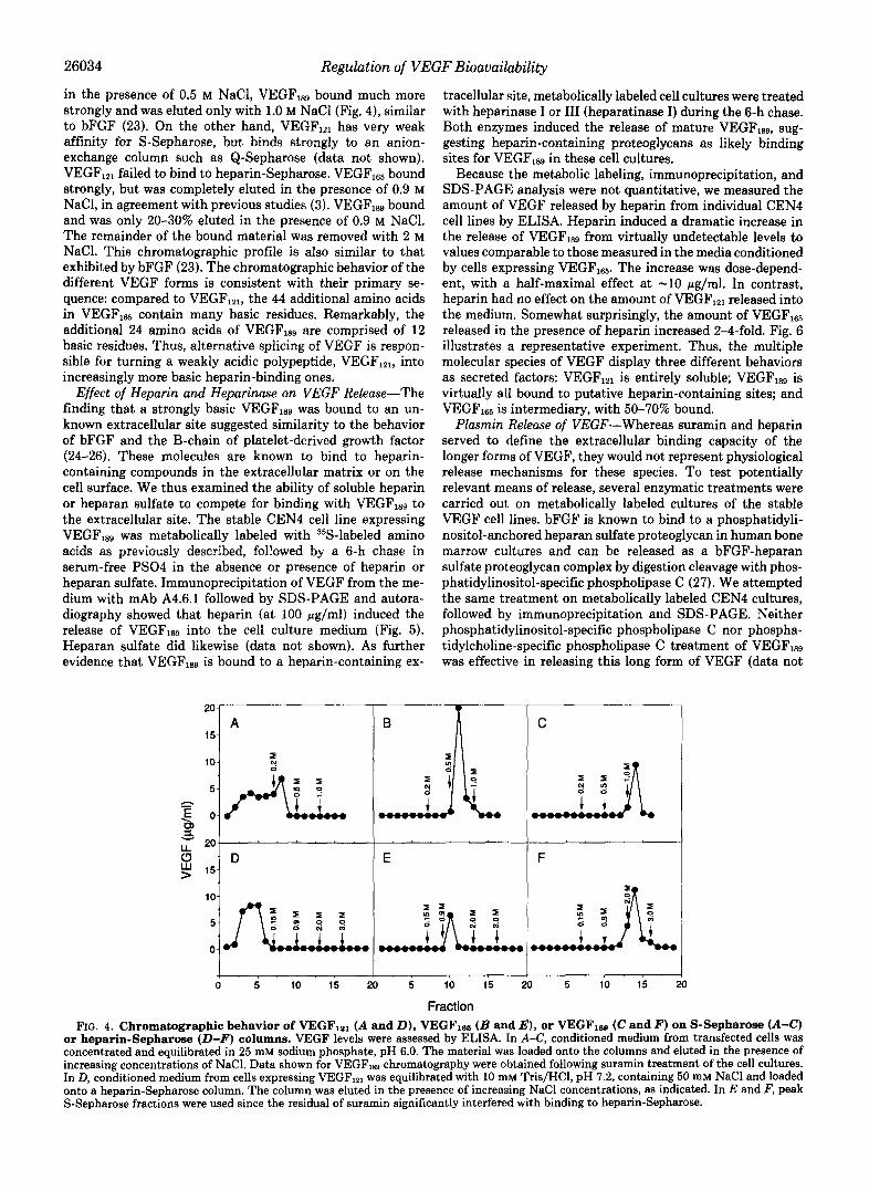
26034 Regulation of VEGF Bioavailability
in the presence of 0.5 M NaC1, VEGF,,, bound much more strongly and was eluted only with 1.0 M NaCl (Fig. 4), similar to bFGF (23). On the other hand, VEGF,,, has very weak affinity for S-Sepharose, but binds strongly to an anion- exchange column such as Q-Sepharose (data not shown). VEGF,,, failed to bind to heparin-Sepharose. VEGF,, bound strongly, but was completely eluted in the presence of 0.9 M NaCl, in agreement with previous studies (3). VEGF,,, bound and was only 20-30% eluted in the presence of 0.9 M NaCl. The remainder of the bound material was removed with 2 M NaC1. This chromatographic profile is also similar to that exhibited by bFGF (23). The chromatographic behavior of the different VEGF forms is consistent with their primary se- quence: compared to VEGFlZ1, the 44 additional amino acids in VEGFlss contain many basic residues. Remarkably, the additional 24 amino acids of VEGF,sg are comprised of 12 basic residues. Thus, alternative splicing of VEGF is respon- sible for turning a weakly acidic polypeptide, VEGFlzl, into increasingly more basic heparin-binding ones.
Effect of Heparin and Heparinase on VEGF Release-The finding that a strongly basic VEGFlsg was bound to an un- known extracellular site suggested similarity to the behavior of bFGF and the B-chain of platelet-derived growth factor (24-26). These molecules are known to bind to heparin- containing compounds in the extracellular matrix or on the cell surface. We thus examined the ability of soluble heparin or heparan sulfate to compete for binding with VEGF,,, to the extracellular site. The stable CEN4 cell line expressing VEGF,,, was metabolically labeled with 35S-labeled amino acids as previously described, followed by a 6-h chase in serum-free PS04 in the absence or presence of heparin or heparan sulfate. Immunoprecipitation of VEGF from the me- dium with mAb A4.6.1 followed by SDS-PAGE and autora- diography showed that heparin (at 100 pg/ml) induced the release of VEGF,,, into the cell culture medium (Fig. 5 ) . Heparan sulfate did likewise (data not shown). As further evidence that VEGF,,, is bound to a heparin-containing ex-
20
15-
10-
5- h
E O -
LL 20" 4 v
5 15'
10-
5-
0-
7
0
A I B A
tracellular site, metabolically labeled cell cultures were treated with heparinase I or I11 (heparatinase I) during the 6-h chase. Both enzymes induced the release of mature VEGFlsg, sug- gesting heparin-containing proteoglycans as likely binding sites for VEGF,, in these cell cultures.
Because the metabolic labeling, immunoprecipitation, and SDS-PAGE analysis were not quantitative, we measured the amount of VEGF released by heparin from individual CEN4 cell lines by ELISA. Heparin induced a dramatic increase in the release of VEGFlS9 from virtually undetectable levels to values comparable to those measured in the media conditioned by cells expressing VEGFls5. The increase was dose-depend- ent, with a half-maximal effect at -10 pg/ml. In contrast, heparin had no effect on the amount of VEGF,,, released into the medium. Somewhat surprisingly, the amount of VEGF165 released in the presence of heparin increased 2-4-fold. Fig. 6 illustrates a representative experiment. Thus, the multiple molecular species of VEGF display three different behaviors as secreted factors: VEGFIPl is entirely soluble; VEGF,,, is virtually all bound to putative heparin-containing sites; and VEGFI6, is intermediary, with 50-70% bound.
Plasmin Release of VEGF-Whereas suramin and heparin served to define the extracellular binding capacity of the longer forms of VEGF, they would not represent physiological release mechanisms for these species. To test potentially relevant means of release, several enzymatic treatments were carried out on metabolically labeled cultures of the stable VEGF cell lines. bFGF is known to bind to a phosphatidyli- nositol-anchored heparan sulfate proteoglycan in human bone marrow cultures and can be released as a bFGF-heparan sulfate proteoglycan complex by digestion cleavage with phos- phatidylinositol-specific phospholipase C (27). We attempted the same treatment on metabolically labeled CEN4 cultures, followed by immunoprecipitation and SDS-PAGE. Neither phosphatidylinositol-specific phospholipase C nor phospha- tidylcholine-specific phospholipase C treatment of VEGF18g was effective in releasing this long form of VEGF (data not
1 0 l a I 311 A I N a n rl I
I
D F E
5 10 15 x) 5 10 15 20 5 10 15 :
Fraction
21 0
FIG. 4. Chromatographic behavior of VEGFlal ( A and D ) , VEGFle6 ( B and E ) , or VEGFlse (C and F) on S-Sepharose (A-C) or heparin-Sepharose (D-F) columns. VEGF levels were assessed by ELISA. In A-C, conditioned medium from transfected cells was concentrated and equilibrated in 25 mM sodium phosphate, pH 6.0. The material was loaded onto the columns and eluted in the presence of increasing concentrations of NaCl. Data shown for VEGFles chromatography were obtained following suramin treatment of the cell cultures. In D, conditioned medium from cells expressing VEGF,,, was equilibrated with 10 mM Tris/HCl, pH 7.2, containing 50 mM NaCl and loaded onto a heparin-Sepharose column. The column was eluted in the presence of increasing NaCl concentrations, as indicated. In E and F, peak S-Sepharose fractions were used since the residual of suramin significantly interfered with binding to heparin-Sepharose.

Regulation of VEGF Bioavailability 26035
43
?9
18.4
FIG. 5. Release of VEGFlsa by heparin and heparinase. The control cell line, pHEBO, or the VEGFls~ cell line was metabolically labeled with [35S]cysteine and [35S]methionine as described under "Experimental Procedures." Heparin was included a t 100 pg/ml dur- ing the 6-h chase period where indicated. Heparinase I or I11 (hepar- atinase) was used a t 10 IU/ml. VEGF was immunoprecipitated with mAb A4.6.1 and analyzed under reducing conditions by SDS-PAGE.
I /I 0- .oca1 .001 .01 .1 1 10
Heparin (mg/ml) FIG. 6. Quantitation of bound VEGF. Each VEGF-expressing
cell line was treated for 24 h in serum-free medium with increasing amounts of heparin. The amount of VEGF present in the conditioned medium was quantitated by ELISA as described under "Experimental Procedures." Points represent the mean of triplicate determinations from a representative experiment. No VEGF could be detected in medium conditioned by the control cell line, pHEBO (data not shown).
shown). However, a 20-min digestion with the serine protease plasmin induced the release of an -17-kDa form and a 15- kDa form of VEGFlsg as well as peptides of the identical size from the VEGF,,, cell line as detected by immunoprecipita- tion and SDS-PAGE analysis under reducing conditions (Fig. 7, upper). These peptides were capable of dimerization as evidenced by the 34- and 30-kDa forms seen under nonreduc- ing conditions (Fig. 7, lower). Following inhibition of protease activity with aprotinin, the conditioned media from these cell lines were tested for activity in the endothelial cell prolifera- tion assay. Both were active as endothelial cell mitogens, whereas no activity could be detected in the medium condi- tioned for 20 min without plasmin (Fig. 8). The plasmin- released VEGF species did not bind to heparin, analogous to VEGF,,, (data not shown). In addition, these conditioned media were tested for induction of vascular permeability in the Miles assay (19). For this assay, serum-free medium was conditioned for 6 h by the stable cell lines, and plasmin was added at 0.1 CU/ml directly to this medium for the last 20 min at 37 "C. Following neutralization of the plasmin activity, media conditioned by the VEGF,,, cell line in the absence or presence of plasmin and by the VEGFls9 cell line only in the presence of plasmin stimulated significant leakage of Evans blue dye from vessels in guinea pig skin (Fig. 9). Media from the control cell line, pHEBO, and the VEGFlsg cell line in the absence of plasmin were inactive.
KRa -68 - 43
- 29 -- 10.4 - 14.3
-68 - 43 - 29
- 14.3 - 18.4
FIG. 7. Proteolytic release of VEGF. Cell lines expressing each form of VEGF were metabolically labeled with [35S]cysteine and [35S] methionine as described under "Experimental Procedures." Following a 6-h chase period in serum-free medium, the cultures were washed with fresh medium, and an additional incubation for 20 min a t 37 "C in the absence or presence of 0.1 casein units/ml plasmin was carried out. Following neutralization of plasmin activity with aprotinin, VEGF was immunoprecipitated from these 20-min incubations as well as from the 6-h chase with mAb A4.6.1. Samples were analyzed by SDS-PAGE under reducing (upper) and nonreducing (lower) conditions, followed by autoradiography. Control indicates the im- munoprecipitated products from the 6-h chase period. For VEGFls9, heparin (100 pg/ml) was included in the serum-free chase medium; cultures not treated with heparin were used for plasmin digestion. The immunoprecipitated products from the 20-min incubation are labeled as -Plasmin and +Plasmin.
12 1 i I O Control I
I
1 PHEBO VEGF165 VEGF189
Cell Line
1 FIG. 8. Mitogenic activity of proteolytically released VEGF.
Cell lines expressing VEGF1a or VEGFls9 or the control cell line was plated at equal densities and 24 h later incubated for 6 h in serum- free medium. Cultures were washed with fresh medium and incubated for 20 min a t 37 "C in the absence or presence of 0.1 CU/ml plasmin. Following neutralization of plasmin activity with aprotinin, the con- ditioned media were tested for mitogenic activity toward bovine capillary endothelial cells as described under "Experimental Proce- dures.'' The bars represent the average of triplicate determinations from a single dose of conditioned medium (100 pl) from a represent- ative experiment.
DISCUSSION
Numerous growth factors and their receptors as well as

26036 Regulation of VEGF Bioauailability
r .- E
h dilution 1:lO 1:lOO 2 No
pHEBO I + I -
FIG. 9. Vascular permeability activity of proteolytically re- leased VEGF. Cell lines expressing VEGFl= or VEGF189 or the control cell line was used to condition serum-free media for 6 h at 37 “C. During the last 20 min of this incubation, plasmin was added at 0.1 CU/ml where indicated. Plasmin activity was neutralized with aprotinin, and the media were tested at the indicated dilution for vascular permeability activity in the Miles assay (19) as described under “Experimental Procedures.”
other proteins are known to exist in multiple forms as a result of alternative splicing of their RNAs. It has recently become evident that these multiple molecular species often exhibit significantly different biological behavior (26, 28, 29). One such family of polypeptides resulting from alternative splicing of RNA is the angiogenic direct-acting endothelial cell mito- gen VEGF (5, 6). In this report, we demonstrated that the information supplied by alternative splicing has profound effects on the behavior of the translated proteins following secretion from the cell. The shortest form, VEGFIPl, which is a 34-36-kDa homodimeric polypeptide, is secreted and freely soluble in the conditioned medium of the human embryonic kidney cell line CEN4 stably transfected with a VEGFlZ1 expression vector. The transcript of VEGF165, the most com- mon form of VEGF, contains an additional 44 codons relative to VEGFIPl, resulting in a homodimeric protein of -45 kDa. These 44 amino acids in the carboxyl-terminal end of the protein convert it to a basic polypeptide with heparin binding capability. In a stably transfected CEN4 cell line, VEGF165 is secreted, but -50-70% of the material binds to as yet uniden- tified components on the cell surface or in the extracellular matrix. VEGFlag, which contains an additional 24 amino acids highly enriched in basic residues, binds much more tightly to cation-exchange columns and to heparin-Sepharose. Little or no VEGFlag is found in a freely soluble form in the conditioned medium of stably transfected CEN4 cells. Release of the bound forms of VEGF,,, and VEGFlag by treatment with heparin, heparan sulfate, or heparinase suggests that the unknown binding site in these cell cultures involves a heparin- containing proteoglycan (30, 31).
A variety of recent studies have demonstrated that heparin sulfate-containing proteoglycans are the constituents of the extracellular matrix that are responsible for binding and concentrating a variety of growth factors in the matrix and at the cell surface. The most studied system of proteoglycan regulation of growth factor availability is that of bFGF. The binding of bFGF to heparin or heparan sulfate, either as
soluble components of the tissue culture medium or as matrix- or cell surface-bound proteoglycans, appears to be necessary for bFGF binding to the high affinity FGF receptor (32). This requirement has not been completely understood. I t may involve protection from degradation of FGF (33, 34); altera- tion of the conformation of FGF to that required for receptor binding (32, 35); or oligomerization of FGF in a manner facilitating dimerization of FGF receptors (36), a necessary step in signal transduction by all tyrosine kinase receptors examined thus far (37). The extracellular matrix may also serve as a reservoir for FGF since active bFGF-glycosamino- glycan complexes can be generated by proteolysis of the proteoglycan core protein (31) or by cleavage of a glycosyl- phosphatidylinositol membrane anchor by phospholipase C (27, 38). A variety of other growth factors also bind heparin or heparan sulfate: interleukin-3 and granulocyte-macrophage colony-stimulating factor (39), pleiotropin (40), heregulin (41), and hepatocyte growth factor (42). Recently, evidence has been provided for the involvement of cell surface-associ- ated heparin-like molecules in enhancing binding of VEGF to high affinity receptors on endothelial cells in a manner similar to that reported for bFGF (43).
Whereas the CEN4 cell line is not known to endogenously express VEGF, heparin-binding sites are an integral compo- nent of many extracellular matrices. Thus, whatever cell type expresses VEGF, the described behavior of the different forms of VEGF could be expected. The source of VEGF secretion in vivo has not yet been thoroughly examined. Cultured aortic smooth muscle cells have been demonstrated to secrete VEGF and would represent a potential source capable of delivering VEGF directly to the endothelium in vivo (6, 44). In situ hybridization experiments demonstrated expression of the RNA transcript for VEGF in adult rat in a variety of well- vascularized organs such as pituitary, brain, heart, kidney, and lung (45,46). Despite this expression, the endothelium is essentially quiescent in such organs, suggesting the VEGF produced does not actively function as a growth factor in this context. Perhaps by binding to the extracellular matrix, VEGFlag and, to a significant extent, also VEGF165 are se- questered in a reservoir providing ready access to angiogenic factors, as has been postulated for bFGF (47). Although cell surface-associated heparin-like molecules are thought to be necessary for VEGF binding to its receptor on endothelial cells and such a complex is an active endothelial cell mitogen (43), if the heparin-binding sites are not contiguous with endothelial cells, then it would be unlikely that the bound VEGF could activate those receptors. During periods of active angiogenesis, the freely soluble VEGF,,, may be especially important. In this context, it is interesting that this form of VEGF appears to be the predominant one found in placenta, an organ with very active angiogenesis (5). In the developing corpus luteum, another tissue undergoing angiogenesis, in situ hybridization demonstrated high levels of VEGF RNA expres- sion, although the specific VEGF molecular species produced was not identified (48).
Binding of the long forms of VEGF to heparan sulfate proteoglycans in the extracellular matrix could provide a reservoir of biologically active VEGF available to endothelial cells following its release. We have demonstrated here that the serine protease plasmin induces the release of proteolyti- cally clipped VEGF species of both VEGF165 and VEGF,, that are freely soluble in the tissue culture medium and are biologically active both as endothelial cell mitogens and as vascular permeability-enhancing agents. The size of the mon- omeric subunits as analyzed under reducing conditions by SDS-PAGE (-17 kDa) is very similar to that of the VEGF,,,

Regulation of VEGF Bioavailability 26037
monomer. In addition, mAb 3.13.1 does not immunoprecipi- tate the plasmin-cleaved forms of VEGFIGS and VEGFlss,2 and it does not recognize VEGFlzl (17). Since VEGF121 lacks 44 amino acids in the carboxyl terminus of the protein relative to VEGFla, this suggests that the plasmin-released species of VEGF resulted from truncation of the proteins in the carboxyl termini, thus eliminating putative heparin-binding sites re- sponsible for sequestration of the proteins in the extracellular matrix or on the plasma membrane.
Extensive studies have demonstrated the integral role of proteases during angiogenesis. The traditional paradigm for angiogenesis suggests that new capillary formation results from a cascade of processes beginning with degradation of the extracellular matrix of a venule following protease release or activation. This is followed by endothelial cell proliferation and migration, processes that also involve protease action through the degradation of the stroma of the tissue undergoing vascularization. Finally, the capillary lumen is formed; and again, proteases may be required (49). A potential key protease involved in these processes is plasmin (50-52). The active serine protease plasmin is generated by plasminogen activator cleavage of proteolytically inactive plasminogen. Plasminogen is present in serum and is relatively abundant in most tissues. Plasminogen activators of either or both the tissue and uro- kinase type are secreted from endothelial cells following stim- ulation with both VEGF and bFGF (53,54). Therefore, secre- tion of plasminogen activators by endothelial cells in response to angiogenic agents would result in locally high concentra- tions of plasmin. In addition to previously demonstrated involvement in degradation of the extracellular matrix, both directly through digestion of such components of the base- ment membrane as fibronectin and laminin and indirectly by activating collagenases from their zymogens (55), plasmin may thus also serve to liberate angiogenic agents including VEGFla and VEGFlm as well as bFGF from the extracellular matrix. It is possible that the spatial distribution of these angiogenic factors sequestered in the extracellular matrix may serve to orient vessel formation during angiogenesis by pro- viding a network of positive growth stimuli.
In conclusion, several different molecular species of VEGF can be generated as a result of alternative splicing of VEGF RNA. The information encoded by this alternative splicing determines the fate of VEGF to be freely soluble or bound to the extracellular matrix or plasma membrane proteins, or properties of both. The proteolytic cascade of plasminogen activation, a key step during angiogenesis, can cleave the bound forms of VEGF, releasing a soluble factor capable of stimulating endothelial cell growth. Examining the nature of growth factors bound to extracellular matrix components in uiuo may provide necessary information in understanding physiological cell growth regulation.
Acknowledgments-We are grateful to Becky Lyon for performing the Miles vascular permeability assay, to Jin Kim and Bing Li for monoclonal antibodies, and to Louis Tamayo and Kerrie Andow for graphics.
REFERENCES 1. Klagsbrun, M., and DAmore, P. A. (1991) Annu. Reu. Physiol. 63, 217-
2. Ferrara, N., Houck, K., Jakeman, L., and Leung, D. W. (1992) Endocr. Reu.
3. Ferrara, N., and Henzel, W. J. (1989) Biochem. Biophys. Res. Commun.
4. Jakeman, L. B., Winer, J., Bennett, G. L., Altar, C. A,, and Ferrara, N.
5. Houck, K. A., Ferrara, N., Winer, J., Cachianes, G., Li, B., and Leung, D.
'K. A. Houck, D. W. Leung, A. M. Rowland, J. Winer, and N.
239
13,18-32
161,851-858
(1992) J. Clin. Inuest. 8 9 , 244-253
Ferrara, unpublished data.
6. Tischer, E., Mitchell, R., Hartman, T., Silva, M., Gospodarowicz, D., Fiddes, W. (1991) Mol. Endocrinol. 6,1806-1814
J. C.. and Abraham. J. A. (1991) J. Biol. Chem. 266,11947-11954 7. Leung,'D. W., Cachianes, G:, Kuang, W.-J., Goeddel,.D. V., and Ferrara,
8. Plouet, J., Schilling, J., and Gospodarowicz, D. (1989) EMBO J. 8, 3801- N. (1989) Sclence 246,1306-1309
R W 9.
10.
11.
12.
13.
14.
15.
16. 17.
18.
19. 20. 21.
22.
23.
24.
25.
C o y i ~ l y , D. T., Olander, J. V., Heuvelman, D., Nelson, R., Monsell, R., Siegel, N., Haymore, B. L., Leimgrulxr, R., and Feder, J. (1989) J. Bwl.
Keck P. J., Hauser, S. D., Krivi, F., Sanzo, K., Warren, T., Feder, J., and Chem. 2 6 4 , 20017-20024
de Vries, C., Escobedo, J. A,, Ueno, H., Houck, K., Ferrara, N., and Cohnolly D. T. (1989) Science 246,1309-1312
Stanley, E., Ralph, S., McEwen, S., Boulet, I., Holtzman, D. A., Lock, P., Williams, L. T. (1992) Science 266,989-991
Ferrara, N., Leung, D. W., Cachianes, G., Winer, J., and Henzel, W. J. and Dunn, A. R. (1991) Mol. CeU. Bwl. 11,3399-3406
Cachianes, G., Ho, C., Weber, R. F., Williams, S. R., Goeddel, D. V., and (1991) Methods Enzymol. 198,391-404
Su, W., Middleton, T., Sugden, B., and Echols, H. (1991) Proc. Natl. Acad. Leung, D. W. (1992) Technique, in press
Andreason, G. L., and Evans, G. A. (1988) BioTechniques 6,650-660 Sci. U. S. A. 88,10870-10874
Kim, K. J., Li, B., Houck, K., Winer, J., and Ferrara, N. (1992) Growth
Nakane, P. K., and Kawaoi, A. (1974) J. Histoehem. Cytochern. 2 2 , 1084- Factors 7 , 5 3 4 4
Miles, A. A., and Miles, E. M. (1952) J. Physiol ( L o n d . ) 118 , 228-257 Papkoff, J. (1989) Mol. Cell. Biol. 9 , 3377-3384 Betaholtz, C., Johnsson, A., Heldin, C.-H., and Westermark, B. (1986) Proc.
Olander, J. V., Connolly, D. T., andDeLarco, J. E. (1991) Biochem. Biophys. Natl. Acad. Sci. U. S. A. 83,6440-6444
Gospodarowicz, D., Cheng, J., Lui, G.-M., Baird, A., and Bohlen, P. (1984) Res. Commun. 176,68-76
Vlodavsky, I., Folkman, J., Sullivan, R., Fridman, R., Ishai-Michaeli, R., Proc. Natl. Acad. Sci. U. S. A. 8 1 , 6963-6967
Sasse, J., and Klagsbrun, M. (1987) Proc. Natl. Acad. Sci. U. S. A. 8 4 , 2292-2296
Beckmann, M. P., Betsholtz, C., Heldin, C.-H., Westermark, B., DiMarco, E., DiFiore, P. P., Robbins, K. C., and Aaronson, S. A. (1988) Science 241.1346-1349
1091
26. Ostman, A., Andersson, M., Betsholtz, C., Westermark, B., and Heldin, C.-
27. Brunner. G.. Gabrilove. J.. Rifkin. D. B.. and Wilson. E. L. (1991) J. Cell
---, " - - ~~ ~~
H. (1991) Cell Regul. 2,503-512
j. Miki, T., Bottaro, D. P., Fleming, T. P.,'Smit and Aaronson, S. A. (1991,
30. Savona, C., Chambaz, E. M., and Feige
31. 32.
33. 34.
35.
36. 37. 38.
39.
40.
282 Saksela, O., and Rifkin, D. B. (1990) J. Cell Biol. 110 , 767-775 Yayon, A., Klagsbrun, M., Esko, J. D., Leder, P., and Ornitz, D. M. (1990)
Gospodarowicz, D., and Cheng, J. (1986) J. Cell. Physiol. 128,475-484 Saksela, O., Moscatelli, D., Sommer, A., and Rifkin, D. B. (1988) J. Cell
Prestrelski, S. J., Fox, G. M., and Arakawa, T. (1992) Arch. Biochem.
Ruoslahti, E., and Yamaguchi, Y. (1991) CeU 64,867-869 Ullrich, A., and Schlessin er, J. (1990) Cell 6 1 , 203-212 Bashkin, P., Neufeld, G., bitay-Goren, H., and Vlodavsky, I. (1992) J. Cell.
Roberts, R., Gallagher, J., Spooncer, E., Allen, T. D., Bloomfield, F., and Physwl. 161 , 126-137
Li, Y.-S., Milner, P. G., Chauhan, A. K., Watson, M. A., Hoffman, R. M., Dexter, T. M. (1992) Nature 332,376-378
Kodner, C. M., Milbrandt, J., and Deuel, T. F. (1990) Science 260,1690-
Cell 64,841-848
Biol. 107,743-751
Biophys. 293,314-319
1 K I A 41. H&x&, W. E., Sliwkowski, M. X., Akita R. W., Henzel, W. J., Lee, J.,
Park, J. W., Yansura, D., Abadi, N., Raa'b, H., Lewis, G. D., Shepard, H. M., Kuang, W.-J., Wood, W. I., Goeddel, D. V., andvandlen, R. L. (1992)
42. Zarnegar, R., and Michalopoulos G. (1989) Cancer Res. 49,3314-3320 43. Gitay-Goren, H., Soker, S., Vlodavsky, I., and Neufeld, G. (1992) J. Biol.
44. Ferrara, N., Winer, J., and Burton, T. (1991) Growth Factors 6, 141-148 45. Ferrara, N:, Leung, D. W., and Phillips, H. S. (1991) in Neuroendocrine
Persmctzues (Muller. E. E.. and MacLeod. R. B.. eds) Vol. 9. nn. 127-
Science 2 6 6 , 1205-1210
Chem. 267,6093-6098
~. ~
161,'Springer-Verla 'New York Inc., New York 46. Berse, B., Brown, L. f., Van De Water, L., Dvorak, H. F., and Senger, D.
R. (1992) Mol. Biol. Cell. 3, 211-220 47. Vlodavsky, I., Fuks, Z. Ishai-Michaeli, R., Bashkin, P., Levi, E., Korne G.,
Bar-Shavit, R., andkla sbrun, M. (1991) J. Cell. Biochem. 4 6 , 167-176 48. Phillips, H. S., Hains, J., L u n g , D. W., and Ferrara, N. (1990) Endocri-
, - - ~ - , ~~- - I rr-
49. Pepper, M. S., Belin, D., Montesano, R., Orci, L., andvassalli, J.-D. (1990) nology 127,965-967
50. Ossowski, L., Quigley, J. P., and Reich, E. (1975) in Proteases and Biolo ical J. Cell Biol. 11 1 , 743-755
Control (Reich, E., Rifkin, D. B., and Shaw, E., eds) pp. 901-903, gold Spring Harbor Laboratory, Cold Sprin Harbor, NY
51. Mignatti, P., Tsuboi, R., Robbins, E., anfiRifkin, D. B. (1989) J. Cell Biol. 108,671-682
52. Tsuboi, R., Sato, Y., and Rifkin, D. B. (1990) J. Cell Biol. 110,511-517 53. Moscatelli D., Presta M., and Rifkin, D. B. (1986) Proc. Natl. Acad. Sci.
54. Pepper, M. S., Ferrara, N., Orci, L., and Montesano, R. (1991) Biochem. U. S. A. 8 3 , 2091-2'095
55. Werb, Z., Banda, M. J., and Jones, P. A. (1980) J. Exp. Med. 162 , 1340- Biophys. Res. Commun. 181,902-906
1357