rapamycin prevents endothelial cell migration by inhibiting … · · 2011-12-24rapamycin...
TRANSCRIPT

Rapamycin prevents endothelial cell migration by inhibiting theendothelial-to-mesenchymal transition and matrixmetalloproteinase-2 and -9: An in vitro study
Hua Gao,1 Jingjing Zhang,2 Ting Liu,1 Weiyun Shi1
(The first two authors contributed equally to this work)
1State Key Lab Cultivation Base, Shandong Provincial Key Lab of Ophthalmology, Shandong Eye Institute, Qingdao, China;2Qingdao University, Qingdao, China
Purpose: To evaluate the influence of rapamycin on endothelial-mesenchymal transition and matrix metalloproteinase(MMP) secretion by human umbilical vein endothelial cell line EA.hy926 and explore rapamycin’s angiogenesis inhibitionmechanism.Methods: EA.hy926 cells were cultivated in vitro. After the cells attained complete confluency, an artificial scratch wasmade through the monolayer with a sterile plastic 100 μl micropipette tip. Cell morphology changes were observed. Theexpression of vascular endothelial (VE)-cadherin, vimentin, and Twist protein were examined by immunofluorescence.After scratching, the cells were treated with 10, 100, and 1,000 ng/ml rapamycin for durations of 24, 48, and 72 h. Cellproliferation was then assessed using methyl thiazolyl tetrazolium assay. Cell migration ability was examined, and theexpression of VE-cadherin, vimentin, and the Twist transcription factor in mRNA levels was evaluated with reversetranscriptase PCR. The expression of gelatinases (MMP-2 and MMP-9) was examined using gelatin zymography.Results: After scratching, the endothelial cells were able to migrate via an endothelial-to-mesenchymal transition, whichwas related to Twist expression. Finally, mesenchymal cells transitioned into endothelial cells and reached cell confluencyagain. The growth of EA.hy926 cells was not affected by rapamycin concentrations of 10 ng/ml or 100 ng/ml duringtreatment periods of 1, 2, and 3 days; however, cell growth was inhibited by 1,000 ng/ml rapamycin with a three-daytreatment period. Rapamycin successfully inhibited cell migration at concentrations of 10 ng/ml, 100 ng/ml, and 1,000ng/ml for a treatment period of up to 8 h. Different concentrations of rapamycin induced the expression of VE-cadherin,inhibited vimentin and Twist expression in the endothelial cells, and inhibited endothelial cell secretion of MMP-2 andMMP-9.Conclusions: Rapamycin inhibited cell migration and extracellular matrix degradation by inhibiting endothelial-to-mesenchymal transition and the endothelial cell secretion of MMP-2 and MMP-9; these may be possible mechanisms forthe inhibition of angiogenesis by rapamycin.
Neovascularization is a complex process and is tightlyregulated by many positive and negative factors [1-5].Endothelial cell migration plays an important role inangiogenesis [6]. Rapamycin is an immunosuppressivemacrolide. Its strong immune inhibition effects, as well as itsability to effectively inhibit corneal neovascularization andtumor angiogenesis [4,7], have gained much attention.Rapamycin has effectively inhibited angiogenesis byinhibiting endothelial cell migration and proliferation [4].However, very little is known about the mechanism by whichrapamycin inhibits endothelial cell migration. This mightoccur by the direct suppression of mammalian target ofrapamycin (mTOR) expression [4] and reduction of vascular
Correspondence to: Professor Weiyun Shi, Shandong Eye Institute,5 Yanerdao Road, Qingdao 266071, P.R. China; Phone:86-531-81276002; FAX: 86-531-81276090; email:[email protected]
endothelial growth factor (VEGF) expression [8] or throughsome other mechanism. The endothelial-to-mesenchymaltransition (EndoMT), whereby endothelial cells cantransdifferentiate into mesenchymal cells accompanied bydecreased endothelial markers (vascular endothelial [VE]-cadherin) and increased mesenchymal markers (vimentin), isan important step of angiogenesis during embryodevelopment [9,10], as well as in kidney fibrosis and otherfibrotic diseases [11,12]. It is still not clear whether rapamycininhibits endothelial cell migration by inhibiting EndoMT.Kwon found that rapamycin had not affected the expressionof MMP-9 mRNA (mRNA) in the alkaline-burned cornea[4], but many reports have also found that rapamycin maysuppress the expression of MMPs [13-15]. Therefore, thisstudy focused on whether EndoMT occurred during the cellmigration process, and whether rapamycin inhibitedendothelial cell migration by inhibiting EndoMT and blockingthe production of MMP-2 and MMP-9.
Molecular Vision 2011; 17:3406-3414 <http://www.molvis.org/molvis/v17/a367>Received 25 October 2010 | Accepted 21 December 2011 | Published 24 December 2011
© 2011 Molecular Vision
3406

METHODSMaterials: An EA.hy926 cell line was generously providedby Cora-Jean S. Edgell from the University of North Carolinaat Chapel Hill and the Tissue Culture Facility in the UnitedStates. Additional materials included fetal bovine serum(FBS; Gibco Company); goat polyclonal antibodies specificfor human Twist (Santa Cruz Company, Santa Cruz, CA); andmouse monoclonal antibodies (Mabs) binding to the humancell type–specific protein VE-cadherin (Santa CruzCompany). Rabbit polyclonal antibodies specific for humanvimentin, fluorescein isothiocyanate (FITC)-labeled goatantirabbit IgG antibody, FITC-labeled rabbit antigoat IgGantibody and tetramethyl rhodamine isothiocyanate (TRITC)-labeled goat antimouse IgG antibody were from ZhongshanGolden Bridge Biotechnology Co. Ltd. (Beijing, China).Primers were purchased from the Invitrogen Corporation(China). Rapamycin was provided by the North ChinaPharmaceutical Group New Drug Research and DevelopmentCenter. All chemical reagents were of analytical grade andwere purchased from Sigma.Cell cultures: The EA.hy926 cell line, a permanentendothelial cell line derived from human umbilical veinendothelial cells (HUVECs) by fusion with the lungcarcinoma cell line A549, were maintained in Dulbecco’sModified Eagle’s Medium (DMEM)-high glucose with4500 mg/l glucose (Gibco, Carlsbad, CA), supplemented with10% FBS at 37 °C in a 5% CO2 incubator. When theexperiment was conducted, EA.hy926 cells were seeded intissue culture plates and maintained in DMEM-high glucosewith 4500 mg/l glucose, supplemented with 2% FBS. A 5 mg/ml rapamycin stock solution was prepared in dimethylsulfoxide (DMSO; Xinxing Chemicals Company, Panjin,China) and stored at −20 °C. The culture medium was replacedwith rapamycin-containing medium (10, 100, and 1,000 ng/ml rapamycin) or vehicle control medium (0.1% DMSO infresh medium).Endothelial cell morphology and immunofluorescencestaining: EA.hy926 cells were seeded in 96-well tissue cultureplates in amounts of 5×10³ cells/well. After completeconfluency, an artificial scratch approximately 300 μm widewas made through the monolayer with a sterile plastic 100 μlmicropipette tip. After 24 h, 48 h, and 72 h of incubation, cellmorphology changes were observed under invertedmicroscope. The expression of VE-cadherin, vimentin, andTwist protein was examined using immunofluorescencestaining. Primary antibodies were VE-cadherin mouseantihuman, vimentin rabbit antihuman, and Twist goatantihuman; secondary antibodies were FITC-labeled goatantirabbit IgG, TRITC-labeled goat antimouse IgG antibody,FITC-labeled rabbit antigoat IgG, and nucleus stained withHoechst (Beyotime Institute of Biotechnology, Shanghai,China). Finally, the cells were visualized and photographedwith an Olympus fluorescence microscope (Olympus BX60,Tokyo, Japan).
Cell proliferation analysis: The cells (5×10³) were inoculatedin 96-well plates (150 µl/well), incubated for 12 h, and serum-starved overnight. Cell proliferation was measured using 3-(4, 5)-dimethylthiahiazo (-z-y1)-3, 5-di-phenytetrazoliumromide (MTT) [16].The cells wereseparately treated with 10, 100, and 1000 ng/ml rapamycin fordurations of 24, 48, and 72 h, with six wells for each group.The cells of the control group were left untreated. 3-(4,5)-dimethylthiahiazo (-z-y1)-3, 5-di- phenytetrazoliumromide(MTT, Sigma; 5 g/l) was used as an incubation solution, andthe wells were incubated for 4 h. DMSO (150 µl) was addedto the culture medium with low-speed oscillation for 10 min.The optical density (A value) was measured at 492 nm with acontinuous spectrum densitometer (Spectra Max M2;Molecular Devices, Sunnyvale, CA).Cell migration ability examination: The cells (5×10³) wereinoculated in 96-well plates (150 µl/well). An artificial scratchwas performed after complete confluency and separatelytreated with 10, 100, or 1000 ng/ml rapamycin for a durationof 8 h. The cells of the control group were left untreated.Photographs of treated cells within the scratch were taken witha phase-contrast microscope (magnification of 100×; NikonDiaphot 300; Nikon, Tokyo, Japan) connected to a digitalcamera. We randomly selected six photographs and countedthe cells that had moved into the wounded area in the selectedphotographs.Evaluation of the gene expression of vascular endothelial-cadherin, vimentin, and Twist: The cells (500×103) wereinoculated in 6-well plates (2 ml/well). After reachingconfluency, the cells were randomly divided into three groups:(1) the no-scratch group, (2) the scratch group, and (3) therapamycin group was further divided into three groups: afterbeing artificially scratched, the cells were separately treatedwith 10, 100, and 1,000 ng/ml of rapamycin for an incubationof 24 h. The cells were collected by centrifugation and thetotal RNA was extracted with the NucleoSpin RNA II System(Company Macherey-Nagel, Düren, Germany) according tothe manufacturer’s protocol. PCR primers were designed onthe basis of the published human gene sequences (Table 1).Reverse transcriptase–PCR was performed to evaluate theexpression of these genes in EA.hy926 cells using thehousekeeping gene glyceraldehyde-3-phosphatedehydrogenase (GAPDH) as an internal control [17].
Gelatin zymography: The endothelial cells of each group wereincubated according to the protocol used for reversetranscriptase–PCR. The culture supernatants were harvestedat 24 h and mixed with a gel sample buffer (0.5M Tris-HCl,glycerol, 10% sodium dodecyl sulfate [SDS], β-mercaptoethanol, and 0.5% bromophenol blue). Tenmicrograms of protein were taken by SDS PAGE (PAGE)separation; the SDS–PAGE gels contained 0.1% gelatin(Sigma) [18]. After electrophoresis, the gels were washed in50 mM Tris buffer containing 2.5% Triton X-100. The gels
Molecular Vision 2011; 17:3406-3414 <http://www.molvis.org/molvis/v17/a367> © 2011 Molecular Vision
3407

were incubated for an additional 18 h in incubation fluid(50 mM Tris buffer [pH 7.6], 10 mM CaCl2, and 200 mmol/lNaCl). The gels were stained with 0.5% Coomassie blue thatcontained 30% methanol and 10% glacial acetic acid andsubsequently destained in 45% methanol/10% acetic acid/H2O. White bands on a blue background indicated zones ofdigestion corresponding to the presence of different MMPs.Gels were scanned, and a density analysis of the bands wasperformed using ImageJ software.Statistical analysis: All values in the figures and text wereexpressed as mean±standard deviation (SD) of threeobservations. All data were analyzed with the SPSS 11.5statistical package. A one-way analysis of independentsamples t-test for two-sample comparisons was used tocompare the means of two groups. A p value less than 0.05was considered statistically significant. The reported resultswere representative of three independent experiments.
RESULTSChanges in endothelial cell morphology during migration andexpression of vascular endothelial cadherin, vimentin, and
Twist: After cell confluency, EA.hy926 cells ultimatelyformed a cobblestone-like monolayer (Figure 1A). Antibodiesthat recognized VE-cadherin yielded continuous, linearstaining around the periphery of EA.hy926 cells (Figure 2A),but there was no expression of Twist protein (Figure 2B) orvimentin protein (Figure 2C).There was a striking change inthe morphology of the endothelial cells that migrated into thescratch wound, which developed a fibroblast-like morphologywith visible pseudopods (Figure 1B); VE-cadherin expressiondecreased at endothelial cell-cell junctions (Figure 2D); andantibodies that recognized the cytoskeletal proteins vimentin(Figure 2E) and Twist (Figure 2F) were localized to thecytoplasm. The cells eventually reached confluence again fordurations of 72 h and only VE-cadherin protein wasexpressed.Effect of rapamycin on the growth and migration of EA.hy926cells: The growth of EA.hy926 cells was not affected by 10ng/ml or 100 ng/ml rapamycin treatment for up to 72 h. Incontrast, when the EA.hy926 cells were exposed to rapamycin
TABLE 1. PCR PRIMERS OF HUMAN GENE SEQUENCES
Gene name Primer sequence (5′-3′) Gene sizeVE-cadherin [40] F: AACTTCCCCTTCTTCACCC 387 bp R: AAAGGCTGCTGGAAAATGA Vimentin [41] F: GGCTCAGATTCAGGAACAGC 327 bp R: GCTTCAACGGCAAAGTTCTC Twist [42] F: GGAGTCCGCAGTCTTACGAG 201 bp R: TCTGGAGGACCTGGTAGAGG GAPDH [17] F: ACCACAGTCCATGCCATCAC 439 bp R: TCCACCACCCTGTGGCTGTA
Abbreviations: GAPDH represents glyceraldehyde 3-phosphate dehydrogenase, VE represents vascular endothelial.
Figure 1. Cell morphology. A: In the no-scratch group, after cell confluency, EA.hy926 cells formed a cobblestone-like monolayer. B: In thescratch group, the endothelial cells that migrated into the scratch wound showed a fibroblast-like morphology with visible pseudopods (contrastphase microscope 100×).
Molecular Vision 2011; 17:3406-3414 <http://www.molvis.org/molvis/v17/a367> © 2011 Molecular Vision
3408

for 72 h, the EA.hy926 cell growth was significantly inhibitedby concentrations of 1000 ng/ml rapamycin (Figure 3).
The effect of rapamycin on the migration of EA.hy926cells was examined by using a scratch-wound assay.Rapamycin at concentrations of 10,100, and 1000 ng/mlinhibited the migration of EA.hy926 cells. There werestatistically significant differences between the control groupand the rapamycin groups (10, 100, 1000 ng/ml; Figure 4).
Effect of rapamycin on gene expression cell marker proteinsand Twist: After full cell confluency, there was onlyexpression of VE-cadherin mRNA. After scratching,expression of VE-cadherin in the mRNA level downregulated,and EA.hy926 cells expressed Twist mRNA and vimentinmRNA, while 10, 100, and 1,000 ng/ml rapamycin inhibitedthe expression of Twist mRNA and vimentin mRNA. Therewere statistically significant differences between the no-scratch group and the scratch group and between the scratch
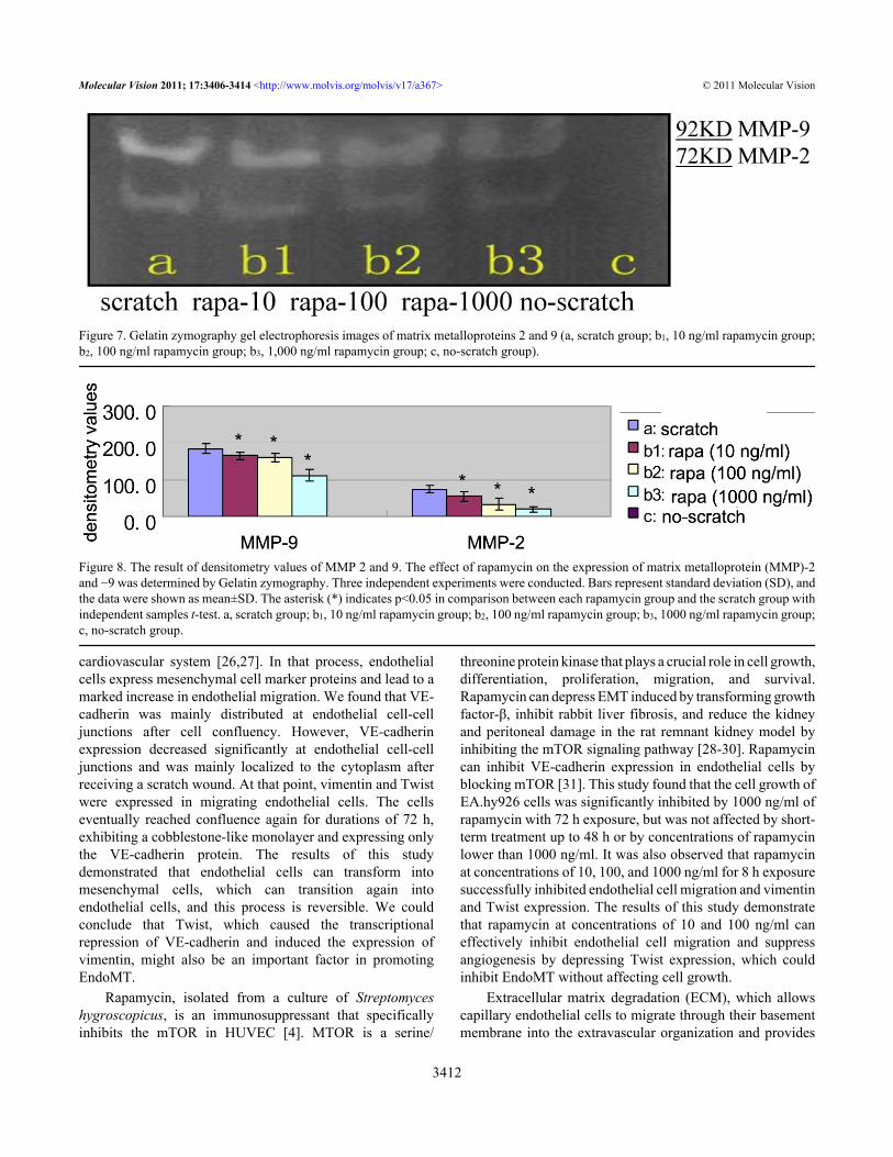
group and the rapamycin groups (10, 100, and 1000 ng/ml)(Figure 5, Figure 6).Effect of rapamycin on matrix metalloproteinase-2 and matrixmetalloproteinase-9 activity: Results from the gelatinzymography assay showed that neither MMP-2 nor MMP-9was detected in the culture supernatants of the no-scratchgroup (Figure 7), while there was expression of MMP-2 andMMP-9 protein in the scratch group and rapamycin groups.Both MMP-2 and MMP-9 were significantly reduced inEA.hy926 cells with a linear scratch wound with increasingconcentration of rapamycin. There were statisticallysignificant differences between the scratch group and therapamycin groups (t=2.954, 5.640, 11.504, 3.056, 3.600,9.349; p<0.05; Figure 7 and Figure 8).
DISCUSSIONExtracellular microenvironmental changes, such as hypoxia,result in increased expression of VEGF, which can activate
Figure 2. Immunofluorescence images. A: In the no-scratch group, after cell confluency, vascular endothelial (VE)-cadherin yieldedcontinuous, linear staining around the periphery of EA.hy926 cells (confocal microscopy 200×). B: In the no-scratch group, after cellconfluency, there was no expression of vimentin protein (confocal microscopy 100×). C: In the no-scratch group, after cell confluency, therewas no expression of the Twist protein (confocal microscopy 100×). D: In the scratch group, the VE-cadherin expression of the endothelialcells that migrated into the scratch wound was decreased at endothelial cell-cell junctions and was mainly localized to the cytoplasm; VE-cadherin yielded continuous, linear staining around the periphery of EA.hy926 cells on the outside of the scratch wound (confocal microscopy100×). E: In the scratch group, the vimentin expression of endothelial cells that migrated into the scratch wound (confocal microscopy 100×).F: In the scratch group, the Twist expression of endothelial cells that migrated into the scratch wound (confocal microscopy 100×).
Molecular Vision 2011; 17:3406-3414 <http://www.molvis.org/molvis/v17/a367> © 2011 Molecular Vision
3409

endothelial cells. Activated endothelial cells can secreteprotease (which dissolves vascular basement membranes) andextracellular matrix proteins, allowing capillary endothelialcells to migrate through their basement membrane into theextracellular matrix to form neovascular buds. This is thebasic pathological process of angiogenesis. Rapamycin caneffectively inhibit corneal neovascularization and tumorangiogenesis [4,7,19]. However, very little is known aboutrapamycin’s mechanism for inhibiting endothelial cellmigration. This study aimed to fill this knowledge gap byobserving rapamycin’s effects on EndoMT and the productionof MMP-2 and MMP-9.
Numerous studies have shown that tumor cells originatedin epithelial cells that transdifferentiated into mesenchymalcells, gained the ability to migrate, and promoted tumor
metastasis [20]. When epithelial cells underwentmesenchymal transformation, their morphology changedfrom a cobblestone-like shape to a spindle-like shape [21,22]. This change in morphology was accompanied bydecreased epithelial markers (E-cadherin) and increasedTwist and mesenchymal markers (vimentin). Vimentin, amesenchymal phenotype marker, was an intermediatefilament cytoskeletal protein and was widely expressed inmesenchymal cells, which was very closely related tomaintaining the fibroblasts’ shape and permitting cellmigration. Vimentin was also expressed in HUVEC [23],which was more especially implicated in cellular motility andplasticity and was exclusively expressed in the migrating ratheart endothelial cell line [24].
Figure 3. The effect of rapamycin on EA.hy926 cell growth. EA.hy926 cells were treated with 0 to 1,000 ng/ml rapamycin for 24, 48, or 72h. Cell growth was determined by methyl thiazolyl tetrazolium assay (MTT). 0 ng/ml of rapamycin represents the control group. Bars representstandard deviation (SD; n=6 wells per measurement). Similar results were obtained in three independent experiments, and the data were shownas mean±SD. The asterisk indicates p<0.05, compared with the control group with independent samples t-test.
Figure 4. The effect of rapamycin on the migration of EA.hy926 cells. EA.hy926 cells were treated with 0 to 1,000 ng/ml rapamycin for 8 h.Cell migration ability was determined by the scratch wound assay method. 0 ng/ml of rapamycin represents the control group. Bars representSD (n=6 photographs per measurement). Similar results were obtained in three independent experiments, and the data were shown as mean±SD. The asterisk indicates p<0.05, compared with the control group with independent samples t-test.
Molecular Vision 2011; 17:3406-3414 <http://www.molvis.org/molvis/v17/a367> © 2011 Molecular Vision
3410

The transcription factor Twist, a master regulator ofembryonic morphogenesis, was recently identified as animportant promoter of epithelial-to-mesenchymal transition(EMT) in breast cancers. In this process, Twist directly andindirectly caused the transcriptional repression of E-cadherinthrough the E-box elements on the E-cadherin promoter,
thereby inducing vimentin expression [20]. Lopez et al. [25]has found that the tumor-induced upregulation of Twistrepressed the activity of the human VE-cadherin promoter.
Endothelial cell migration plays an important role inangiogenesis [6]. Endothelial cells can change intomesenchymal cells during the embryonic development of the
Figure 5. Reverse transcriptase–PCR gel electrophoresis images of vascular endothelial–cadherin/vimentin/Twist/glyceraldehyde 3-phosphatedehydrogenase mRNA expression in EA.hy926 cells. The effect of rapamycin on the gene expression of vascular endothelial (VE)-cadherin,vimentin, and Twist. (a, scratch group; b1, 10 ng/ml rapamycin group; b2, 100 ng/ml rapamycin group; b3, 1,000 ng/ml rapamycin group; c,no-scratch group).
Figure 6. Relative expression ratio of the target gene versus the housekeeping gene (glyceraldehyde 3-phosphate dehydrogenase). The ratiofor the vertical axis is relative expression ratio of the target gene versus the house-keeping gene (glyceraldehyde 3-phosphate dehydrogenase[GAPDH]). Three independent experiments were conducted. Bars represent standard deviation (SD), and the data were shown as mean±SD.The asterisk indicates p<0.05 in comparison between each rapamycin group and the scratch group with independent samples t-test. Poundsign (#) indicates p<0.05 in comparison between the scratch group and the no-scratch group with independent samples t-test. a, scratch group;b1, 10 ng/ml rapamycin group; b2, 100 ng/ml rapamycin group; b3, 1,000 ng/ml rapamycin group; c, no-scratch group.
Molecular Vision 2011; 17:3406-3414 <http://www.molvis.org/molvis/v17/a367> © 2011 Molecular Vision
3411
cardiovascular system [26,27]. In that process, endothelialcells express mesenchymal cell marker proteins and lead to amarked increase in endothelial migration. We found that VE-cadherin was mainly distributed at endothelial cell-celljunctions after cell confluency. However, VE-cadherinexpression decreased significantly at endothelial cell-celljunctions and was mainly localized to the cytoplasm afterreceiving a scratch wound. At that point, vimentin and Twistwere expressed in migrating endothelial cells. The cellseventually reached confluence again for durations of 72 h,exhibiting a cobblestone-like monolayer and expressing onlythe VE-cadherin protein. The results of this studydemonstrated that endothelial cells can transform intomesenchymal cells, which can transition again intoendothelial cells, and this process is reversible. We couldconclude that Twist, which caused the transcriptionalrepression of VE-cadherin and induced the expression ofvimentin, might also be an important factor in promotingEndoMT.
Rapamycin, isolated from a culture of Streptomyceshygroscopicus, is an immunosuppressant that specificallyinhibits the mTOR in HUVEC [4]. MTOR is a serine/
threonine protein kinase that plays a crucial role in cell growth,differentiation, proliferation, migration, and survival.Rapamycin can depress EMT induced by transforming growthfactor-β, inhibit rabbit liver fibrosis, and reduce the kidneyand peritoneal damage in the rat remnant kidney model byinhibiting the mTOR signaling pathway [28-30]. Rapamycincan inhibit VE-cadherin expression in endothelial cells byblocking mTOR [31]. This study found that the cell growth ofEA.hy926 cells was significantly inhibited by 1000 ng/ml ofrapamycin with 72 h exposure, but was not affected by short-term treatment up to 48 h or by concentrations of rapamycinlower than 1000 ng/ml. It was also observed that rapamycinat concentrations of 10, 100, and 1000 ng/ml for 8 h exposuresuccessfully inhibited endothelial cell migration and vimentinand Twist expression. The results of this study demonstratethat rapamycin at concentrations of 10 and 100 ng/ml caneffectively inhibit endothelial cell migration and suppressangiogenesis by depressing Twist expression, which couldinhibit EndoMT without affecting cell growth.
Extracellular matrix degradation (ECM), which allowscapillary endothelial cells to migrate through their basementmembrane into the extravascular organization and provides
Figure 7. Gelatin zymography gel electrophoresis images of matrix metalloproteins 2 and 9 (a, scratch group; b1, 10 ng/ml rapamycin group;b2, 100 ng/ml rapamycin group; b3, 1,000 ng/ml rapamycin group; c, no-scratch group).
Figure 8. The result of densitometry values of MMP 2 and 9. The effect of rapamycin on the expression of matrix metalloprotein (MMP)-2and −9 was determined by Gelatin zymography. Three independent experiments were conducted. Bars represent standard deviation (SD), andthe data were shown as mean±SD. The asterisk (*) indicates p<0.05 in comparison between each rapamycin group and the scratch group withindependent samples t-test. a, scratch group; b1, 10 ng/ml rapamycin group; b2, 100 ng/ml rapamycin group; b3, 1000 ng/ml rapamycin group;c, no-scratch group.
Molecular Vision 2011; 17:3406-3414 <http://www.molvis.org/molvis/v17/a367> © 2011 Molecular Vision
3412

the necessary space for new vessel formation, is an importantprocess required during angiogenesis. MMPs are a class ofendogenous proteolytic enzymes that are involved in ECMdegradation. In particular, MMP-2 and MMP-9, which candegrade ECM and activate many growth factors, play animportant role in tumor invasion and metastasis [32,33].Rapamycin can suppress glioma invasion by blocking theproduction of MMP-2 and MMP-9 [13]. Endothelial cellsproduce MMPs at lower levels under normal circumstances,but endothelial cells activated by inflammation, trauma, andtumor growth can secrete a large number of MMPs that areactivated quickly [34,35]. Some experimental studies haveshown that normal corneal tissues produce MMPs at lowerlevels, which can be directly induced by some cytokines suchas VEGF, fibroblast growth factor, and transforming growthfactor and indirectly activated by active urokinase to promoteangiogenesis in a disease state [4,36]. Kwon et al. found thatrapamycin did not change MMP-9 expression in alkaline-burned corneal tissue [4], and this was possible becauseangiogenesis is a complex process regulated by multiplestimulatory and inhibitory factors in vivo. Some studies havefound that HUVECs could produce MMP-2 and MMP-9 in aninactive form [37,38]. This experiment found that EA.hy926cells secreted neither MMP-2 nor MMP-9 when they reachedfull confluence, but EA.hy926 cells activated by the scratchcould produce MMP-2 and MMP-9 to increase cell migration.MMP-2 and MMP-9 were inhibited by 10, 100, and 1000 ng/ml doses of rapamycin to repress ECM degradation and cellmigration. MMP-2 played an essential role in producing EMT[39]. Therefore, rapamycin might inhibit EndoMT bydepressing MMP-2.
Our data indicate that Twist may also be an importantfactor in promoting EndoMT. The possible mechanism for theinhibition of angiogenesis by rapamycin is as follows:Rapamycin inhibited cell migration and extracellular matrixdegradation by inhibiting EndoMT and endothelial cellsecretion of MMP-2 and MMP-9. These are possiblemechanisms for the inhibition of angiogenesis by Rapamycin.
ACKNOWLEDGMENTSThis study was supported by the National Natural ScienceFoundation of China (30700923), the Department of Scienceand Technology of Shandong Province (2006GG1102020,2009GG20002015), and the Taishan Scholar Program(20081148). The authors thank Ms. Ping Lin for her assistancein the preparation of this manuscript.
REFERENCES1. Usui T, Sugisaki K, Iriyama A, Yokoo S, Yamagami S, Nagai
N, Ishida S, Amano S. Inhibition of cornealneovascularization by blocking the angiotensin II type 1receptor. Invest Ophthalmol Vis Sci 2008; 49:4370-6.[PMID: 18829859]
2. Mochimaru H, Usui T, Yaguchi T, Nagahama Y, Hasegawa G,Usui Y, Shimmura S, Tsubota K, Amano S, Kawakami Y,
Ishida S. Suppression of alkali burn-induced cornealneovascularization by dendritic cell vaccination targetingVEGF receptor 2. Invest Ophthalmol Vis Sci 2008;49:2172-7. [PMID: 18263815]
3. Lai LJ, Xiao X, Wu JH. Inhibition of corneal neovascularizationwith endostatin delivered by adeno-associated viral (AAV)vector in a mouse corneal injury model. J Biomed Sci 2007;14:313-22. [PMID: 17373573]
4. Kwon YS, Hong HS, Kim JC, Shin JS, Son Y. Inhibitory effectof rapamycin on corneal neovascularization in vitro and invivo. Invest Ophthalmol Vis Sci 2005; 46:454-60. [PMID:15671269]
5. Chen P, Yin H, Wang Y, Mi J, He W, Xie L, Wang Y. Multi-gene targeted antiangiogenic therapies for experimentalcorneal neovascularization. Mol Vis 2010; 16:310-9. [PMID:20208988]
6. Lamalice L, Le Boeuf F, Huot J. Endothelial cell migrationduring angiogenesis. Circ Res 2007; 100:782-94. [PMID:17395884]
7. Guba M, von Breitenbuch P, Steinbauer M, Koehl G, Flegel S,Hornung M, Bruns CJ, Zuelke C, Farkas S, Anthuber M,Jauch KW, Geissler EK. Rapamycin inhibits primary andmetastatic tumor growth by antiangiogenesis: involvement ofvascular endothelial growth factor. Nat Med 2002;8:128-35. [PMID: 11821896]
8. Stahl A, Paschek L, Martin G, Gross NJ, Feltgen N, Hansen LL,Agostini HT. Rapamycin reduces VEGF expression in retinalpigment epithelium (RPE) and inhibits RPE-inducedsprouting angiogenesis in vitro. FEBS Lett 2008;582:3097-102. [PMID: 18703055]
9. Arciniegas E, Neves CY, Carrillo LM, Zambrano EA, RamirezR. Endothelial-mesenchymal transition occurs duringembryonic pulmonary artery development. Endothelium2005; 12:193-200. [PMID: 16162442]
10. Arciniegas E, Servin M, Arguello C, Mota M. Development ofthe aorta in the chick embryo: structural and ultrastructuralstudy. Atherosclerosis 1989; 76:219-35. [PMID: 2730719]
11. Zeisberg EM, Potenta SE, Sugimoto H, Zeisberg M, Kalluri R.Fibroblasts in kidney fibrosis emerge via endothelial-to-mesenchymal transition. J Am Soc Nephrol 2008;19:2282-7. [PMID: 18987304]
12. Zeisberg EM, Tarnavski O, Zeisberg M, Dorfman AL,McMullen JR, Gustafsson E, Chandraker A, Yuan X, Pu WT,Roberts AB, Neilson EG, Sayegh MH, Izumo S, Kalluri R.Endothelial-to-mesenchymal transition contributes to cardiacfibrosis. Nat Med 2007; 13:952-61. [PMID: 17660828]
13. Heimberger AB, Wang E, McGary EC, Hess KR, Henry VK,Shono T, Cohen Z, Gumin J, Sawaya R, Conrad CA, LangFF. Mechanisms of action of rapamycin in gliomas. Neuro-oncol 2005; 7:1-11. [PMID: 15701277]
14. Mabuchi S, Altomare DA, Connolly DC, Klein-Szanto A,Litwin S, Hoelzle MK, Hensley HH, Hamilton TC, Testa JR.RAD001 (Everolimus) delays tumor onset and progression ina transgenic mouse model of ovarian cancer. Cancer Res2007; 67:2408-13. [PMID: 17363557]
15. Busch S, Renaud SJ, Schleussner E, Graham CH, Markert UR.mTOR mediates human trophoblast invasion throughregulation of matrix-remodeling enzymes and is associatedwith serine phosphorylation of STAT3. Exp Cell Res 2009;315:1724-33. [PMID: 19331815]
Molecular Vision 2011; 17:3406-3414 <http://www.molvis.org/molvis/v17/a367> © 2011 Molecular Vision
3413

16. Yang L, Wang Y, Zhou Q, Chen P, Wang Y, Wang Y, Liu T,Xie L. Inhibitory effects of polysaccharide extract fromSpirulina platensis on corneal neovascularization. Mol Vis2009; 15:1951-61. [PMID: 19784394]
17. Song Z, Wang Y, Xie L, Zang X, Yin H. Expression ofsenescence-related genes in human corneal endothelial cells.Mol Vis 2008; 14:161-70. [PMID: 18334933]
18. Hawkes SP, Li H, Taniguchi GT. Zymography and reversezymography for detecting MMPs and TIMPs. Methods MolBiol 2010; 622:257-69. [PMID: 20135288]
19. Liu L, Chen L, Chung J, Huang S. Rapamycin inhibits F-actinreorganization and phosphorylation of focal adhesionproteins. Oncogene 2008; 27:4998-5010. [PMID: 18504440]
20. Yang J, Mani SA, Donaher JL, Ramaswamy S, Itzykson RA,Come C, Savagner P, Gitelman I, Richardson A, WeinbergRA. Twist, a master regulator of morphogenesis, plays anessential role in tumor metastasis. Cell 2004; 117:927-39.[PMID: 15210113]
21. Veveris-Lowe TL, Lawrence MG, Collard RL, Bui L,Herington AC, Nicol DL, Clements JA. Kallikrein 4 (hK4)and prostate-specific antigen (PSA) are associated with theloss of E-cadherin and an epithelial-mesenchymal transition(EMT)-like effect in prostate cancer cells. Endocr RelatCancer 2005; 12:631-43. [PMID: 16172196]
22. Whitbread AK, Veveris-Lowe TL, Lawrence MG, Nicol DL,Clements JA. The role of kallikrein-related peptidases inprostate cancer: potential involvement in an epithelial tomesenchymal transition. Biol Chem 2006; 387:707-14.[PMID: 16800731]
23. Bruneel A, Labas V, Mailloux A, Sharma S, Vinh J,Vaubourdolle M, Baudin B. Proteomic study of humanumbilical vein endothelial cells in culture. Proteomics 2003;3:714-23. [PMID: 12748950]
24. Obermeyer N, Janson N, Bergmann J, Buck F, Ito WD.Proteome analysis of migrating versus nonmigrating rat heartendothelial cells reveals distinct expression patterns.Endothelium 2003; 10:167-78. [PMID: 13129820]
25. Lopez D, Niu G, Huber P, Carter WB. Tumor-inducedupregulation of Twist, Snail, and Slug represses the activityof the human VE-cadherin promoter. Arch Biochem Biophys2009; 482:77-82. [PMID: 19046938]
26. Frid MG, Kale VA, Stenmark KR. Mature vascular endotheliumcan give rise to smooth muscle cells via endothelial-mesenchymal transdifferentiation: in vitro analysis. Circ Res2002; 90:1189-96. [PMID: 12065322]
27. Ishisaki A, Hayashi H, Li AJ, Imamura T. Human umbilicalvein endothelium-derived cells retain potential todifferentiate into smooth muscle-like cells. J Biol Chem 2003;278:1303-9. [PMID: 12417591]
28. Patel P, Sekiguchi Y, Oh KH, Patterson SE, Kolb MR, MargettsPJ. Smad3-dependent and -independent pathways areinvolved in peritoneal membrane injury. Kidney Int 2010;77:319-28. [PMID: 19956083]
29. Bridle KR, Popa C, Morgan ML, Sobbe AL, Clouston AD,Fletcher LM, Crawford DH. Rapamycin inhibits hepaticfibrosis in rats by attenuating multiple profibrogenicpathways. Liver Transpl 2009; 15:1315-24. [PMID:19790156]
30. Esposito C, Villa L, Grosjean F, Mangione F, Esposito V,Castoldi F, Serpieri N, Arra M, Pertile E, Maggi N, ValentinoR, Dal Canton A. Rapamycin reduces proteinuria and renaldamage in the rat remnant kidney model. Transplant Proc2009; 41:1370-1. [PMID: 19460562]
31. Bieri M, Oroszlan M, Zuppinger C, Mohacsi PJ. Biosynthesisand expression of VE-cadherin is regulated by the PI3K/mTOR signaling pathway. Mol Immunol 2009; 46:866-72.[PMID: 18990449]
32. Klein G, Vellenga E, Fraaije MW, Kamps WA, de Bont ES. Thepossible role of matrix metalloproteinase (MMP)-2 andMMP-9 in cancer, e.g. acute leukemia. Crit Rev OncolHematol 2004; 50:87-100. [PMID: 15157658]
33. Sluijter JP, de Kleijn DP, Pasterkamp G. Vascular remodelingand protease inhibition–bench to bedside. Cardiovasc Res2006; 69:595-603. [PMID: 16387286]
34. Gharagozlian S, Henriksen T, Kolset SO. High glucose andNepsilon-(carboxymethyl) lysine bovine serum albuminmodulate release of matrix metalloproteinases in culturedhuman endothelial cells. Eur J Nutr 2006; 45:283-90. [PMID:16705353]
35. Hanemaaijer R, Koolwijk P, le Clercq L, de Vree WJ, vanHinsbergh VW. Regulation of matrix metalloproteinaseexpression in human vein and microvascular endothelial cells.Effects of tumour necrosis factor alpha, interleukin 1 andphorbol ester. Biochem J 1993; 296:803-9. [PMID: 8280080]
36. Zhai HL, Xie LX, Dong XG. The roles of gelatinases inpathological changes of fungal keratitis in experimentalrabbits. Zhonghua Yan Ke Za Zhi 2007; 43:817-22. [PMID:18070528]
37. Montiel M, Urso L, de la Blanca EP, Marsigliante S, JimenezE. Cisplatin reduces endothelial cell migration via regulationof type 2-matrix metalloproteinase activity. Cell PhysiolBiochem 2009; 23:441-8. [PMID: 19471112]
38. Mauro A, Buscemi M, Gerbino A. Immunohistochemical andtranscriptional expression of matrix metalloproteinases infull-term human umbilical cord and human umbilical veinendothelial cells. J Mol Histol 2010; 41:367-77. [PMID:20936527]
39. Duong TD, Erickson CA. MMP-2 plays an essential role inproducing epithelial-mesenchymal transformations in theavian embryo. Dev Dyn 2004; 229:42-53. [PMID: 14699576]
40. Schäfer R, Abraham D, Paulus P, Blumer R, Grimm M, WojtaJ, Aharinejad S. Impaired VE-cadherin/beta-cateninexpression mediates endothelial cell degeneration in dilatedcardiomyopathy. Circulation 2003; 108:1585-91. [PMID:12963640]
41. Uchida S, Yokoo S, Yanagi Y, Usui T, Yokota C, Mimura T,Araie M, Yamagami S, Amano S. Sphere formation andexpression of neural proteins by human corneal stromal cellsin vitro. Invest Ophthalmol Vis Sci 2005; 46:1620-5. [PMID:15851560]
42. Cheng GZ, Zhang WZ, Sun M, Wang Q, Coppola D, MansourM, Xu LM, Costanzo C, Cheng JQ, Wang LH. Twist istranscriptionally induced by activation of STAT3 andmediates STAT3 oncogenic function. J Biol Chem 2008;283:14665-73. [PMID: 18353781]
Molecular Vision 2011; 17:3406-3414 <http://www.molvis.org/molvis/v17/a367> © 2011 Molecular Vision
Articles are provided courtesy of Emory University and the Zhongshan Ophthalmic Center, Sun Yat-sen University, P.R. China.The print version of this article was created on 24 December 2011. This reflects all typographical corrections and errata to thearticle through that date. Details of any changes may be found in the online version of the article.
3414