dubai owc 2012 - sicotlhcnews.sicot.org/resources/file/newsletter/nl132.pdfthe aaos, ao foundation,...
TRANSCRIPT
June 2012 - No. 132
In this issueCase of the Month 2 / Editorial by Hashem Al Khatib 3 / Worldwide News: Radiologic Predictors of Functional Outcome Following Fractures of the Distal Radius 4Scientific Debate: High Tibial Osteotomy versus Unicompartmental Knee Arthroplasty 6Young Surgeons: 2011 International Travelling Fellowship Report 9 Conference News: Plenary Speakers 10 / Social & Sports Programmes 12
Dubai OWC 2012Social & Sports
Programmes
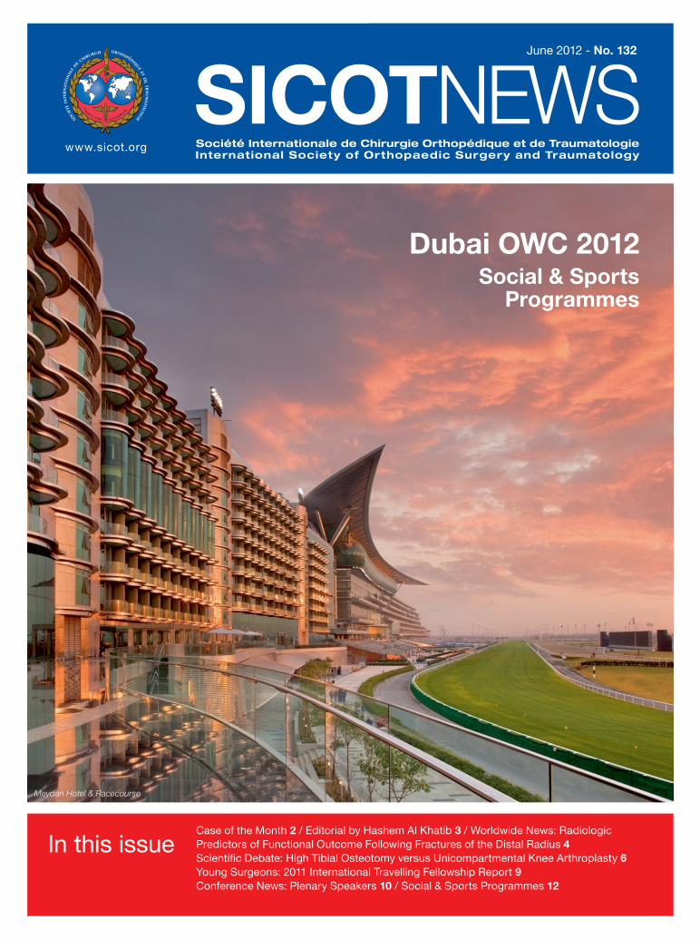
Meydan Hotel & Racecourse
2 SICOTNEWS | June 2012 - No. 132
Multiple swellings in a three-year-old girl
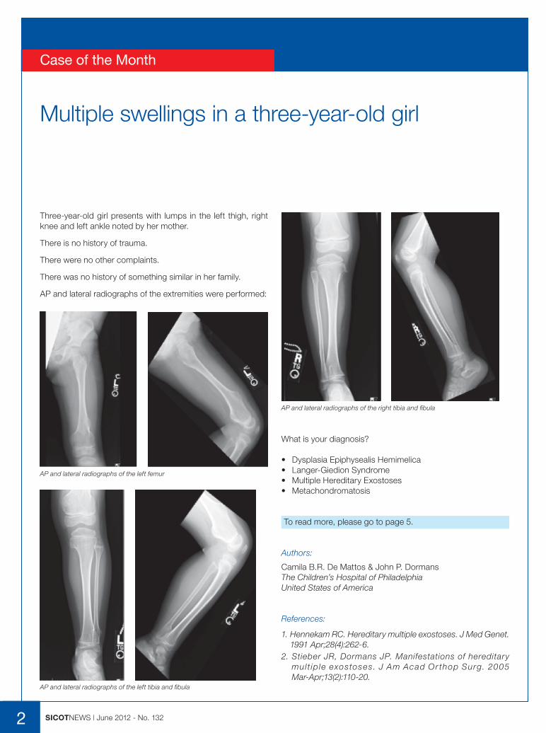
Three-year-old girl presents with lumps in the left thigh, right knee and left ankle noted by her mother.
There is no history of trauma.
There were no other complaints.
There was no history of something similar in her family.
AP and lateral radiographs of the extremities were performed:
AP and lateral radiographs of the left femur
Case of the Month
What is your diagnosis?
• Dysplasia Epiphysealis Hemimelica• Langer-Giedion Syndrome• Multiple Hereditary Exostoses• Metachondromatosis
To read more, please go to page 5.
Authors:
Camila B.R. De Mattos & John P. DormansThe Children’s Hospital of PhiladelphiaUnited States of America
References:
1. Hennekam RC. Hereditary multiple exostoses. J Med Genet. 1991 Apr;28(4):262-6.
2. Stieber JR, Dormans JP. Manifestations of hereditary mul t ip le exostoses. J Am Acad Or thop Surg. 2005 Mar-Apr;13(2):110-20.
AP and lateral radiographs of the left tibia and fibula
AP and lateral radiographs of the right tibia and fibula
Welcome to Dubai
I would like to welcome you to Dubai, United Arab Emirates, the host city for the 33rd SICOT and 17th PAOA Orthopaedic World Conference. We are working hard to provide a conference that covers the latest advances in orthopaedic and trauma care as well as other exciting events for your entertainment and leisure. Thanks to Dubai’s excellent infrastructure, tourism options and a host of other facilities, this conference makes for an experience that will be extremely rewarding for all attending delegates.
About 2,500 abstracts from all over the globe were submitted for our conference, which shows the great interest of the entire orthopaedic surgeon community in this event. It is also worth mentioning that this broke the record for the number of abstracts submitted to any previous SICOT meeting.
In this three full-day conference to be held from 28 to 30 November 2012, there will be:• four plenary lectures which will be given by Freddie Fu, Gamal Hosny, Chitranjan Ranawat, and C.
Niek van Dijk; • 11 instructional courses covering sports injuries, polytrauma, fractures, arthroscopies, failed
back, paediatrics, joint replacements, and so on; • 30 symposia covering many aspects of orthopaedics and trauma; • 700 free papers along with 700 e-poster presentations.
We are pleased to announce that other orthopaedic societies will be joining our meeting including the AAOS, AO Foundation, ASAMI, IGASS, SOFCOT, and WOC.
On 27 November 2012, a day before the conference starts, an Educational Day has been planned. This year the topic will be orthopaedic problems related to the knee. On the same day, the oral part of the SICOT Diploma Examination will also be held.
Dubai’s geographical location makes it easily accessible from all over the world. Direct flights are available from every continent. More than 120 airlines connect Dubai to over 200 destinations with most flights from Europe, Asia, the Indian subcontinent and parts of Africa lasting less than six hours. Individuals of more than 130 different nationalities call Dubai home. This has resulted in a city that is cosmopolitan in nature and rich in culture. This coupled with a multitude of ultra-modern and exciting shopping malls result in a dining and entertainment experience like no other. As the conference will be hosted towards the end of November, when the weather in the Middle East is at its best, delegates will be able to experience some of the best sporting and leisure facilities, including spectacular golf courses, in the world.
Dubai is also the home of the world’s tallest structure, the Burj Khalifa, an iconic building representing the innovation and architectural vision that the emirate is famous for.
All of these factors culminate in providing a conference that will be extremely memorable for all who attend.
Hashem Al Khatib Dubai Conference President
3SICOTNEWS | June 2012 - No. 132
Editorial

Kareem El-Sorafy SICOT Member - London, United Kingdom
Radiologic Predictors of Functional Outcome Following Fractures of the Distal Radius
Worldwide News
4 SICOTNEWS | June 2012 - No. 132
This is a summary for a paper which reviews various radiological indices that are relevant to radial fractures and identifies potential predictors of functional outcome [1].
Abstract:
“The fracture most commonly treated by orthopaedic surgeons is that of the distal radius. However, as yet there is no consensus on what constitutes an ‘acceptable’ radiological position before or after treatment. This should be defined as the position that will predict good function in the majority of cases. In this paper we review the radiological indices that can be measured in fractures of the distal radius and try to identify potential predictors of functional outcome. In patients likely to have high functional demands, we recommend that the articular reconstruction be achieved with less than 2 mm of gap or step-off, the radius be restored to within 2 mm of its normal length, and that carpal alignment be restored. The ultimate aim of treatment is a pain-free, mobile wrist joint without functional limitation.”
Distal radius fractures are one of the commonest fractures treated by orthopaedic surgeons constituting a sixth of total fractures [2]. There is little consensus on what constitutes an acceptable radiological position. A perfect anatomical reduction is not always achievable, nor is it always necessary for a satisfactory result. Some radiological parameters are commonly used in the assessment of distal radial fractures, including radial height, ulnar variance, dorsal/palmar tilt, carpal alignment, and intra-articular gaps and steps [1].
The article explains the methods for obtaining a PA radiograph of the wrist, with the shoulder in 90° of abduction and 90° of elbow flexion with a neutral wrist and forearm. The lateral radiographs are obtained by adducting the arm while the elbow is flexed to 90° with the hand positioned in the same plane as the humerus. A rotational change can significantly affect the palmar tilt.
There is a clear detailed explanation of how to measure radiologic parameters including carpal alignment, teardrop angle and anteroposterior distance. Below is a summary of the acceptable parameters.
Table: minimum radiologic assessment for distal radius fractures
Radiological view Normal Range
Posteroanterior
Radial height (mm) 11 to 12 (8 to 18)
Ulnar variance (mm) -2 (-4 to 2)
Radial inclination (°) 22 to 23 (13 to 30)
Gap or step in joint Nil Nil
Lateral
Dorsal/palmar tilt (°) 11 to 12 (0 to 28)
Carpal alignment N/A* N/A*
Gap or step in joint Nil Nil
Radiological Parameters
Radial height: Metaphyseal comminution and shortening is the most significant factor affecting outcomes. This is either judged by comparison to the contralateral side or in relation to the uninjured ulnar height. Radial shortening affects the kinematics of the DRUJ (distal radioulnar joint) and results in distortion of the triangular fibrocartilage [3]. Shortening ≥4mm is associated with wrist pain [4]. These effects of radial shortening apply to young active individuals and diminish with advancing age [5]. A positive ulnar variance of >3mm compared to the contralateral side is associated with reduction in grip strength [6].
Radial inclination: There are conflicting messages in the literature due to difference in methodologies used by different investigators.
Articular incongruity: A step of >2mm is associated with development of degenerative changes within the joint, but this is not necessarily associated with loss of function due to adaptation of the functional demands.
Dorsal/Palmar tilt: The effect of this is less clear and measuring carpal malalignment is more accurate and if present implies significant dorsal tilt. The minimum acceptable dorsal tilt is neutral.
*N/A = not available
5SICOTNEWS | June 2012 - No. 132
Ulnar styloid fractures: Evidence does not recommend intervention in case of ulnar styloid fractures as long as the radius is treated satisfactorily with an intact DRUJ.
This article summarises the up-to-date evidence regarding the management of distal radius fractures, which is dependent on obtaining adequate radiographs. The tear drop angle of <70° may signify an articular step.
References
1. Ng CY, McQueen MM. What are the radiological predictors of functional outcome following fractures of the distal radius? J Bone Joint Surg Br. 2011;93:145-150.
2. PR P. Fraturas do rádio distal. 2004. 3. Adams BD. Effects of radial deformity on distal radioulnar
joint mechanics. J Hand Surg Am. 1993;18:492-498. 4. Jenkins NH, Mintowt-Czyz WJ. Mal-union and dysfunction
in Colles’ fracture. J Hand Surg Br. 1988;13:291-293. 5. Grewal R, MacDermid JC, Pope J, Chesworth BM.
Baseline predictors of pain and disability one year following extra-articular distal radius fractures. Hand (N Y). 2007;2:104-111.
6. McQueen MM, Hajducka C, Court-Brown CM. Redisplaced unstable fractures of the distal radius: a prospective randomised comparison of four methods of treatment. J Bone Joint Surg Br. 1996;78:404-409.
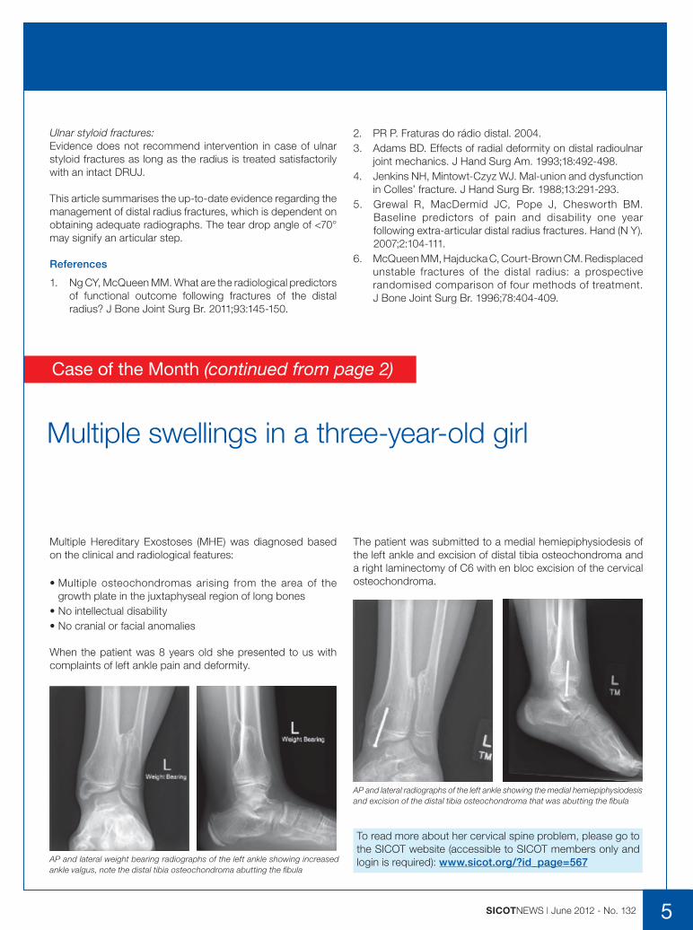
Multiple Hereditary Exostoses (MHE) was diagnosed based on the clinical and radiological features:
• Multiple osteochondromas arising from the area of the growth plate in the juxtaphyseal region of long bones
• No intellectual disability • No cranial or facial anomalies
When the patient was 8 years old she presented to us with complaints of left ankle pain and deformity.
The patient was submitted to a medial hemiepiphysiodesis of the left ankle and excision of distal tibia osteochondroma and a right laminectomy of C6 with en bloc excision of the cervical osteochondroma.
AP and lateral radiographs of the left ankle showing the medial hemiepiphysiodesis and excision of the distal tibia osteochondroma that was abutting the fibula
To read more about her cervical spine problem, please go to the SICOT website (accessible to SICOT members only and login is required): www.sicot.org/?id_page=567 AP and lateral weight bearing radiographs of the left ankle showing increased
ankle valgus, note the distal tibia osteochondroma abutting the fibula
Multiple swellings in a three-year-old girl
Case of the Month (continued from page 2)
High Tibial Osteotomy versus Unicompartmental Knee Arthroplasty
6 SICOTNEWS | June 2012 - No. 132
Scientific Debate
There is no doubt that a young patient less than 45 years of age with medial OA of the knee and axial varus deformity would need a high tibial osteotomy (HTO), whereas an elderly patient above 65 with advanced OA would need a total knee replacement. The debate arises in the mid zone of patients 45-65 years of age who can be offered an HTO or a unicompartmental knee arthroplasty (UKA), according to multiple factors. We present two views from highly active centres, who will try to convince you of the procedure they perform and the benefits of each. The final decision lies in the hands of the surgeon and the patient, according to the facilities, experience and ultimate patient requirements from the procedure.
Join us also on Facebook, and vote for your decision.
High Tibial Osteotomy
Abdel Rahman Babaqi & Hatem Said Assiut, Egypt
High tibial osteotomy is a commonly used surgical procedure for the treatment of medial compartmental osteoarthritis (OA) of the knee and has wide appeal because of the preservation of the knee joint with this method relative to the use of total knee arthroplasty (TKA) or UKA.
The principal advantages of opening wedge high tibial osteotomy include maintenance of the bone stock, correction of the deformity close to its origin, and no requirement for a fibular osteotomy [1,2]. Although it is a good option for young patients with isolated medial compartment OA and varus deformity, it can be done even for older active people over 65 [3].
The biomechanical principle of HTO in medial compartment OA is to redistribute the weight-bearing forces from the worn medial compartment across to the lateral compartment to relieve pain and to slow disease progression [4-6]. This can be accompanied by pain relief and improvements in gait and function. A correction of axial misalignment is seldom possible in UKA [7].
Mechanical release alone or a combination of HTO with arthroscopic measures (debridement, synovectomy or microfracture), chondral resurfacing or meniscal transplantation can also improve these results [8,9]. The main benefit for the
patient is the preservation of the natural joint and the main advantage for the patient is that potential physical loading (professional or sports-related) is almost entirely unaffected [7].
The surgical technique underwent many variations in the fixation technique and augmentation with bone graft or bone substitutes. Although autogenous iliac graft has been used routinely as part of the technique, recent studies showed successful results after HTO up to 14mm without bone graft [10] or at least similar results to osteotomy with bone graft [11].
Biopsy and second-look arthroscopic and open procedures have shown that there is regrowth of fibrocartilage in the worn medial compartment with a predilection for the ulcerated regions of wear in the weight-bearing portion of the medial femoral condyle [12-15].
It has been shown that when HTO for medial compartment arthrosis is performed in an otherwise healthy knee, no degenerative changes occur in the lateral or patellofemoral compartments [16].
Some authors reported that with an early and active rehabilitation programme, OA patients can walk with full weight bearing at two weeks after their OWHTO procedure [17,18].
The long-term follow-up for HTO patients has also been well documented. Billings et al reported that 43 of 64 knees had good clinical results with an average HSS knee score of 94 points at an average of 8.5 years after HTO [19]. Koshino et al reported long-term results (15~28 years) from their analysis of 75 knees in a group of 53 patients and found that the mean KSS score improved from 37 to 87 [20]. HTO gives high patient satisfaction and affords patients unrestricted activity for many years [3].
One of the beneficial outcomes after HTO is the style of sitting. One study comparing HTO versus UKA stated that [20]: “although none of the patients in the OWHTO group could sit in the Japanese style before surgery, 17 of these 24 patients (71%) could do so after this procedure. This is an important outcome that has only been achieved thus far using OWHTO”, and finally concluded: “although there were no significant differences found between OWHTO and UKA procedures in terms of the postoperative KSS score, we found that the operative function score for OWHTO was significantly better than that for UKA”.
7SICOTNEWS | June 2012 - No. 132
However, after recognising the need to avoid overcorrection of the mechanical axis, surgeons ultimately were able to reduce the risk that over tensioning in the “normal” compartment would cause early failure.
Although, in 1994, the Swedish Joint Replacement Registry reported a high percentage of poor results for UKA, these outcomes mainly reflected the fact that the surgery was performed on patients with chronic inflammatory arthritis [7].
Interest in UKA was stimulated by the introduction of the mobile-bearing implant. Goodfellow and O’Connor, along with others, published excellent long-term survivorship rates associated with this technique [8]. Their rationale for the procedure’s success was clearly stated: a mobile bearing (also called a meniscal bearing) provides the unique combination of complete congruency of the articular surface (to minimise wear) and total freedom of movement (to accommodate the preferred motion pattern of the retained natural compartment).
The past few years have again seen a renewed interest in unicompartmental arthroplasty, particularly because of the introduction of the minimally invasive parapatellar technique. This form of the procedure potentially can reduce morbidity, complications, and length of hospital stay.
Many now view UKA as an alternative to HTO in relatively young patients with medial compartment arthritis.
Indications
Appropriate patient selection is critical for UKA if reliable results are to be achieved. Criteria include:
• arthritis confined to a single compartment (medial compartment osteoarthritis is usually on the anteromedial aspect of the tibial plateau, and lateral compartment osteoarthritis is typically on the femoral side);
• intact anterior and posterior cruciate ligaments; • passively correctable malalignment - varus deformity <15° or
a valgus deformity < 20°; • fixed flexion < 15°; • knee able to flex to 120°. Key for the preparation of the
femoral condyle preoperatively.
Contraindications
Contraindications to a UKA include the following:
• inflammatory arthropathy; • previous HTO with overcorrection; • sepsis; • tibial or femoral shaft deformity (malunion post fracture).
UKA is controversial in the presence of patellofemoral joint arthritis, youth and high activity level, obesity, chondrocalcinosis, and crystalline arthropathy.
Converting HTO to knee arthroplasty is not a problem like UKA, as previous HTO does not influence the function or survival of a knee in the long term [21]. One study stated that knee arthroplasty after proximal tibial osteotomy was satisfactory in 96.5% of cases over a mean follow-up of 97 months. They compare between primary knee arthroplasty with and without previous HTO, and they found there is no significant difference in clinical and radiological results apart from a greater rate of anterior knee pain and revision for secondary resurfacing of the patella in the patients with previous HTO [22].
To conclude, OWHTO is the more appropriate treatment for active patients even those aged over 65 who demand a good range of motion of the knee.
Unicompartmental Knee Arthroplasty
Simon Cockshott & Nadim AslamWorcester, United Kingdom
Introduction
The concept of unicompartmental knee arthroplasty was introduced over 30 years ago. The theoretical advantages of this procedure include:
• preservation of bone; • reduced operative time; • better range of motion;• improved gait; • increased patient satisfaction.
With careful patient selection, surgical technique, and proper implant design, UKA can now be viewed as a procedure with reliable medium to long-term success [1,2].
History
In the early 1970s, Gunston and Marmor independently introduced the cemented unicompartmental knee arthroplasty. Gunston employed a circular runner of stainless steel and a track of high-molecular-weight polyethylene, for use as either a bicompartmental or unicompartmental replacement [3].
In the late 1980’s the popularity of unicompartmental knee replacements began to fall. This was due to high failure rates. Marmor in 1988 reported 21 treatment failures in 97 cases at 10- to 13-year follow-up [4].
Laskin reported that 35% of his knees had a fair to poor result with a high incidence of loosening of the implant and degeneration of the opposite compartment with a revision rate of 22% in a two-year follow-up of patients using the Marmor modular knee replacement [5].
In 1980, Insall published a series in which the conversion rate to a total knee arthroplasty was 28% at 5-7 years [6].
SICOTNEWS | June 2012 - No. 1328
Scientific Debate (continued)
Procedure Comparisons
Unicompartmental Knee Arthroplasty versus High Tibial Osteotomy
Resur facing methods are gaining popular ity. Results comparing HTO with UKA favour the latter.
Broughton et al demonstrated good results in 76% of patients in a replacement group and in 43% of patients in an osteotomy group [9]. Range of motion, speed of rehabilitation, and perioperative morbidity were significantly better for UKA, and no signs of late deterioration were present.
Weale and Newman, after a 12- to 17-year follow-up period, also reported better function and longer survival in the unicompartmental group [10].
Other publications have similarly shown more favourable results with arthroplasty [11,12]. The functional benefits of UKA over HTO have also been demonstrated using gait analysis, with patients displaying a more normal gait and better stair-climbing ability following UKA than they did after HTO.
If a revision to a total knee arthroplasty becomes necessary, the results are now believed to be generally better if the revision occurs after a failed UKA than they are following a failed HTO.
Unicompartmental Knee Arthroplasty versus Total Knee Arthroplasty
Laurencin et al reported that UKA results in less pain, more stability, and better stair-climbing ability than in total knee arthroplasty [13].
In addition, the cost of the unicompartmental procedure is about 57% that of total knee arthroplasty.
In a study, Dalury et al found little or no difference in outcome between patients who received a total knee arthroplasty and those who received a UKA, except for a slightly improved range of motion with UKA over the total knee procedure (123º ± 9º vs 119.8º ± 7º, respectively). In their investigation, the authors identified 23 patients with osteoarthritis who had undergone a total knee arthroplasty on one side and a UKA on the other, for medial compartment disease, and measured Knee Society scores, radiographic analysis, and patient preferences. Average follow-up was 46 months for total knee arthroplasty and 42 months for UKA. Of the 23 patients, 11 expressed no preference between either knee and 12 preferred the unicompartmental knee; no patient preferred the total knee [14].
A prospective, randomised, controlled trial in England compared unicompartmental knee replacement with TKA over 8, 10, 12, and 15 years’ follow-up. At 5 years, the numbers of failures were equal in the two groups. At 15 years’ follow-up, the survivorship rate was 89.8% for unicompartmental knee
replacement and 78.7% for TKA. Four of the unicompartmental knees failed, and 6 of the TKA knees failed. Newman et al determined from their findings that the results of their study justify increased use of unicompartmental replacement [15].
Results
When nonoperative and, possibly, arthroscopic procedures fail, the surgeon may consider HTO, unicompartmental arthroplasty, or total knee arthroplasty. UKA appears to result in better function and pain relief, less morbidity, and higher patient satisfaction than do HTO and total knee arthroplasty.
The long-term survival rate for UKA is better than that for HTO and comparable to the long-term survival rate for a total knee arthroplasty. With strict indications, newer prostheses, and attention to surgical technique, UKA has become a valuable treatment for unicompartmental knee arthritis.
In a prospective study, Berger et al found that in a 3-month period following knee arthroplasty, no patients in the investigation who had undergone the unicompartmental knee procedure required hospital readmission, compared with 9% of patients who received total knee arthroplasty. In the study, the authors looked at 111 patients who had undergone primary knee arthroplasty, 25 of whom had UKA and 86 of whom underwent total knee arthroplasty. Of the 111 patients, 104 (24 with UKA and 80 with total knee arthroplasty) met discharge criteria and were discharged directly to home [16].
Saenz et al evaluated the clinical and radiographic outcomes of the EIUS unicompartmental prosthesis and found it to be associated with higher revision rates than were metal-backed implants. The implant survival rate was 89%, with 16 knees either revised or scheduled for revision. The reasons for revision included aseptic loosening of the tibial component, progressive symptomatic patellofemoral disease, and tibial component subsidence [17].
Revision
Converting a UKA to a total knee arthroplasty is more difficult than performing a primary total knee arthroplasty. The results are acceptable but debatably not as good as they are with primary total knee arthroplasty. Despite the benefit of the conservative bone cuts used for unicompartmental knee replacement, stemmed components and augments may be needed for bone loss associated with component removal or osteolysis.
Bone defects, if present, usually can be treated with a local autograft. The cumulative revision rate at 10 years is more than 3 times higher for patients in whom a failed UKA has been revised to a further UKA than for those in whom it has been revised to a total knee arthroplasty [12].
To read more and for a list of references, please go to the SICOT website: www.sicot.org/?id_page=572
Alfredo Pozzo SICOT Member - La Paz, Bolivia
2011 International Travelling Fellowship Report
SICOTNEWS | June 2012 - No. 132 9
I had the great honour of being awarded the 2011 SICOT International Travell ing Fellowship, which gave me the opportunity to visit Mexico City, a charming city for both its architecture and its people.
Mexico City is one of the largest metropolitan areas with a population of more than 20 million people, located in the valley of Mexico at an altitude of 2,240 metres, where you can find many beautiful places to visit and admire.
My stay in Mexico began in October 2011 and lasted until January 2012. I had the opportunity to be under the leadership of Dr Felix Gil Orbezo, who is the SICOT National Representative of Mexico and Chief of the Orthopaedics Department of the Hospital Español de Mexico, a modern hospital with all the latest resources and equipment for the clinical and surgical practices of orthopaedics. There, I had the opportunity to share and interact with the faculty and residents who were kind enough to share their knowledge with me.
I participated in many hip arthroplasty surgeries, in addition to knee and shoulder arthroplasty. I was also fortunate to gain experience in revision arthroplasty surgery.
My stay in the hospital also served to update my concepts of treating common orthopaedic problems, which I can apply in my daily practice.
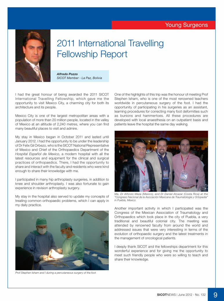
One of the highlights of this trip was the honour of meeting Prof Stephen Isham, who is one of the most renowned teachers worldwide in percutaneous surgery of the foot. I had the opportunity of participating in his surgeries as an assistant, learning procedures for correcting many foot deformities such as bunions and hammertoes. All these procedures are developed with local anaesthesia on an outpatient basis and patients leave the hospital the same day walking.
Young Surgeons
Prof Stephen Isham and I during a percutaneous surgery of the foot
Me, Dr Alfonzo Meza (Mexico), and Dr Daniel Alcazar (Costa Rica) at the “Congreso Nacional de la Asociación Mexicana de Traumatología y Ortopedia” in Puebla, Mexico
Another important activity in which I participated was the Congress of the Mexican Association of Traumatology and Orthopaedics which took place in the city of Puebla, a very traditional and beautiful colonial city. The meeting was attended by renowned faculty from around the world and addressed issues that were very interesting in terms of the evolution of orthopaedic surgery and the latest treatments in the management of oncological patients.
I deeply thank SICOT and the fellowships department for this wonderful experience and for giving me the opportunity to meet such friendly people who were so willing to teach and share their knowledge.
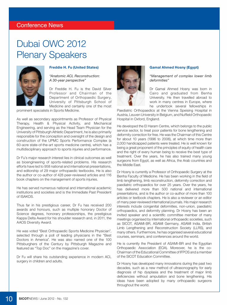
Gamal Ahmed Hosny (Egypt)
“Management of complex lower limb deformities”
Dr Gamal Ahmed Hosny was born in Cairo and graduated from Benha University. He then travelled abroad to work in many centres in Europe, where he undertook several fellowships in
Paediatric Orthopaedics at the Vienna Speising Hospital in Austria, Leuven University in Belgium, and Nuffield Orthopaedic Hospital in Oxford, England.
He developed the El Haram Centre, which belongs to the public service sector, to treat poor patients for bone lengthening and deformity correction for free. He was the Chairman of this Centre for about 10 years (1996 to 2006), at which time more than 2,000 handicapped patients were treated. He is well known for being a great proponent of the principles of equity of health care and the right of every human being to receive the best type of treatment. Over the years, he has also trained many young surgeons from Egypt, as well as Africa, the Arab countries and the Middle East.
Dr Hosny is currently a Professor of Orthopaedic Surgery at the Benha Faculty of Medicine. He has been working in the field of limb lengthening, limb reconstruction, deformity correction and paediatric orthopaedics for over 25 years. Over the years, he has delivered more than 500 national and international presentations, and is the author or co-author of more than 100 articles or textbook chapters. He is also a reviewer or an editor of many peer-reviewed international journals. His major research interests include congenital deformities, non-union, paediatric orthopaedics, and deformity planning. Dr Hosny has been an invited speaker and a scientific committee member of many meetings organised by international orthopaedic societies, such as SICOT, ASAMI-BR, ASAMI Germany, ASAMI India, British Limb Lengthening and Reconstruction Society (LLRS), and many others. Furthermore, he has organised several educational courses, seminars, and conferences around the world.
He is currently the President of ASAMI-BR and the Egyptian Orthopaedic Association (EOA). Moreover, he is the co-Chairman of the Educational Committee of IFPOS and a member of the SICOT Education Committee.
Dr Hosny has developed many innovations during the past two decades, such as a new method of ultrasonography for early diagnosis of hip dysplasia and the treatment of major limb deficiencies without amputation and bone lengthening. His ideas have been adopted by many orthopaedic surgeons throughout the world.
10 SICOTNEWS | June 2012 - No. 132
Conference News
Dubai OWC 2012Plenary Speakers
Freddie H. Fu (United States)
“Anatomic ACL Reconstruction: A 30-year perspective”
Dr Freddie H. Fu is the David Silver Professor and Chairman of the Department of Orthopaedic Surgery, University of Pittsburgh School of Medicine and certainly one of the most
prominent specialists in Sports Medicine.
As well as secondary appointments as Professor of Physical Therapy, Health & Physical Activity, and Mechanical Engineering, and serving as the Head Team Physician for the University of Pittsburgh Athletic Department, he is also primarily responsible for the conception and oversight of the design and construction of the UPMC Sports Performance Complex (a 60-acre state-of-the-art sports medicine centre), which has a multidisciplinary approach to sports injuries and performance.
Dr Fu’s major research interest lies in clinical outcomes as well as bioengineering of sports-related problems. His research efforts have led to 958 national and international presentations, and editorship of 29 major orthopaedic textbooks. He is also the author or co-author of 428 peer-reviewed articles and 116 book chapters on the management of sports injuries.
He has served numerous national and international academic institutions and societies and is the Immediate Past President of ISAKOS.
Thus far in his prestigious career, Dr Fu has received 200 awards and honours, such as multiple honorary Doctor of Science degrees, honorary professorships, the prestigious Kappa Delta Award for his shoulder research and, in 2011, the AAOS Diversity Award.
He was voted “Best Orthopaedic Sports Medicine Physician”‚ selected through a poll of leading physicians in the “Best Doctors in America”. He was also named one of the 100 Pittsburghers of the Century by Pittsburgh Magazine and featured as “Top Doc” on the magazine’s cover.
Dr Fu will share his outstanding experience in modern ACL surgery in children and adults.
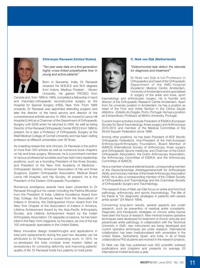
Chitranjan Ranawat (United States)
“Ten year wear data on a first generation highly cross-linked polyethylene liner in young and active patients”
Born in Sarwania, India, Dr Ranawat received his M.B.B.S and M.S degrees from Indore, Madhya Pradesh - Vikram University. He gained FRCS(C) from
Canada and, from 1966 to 1969, completed a fellowship in hand and rheumato-orthopaedic reconstructive surgery at the Hospital for Special Surgery (HSS), New York. From 1969 onwards, Dr Ranawat was appointed attending surgeon and later the director of the hand service and director of the comprehensive arthritis service. In 1994, he moved to Lenox Hill Hospital (LHH) as a Chairman of the Department of Orthopaedic Surgery until 2008 when he returned to HSS. As well as being Director of the Ranawat Orthopaedic Center (ROC) from 1988 to present, he is also a Professor of Orthopaedic Surgery at the Weill Medical College of Cornell University and has been visiting professor at different universities over 30 times.
As a leading researcher and clinician, Dr Ranawat is the author of more than 300 articles as well as numerous book chapters on hip and knee surgery. Moreover, he is a prominent member of various professional societies and has held many leadership positions, such as a founding President of the Knee Society, and President of the New York Academy of Medicine - Orthopaedic Section, American Association of Hip and Knee Surgeons, Eastern Orthopaedic Association, Medical Board Lenox Hill Hospital, and Hip Society. At present, he is the President of the Eastern Orthopaedic Foundation.
Numerous prestigious awards have been presented to Dr Ranawat throughout his career including the Padma Bhushan from the President of India, Lifetime Excellence Award from Daly College, the Shushruta Award from the Association of Indians in America, the Distinguished Honor Award from the New York Chapter of the Association of Indians in America, Lifetime Achievement Award by the Asia-Pacific Arthroplasty Society, and Lifetime Achievement Award by the Indian Arthroplasty Association. On separate occasions, he has been cited in the New York magazine, by his peers, as one of the top 100 orthopaedic specialists in the United States.
Many innovative design breakthroughs and applications in total joint replacements during the past two decades can be attributed to Dr Ranawat’s outstanding work. In 1974, he co-developed the total condylar knee implant hailed as revolutionary for correcting deformity and improving patients’ quality of life. Dr Ranawat holds four patents on total joints.
11SICOTNEWS | June 2012 - No. 132
C. Niek van Dijk (Netherlands)
“Osteochondral talar defect: the rationale for diagnosis and treatment”
Dr Niek van Di jk is fu l l Professor in Orthopaedics and head of the Orthopaedic Depar tment of the AMC-hospita l (Academic Medical Centre Amsterdam, University of Amsterdam) and is specialised in surgery of the ankle and knee, sport
traumatology and arthroscopic surgery. He is founder and director of the Orthopaedic Research Centre Amsterdam. Apart from his university position in Amsterdam, he has a position as head of the Foot and Ankle Section in the Clínica Saúde Atlântica - Estádio do Dragão, Porto, Portugal. He has a position as Extraordinary Professor at Minho University, Portugal.
Current board activities include President of ESSKA (European Society for Sport traumatology, Knee surgery and Arthroscopy) 2010-2012 and member of the Medical Committee of the World Squash Federation since 1996.
Among other positions, he has been President of NOF (Nordic Orthopaedic Federation), Vice-President of the Indo-European Arthroscopy/Arthroplasty Foundation, Board Member of ISAKOS (International Society of Arthroscopy, Knee surgery and Orthopaedic Sports medicine), and Chairman of the Dutch Orthopaedic Association, the Dutch Arthroscopy Association, the Arthroscopy Committee of ESSKA, and the Arthroscopy Committee of ISAKOS.
He is a member of several editorial boards, corresponding member of the Deutschprachige Arbeitsgemeinschaft für Arthroskopie (AGA), and honorary member of the Greek Arthroscopy Association (HAA). He is also a corresponding member of the Chilean Society of Orthopaedics and Traumatology and the Colombian Society of Orthopaedic Surgery and Traumatology.
The research lines of Niek van Dijk focus on ankle and hind foot pathology, arthroscopy and sports traumatology. The title of his thesis is “On diagnostic strategies in patients with severe ankle sprain” (24 March 1994).
Concerning long-term results, several aspects are under research, such as prevention of residual ankle complaints. Diagnostic and therapeutic strategies on chronic ankle injuries have been the focus of research. New minimal invasive operative techniques were developed for treatment of chronic articular and periarticular ankle pathology. In collaboration with the Technical University in Delft, new instruments are being developed and current operative techniques are under research. International collaboration has been institutionalised with universities in the United States, Switzerland, Spain, and India. In all of these collaborations PhD students are involved in the research projects.
Dr Niek van Dijk has published over 250 scientific indexed publications and chapters and presents on average 25 international invited lectures a year.
Combined 33rd SICOT & 17th PAOA Orthopaedic World ConferenceDubai, United Arab Emirates
More information about Dubai OWC 2012 is available on the SICOT website: www.sicot.org
Editorial DepartmentEditorial Secretary: Hatem SaidEditorial Production: Linda RidefjordEditorial Board: Ahmed Abdel Azeem, Syah Bahari, Kamal Bali, Bassel El-Osta, Anthony Hall, Maximilian Rudert
Rue Washington 40-b.9, 1050 Brussels, BelgiumTel.: +32 2 648 68 23 | Fax: +32 2 649 86 01E-mail: [email protected] | Website: www.sicot.org
Social & Sports Programmes
28-30 November 2012
LE 10 OCTOBRE 1
929
FO
NDÉE À PARIS
Social Events
• Opening Ceremony & Welcome Buffet
Date: Wednesday, 28 November 2012 (evening)
Fee: EUR 10
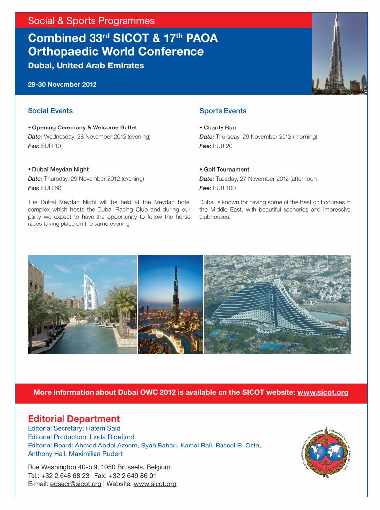
• Dubai Meydan Night
Date: Thursday, 29 November 2012 (evening)
Fee: EUR 60
The Dubai Meydan Night will be held at the Meydan hotel complex which hosts the Dubai Racing Club and during our party we expect to have the opportunity to follow the horse races taking place on the same evening.
Sports Events
• Charity Run
Date: Thursday, 29 November 2012 (morning)
Fee: EUR 20
• Golf Tournament
Date: Tuesday, 27 November 2012 (afternoon)
Fee: EUR 100
Dubai is known for having some of the best golf courses in the Middle East, with beautiful sceneries and impressive clubhouses.