dysregulated left inferior parietal activity in
TRANSCRIPT
Florida International UniversityFIU Digital Commons
Department of Physics College of Arts, Sciences & Education
6-12-2013
Dysregulated left inferior parietal activity inschizophrenia and depression: functionalconnectivity and characterizationVeronika I. MüllerHeinrich Heine University; Research Center Jülich; RWTH Aachen University
Edna C. CieslikHeinrich Heine University; Research Center Jülich; RWTH Aachen University
Angela LairdDepartment of Physics, Florida International University;University of Texas Health Science Center at San Antonio,[email protected]
Peter T. FoxUniversity of Texas Health Science Center at San Antonio; South Texas Veterans Administration Medical Center
Simon B. EickhoffHeinrich Heine University; Research Center Jülich; RWTH Aachen University
Follow this and additional works at: https://digitalcommons.fiu.edu/physics_fac
Part of the Physics Commons
This work is brought to you for free and open access by the College of Arts, Sciences & Education at FIU Digital Commons. It has been accepted forinclusion in Department of Physics by an authorized administrator of FIU Digital Commons. For more information, please contact [email protected].
Recommended CitationMüller VI, Cieslik EC, Laird AR, Fox PT and Eickhoff SB (2013) Dysregulated left inferior parietal activity in schizophrenia anddepression: functional connectivity and characterization. Front. Hum. Neurosci. 7:268. doi: 10.3389/fnhum.2013.00268
ORIGINAL RESEARCH ARTICLEpublished: 12 June 2013
doi: 10.3389/fnhum.2013.00268
Dysregulated left inferior parietal activity in schizophreniaand depression: functional connectivity andcharacterizationVeronika I. Müller1,2,3*, Edna C. Cieslik1,2,3, Angela R. Laird 4,5, Peter T. Fox4,6 and Simon B. Eickhoff1,2,3
1 Institute of Clinical Neuroscience and Medical Psychology, Heinrich Heine University, Düsseldorf, Germany2 Department of Neuroscience and Medicine, Research Center Jülich, INM-1, Jülich, Germany3 Department of Psychiatry, Psychotherapy, and Psychosomatics, Medical School, RWTH Aachen University, Aachen, Germany4 Research Imaging Institute, University of Texas Health Science Center at San Antonio, San Antonio, TX, USA5 Department of Physics, Florida International University, Miami, FL, USA6 South Texas Veterans Administration Medical Center, San Antonio, TX, USA
Edited by:
Martin Klasen, RWTH AachenUniversity, Germany
Reviewed by:
Jennifer L. Robinson, AuburnUniversity, USACorrado Corradi-Dell’Acqua,University of Geneva, SwitzerlandJulia Sacher, University Leipzig,Germany
*Correspondence:
Veronika I. Müller, Department ofNeuroscience and Medicine,Research Center Jülich, INM-1,Leo-Brandt-Straße, D-52428Jülich, Germanye-mail: [email protected]
The inferior parietal cortex (IPC) is a heterogeneous region that is known to beinvolved in a multitude of diverse different tasks and processes, though its contributionto these often-complex functions is yet poorly understood. In a previous study wedemonstrated that patients with depression failed to deactivate the left IPC duringprocessing of congruent audiovisual information. We now found the same dysregulation(same region and condition) in schizophrenia. By using task-independent (resting state)and task-dependent meta-analytic connectivity modeling (MACM) analyses we aimed atcharacterizing this particular region with regard to its connectivity and function. Acrossboth approaches, results revealed functional connectivity of the left inferior parietal seedregion with bilateral IPC, precuneus and posterior cingulate cortex (PrC/PCC), medialorbitofrontal cortex (mOFC), left middle frontal (MFG) as well as inferior frontal (IFG)gyrus. Network-level functional characterization further revealed that on the one hand, allinterconnected regions are part of a network involved in memory processes. On the otherhand, sub-networks are formed when emotion, language, social cognition and reasoningprocesses are required. Thus, the IPC-region that is dysregulated in both depression andschizophrenia is functionally connected to a network of regions which, depending on taskdemands may form sub-networks. These results therefore indicate that dysregulationof left IPC in depression and schizophrenia might not only be connected to deficits inaudiovisual integration, but is possibly also associated to impaired memory and deficits inemotion processing in these patient groups.
Keywords: functional connectivity, depression, schizophrenia, inferior parietal cortex, resting-state
INTRODUCTIONDepression and schizophrenia are both associated with social andaffective dysfunctions as well as deficits in emotional process-ing (Bach et al., 2009; Bourke et al., 2010; Kohler et al., 2010;Comparelli et al., 2013). Research on affective deficits in psychi-atric populations to date, however, has mainly focused on uni-modal emotion processing, while in daily life emotion perceptionis generally based on the multimodal evaluation of information,such as hearing a laugh and seeing a smiling face. Importantly, inthis context, emotional information from different sensory chan-nels can be either congruent or incongruent, leading to fasterresponses when processing emotional congruent compared toincongruent information (De Gelder and Vroomen, 2000; Dolanet al., 2001; Collignon et al., 2008). Clinical studies have shownthat patients with schizophrenia show aberrant audiovisual inte-gration (De Gelder et al., 2005; De Jong et al., 2009; Van DenStock et al., 2011). In contrast, Müller et al. (2012) and Mülleret al. (2013) could not find any significant group difference inthe behavioral rating of emotional faces while distracted by con-gruent and incongruent sounds, neither in a group of patients
with schizophrenia, nor in depression. Importantly, the neuronalcorrelates of crossmodal emotional processing in clinical pop-ulations are still sparse. In a recent study (Müller et al., 2013)investigating audiovisual (in) congruence processing in depres-sion, we showed that compared to healthy controls, patients failedto deactivate the left inferior parietal cortex (IPC) and inferiorfrontal cortex when confronted with congruent happy audiovi-sual information, while there was no difference between groupsfor incongruent pairs. As we will show in this paper by usingthe same paradigm, a similar effect in the same region of theleft IPC in patients with schizophrenia can be observed. In par-ticular, schizophrenic patients also reveal decreased deactivationin congruent audiovisual conditions compared to controls. Thus,both schizophrenia and depression go along with IPC dysregula-tion during congruent audiovisual emotional processing, possiblyindicating increased processing of unambiguous stimuli in thesepatient groups (Müller et al., 2013).
However, when interpreting these findings, one has toacknowledge that the IPC is a heterogeneous region, which isinvolved in a wide range of different functions ranging from
Frontiers in Human Neuroscience www.frontiersin.org June 2013 | Volume 7 | Article 268 | 1
HUMAN NEUROSCIENCE
Müller et al. Functional characterization of posterior IPC
language and memory to action planning, higher social-cognitionand other integrative processes (Glover, 2004; Wagner et al., 2005;Daselaar et al., 2006; Buckner et al., 2008; Binder et al., 2009;Spreng et al., 2009; Caspers et al., 2010; Arsalidou and Taylor,2011; Bzdok et al., 2012; Schilbach et al., 2012; Seghier, 2013).Hence, it has been suggested that the IPC can be subdividedin different sub regions. Based on cytoarchitectonic mapping,Caspers et al. (2006, 2008) divided the IPC into seven differ-ent sub-regions. With regard to these divisions, the area that wefound to be dysregulated in depression as well as in schizophre-nia strongly overlaps with area PGp. Based on its anatomicaland functional connections with temporal and lateral occipital(Caspers et al., 2011) as well as with frontal and parahippocam-pal areas (Uddin et al., 2010) it has been argued that PGp maymainly be involved in auditory-sensory integration and mem-ory processes. Given the size and potential heterogeneity of thisregion in the posterior IPC, however, it remains open as to howthe specific location disturbed in depression and schizophreniarelates to these roles. Furthermore, as the function of a spe-cific brain area depends on those regions it interacts with, itsrole should not only be assessed in isolation but also togetherwith regions it stands in interplay with (Stephan, 2004; Seghier,2013). Therefore, the present study aims to investigate connectiv-ity and function of the particular region, which has been foundto be dysregulated in cross-modal affective integration acrosstwo different clinical groups, i.e., depression and schizophre-nia. In particular, task-dependent and task-independent func-tional connectivity as well as behavioral characterization of theregion of interest was carried out in healthy subjects in orderto gain better insight of the role of this region from a systemperspective.
METHODSVOLUME OF INTERESTThe volume of interest used in the current study is based ontwo fMRI studies investigating the neural correlates of audiovi-sual incongruence processing in patients with depression as wellas patients with schizophrenia. Before, describing the methods ofthe current study, we first describe the patient samples on whichthe VOI is based on as well as the experimental procedure of theaudiovisual paradigm.
Audiovisual paradigm and fMRI analysisThe stimuli and procedure used in the fMRI studies is the sameas previously described (Müller et al., 2011, 2013). Thirty dif-ferent pictures of faces from five males and five females, eachshowing a happy, neutral, and fearful expression (FEBA; Guret al., 2002) were combined with 30 auditory stimuli, con-sisting of 10 yawns, 10 laughs, and 10 screams. Additionally10 different blurred faces served as masks. This resulted in180 stimulus pairs with 9 different conditions (fearful/scream,fearful/yawn, fearful/laugh, neutral/scream, neutral/yawn, neu-tral/laugh, happy/scream, happy/yawn, happy/laugh). Every trialstarted with the presentation of a sound concurrently with amask. After 1000 ms the mask was displaced by an either neu-tral or emotional face and presented with the continuing soundfor another 500 ms. Subjects had to ignore the sound and to just
rate the facial expression on an eight-point scale from extremelyfearful to extremely happy.
fMRI data acquisition and statistical analysis was done asdescribed in Müller et al. (2013). Images were acquired on aSiemens Trio 3T whole-body scanner (Erlangen, Germany) inthe RWTH Aachen University hospital using blood-oxygen-level-dependent (BOLD) contrast [Gradient-echo EPI pulse sequence,TR = 2.2 s, in plane resolution = 3.1 × 3.1 mm, 36 axial slices(3.1 mm thickness)] covering the entire brain. Echo-planar imag-ing (EPI) images were corrected for head movement, normalizedto the Montreal Neurological Institute (MNI) single subject tem-plate and spatially smoothed using an 8 mm FWHM Gaussiankernel. Data were then analysed using a General Linear Model asimplemented in SPM8. For each subject, each experimental con-dition as well as the response event were separately modeled andsimple main effects for each of the conditions computed by apply-ing appropriate baseline contrasts. These individual first-levelcontrasts were then entered into a second-level group-analysisusing an analysis of variance (ANOVA) employing a random-effects model. Based on these estimates of the second-levelanalysis, separate t-contrasts were calculated for the interactioncongruence × group by applying the respective contrast to the2nd level parameter estimates. The resulting SPM (T) maps werethen thresholded at a cluster level FWE rate of p < 0.05 (clusterforming threshold: p < 0.001 at voxel level).
SubjectsThe demographic and clinical characteristics of the patients withdepression and the corresponding controls can be found in Mülleret al. (2013).
Using the same audiovisual paradigm as in the previous study,we now tested a sample of 18 patients with schizophrenia and18 healthy controls matched for age, gender, and education. Twopatients and the corresponding controls were excluded from fur-ther analysis due to abnormal anatomy or an inability to under-stand the task. All participants were right handed, as confirmed bythe Edinburgh Inventory (Oldfield, 1971) and reported normal orcorrected-to-normal vision. Table 1 presents the clinical profile ofthe schizophrenic patient group. Patients were recruited from theinpatient and outpatient units of the Department of Psychiatry,Psychotherapy and Psychosomatics, RWTH University Hospital.Of the 16 patients included in the final analysis, 14 met ICD-10criteria for paranoid schizophrenia (F 20.0), whereas two were
Table 1 | Demographic and clinical characteristics of the patient and
control groups.
Patients Controls
Gender 6 female/ 10 male 6 female/ 10 male
Age 38.37 ± 10.21 37.94 ± 10.38
Education in years 11.75 ± 2.29 12.93 ± 2.20
Age of onset 29.19 ± 8.65
Duration of illness 9.25 ± 6.59
PANSS positive 10.63 ± 3.83
PANSS negative 12.75 ± 5.59
PANSS general 22.31 ± 6.33
Frontiers in Human Neuroscience www.frontiersin.org June 2013 | Volume 7 | Article 268 | 2
Müller et al. Functional characterization of posterior IPC
diagnosed with the residual subtype (F 20.4). Only patients withno comorbid psychiatric or neurological illness and no substanceaddiction in the last 6 months were included in the study. Allpatients were medicated, in particular all of them were treatedwith atypical antipsychotics with one additionally taking typ-ical antipsychotic medication. Furthermore, five patients weretaking antidepressant agents and one was taking anticholinergicdrugs.
Healthy controls had no history of neurological or psychi-atric disorder and did not take any mood- or cognition-alteringmedication.
All subjects gave informed consent into the study which wasapproved by the ethics committee of the School of Medicine ofthe RWTH Aachen University.
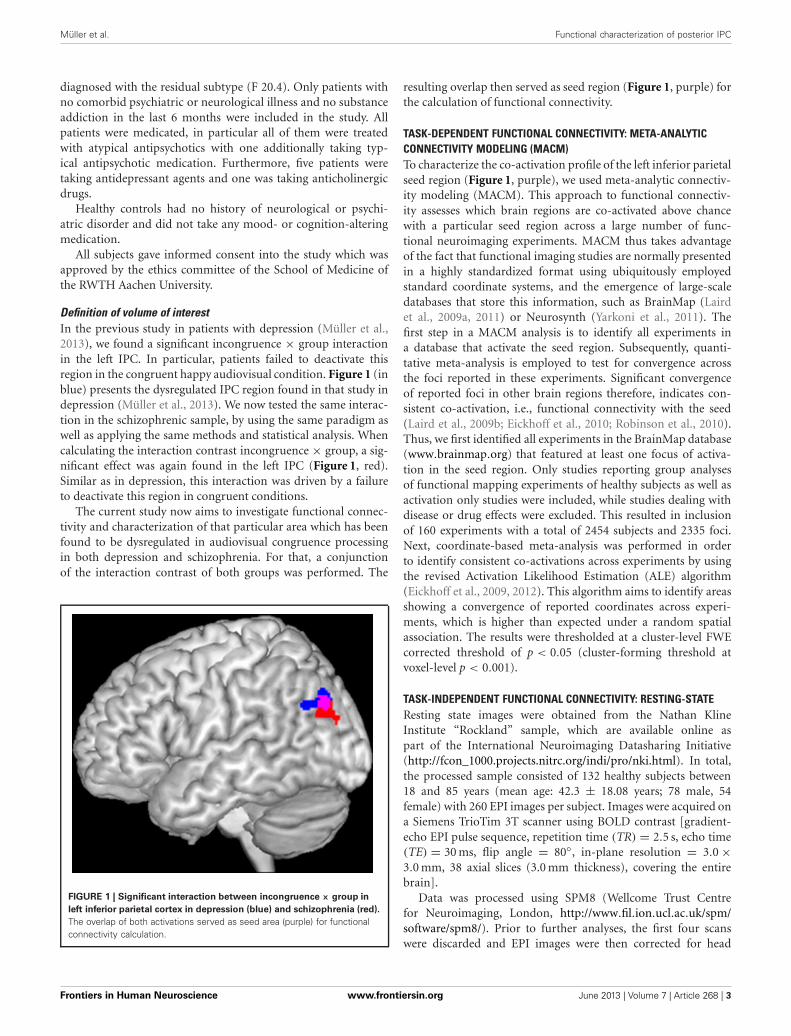
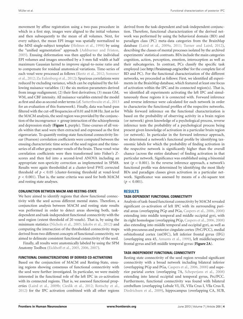
Definition of volume of interestIn the previous study in patients with depression (Müller et al.,2013), we found a significant incongruence × group interactionin the left IPC. In particular, patients failed to deactivate thisregion in the congruent happy audiovisual condition. Figure 1 (inblue) presents the dysregulated IPC region found in that study indepression (Müller et al., 2013). We now tested the same interac-tion in the schizophrenic sample, by using the same paradigm aswell as applying the same methods and statistical analysis. Whencalculating the interaction contrast incongruence × group, a sig-nificant effect was again found in the left IPC (Figure 1, red).Similar as in depression, this interaction was driven by a failureto deactivate this region in congruent conditions.
The current study now aims to investigate functional connec-tivity and characterization of that particular area which has beenfound to be dysregulated in audiovisual congruence processingin both depression and schizophrenia. For that, a conjunctionof the interaction contrast of both groups was performed. The
FIGURE 1 | Significant interaction between incongruence × group in
left inferior parietal cortex in depression (blue) and schizophrenia (red).
The overlap of both activations served as seed area (purple) for functionalconnectivity calculation.
resulting overlap then served as seed region (Figure 1, purple) forthe calculation of functional connectivity.
TASK-DEPENDENT FUNCTIONAL CONNECTIVITY: META-ANALYTICCONNECTIVITY MODELING (MACM)To characterize the co-activation profile of the left inferior parietalseed region (Figure 1, purple), we used meta-analytic connectiv-ity modeling (MACM). This approach to functional connectiv-ity assesses which brain regions are co-activated above chancewith a particular seed region across a large number of func-tional neuroimaging experiments. MACM thus takes advantageof the fact that functional imaging studies are normally presentedin a highly standardized format using ubiquitously employedstandard coordinate systems, and the emergence of large-scaledatabases that store this information, such as BrainMap (Lairdet al., 2009a, 2011) or Neurosynth (Yarkoni et al., 2011). Thefirst step in a MACM analysis is to identify all experiments ina database that activate the seed region. Subsequently, quanti-tative meta-analysis is employed to test for convergence acrossthe foci reported in these experiments. Significant convergenceof reported foci in other brain regions therefore, indicates con-sistent co-activation, i.e., functional connectivity with the seed(Laird et al., 2009b; Eickhoff et al., 2010; Robinson et al., 2010).Thus, we first identified all experiments in the BrainMap database(www.brainmap.org) that featured at least one focus of activa-tion in the seed region. Only studies reporting group analysesof functional mapping experiments of healthy subjects as well asactivation only studies were included, while studies dealing withdisease or drug effects were excluded. This resulted in inclusionof 160 experiments with a total of 2454 subjects and 2335 foci.Next, coordinate-based meta-analysis was performed in orderto identify consistent co-activations across experiments by usingthe revised Activation Likelihood Estimation (ALE) algorithm(Eickhoff et al., 2009, 2012). This algorithm aims to identify areasshowing a convergence of reported coordinates across experi-ments, which is higher than expected under a random spatialassociation. The results were thresholded at a cluster-level FWEcorrected threshold of p < 0.05 (cluster-forming threshold atvoxel-level p < 0.001).
TASK-INDEPENDENT FUNCTIONAL CONNECTIVITY: RESTING-STATEResting state images were obtained from the Nathan KlineInstitute “Rockland” sample, which are available online aspart of the International Neuroimaging Datasharing Initiative(http://fcon_1000.projects.nitrc.org/indi/pro/nki.html). In total,the processed sample consisted of 132 healthy subjects between18 and 85 years (mean age: 42.3 ± 18.08 years; 78 male, 54female) with 260 EPI images per subject. Images were acquired ona Siemens TrioTim 3T scanner using BOLD contrast [gradient-echo EPI pulse sequence, repetition time (TR) = 2.5 s, echo time(TE) = 30 ms, flip angle = 80◦, in-plane resolution = 3.0 ×3.0 mm, 38 axial slices (3.0 mm thickness), covering the entirebrain].
Data was processed using SPM8 (Wellcome Trust Centrefor Neuroimaging, London, http://www.fil.ion.ucl.ac.uk/spm/software/spm8/). Prior to further analyses, the first four scanswere discarded and EPI images were then corrected for head
Frontiers in Human Neuroscience www.frontiersin.org June 2013 | Volume 7 | Article 268 | 3
Müller et al. Functional characterization of posterior IPC
movement by affine registration using a two-pass procedure inwhich in a first step, images were aligned to the initial volumesand then subsequently to the mean of all volumes. Next, forevery subject, the mean EPI image was spatially normalized tothe MNI single-subject template (Holmes et al., 1998) by usingthe “unified segmentation” approach (Ashburner and Friston,2005). Ensuing deformation was then applied to the individualEPI volumes and images smoothed by a 5-mm full width at halfmaximum Gaussian kernel to improve signal-to-noise ratio andto compensate for residual anatomical variations. Time-series ofeach voxel were processed as follows (Reetz et al., 2012; Sommeret al., 2012; Zu Eulenburg et al., 2012): Spurious correlations werereduced by excluding variance, which can be explained by the fol-lowing nuisance variables: (1) the six motion parameters derivedfrom image realignment; (2) their first derivatives; (3) mean GM,WM, and CBF intensity. All nuisance variables entered the modelas first and also as second order terms (cf. Satterthwaite et al., 2013for an evaluation of this framework). Finally, data was band-passfiltered with the cut-off frequencies of 0.01 and 0.08 Hz. Just as forthe MACM analysis, the seed region was provided by the conjunc-tion of the incongruence × group interaction of the schizophreniaand depression study (Figure 1, purple). Time-courses of all vox-els within that seed were then extracted and expressed as the firsteigenvariate. To quantify resting-state functional connectivity lin-ear (Pearson) correlation coefficients were computed between theensuing characteristic time series of the seed region and the time-series of all other gray matter voxels of the brain. These voxel-wisecorrelation coefficients were then transformed into Fisher’s Z-scores and then fed into a second-level ANOVA including anappropriate non-spericity correction as implemented in SPM8.Results were again thresholded at a cluster-level FWE correctedthreshold of p < 0.05 (cluster-forming threshold at voxel-levelp < 0.001). That is, the same criteria was used for both MACMand resting state analyses.
CONJUNCTION BETWEEN MACM AND RESTING-STATEWe here aimed to identify regions that show functional connec-tivity with the seed across different mental states. Therefore, aconjunction analysis between MACM and resting state resultswas performed in order to detect areas showing both, task-dependent and task-independent functional connectivity with theseed region (extent threshold of 20 voxels). That is, by using theminimum statistics (Nichols et al., 2005; Jakobs et al., 2012) andcomputing the intersection of the thresholded connectivity mapsderived from two different concepts of functional connectivity, weaimed to delineate consistent functional connectivity of the seed.
Finally, all results were anatomically labeled by using the SPMAnatomy Toolbox (Eickhoff et al., 2005, 2006, 2007).
FUNCTIONAL CHARACTERIZATIONS OF DERIVED CO-ACTIVATIONSBased on the conjunction of MACM and Resting-State, ensu-ing regions showing consensus of functional connectivity withthe seed were further investigated. In particular, we were mainlyinterested in the functional role of the left IPC in co-activationwith its connected regions. That is, we assessed functional prop-erties (Laird et al., 2009b; Cieslik et al., 2012; Rottschy et al.,2012) for the IPC activation combined with all other regions
derived from the task-dependent and task-independent conjunc-tion. Therefore, functional characterization of the derived net-work was performed by using the behavioral domain (BD) andparadigm class (PC) meta-data categories from the BrainMapdatabase (Laird et al., 2009a, 2011; Turner and Laird, 2012),describing the classes of mental processes isolated by the archivedexperiments’ statistical contrasts. BDs include the main categoriescognition, action, perception, emotion, interoception as well astheir subcategories. In contrast, PCs classify the specific taskemployed (see http://brainmap.org/scribe/ for the complete list ofBD and PC). For the functional characterization of the differentnetworks, we proceeded as follows: First, we identified all experi-ments in the BrainMap database, which featured at least one focusof activation within the IPC and its connected region(s). That is,we identified all experiments activating the left IPC and simul-taneously those regions it is connected with. Forward inferenceand reverse inference were calculated for each network in orderto characterize the functional profiles of the respective networks.While forward inference on the functional characterization isbased on the probability of observing activity in a brain region(or network) given knowledge of a psychological process, reverseinference tests the probability of a psychological process beingpresent given knowledge of activation in a particular brain region(or network). In particular in the forward inference approach,we determined a network’s functional profile by identifying tax-onomic labels for which the probability of finding activation inthe respective network is significantly higher than the overallchance (across the entire database) of finding activation in thatparticular network. Significance was established using a binomialtest (p < 0.001). In the reverse inference approach, a network’sfunctional profile was determined by identifying the most likelyBDs and paradigm classes given activation in a particular net-work. Significance was assessed by means of a chi-square test(p < 0.001).
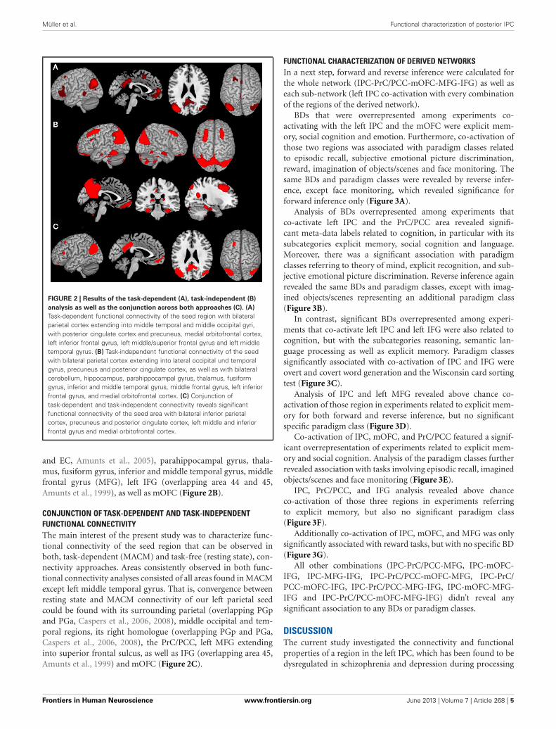
RESULTSTASK-DEPENDENT FUNCTIONAL CONNECTIVITYAnalysis of task-based functional connectivity by MACM revealedsignificant co-activation of left IPC with its surrounding pari-etal areas (overlapping PGp and PGa, Caspers et al., 2006, 2008)extending into middle temporal and middle occipital gyri, withits right homologue (overlapping PGp, Caspers et al., 2006, 2008)also extending into middle temporal and occipital gyri, as well aswith precuneus and posterior cingulate cortex (PrC/PCC), medialorbitofrontal cortex (mOFC), left inferior frontal gyrus (IFG)(overlapping area 45, Amunts et al., 1999), left middle/superiorfrontal gyrus and left middle temporal gyrus (Figure 2A).
TASK-INDEPENDENT FUNCTIONAL CONNECTIVITYResting state connectivity of the seed region revealed significantconnectivity with a broad network including bilateral inferior(overlapping PGp and PGa, Caspers et al., 2006, 2008) and supe-rior parietal cortex (overlapping 7A, Scheperjans et al., 2008)extending into lateral occipital und temporal gyrus, Prc/PCC.Furthermore, functional connectivity was found with bilateralcerebellum (overlapping Lobule VI, IX, VIIa Crus I, Vlla Crus ll,Diedrichsen et al., 2009), hippocampus (overlapping CA, SUB,
Frontiers in Human Neuroscience www.frontiersin.org June 2013 | Volume 7 | Article 268 | 4
Müller et al. Functional characterization of posterior IPC
FIGURE 2 | Results of the task-dependent (A), task-independent (B)
analysis as well as the conjunction across both approaches (C). (A)
Task-dependent functional connectivity of the seed region with bilateralparietal cortex extending into middle temporal and middle occipital gyri,with posterior cingulate cortex and precuneus, medial orbitofrontal cortex,left inferior frontal gyrus, left middle/superior frontal gyrus and left middletemporal gyrus. (B) Task-independent functional connectivity of the seedwith bilateral parietal cortex extending into lateral occipital und temporalgyrus, precuneus and posterior cingulate cortex, as well as with bilateralcerebellum, hippocampus, parahippocampal gyrus, thalamus, fusiformgyrus, inferior and middle temporal gyrus, middle frontal gyrus, left inferiorfrontal gyrus, and medial orbitofrontal cortex. (C) Conjunction oftask-dependent and task-independent connectivity reveals significantfunctional connectivity of the seed area with bilateral inferior parietalcortex, precuneus and posterior cingulate cortex, left middle and inferiorfrontal gyrus and medial orbitofrontal cortex.
and EC, Amunts et al., 2005), parahippocampal gyrus, thala-mus, fusiform gyrus, inferior and middle temporal gyrus, middlefrontal gyrus (MFG), left IFG (overlapping area 44 and 45,Amunts et al., 1999), as well as mOFC (Figure 2B).
CONJUNCTION OF TASK-DEPENDENT AND TASK-INDEPENDENTFUNCTIONAL CONNECTIVITYThe main interest of the present study was to characterize func-tional connectivity of the seed region that can be observed inboth, task-dependent (MACM) and task-free (resting state), con-nectivity approaches. Areas consistently observed in both func-tional connectivity analyses consisted of all areas found in MACMexcept left middle temporal gyrus. That is, convergence betweenresting state and MACM connectivity of our left parietal seedcould be found with its surrounding parietal (overlapping PGpand PGa, Caspers et al., 2006, 2008), middle occipital and tem-poral regions, its right homologue (overlapping PGp and PGa,Caspers et al., 2006, 2008), the PrC/PCC, left MFG extendinginto superior frontal sulcus, as well as IFG (overlapping area 45,Amunts et al., 1999) and mOFC (Figure 2C).
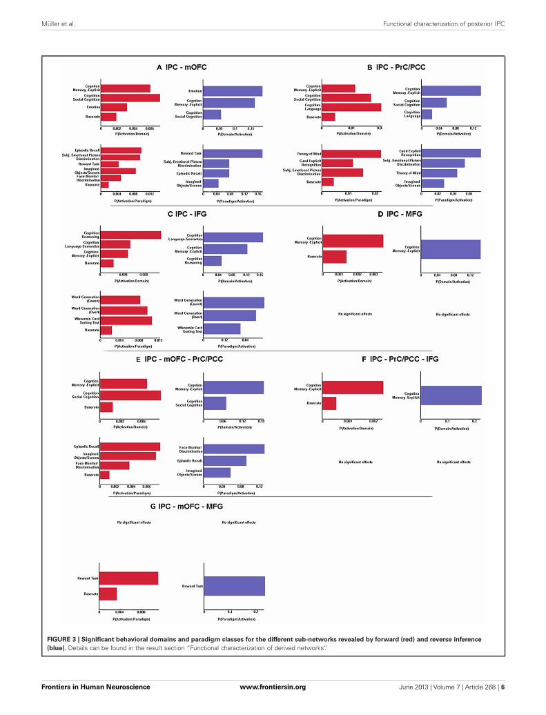
FUNCTIONAL CHARACTERIZATION OF DERIVED NETWORKSIn a next step, forward and reverse inference were calculated forthe whole network (IPC-PrC/PCC-mOFC-MFG-IFG) as well aseach sub-network (left IPC co-activation with every combinationof the regions of the derived network).
BDs that were overrepresented among experiments co-activating with the left IPC and the mOFC were explicit mem-ory, social cognition and emotion. Furthermore, co-activation ofthose two regions was associated with paradigm classes relatedto episodic recall, subjective emotional picture discrimination,reward, imagination of objects/scenes and face monitoring. Thesame BDs and paradigm classes were revealed by reverse infer-ence, except face monitoring, which revealed significance forforward inference only (Figure 3A).
Analysis of BDs overrepresented among experiments thatco-activate left IPC and the PrC/PCC area revealed signifi-cant meta-data labels related to cognition, in particular with itssubcategories explicit memory, social cognition and language.Moreover, there was a significant association with paradigmclasses referring to theory of mind, explicit recognition, and sub-jective emotional picture discrimination. Reverse inference againrevealed the same BDs and paradigm classes, except with imag-ined objects/scenes representing an additional paradigm class(Figure 3B).
In contrast, significant BDs overrepresented among experi-ments that co-activate left IPC and left IFG were also related tocognition, but with the subcategories reasoning, semantic lan-guage processing as well as explicit memory. Paradigm classessignificantly associated with co-activation of IPC and IFG wereovert and covert word generation and the Wisconsin card sortingtest (Figure 3C).
Analysis of IPC and left MFG revealed above chance co-activation of those region in experiments related to explicit mem-ory for both forward and reverse inference, but no significantspecific paradigm class (Figure 3D).
Co-activation of IPC, mOFC, and PrC/PCC featured a signif-icant overrepresentation of experiments related to explicit mem-ory and social cognition. Analysis of the paradigm classes furtherrevealed association with tasks involving episodic recall, imaginedobjects/scenes and face monitoring (Figure 3E).
IPC, PrC/PCC, and IFG analysis revealed above chanceco-activation of those three regions in experiments referringto explicit memory, but also no significant paradigm class(Figure 3F).
Additionally co-activation of IPC, mOFC, and MFG was onlysignificantly associated with reward tasks, but with no specific BD(Figure 3G).
All other combinations (IPC-PrC/PCC-MFG, IPC-mOFC-IFG, IPC-MFG-IFG, IPC-PrC/PCC-mOFC-MFG, IPC-PrC/PCC-mOFC-IFG, IPC-PrC/PCC-MFG-IFG, IPC-mOFC-MFG-IFG and IPC-PrC/PCC-mOFC-MFG-IFG) didn’t reveal anysignificant association to any BDs or paradigm classes.
DISCUSSIONThe current study investigated the connectivity and functionalproperties of a region in the left IPC, which has been found to bedysregulated in schizophrenia and depression during processing
Frontiers in Human Neuroscience www.frontiersin.org June 2013 | Volume 7 | Article 268 | 5
Müller et al. Functional characterization of posterior IPC
FIGURE 3 | Significant behavioral domains and paradigm classes for the different sub-networks revealed by forward (red) and reverse inference
(blue). Details can be found in the result section “Functional characterization of derived networks”.
Frontiers in Human Neuroscience www.frontiersin.org June 2013 | Volume 7 | Article 268 | 6
Müller et al. Functional characterization of posterior IPC
of audiovisual emotional stimuli. That is, we investigated neu-ronal networks and their associated functions that center onan inferior parietal region showing aberrant responses during amulti-modal affective processing task in two major psychiatricdisorders. First, results revealed that the seed region in the leftposterior IPC functionally connects with PrC/PCC, mOFC aswell as MFG and inferior (IFG) frontal gyrus. Quantitative func-tional characterization further indicates that on the one hand,all areas are engaged during experiments related to memory.On the other hand, sub-networks that relate to social cog-nition, reasoning, emotional, or language processing are alsodiscernible.
THE ROLE OF THE LEFT IPCThe IPC is a large and heterogeneous region that has been foundto be involved in a wide range of different processes, ranging fromaction, memory, and language to mathematical problem solvingand social cognition. Furthermore, it has been described as partof the default mode network (Glover, 2004; Wagner et al., 2005;Daselaar et al., 2006; Buckner et al., 2008; Binder et al., 2009;Spreng et al., 2009; Caspers et al., 2010; Arsalidou and Taylor,2011; Bzdok et al., 2012; Schilbach et al., 2012; Seghier, 2013).Apart from this functional diversity, the IPC is also heterogeneouswith regard to its macroanatomy. It has for instance been classi-fied into BA39, the angular gyrus and BA40, the supramarginalgyrus. In addition, based on cyto-architectonic mapping, Casperset al. (2006, 2008) subdivided the IPC into seven different regions.Thus, both the functional as well as architectonical diversitystrongly suggest that the IPC as a whole is too large and heteroge-neous to allow any meaningful interpretations of IPC activity orIPC dysregulation.
Going more into detail, the IPC area which has been ana-lyzed in the current study demonstrates correspondence withthe angular gyrus and more precisely with area PGp (based onthe subdivisions of Caspers et al., 2006, 2008). This part of theIPC is mainly involved in language processing (Hall et al., 2005;Binder et al., 2009; Price, 2010; Clos et al., 2012) and has in thiscontext been suggested to act as a high-level supramodal con-ceptual integration area (Binder et al., 2009). In addition and inline with this view, investigation of audiovisual speech integra-tion (Bernstein et al., 2008) point to angular gyrus involvementin crossmodal binding. Furthermore, Joassin et al. (2011) reportgreater left angular gyrus activity when presenting bimodal face–voice pairs compared to faces or voices alone, indicating a generaland not only speech-specific role of this region in audiovisualbinding. However, apart from crossmodal binding, the left angu-lar region has also been associated with memory, the defaultmode network and social cognition (Seghier, 2013). Therefore,the common process involved in all these different tasks anddomains associated with the angular gyrus may lie in the inte-gration of information and concepts from different modalitiesand subsystems (Seghier, 2013). This indicates that it might bemore informative to assess the functional role of the IPC from anetwork-based perspective. This idea is in line with the view thata specific role of a brain area cannot completely be determinedby looking at it in isolation but should ideally be investigatedtogether with regions it stands in interplay with (Stephan, 2004;
Seghier, 2013). Therefore, it may be argued that, in order to beable to interpret activity in such a complex multimodal region asthe IPC as well as its dysregulation in schizophrenia and depres-sion, it is crucial to investigate its functional role from a systemperspective.
CONNECTIVITY OF THE LEFT POSTERIOR IPCBy using two different approaches, investigation of functionalconnectivity revealed left IPC functional connectivity withnumerous cortical and subcortical structures. In previous stud-ies, area PGp, which overlaps with our seed, has been shownto structurally connect to temporal and lateral occipital areas(Caspers et al., 2011) but also with frontal (Caspers et al., 2011)and (para)hippocampal regions (Uddin et al., 2010). In addition,investing resting-state functional connectivity, Uddin et al. (2010)also investigated functional connectivity of area PGp, demon-strating on the one hand similar but also different connectionsas found in anatomical studies. With a few subtle differences,possibly due to a larger seed in the former study, the resultsof the task-independent functional connectivity of the presentstudy greatly resembles the results of Uddin et al. (2010), indicat-ing a consistent network across different samples. In addition, asFigure 2A shows, task based co-activation of area PGp demon-strated co-activation with regions which have also been foundto be functionally connected in the task-independent analysis,except left middle temporal gyrus. Therefore, left IPC, PrC/PCC,mOFC, MFG, and IFG may be regarded as a core network,as convergent connectivity patterns across both task dependentand task-independent approaches could be demonstrated andtherefore they reflect regions that show coupling across two fun-damentally different mental states (Jakobs et al., 2012). Now thequestion arises if this core network as a whole is involved in cer-tain functions or if only the interplay of the left posterior IPC withspecific regions of this core network is associated with particu-lar functions. Therefore, we further analyzed the derived networkwith regard to the functional characterization of the different sub-networks. In this context, sub-networks are defined as associationof specific functions to co-activation of the left IPC not with thewhole but rather with only some (or only one) regions of thenetwork.
COMMON AND DIFFERENTIAL FUNCTIONAL ROLES OF DERIVEDNETWORKSCommon role: explicit memoryFunctional investigation of the sub-networks derived from thefunctional connectivity analyses reveals that all regions areinvolved in explicit memory processing. That is, memory process-ing was found to be above chance associated with co-activationof IPC-PrC/PCC, IPC-mOFC, IPC-IFG, IPC-MFG but also withco-activation of IPC-PrC/PCC-mOFG and IPC-Prc/PCC-IFG.All regions of this core network have already been associatedwith memory (Wagner et al., 2005; Brand and Markowitsch,2008; Spreng et al., 2009). However, functional characterizationof co-activation of all five regions together didn’t reveal anysignificant result. This indicates that there is not only one spe-cific memory process in which the whole network is involved,but rather, different sub-networks are associated with differential
Frontiers in Human Neuroscience www.frontiersin.org June 2013 | Volume 7 | Article 268 | 7
Müller et al. Functional characterization of posterior IPC
memory processes. This may depend on the stimulus mate-rial used, for instance IPC-PrC/PCC-mOFC co-activation mayplay a role when social stimuli are processed, whereas con-joint activity of IPC, PrC/PCC, and IFG might be associatedwith recollection of verbal material. Furthermore, it shouldbe noted that these results might also indicate that the label“explicit memory” in BrainMap is in some way too broad andtherefore combines memory processes which are still heteroge-neous.
In terms of processing of social stimuli it may be further spec-ulated that co-activation of IPC and PrC/PCC mainly denotesrecollection of the content of explicit memory. In contrast, IPCand mOFC interactions on the other side might be more involvedin the emotional connotation of explicit memory. In line with thisidea, Brand and Markowitsch (2006) suggested that OFC is alwaysinvolved when a memory has a personal/emotional connota-tion. As the recall of explicit memory, especially autobiographicalinformation, often involves both recollection of facts itself aswell as the associated emotional hue, the result of elevated likeli-hood of IPC-mOFC-PrC/PCC activation during explicit memoryexperiments is not surprising.
In sum, current results suggest that even though the wholederived network is related to memory, different sub-networks areinvolved in differential memory processes.
Specific roles of sub-networksBesides the common process of memory, functional characteriza-tion also revealed specific roles of the different sub-networks. Firstof all, our results reveal that emotional processing as well as rea-soning is sub-network specific. That is, emotion was associatedwith co-activation of IPC and mOFC only, while reasoning wasexclusively overrepresented among experiments co-activating IPCand IFG. Thus, this results extent previous reports of involvementof mOFC in emotional processing (Phan et al., 2002; Kellermannet al., 2012) and IFG in reasoning (Prado et al., 2011), by showingboth regions to be associated to these processes in co-activationwith the left IPC.
Furthermore, with regard to our investigated networks, lan-guage was found to be associated with IPC and IFG as wellas with IPC and PrC/PCC, but not with IPC, PrC/PCC, andIFG co-activation. This result is hence reasonable, consideringthat IPC and IFG co-activation was mainly associated to thesubcategory semantic language processing while IPC-PrC/PCCco-activation was not related to any specific subcategory. On theone hand, IPC and IFG have already been reported to play amajor role in semantic processing and resting state functionalconnectivity between those two regions have been demonstratedto correlate with reading comprehension (Hampson et al., 2006).On the other hand, IPC and PrC/PCC are important nodesof the mentalizing system (Mar, 2011). Therefore, it may beargued that IPC and IFG co-activation is more involved ingeneral language comprehension, whereas conjoint activationof IPC, PrC/PCC mainly denotes language processes requir-ing theory of mind. In line with this view, Mar (2011) reportIPC and precuneus activity only during story-based and non-story based theory of mind tasks but not just for narrativecomprehension.
Moreover, in line with studies reporting a role of IPC,PrC/PCC, and mOFC in mentalizing and self-referential pro-cessing (Ochsner et al., 2005; Mar, 2011; Bzdok et al., 2012),functional characterization further reveal that IPC-PrC/PCC andIPC-mOFC, but also co-activation of all three regions togetherare related to social cognition. Thus, the two sub-networks(IPC-PrC/PCC and IPC-mOFC) may differently contribute tothe overall process of social cognition. That is, IPC-mOFCco-activation may subserve the affective component of socialcognition whereas IPC-PrC/PCC is more associated with intro-spection and self/other referential processing. As social cognitionusually involves both, emotional but also perspective taking,co-activation of all three regions may be necessary for this process.
In sum, given the suggested role of the left posterior IPC as ahigher-level integration area (Binder et al., 2009; Seghier, 2013),it may be speculated that depending on the specific regions theIPC co-activates with, the aspects which have to be integratedchange, leading to association of different functions with differ-ential sub-networks. These results therefore further highlight theimportance of network-based investigations, indicating that thefunctional role of a specific brain area is highly dependent onthose regions it interacts with.
POSTERIOR INFERIOR PARIETAL DYSFUNCTION IN PSYCHIATRICDISORDERSStructural and functional deficits of the IPC have already beendemonstrated, in schizophrenia as well as in depression (Canliet al., 2004; Torrey, 2007; Wang et al., 2008; Palaniyappanand Liddle, 2012; Zeng et al., 2012). Furthermore, in termsof schizophrenia, dysfunctions of this region have been associ-ated with symptoms of thought disorder and depersonalization(Torrey, 2007). We investigated audiovisual emotional integrationin schizophrenia and depression and found dysregulation of leftposterior IPC in both patient groups. From a regional perspec-tive, given the role of the IPC in combining information fromdifferent subsystems and in cross-sensory binding (Joassin et al.,2011; Seghier, 2013), dysregulation of this region in schizophre-nia and depression suggests a deficit in audiovisual integration inboth patient groups. In particular, deactivation possibly in orderto inhibit binding of acoustic information with congruent visualtarget information was found to be impaired in schizophrenia andin depression. As especially deactivation of the congruent condi-tion is impaired it may be suggested that patients show increasedbinding of congruent information compared to controls. Thisincreased binding might in some way have a positive effect, possi-bly leading to increased salience of congruent pairs and as a resultto normal face processing strategies. This assumption fits wellwith a previous EEG study in schizophrenia (Müller et al., 2012),demonstrating similar P1 amplitudes between patients and con-trols in emotional congruent audiovisual conditions, whereas inincongruent conditions patients showed a reduced P1 response.
However, as the current study now demonstrates that the leftIPC is functionally connected with frontal cortices and PrC/PCC,which depending on task demands, form sub-networks, it may bespeculated that deficits in this area might not only affect audiovi-sual processing of emotions but rather be associated to diverseareas of functioning. In particular, even though the seed was
Frontiers in Human Neuroscience www.frontiersin.org June 2013 | Volume 7 | Article 268 | 8
Müller et al. Functional characterization of posterior IPC
defined by an area dysregulated specifically in an audiovisualtask, this left posterior IPC deficit might rather reflect impairedintegration in general. This might then further result in deficitsalso in those domains that are associated with the differentsub-networks the IPC interacts with. That is, inferior parietaldysregulations might affect processing in the whole connectednetwork and therefore also be associated with cognitive deficitsand impairments in emotion processing. In line with this view,both disorders, schizophrenia as well as depression, go along withcognitive, social and emotional impairments (Bhalla et al., 2005;Lee et al., 2005; Bach et al., 2009; Bourke et al., 2010; Kohleret al., 2010; Wolkenstein et al., 2011; Young et al., 2011; Dimaggioet al., 2012; Fioravanti et al., 2012; Comparelli et al., 2013; Snyder,2013), as well as with changes in connectivity within parts of thenetwork (Karlsgodt et al., 2008; Zhou et al., 2010). In addition,illness severity in both, schizophrenia and depression correlateswith the severity of impairments in cognition, social skills aswell as emotional impairment (McDermott and Ebmeier, 2009;Gollan et al., 2010; Tanaka et al., 2012; Ventura et al., 2013).Interestingly, in schizophrenia associations of functions withinthese domains are more often found or larger for negative thanfor positive symptoms (Tanaka et al., 2012; Ventura et al., 2013).However, how these associations relate to impairments in pos-terior IPC and its related network remains an open question.Therefore, for future studies, it would be of interest to investigatethe network derived in the current study by comparing functional
connectivity between patients and healthy controls, as well as cor-relate functional connectivity measures between these nodes withneuropsychological scores and symptomology.
CONCLUSIONIn summary, the present results demonstrate functional con-nectivity of left IPC with PrC/PCC, mOFC, left IFG, and MFGacross task-dependent and task-independent approaches, which,depending on task demands, form sub-networks. While thewhole network is associated with memory processes, specific sub-networks are involved when social cognition, reasoning, as wellas emotional and language processes are required. Results there-fore indicate that dysregulation of left IPC in depression andschizophrenia might not only reflect deficits in audiovisual inte-gration, but is possibly also connected to impaired emotional andcognitive processing in these patient groups. Thus, the currentstudy highlights the fact that in order to gain a better under-standing of a region (and the meaning of its dysregulation), it isimportant to investigate its functional role from a system ratherthan a regional perspective.
ACKNOWLEDGMENTSThis study was supported by the Deutsche Forschungsgemeinschaft(DFG, IRTG 1328), by the National Institute of Mental Health(R01-MH074457), and the Helmholtz Initiative on systemsbiology (The Human Brain Model).
REFERENCESAmunts, K., Kedo, O., Kindler,
M., Pieperhoff, P., Mohlberg,H., Shah, N. J., et al. (2005).Cytoarchitectonic mapping of thehuman amygdala, hippocampalregion and entorhinal cortex: inter-subject variability and probabilitymaps. Anat. Embryol. (Berl.) 210,343–352. doi: 10.1007/s00429-005-0025-5
Amunts, K., Schleicher, A., Burgel,U., Mohlberg, H., Uylings,H. B., and Zilles, K. (1999).Broca’s region revisited: cytoar-chitecture and intersubjectvariability. J. Comp. Neurol. 412,319–341. doi: 10.1002/(SICI)1096-9861(19990920)412:2<319::AID-CNE10>3.0.CO;2-7
Arsalidou, M., and Taylor, M.J. (2011). Is 2+2=4? Meta-analyses of brain areas neededfor numbers and calculations.Neuroimage 54, 2382–2393. doi:10.1016/j.neuroimage.2010.10.009
Ashburner, J., and Friston, K. J.(2005). Unified segmentation.Neuroimage 26, 839–851. doi:10.1016/j.neuroimage.2005.02.018
Bach, D. R., Buxtorf, K., Grandjean,D., and Strik, W. K. (2009). Theinfluence of emotion clarity onemotional prosody identifica-tion in paranoid schizophrenia.
Psychol. Med. 39, 927–938. doi:10.1017/S0033291708004704
Bernstein, L. E., Auer, E. T. Jr.,Wagner, M., and Ponton, C. W.(2008). Spatiotemporal dynamicsof audiovisual speech processing.Neuroimage 39, 423–435. doi:10.1016/j.neuroimage.2007.08.035
Bhalla, R. K., Butters, M. A.,Zmuda, M. D., Seligman, K.,Mulsant, B. H., Pollock, B. G.,et al. (2005). Does educationmoderate neuropsychologicalimpairment in late-life depres-sion? Int. J. Geriatr. Psychiatry 20,413–417. doi: 10.1002/gps.1296
Binder, J. R., Desai, R. H., Graves,W. W., and Conant, L. L. (2009).Where is the semantic system? Acritical review and meta-analysisof 120 functional neuroimag-ing studies. Cereb. Cortex 19,2767–2796. doi: 10.1093/cer-cor/bhp055
Bourke, C., Douglas, K., and Porter,R. (2010). Processing of facialemotion expression in majordepression: a review. Aust. N.Z.J. Psychiatry 44, 681–696. doi:10.3109/00048674.2010.496359
Brand, M., and Markowitsch, H.J. (2006). “Memory processesand the orbitofrontal cortex,”in The Orbitofrontal Cortex,eds D. Zald and S. Rauch.
(Oxford: Oxford University Press),285–306. doi: 10.1093/acprof:oso/9780198565741.003.0011
Brand, M., and Markowitsch, H. J.(2008). “The role of the prefrontalcortex in episodic memory,” inHandbook of Episodic Memory, edsE. Dere, A. Easton, L. Nadel, andJ. P. Huston (Amsterdam: Elsevier),317–341.
Buckner, R. L., Andrews-Hanna, J. R.,and Schacter, D. L. (2008). Thebrain’s default network: anatomy,function, and relevance to disease.Ann. N.Y. Acad. Sci. 1124, 1–38. doi:10.1196/annals.1440.011
Bzdok, D., Schilbach, L., Vogeley, K.,Schneider, K., Laird, A. R., Langner,R., et al. (2012). Parsing the neu-ral correlates of moral cognition:ALE meta-analysis on morality, the-ory of mind, and empathy. BrainStruct. Funct. 217, 783–796. doi:10.1007/s00429-012-0380-y
Canli, T., Sivers, H., Thomason, M.E., Whitfield-Gabrieli, S., Gabrieli,J. D., and Gotlib, I. H. (2004).Brain activation to emotionalwords in depressed vs healthy sub-jects. Neuroreport 15, 2585–2588.doi: 10.1097/00001756-200412030-00005
Caspers, S., Eickhoff, S. B., Geyer,S., Scheperjans, F., Mohlberg, H.,Zilles, K., et al. (2008). The human
inferior parietal lobule in stereo-taxic space. Brain Struct. Funct. 212,481–495. doi: 10.1007/s00429-008-0195-z
Caspers, S., Eickhoff, S. B., Rick, T., VonKapri, A., Kuhlen, T., Huang, R.,et al. (2011). Probabilistic fibre tractanalysis of cytoarchitectonicallydefined human inferior parietallobule areas reveals similarities tomacaques. Neuroimage 58, 362–380.doi: 10.1016/j.neuroimage.2011.06.027
Caspers, S., Geyer, S., Schleicher,A., Mohlberg, H., Amunts,K., and Zilles, K. (2006). Thehuman inferior parietal cortex:cytoarchitectonic parcellationand interindividual variability.Neuroimage 33, 430–448. doi:10.1016/j.neuroimage.2006.06.054
Caspers, S., Zilles, K., Laird, A. R.,and Eickhoff, S. B. (2010). ALEmeta-analysis of action observationand imitation in the human brain.Neuroimage 50, 1148–1167. doi:10.1016/j.neuroimage.2009.12.112
Cieslik, E. C., Zilles, K., Caspers, S.,Roski, C., Kellermann, T. S., Jakobs,O., et al. (2012). Is there “One”DLPFC in cognitive action control?evidence for heterogeneity from co-activation-based parcellation. Cereb.Cortex. doi: 10.1093/cercor/bhs256.[Epub ahead of print].
Frontiers in Human Neuroscience www.frontiersin.org June 2013 | Volume 7 | Article 268 | 9
Müller et al. Functional characterization of posterior IPC
Clos, M., Langner, R., Meyer, M.,Oechslin, M. S., Zilles, K., andEickhoff, S. B. (2012). Effects ofprior information on decodingdegraded speech: an fMRI study.Hum. Brain Mapp. doi: 10.1002/hbm.22151. [Epub ahead of print].
Collignon, O., Girard, S., Gosselin, F.,Roy, S., Saint-Amour, D., Lassonde,M., et al. (2008). Audio-visualintegration of emotion expression.Brain Res. 1242, 126–135. doi:10.1016/j.brainres.2008.04.023
Comparelli, A., Corigliano, V., DeCarolis, A., Mancinelli, I., Trovini,G., Ottavi, G., et al. (2013).Emotion recognition impairment ispresent early and is stable through-out the course of schizophrenia.Schizophr. Res. 143, 65–69. doi:10.1016/j.schres.2012.11.005
Daselaar, S. M., Fleck, M. S., andCabeza, R. (2006). Triple dissocia-tion in the medial temporal lobes:recollection, familiarity, and nov-elty. J. Neurophysiol. 96, 1902–1911.doi: 10.1152/jn.01029.2005
De Gelder, B., and Vroomen, J.(2000). The perception ofemotions by ear and by eye.Cogn. Emotion 14, 289–311. doi:10.1080/026999300378824
De Gelder, B., Vroomen, J., DeJong, S. J., Masthoff, E. D.,Trompenaars, F. J., and Hodiamont,P. (2005). Multisensory inte-gration of emotional facesand voices in schizophrenics.Schizophr. Res. 72, 195–203. doi:10.1016/j.schres.2004.02.013
De Jong, J. J., Hodiamont, P. P., VanDen Stock, J., and De Gelder, B.(2009). Audiovisual emotion recog-nition in schizophrenia: reducedintegration of facial and vocal affect.Schizophr. Res. 107, 286–293. doi:10.1016/j.schres.2008.10.001
Diedrichsen, J., Balsters, J. H., Flavell,J., Cussans, E., and Ramnani,N. (2009). A probabilistic MRatlas of the human cerebel-lum. Neuroimage 46, 39–46. doi:10.1016/j.neuroimage.2009.01.045
Dimaggio, G., Salvatore, G., Popolo,R., and Lysaker, P. H. (2012).Autobiographical memory andmentalizing impairment in person-ality disorders and schizophrenia:clinical and research implica-tions. Front. Psychol. 3:529. doi:10.3389/fpsyg.2012.00529
Dolan, R. J., Morris, J. S., and DeGelder, B. (2001). Crossmodalbinding of fear in voice andface. Proc. Natl. Acad. Sci.U.S.A 98, 10006–10010. doi:10.1073/pnas.171288598
Eickhoff, S. B., Bzdok, D., Laird,A. R., Kurth, F., and Fox, P. T.
(2012). Activation likelihood esti-mation meta-analysis revisited.Neuroimage 59, 2349–2361. doi:10.1016/j.neuroimage.2011.09.017
Eickhoff, S. B., Heim, S., Zilles,K., and Amunts, K. (2006).Testing anatomically specifiedhypotheses in functional imagingusing cytoarchitectonic maps.Neuroimage 32, 570–582. doi:10.1016/j.neuroimage.2006.04.204
Eickhoff, S. B., Jbabdi, S., Caspers,S., Laird, A. R., Fox, P. T., Zilles,K., et al. (2010). Anatomicaland functional connectivity ofcytoarchitectonic areas withinthe human parietal operculum.J. Neurosci. 30, 6409–6421. doi:10.1523/JNEUROSCI.5664-09.2010
Eickhoff, S. B., Laird, A. R., Grefkes,C., Wang, L. E., Zilles, K., and Fox,P. T. (2009). Coordinate-based ALEmeta-analysis of neuroimagingdata: a random-effects approachbased on empirical estimatesof spatial uncertainty. Hum.Brain Mapp. 30, 2907–2926. doi:10.1002/hbm.20718
Eickhoff, S. B., Paus, T., Caspers, S.,Grosbras, M. H., Evans, A. C.,Zilles, K., et al. (2007). Assignmentof functional activations toprobabilistic cytoarchitectonicareas revisited. Neuroimage 36,511–521. doi: 10.1016/j.neuroimage.2007.03.060
Eickhoff, S. B., Stephan, K. E.,Mohlberg, H., Grefkes, C., Fink,G. R., Amunts, K., et al. (2005). Anew SPM toolbox for combiningprobabilistic cytoarchitectonicmaps and functional imaging data.Neuroimage 25, 1325–1335. doi:10.1016/j.neuroimage.2004.12.034
Fioravanti, M., Bianchi, V., andCinti, M. E. (2012). Cognitivedeficits in schizophrenia: anupdated metanalysis of the sci-entific evidence. BMC Psychiatry12:64. doi: 10.1186/1471-244X-12-64
Glover, S. (2004). Separate visual repre-sentations in the planning and con-trol of action. Behav. Brain Sci. 27,3–24. discussion: 24–78.
Gollan, J. K., McCloskey, M., Hoxha,D., and Coccaro, E. F. (2010). Howdo depressed and healthy adultsinterpret nuanced facial expres-sions? J. Abnorm. Psychol. 119,804–810. doi: 10.1037/a0020234
Gur, R. C., Sara, R., Hagendoorn, M.,Marom, O., Hughett, P., Macy, L.,et al. (2002). A method for obtain-ing 3-dimensional facial expressionsand its standardization for use inneurocognitive studies. J. Neurosci.Methods 115, 137–143. doi:10.1016/S0165-0270(02)00006-7
Hall, D. A., Fussell, C., andSummerfield, A. Q. (2005).Reading fluent speech fromtalking faces: typical brain net-works and individual differences.J. Cogn. Neurosci. 17, 939–953. doi:10.1162/0898929054021175
Hampson, M., Tokoglu, F., Sun,Z., Schafer, R. J., Skudlarski,P., Gore, J. C., et al. (2006).Connectivity-behavior analysisreveals that functional connectivitybetween left BA39 and Broca’sarea varies with reading ability.Neuroimage 31, 513–519. doi:10.1016/j.neuroimage.2005.12.040
Holmes, C. J., Hoge, R., Collins,L., Woods, R., Toga, A. W., andEvans, A. C. (1998). Enhancementof MR images using registrationfor signal averaging. J. Comput.Assist. Tomogr. 22, 324–333.doi: 10.1097/00004728-199803000-00032
Jakobs, O., Langner, R., Caspers, S.,Roski, C., Cieslik, E. C., Zilles, K.,et al. (2012). Across-study andwithin-subject functional connec-tivity of a right temporo-parietaljunction subregion involved instimulus-context integration.Neuroimage 60, 2389–2398. doi:10.1016/j.neuroimage.2012.02.037
Joassin, F., Pesenti, M., Maurage,P., Verreckt, E., Bruyer, R., andCampanella, S. (2011). Cross-modal interactions between humanfaces and voices involved in personrecognition. Cortex 47, 367–376.doi: 10.1016/j.cortex.2010.03.003
Karlsgodt, K. H., Van Erp, T. G.,Poldrack, R. A., Bearden, C.E., Nuechterlein, K. H., andCannon, T. D. (2008). Diffusiontensor imaging of the superiorlongitudinal fasciculus and work-ing memory in recent-onsetschizophrenia. Biol. Psychiatry 63,512–518. doi: 10.1016/j.biopsych.2007.06.017
Kellermann, T. S., Sternkopf, M. A.,Schneider, F., Habel, U., Turetsky,B. I., Zilles, K., et al. (2012).Modulating the processing of emo-tional stimuli by cognitive demand.Soc. Cogn. Affect. Neurosci. 7,263–273. doi: 10.1093/scan/nsq104
Kohler, C. G., Walker, J. B., Martin, E.A., Healey, K. M., and Moberg,P. J. (2010). Facial emotionperception in schizophre-nia: a meta-analytic review.Schizophr. Bull. 36, 1009–1019.doi: 10.1093/schbul/sbn192
Laird, A. R., Eickhoff, S. B., Fox, P.M., Uecker, A. M., Ray, K. L.,Saenz, J. J. Jr., et al. (2011). TheBrainmap strategy for standardiza-tion, sharing, and meta-analysis of
neuroimaging data. BMC Res. Notes4:349. doi: 10.1186/1756-0500-4-349
Laird, A. R., Eickhoff, S. B., Kurth, F.,Fox, P. M., Uecker, A. M., Turner, J.A., et al. (2009a). ALE meta-analysisworkflows via the brainmapdatabase: progress towards a prob-abilistic functional brain atlas.Front. Neuroinform. 3:23. doi:10.3389/neuro.11.023.2009
Laird, A. R., Eickhoff, S. B., Li, K.,Robin, D. A., Glahn, D. C., and Fox,P. T. (2009b). Investigating the func-tional heterogeneity of the defaultmode network using coordinate-based meta-analytic modeling.J. Neurosci. 29, 14496–14505. doi:10.1523/JNEUROSCI.4004-09.2009
Lee, L., Harkness, K. L., Sabbagh,M. A., and Jacobson, J. A.(2005). Mental state decodingabilities in clinical depression.J. Affect. Disord. 86, 247–258. doi:10.1016/j.jad.2005.02.007
Mar, R. A. (2011). The neural basesof social cognition and story com-prehension. Annu. Rev. Psychol. 62,103–134. doi: 10.1146/annurev-psych-120709-145406
McDermott, L. M., and Ebmeier, K. P.(2009). A meta-analysis of depres-sion severity and cognitive func-tion. J. Affect. Disord. 119, 1–8. doi:10.1016/j.jad.2009.04.022
Müller, V. I., Cieslik, E. C., Kellermann,T. S., and Eickhoff, S. B. (2013).Crossmodal emotional integrationin major depression. Soc. Cogn.Affect. Neurosci. doi: 10.1093/scan/nst057. [Epub ahead of print].
Müller, V. I., Habel, U., Derntl,B., Schneider, F., Zilles, K.,Turetsky, B. I., et al. (2011).Incongruence effects in cross-modal emotional integration.Neuroimage 54, 2257–2266. doi:10.1016/j.neuroimage.2010.10.047
Müller, V. I., Kellermann, T. S.,Seligman, S. C., Turetsky, B. I., andEickhoff, S. B. (2012). Modulationof affective face processing deficitsin schizophrenia by congruentemotional sounds. Soc. Cogn. Affect.Neurosci. doi: 10.1093/scan/nss107.[Epub ahead of print].
Nichols, T., Brett, M., Andersson,J., Wager, T., and Poline, J. B.(2005). Valid conjunction infer-ence with the minimum statis-tic. Neuroimage 25, 653–660. doi:10.1016/j.neuroimage.2004.12.005
Ochsner, K. N., Beer, J. S., Robertson,E. R., Cooper, J. C., Gabrieli, J.D., Kihsltrom, J. F., et al. (2005).The neural correlates of directand reflected self-knowledge.Neuroimage 28, 797–814. doi:10.1016/j.neuroimage.2005.06.069
Frontiers in Human Neuroscience www.frontiersin.org June 2013 | Volume 7 | Article 268 | 10
Müller et al. Functional characterization of posterior IPC
Oldfield, R. C. (1971). The assessmentand analysis of handedness:the Edinburgh inventory.Neuropsychologia 9, 97–113. doi:10.1016/0028-3932(71)90067-4
Palaniyappan, L., and Liddle, P. F.(2012). Dissociable morphometricdifferences of the inferior pari-etal lobule in schizophrenia. Eur.Arch. Psychiatry Clin. Neurosci. 262,579–587. doi: 10.1007/s00406-012-0314-y
Phan, K. L., Wager, T., Taylor, S. F.,and Liberzon, I. (2002). Functionalneuroanatomy of emotion: ameta-analysis of emotion acti-vation studies in PET and fMRI.Neuroimage 16, 331–348. doi:10.1006/nimg.2002.1087
Prado, J., Chadha, A., and Booth,J. R. (2011). The brain net-work for deductive reasoning: aquantitative meta-analysis of 28neuroimaging studies. J. Cogn.Neurosci. 23, 3483–3497. doi:10.1162/jocn_a_00063
Price, C. J. (2010). The anatomy oflanguage: a review of 100 fMRIstudies published in 2009. Ann.N.Y. Acad. Sci. 1191, 62–88. doi:10.1111/j.1749-6632.2010.05444.x
Reetz, K., Dogan, I., Rolfs, A.,Binkofski, F., Schulz, J. B., Laird,A. R., et al. (2012). Investigatingfunction and connectivity of mor-phometric findings–exemplified oncerebellar atrophy in spinocere-bellar ataxia 17 (SCA17).Neuroimage 62, 1354–1366. doi:10.1016/j.neuroimage.2012.05.058
Robinson, J. L., Laird, A. R., Glahn,D. C., Lovallo, W. R., and Fox, P.T. (2010). Metaanalytic connectiv-ity modeling: delineating the func-tional connectivity of the humanamygdala. Hum. Brain Mapp. 31,173–184. doi: 10.1002/hbm.20854
Rottschy, C., Caspers, S., Roski, C.,Reetz, K., Dogan, I., Schulz, J. B.,et al. (2012). Differentiated parietalconnectivity of frontal regions for“what” and “where” memory. BrainStruct. Funct. doi: 10.1007/s00429-012-0476-4. [Epub ahead of print].
Satterthwaite, T. D., Elliott, M.A., Gerraty, R. T., Ruparel, K.,Loughead, J., Calkins, M. E., et al.(2013). An improved frameworkfor confound regression and filter-ing for control of motion artifact
in the preprocessing of resting-state functional connectivity data.Neuroimage 64, 240–256. doi:10.1016/j.neuroimage.2012.08.052
Scheperjans, F., Hermann, K.,Eickhoff, S. B., Amunts, K.,Schleicher, A., and Zilles, K.(2008). Observer-independentcytoarchitectonic mapping of thehuman superior parietal cortex.Cereb. Cortex 18, 846–867. doi:10.1093/cercor/bhm116
Schilbach, L., Bzdok, D., Timmermans,B., Fox, P. T., Laird, A. R., Vogeley,K., et al. (2012). Introspectiveminds: using ALE meta-analysesto study commonalities in theneural correlates of emotionalprocessing, social and uncon-strained cognition. PLoS ONE7:e30920. doi: 10.1371/journal.pone.0030920
Seghier, M. L. (2013). The angu-lar gyrus: multiple functionsand multiple subdivisions.Neuroscientist 19, 43–61. doi:10.1177/1073858412440596
Snyder, H. R. (2013). Major depressivedisorder is associated with broadimpairments on neuropsychologi-cal measures of executive func-tion: a meta-analysis and review.Psychol. Bull. 139, 81–132. doi:10.1037/a0028727
Sommer, I. E., Clos, M., Meijering,A. L., Diederen, K. M., andEickhoff, S. B. (2012). Restingstate functional connectivity inpatients with chronic hallucina-tions. PLoS ONE 7:e43516. doi:10.1371/journal.pone.0043516
Spreng, R. N., Mar, R. A., and Kim,A. S. (2009). The common neuralbasis of autobiographical memory,prospection, navigation, theoryof mind, and the default mode:a quantitative meta-analysis.J. Cogn. Neurosci. 21, 489–510. doi:10.1162/jocn.2008.21029
Stephan, K. E. (2004). On therole of general system theoryfor functional neuroimag-ing. J. Anat. 205, 443–470. doi:10.1111/j.0021-8782.2004.00359.x
Tanaka, T., Tomotake, M., Ueoka,Y., Kaneda, Y., Taniguchi, K.,Nakataki, M., et al. (2012). Clinicalcorrelates associated with cog-nitive dysfunction in peoplewith schizophrenia. Psychiatry
Clin. Neurosci. 66, 491–498. doi:10.1111/j.1440-1819.2012.02390.x
Torrey, E. F. (2007). Schizophreniaand the inferior parietal lobule.Schizophr. Res. 97, 215–225. doi:10.1016/j.schres.2007.08.023
Turner, J. A., and Laird, A. R. (2012).The cognitive paradigm ontol-ogy: design and application.Neuroinformatics 10, 57–66. doi:10.1007/s12021-011-9126-x
Uddin, L. Q., Supekar, K., Amin,H., Rykhlevskaia, E., Nguyen, D.A., Greicius, M. D., et al. (2010).Dissociable connectivity withinhuman angular gyrus and intra-parietal sulcus: evidence fromfunctional and structural connec-tivity. Cereb. Cortex 20, 2636–2646.doi: 10.1093/cercor/bhq011
Van Den Stock, J., De Jong, S. J.,Hodiamont, P. P., and De Gelder,B. (2011). Perceiving emotionsfrom bodily expressions andmultisensory integration ofemotion cues in schizophrenia.Soc. Neurosci. 6, 537–547. doi:10.1080/17470919.2011.568790
Ventura, J., Tom, S. R., Jetton,C., and Kern, R. S. (2013).Memory functioning and neg-ative symptoms as differentialpredictors of social problemsolving skills in schizophrenia.Schizophr. Res. 143, 307–311. doi:10.1016/j.schres.2012.10.043
Wagner, A. D., Shannon, B. J., Kahn,I., and Buckner, R. L. (2005).Parietal lobe contributionsto episodic memory retrieval.Trends Cogn. Sci. 9, 445–453. doi:10.1016/j.tics.2005.07.001
Wang, L., Krishnan, K. R., Steffens, D.C., Potter, G. G., Dolcos, F., andMcCarthy, G. (2008). Depressivestate- and disease-related alter-ations in neural responses toaffective and executive challengesin geriatric depression. Am. J.Psychiatry 165, 863–871. doi:10.1176/appi.ajp.2008.07101590
Wolkenstein, L., Schonenberg, M.,Schirm, E., and Hautzinger, M.(2011). I can see what you feel,but I can’t deal with it: impairedtheory of mind in depression.J. Affect. Disord. 132, 104–111. doi:10.1016/j.jad.2011.02.010
Yarkoni, T., Poldrack, R. A., Nichols, T.E., Van Essen, D. C., and Wager, T.
D. (2011). Large-scale automatedsynthesis of human functionalneuroimaging data. Nat. Methods 8,665–670. doi: 10.1038/nmeth.1635
Young, K. D., Erickson, K., Nugent, A.C., Fromm, S. J., Mallinger, A. G.,Furey, M. L., et al. (2011). Functionalanatomy of autobiographical mem-ory recall deficits in depression.Psychol. Med. 42, 345–357. 10.1017/S0033291711001371
Zeng, L. L., Liu, L., Liu, Y., Shen,H., Li, Y., and Hu, D. (2012).Antidepressant treatment nor-malizes white matter volumein patients with major depres-sion. PLoS ONE 7:e44248. doi:10.1371/journal.pone.0044248
Zhou, Y., Yu, C., Zheng, H., Liu,Y., Song, M., Qin, W., et al.(2010). Increased neural resourcesrecruitment in the intrinsic orga-nization in major depression.J. Affect. Disord. 121, 220–230. doi:10.1016/j.jad.2009.05.029
Zu Eulenburg, P., Caspers, S.,Roski, C., and Eickhoff, S. B.(2012). Meta-analytical defini-tion and functional connectivityof the human vestibular cortex.Neuroimage 60, 162–169. doi:10.1016/j.neuroimage.2011.12.032
Conflict of Interest Statement: Theauthors declare that the researchwas conducted in the absence of anycommercial or financial relationshipsthat could be construed as a potentialconflict of interest.
Received: 21 March 2013; accepted: 24May 2013; published online: 12 June2013.Citation: Müller VI, Cieslik EC, LairdAR, Fox PT and Eickhoff SB (2013)Dysregulated left inferior parietal activ-ity in schizophrenia and depression:functional connectivity and characteri-zation. Front. Hum. Neurosci. 7:268. doi:10.3389/fnhum.2013.00268Copyright © 2013 Müller, Cieslik,Laird, Fox and Eickhoff. This is anopen-access article distributed underthe terms of the Creative CommonsAttribution License, which permits use,distribution and reproduction in otherforums, provided the original authorsand source are credited and subject to anycopyright notices concerning any third-party graphics etc.
Frontiers in Human Neuroscience www.frontiersin.org June 2013 | Volume 7 | Article 268 | 11